Embed Size (px)
Citation preview
418 J Psychiatry Neurosci 2009;34(6)
© 2009 Canadian Medical Association
Background: Most of our social interactions involve perception of emotional information from the faces of other people. Furthermore,such emotional processes are thought to be aberrant in a range of clinical disorders, including psychosis and depression. However, theexact neurofunctional maps underlying emotional facial processing are not well defined. Methods: Two independent researchers con-ducted separate comprehensive PubMed (1990 to May 2008) searches to find all functional magnetic resonance imaging (fMRI) stud-ies using a variant of the emotional faces paradigm in healthy participants. The search terms were: “fMRI AND happy faces,” “fMRIAND sad faces,” “fMRI AND fearful faces,” “fMRI AND angry faces,” “fMRI AND disgusted faces” and “fMRI AND neutral faces.” We ex-tracted spatial coordinates and inserted them in an electronic database. We performed activation likelihood estimation analysis forvoxel-based meta-analyses. Results: Of the originally identified studies, 105 met our inclusion criteria. The overall database consistedof 1785 brain coordinates that yielded an overall sample of 1600 healthy participants. Quantitative voxel-based meta-analysis of brainactivation provided neurofunctional maps for 1) main effect of human faces; 2) main effect of emotional valence; and 3) modulatory ef-fect of age, sex, explicit versus implicit processing and magnetic field strength. Processing of emotional faces was associated with in-creased activation in a number of visual, limbic, temporoparietal and prefrontal areas; the putamen; and the cerebellum. Happy, fearfuland sad faces specifically activated the amygdala, whereas angry or disgusted faces had no effect on this brain region. Furthermore,amygdala sensitivity was greater for fearful than for happy or sad faces. Insular activation was selectively reported during processing ofdisgusted and angry faces. However, insular sensitivity was greater for disgusted than for angry faces. Conversely, neural response inthe visual cortex and cerebellum was observable across all emotional conditions. Limitations: Although the activation likelihood esti-mation approach is currently one of the most powerful and reliable meta-analytical methods in neuroimaging research, it is insensitiveto effect sizes. Conclusion: Our study has detailed neurofunctional maps to use as normative references in future fMRI studies ofemotional facial processing in psychiatric populations. We found selective differences between neural networks underlying the basicemotions in limbic and insular brain regions.
Review Paper
Functional atlas of emotional faces processing: a voxel-based meta-analysis of 105 functional
magnetic resonance imaging studies
Paolo Fusar-Poli, MD; Anna Placentino, MD; Francesco Carletti, MD; Paola Landi, MD; Paul Allen, PhD; Simon Surguladze, PhD; Francesco Benedetti, PhD;
Marta Abbamonte, MD; Roberto Gasparotti, PhD; Francesco Barale, PhD; Jorge Perez, PhD;Philip McGuire, PhD; Pierluigi Politi, PhD
Fusar-Poli, Carletti, Allen, McGuire — Section of Neuroimaging, Department of Psychological Medicine, Institute of Psychiatry,King’s College London, United Kingdom; Fusar-Poli, Landi, Abbamonte, Barale, Politi — Section of Psychiatry, Department ofHealth Sciences, University of Pavia; Placentino, Perez — Biological Psychiatry Unit and Dual Diagnosis Ward IRCCS, CentroSan Giovanni di Dio, Fatebenefratelli, Brescia; Carletti, Gasparotti — Department of Diagnostic Imaging, Neuroradiology Unit,University of Brescia, Italy; Surguladze — Section of Affective Neuroscience, Department of Psychological Medicine, Instituteof Psychiatry, King’s College London, United Kingdom; Benedetti — Department of Neuropsychiatric Sciences, Scientific Insti-tute and University Vita-Salute San Raffaele, Milan, Italy.
Correspondence to: Dr. P. Fusar-Poli, Neuroimaging Section, Division of Psychological Medicine PO67, Institute of Psychiatry, De CrespignyPark 16, SE58AF London UK; fax 44 (0)20 7848 0976; [email protected]
J Psychiatry Neurosci 2009;34(6):418-32.
Submitted Nov. 20, 2008; Revised Apr. 6, Jun. 12, 16, 2009; Accepted Jun. 22, 2009.
Introduction
Humans, like other primates,1 are intensely social creatures
and their lives are intertwined with those of other people.Most of our social interactions involve recognizing other peo-ple’s identities, actions, emotions and intentions. Much of this
Emotional faces processing
J Psychiatry Neurosci 2009;34(6) 419
information is available from their facial expressions. Facialexpressions are powerful nonverbal displays of emotion thatsignal valence information to others and contain informationthat is vital in the complex social world.2 Recognizing facialexpressions permits us to detect another person’s emotionalstate and provides cues on how to respond in these social in-teractions.3,4 Some basic emotions can be most reliably recog-nized from facial expressions (i.e., fear, disgust, anger, happi-ness, sadness) and have been shown to be universal in theirperformance and perception.5 Facial perception is defined as“any higher-level visual processing of faces,”6 which involvesboth perceptual processing — identifying the geometric con-figuration of facial features to discriminate among differentstimuli on the basis of their appearance — and recognition ofthe emotional meaning of a stimulus.5 Thus, facial emotionperception combines current visual sensory input with re-trievable memory and is an important inherited ability evi-dent since the neonatal stages.4
Given the crucial role played by human emotional facesprocessing in social function, over the past 2 decades affec-tive neurosciences have shown an intense interest in under-standing the neural mechanisms that support face percep-tion.7 In particular, functional brain imaging techniques suchas functional magnetic resonance imaging (fMRI), which al-lows the in vivo investigation of the human brain, have beenemployed to address the neurophysiological substrates ofemotional processing. Despite the growing number of fMRIstudies in the field, when taken separately these individualimaging studies indicate contrasting findings8 and are unableto definitively characterize what brain regions are associatedwith each specific emotional condition. Although method-ological factors such as different task designs, imaging meth-ods and analysis may be a source of heterogeneity acrossstudies, the major limitations of current literature are thesmall sample sizes and the associated low statistical power ofmost fMRI studies. Furthermore, the modulatory effect ofother confounding factors such as age,9–13 sex14–17 and type ofemotional processing (i.e., explicit or implicit)18–22 is not com-pletely clear.
The above variations have made it particularly difficult tointerpret the differences in activation patterns found in clin -ical populations such as people with depression or psy-chosis. Thus, the analysis of the consistency and conver-gence of results across experiments is a crucial prerequisitefor correct generalizations about human brain functions.8
Without normative maps, it is difficult to definitively ascer-tain which of the fMRI alterations observed in depressed orpsychotic patients are due to neurobiological disruptionsand which are due to other external methodological incon-sistencies. Although previous reviews23–30 of facial emotionalprocessing and some meta-analyses31,32 of functional neuro -anatomy of emotions have been published, to our know -ledge no activation likelihood estimation meta-analysis offMRI studies during facial emotional processing is currentlyavailable. In coordinate-based meta-analyses such as the ac-tivation likelihood estimation, activation coordinates re-ported from independently performed imaging experimentsare analyzed in search of functional cortical areas that are
relevant for the investigated function.8 Therefore, activationfoci do not represent single points but rather localizationprobability distributions centered on the particular coordin -ates. In activation likelihood estimation, the foci reported byeach study are modelled as a probability distribution. Forthese reasons, the activation likelihood estimation approachis currently one of the most powerful and reliable meta- analytical methods in imaging research and offers consistentadvantages over other approaches.
The aim of our study was to clarify the participation of thedifferent neural regions and networks in the processing ofhuman emotional faces. In line with this premise, the firstpurpose of this meta-analysis was to provide a normativefMRI atlas of human faces processing in a standard stereotac-tic space.33 The second was to clarify what brain structuresparticipate in the processing of different types of basic emo-tional faces (i.e., sadness, fear, happiness, disgust). Specific -ally, we wanted to clarify whether basic emotional faces were1) encoded in specific brain regions or 2) were a property ofthe whole brain neural network or 3) whether there wereoverlapping neural networks processing different facial emo-tions. Finally, we aimed at addressing the modulatory effectof age, sex and implicit or explicit processing on the neuro-physiological response to emotional faces.
Methods
Emotional faces paradigms
Emotion research uses different types of stimuli (e.g., Ekmanfaces and Gur faces) to probe affective processing. Facial ex-pressions portraying specific emotions (e.g., happiness, sad-ness, anger, fear) are universally recognized. Even thoughfacial expressions are used frequently as probes of emotionrecognition, some studies have shown that faces can be in-ducers of emotion.34 The Ekman 60 Faces Test uses a range ofphotographs from the Ekman and Friesen series of Picturesof Facial Affect.35 The Ekman faces have been the mostwidely used series of photographs. However, facial stimuliusually encountered are typically of a restricted race and agerange and they are mostly 2-dimensional (2-D) photographs.Since 2-D stimuli are not amenable to manipulations of angleand orientation and raise methodological concerns when ap-plied to examination of facial asymmetries that could be relatedto hemispheric specialization, Gur and colleagues36 developed aset of 3-dimensional (3-D) images to overcome these limitations.
Selection of studies and contrasts for analyses
Two independent researchers (P.F.-P., P.L.) conducted a 2-step literature search. First, they conducted 2 separate com-prehensive MEDLINE (January 1990 to May 2008) searches inthe English-language literature to identify putative functionalneuroimaging (fMRI) studies employing a variant of theemotional faces paradigm that had reported data on healthyparticipants. The search terms entered in MEDLINE were“fMRI AND happy faces,” “fMRI AND sad faces,” “fMRIAND fearful faces,” “fMRI AND angry faces,” “fMRI AND
disgusted faces” and “fMRI AND neutral faces.” To qualifyfor inclusion in the review, studies must have 1) been an orig-inal paper published in a peer-reviewed journal, 2) used avariant of the emotional faces paradigm in healthy par -ticipants, 3) studied participants using fMRI, 4) used the image subtraction methodology to determine activation/deactivation foci (e.g., happy v. fixation or happy v. neutral)and 5) reported data in standard stereotactic coordinates (either Talairach or Montreal Neurological Institute [MNI]space).37,38 The 2 researchers assessed the inclusion criteria byconsensus. In a second step, we checked the reference lists ofthe articles included in the review for relevant studies notidentified by computerized literature searching. Finally, wecontacted authors of studies where Talairach or MNI coord -inates were not reported to reduce the possibility of a biasedsample set.
We included studies reporting single group data (i.e.,healthy volunteers only). Thus, we did not consider spatialcoordinates reporting a main effect of emotional faces pro-cessing across the control and a clinical group, nor did weconsider coordinates relative to functional, psychophysio -logical or psychopathological correlations. We did not in-clude fMRI studies investigating processes other than emo-tional processing (i.e., working memory,39 attention) by usingsimilar emotional faces stimuli. To avoid an unwanted sys-tematic confounding effect, we excluded other types of emo-tional faces such as schematic faces. Finally, we excludedpositron emission tomography studies to prevent themethodological heterogeneity underlying these differentfunctional imaging techniques.
To assist the reader in forming an independent view ofthe results, we collected meta-analytical data such as meanage, sex, intensity of magnet, type of analysis (whole brainor region of interest [ROI]) and type of emotional facialparadigms employed across all studies (Table 1). Specific -ally, when a study included young/old subgroups, differ-ent ages were listed for each specific contrast. The emo-tional faces category included specific paradigms otherthan the Ekman/Gur faces or a mixture of the original Ekman/Gur stimuli and unstandardized sets of emotionalhuman faces. We coded the procedure as ROI only whenspecific ROI procedures (i.e., MarsBar) were applied to ex-tract the blood oxygenation–level dependent response inselected brain areas.
Quantitative meta-analytical voxel-based procedure
Because the Talairach system is defined such that left is neg-ative, we transformed coordinates based on the radiologicalconvention. In addition, we determined the spatial normal-ization template for each study and converted all foci re-ported in MNI space to Talairach space with the foci con-version option available in the software used for themeta-analytical procedure. In the past, the Brett transformwas used to convert MNI coordinates to Talairach space;136
however, recent findings show that MNI/Talairach coordi-nate bias associated with reference frame (position and ori-entation) and scale (brain size) can be substantially reduced
using the best-fit MNI-to-Talairach transform.137 This trans-form has been validated and shown to provide improved fitover the Brett transform.136 We used the Talairach atlas toidentify the anatomic landmarks of the activation results.
The main meta-analysis (all faces v. baseline) comprised allcoordinates of emotional faces task-related activation re-ported in the primary literature, regardless of task variationor stimuli type. To distinguish the neural networks underly-ing specific emotional condition, we subdivided coordinatesfor the meta-analysis into different sub-groups: 1) neutral >baseline, 2) happy > baseline, 3) sad > baseline, 4) angry >baseline, 5) fearful > baseline and 6) disgusted > baseline. Wedefined baseline condition as fixation of a crosshair on thescreen. Furthermore, to clarify what areas were active in re-sponse to each emotion in particular, we computed the fol-lowing contrasts: 1) happy versus neutral, 2) sad versus neu-tral, 3) angry versus neutral, 4) disgusted versus neutral and5) fearful versus neutral.
We used the equally weighted coordinates to form esti-mates of the activation likelihood for each voxel in thebrain, as described by Turkeltaub and colleagues.38 In brief,to allow for error in spatial localization related to intersub-ject variation in functional anatomy and interstudy differ-ences in data smoothing and registration, we modelled thereported loci of maximal activation as the peaks of 3-DGaussian probability density functions with a full-width athalf-maximum of 10 mm. Then we combined the probabil -ities of each voxel in standard space representing each pri-mary locus of activation to form a map of the activationlikelihood estimation score at each voxel. We assessed sta-tistical significance using a permutation test with 5000 per-mutations, corrected for multiple comparisons (the falsediscovery rate [FDR] was set at p = 0.01). We defined clus-ters of suprathreshold voxels exceeding 200 mm3 in volumeas loci of brain activation in common across all studies in-cluded in the meta-analysis;37 the resultant activation likeli-hood estimation maps were thresholded at p = 0.05, in linewith previous studies.138 To investigate the effect of explicit/implicit emotional processing, sex and magnet field, we carried out meta-analytic comparisons between subgroupsof studies (explicit v. implicit, male v. female participantss,magnet ≥ 3 T v. magnet ≤ 1.5 T) using the permutation testdescribed in more detail by Laird and colleagues37 after ensuring that there were no significant differences in thenumber of coordinates found in each group. Similarly, weselected coordinates relative to studies investigating youngparticipants (< 20 yr) and contrasted them with that of studies involving older participants (> 40 yr) to addressthe influence of age on emotional processing. Finally, to better clarify the main effect of valence, we contrasted spe-cific emotions: 1) happy versus sad, 2) angry versus dis-gusted, 3) sad versus fearful and 4) happy versus fearful.The overall fMRI meta-analytical approach33 has beenwidely used in a number of studies.138–143 We importedwhole-brain maps of the activation likelihood estimationvalues into the MRIcron software program (www .sph .sc.edu /comd /rorden /mricron) and overlaid them onto thebrain template for presentation purposes.
Fusar-Poli et al.
420 J Psychiatry Neurosci 2009;34(6)
Emotional faces processing
J Psychiatry Neurosci 2009;34(6) 421
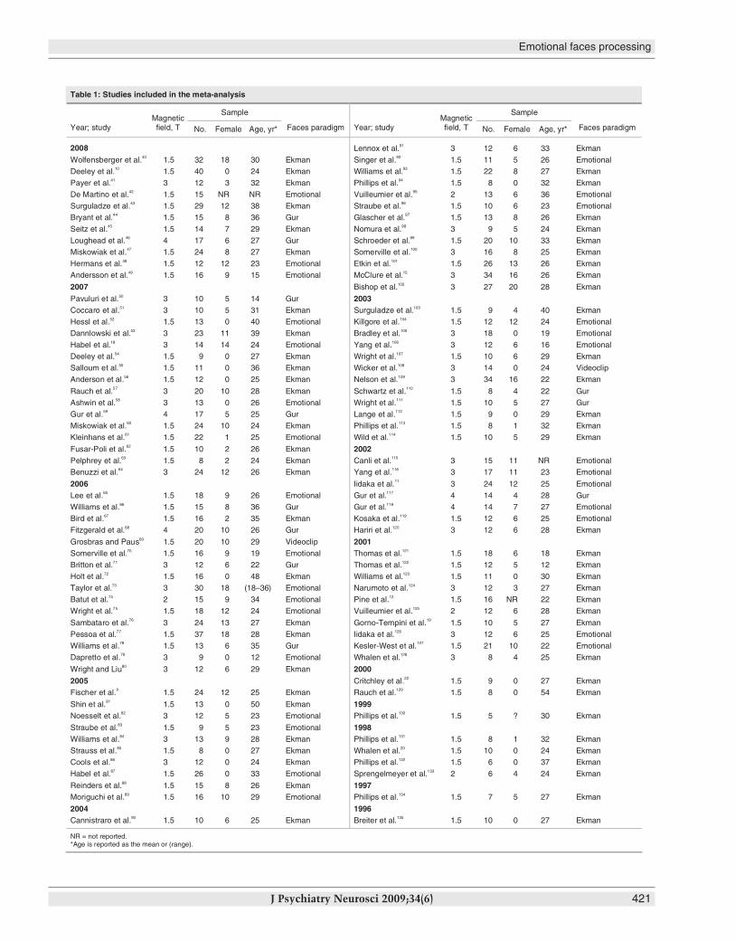
Table 1: Studies included in the meta-analysis
Sample Sample
Year; studyMagneticfield, T No. Female Age, yr* Faces paradigm Year; study
Magneticfield, T No. Female Age, yr* Faces paradigm
2008 Lennox et al.91 3 12 6 33 Ekman
Wolfensberger et al.40 1.5 32 18 30 Ekman Singer et al.92 1.5 11 5 26 Emotional
Deeley et al.10 1.5 40 0 24 Ekman Williams et al.93 1.5 22 8 27 Ekman
Payer et al.41 3 12 3 32 Ekman Phillips et al.94 1.5 8 0 32 Ekman
De Martino et al.42 1.5 15 NR NR Emotional Vuilleumier et al.95 2 13 6 36 Emotional
Surguladze et al.43 1.5 29 12 38 Ekman Straube et al.96 1.5 10 6 23 Emotional
Bryant et al.44 1.5 15 8 36 Gur Glascher et al.97 1.5 13 8 26 Ekman
Seitz et al.45 1.5 14 7 29 Ekman Nomura et al.98 3 9 5 24 Ekman
Loughead et al.46 4 17 6 27 Gur Schroeder et al.99 1.5 20 10 33 Ekman
Miskowiak et al.47 1.5 24 8 27 Ekman Somerville et al.100 3 16 8 25 Ekman
Hermans et al.48 1.5 12 12 23 Emotional Etkin et al.101 1.5 26 13 26 Ekman
Andersson et al.49 1.5 16 9 15 Emotional McClure et al.15 3 34 16 26 Ekman
2007 Bishop et al.102 3 27 20 28 Ekman
Pavuluri et al.50 3 10 5 14 Gur 2003Coccaro et al.51 3 10 5 31 Ekman Surguladze et al.103 1.5 9 4 40 Ekman
Hessl et al.52 1.5 13 0 40 Emotional Killgore et al.104 1.5 12 12 24 Emotional
Dannlowski et al.53 3 23 11 39 Ekman Bradley et al.105 3 18 0 19 Emotional
Habel et al.18 3 14 14 24 Emotional Yang et al.106 3 12 6 16 Emotional
Deeley et al.54 1.5 9 0 27 Ekman Wright et al.107 1.5 10 6 29 Ekman
Salloum et al.55 1.5 11 0 36 Ekman Wicker et al.108 3 14 0 24 Videoclip
Anderson et al.56 1.5 12 0 25 Ekman Nelson et al.109 3 34 16 22 Ekman
Rauch et al.57 3 20 10 28 Ekman Schwartz et al.110 1.5 8 4 22 Gur
Ashwin et al.58 3 13 0 26 Emotional Wright et al.111 1.5 10 5 27 Gur
Gur et al.59 4 17 5 25 Gur Lange et al.112 1.5 9 0 29 Ekman
Miskowiak et al.60 1.5 24 10 24 Ekman Phillips et al.113 1.5 8 1 32 Ekman
Kleinhans et al.61 1.5 22 1 25 Emotional Wild et al.114 1.5 10 5 29 Ekman
Fusar-Poli et al.62 1.5 10 2 26 Ekman 2002Pelphrey et al.63 1.5 8 2 24 Ekman Canli et al.115 3 15 11 NR Emotional
Benuzzi et al.64 3 24 12 26 Ekman Yang et al.116 3 17 11 23 Emotional
2006 Iidaka et al.11 3 24 12 25 Emotional
Lee et al.65 1.5 18 9 26 Emotional Gur et al.117 4 14 4 28 Gur
Williams et al.66 1.5 15 8 36 Gur Gur et al.118 4 14 7 27 Emotional
Bird et al.67 1.5 16 2 35 Ekman Kosaka et al.119 1.5 12 6 25 Emotional
Fitzgerald et al.68 4 20 10 26 Gur Hariri et al.120 3 12 6 28 Ekman
Grosbras and Paus69 1.5 20 10 29 Videoclip 2001Somerville et al.70 1.5 16 9 19 Emotional Thomas et al.121 1.5 18 6 18 Ekman
Britton et al.71 3 12 6 22 Gur Thomas et al.122 1.5 12 5 12 Ekman
Holt et al.72 1.5 16 0 48 Ekman Williams et al.123 1.5 11 0 30 Ekman
Taylor et al.73 3 30 18 (18–36) Emotional Narumoto et al.124 3 12 3 27 Ekman
Batut et al.74 2 15 9 34 Emotional Pine et al.12 1.5 16 NR 22 Ekman
Wright et al.75 1.5 18 12 24 Emotional Vuilleumier et al.125 2 12 6 28 Ekman
Sambataro et al.76 3 24 13 27 Ekman Gorno-Tempini et al.19 1.5 10 5 27 Ekman
Pessoa et al.77 1.5 37 18 28 Ekman Iidaka et al.126 3 12 6 25 Emotional
Williams et al.78 1.5 13 6 35 Gur Kesler-West et al.127 1.5 21 10 22 Emotional
Dapretto et al.79 3 9 0 12 Emotional Whalen et al.128 3 8 4 25 Ekman
Wright and Liu80 3 12 6 29 Ekman 20002005 Critchley et al.22 1.5 9 0 27 Ekman
Fischer et al.9 1.5 24 12 25 Ekman Rauch et al.129 1.5 8 0 54 Ekman
Shin et al.81 1.5 13 0 50 Ekman 1999Noesselt et al.82 3 12 5 23 Emotional Phillips et al.130 1.5 5 ? 30 Ekman
Straube et al.83 1.5 9 5 23 Emotional 1998Williams et al.84 3 13 9 28 Ekman Phillips et al.131 1.5 8 1 32 Ekman
Strauss et al.85 1.5 8 0 27 Ekman Whalen et al.20 1.5 10 0 24 Ekman
Cools et al.86 3 12 0 24 Ekman Phillips et al.132 1.5 6 0 37 Ekman
Habel et al.87 1.5 26 0 33 Emotional Sprengelmeyer et al.133 2 6 4 24 Ekman
Reinders et al.88 1.5 15 8 26 Ekman 1997Moriguchi et al.89 1.5 16 10 29 Emotional Phillips et al.134 1.5 7 5 27 Ekman
2004 1996Cannistraro et al.90 1.5 10 6 25 Ekman Breiter et al.135 1.5 10 0 27 Ekman
NR = not reported.*Age is reported as the mean or (range).
Results
Study selection
Our combined 1990–May 2008 literature search uncovered551 potential articles. The number of published articles relatingto each emotional condition of interest were as follows: “fMRIsad faces” returned 42 published papers, “fMRI angry faces”returned 65, “fMRI disgust faces” returned 22, “fMRI fearfulfaces” returned 142, “fMRI happy faces” returned 106 and“fMRI neutral faces” found 174. Most of the excluded studies(94%) did not meet inclusion criteria 2 and 3, whereas 2% didnot meet inclusion criterion 4 and 4% did not meet criterion 5.
Overall, 105 articles satisfied the inclusion criteria and weconsidered them for the meta-analysis. The final database of105 studies corresponded to a whole sample of 1600 healthyparticipants (of whom 639 were female, 40%). The mean ageof the sample was of 27 (standard deviation [SD] 6.8) years.Forty-seven of 105 studies (45%) reported coordinates in MNIspace. Fifty-five of 105 studies (52%) adopted an ROI ap-proach in the analysis of functional activation. Sixty percentof studies employed a classical Ekman faces paradigm, 10%of them used a Gur faces paradigm and the final 30% em-ployed other emotional faces paradigms. We used a total of1785 brain voxels for the meta-analytical procedure. Table 1lists all of the studies included in the meta-analysis by firstauthor and year of publication.
Main effect for faces processing independent of emotional valence
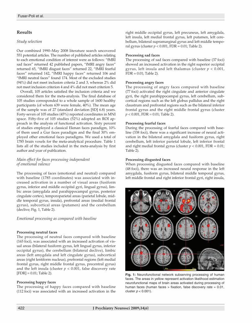
The processing of faces (emotional and neutral) comparedwith baseline (1785 coordinates) was associated with in-creased activation in a number of visual areas (fusiformgyrus, inferior and middle occipital gyri, lingual gyrus), lim-bic areas (amygdala and parahippocampal gyrus, posteriorcingulate cortex), temporoparietal areas (parietal lobule, mid-dle temporal gyrus, insula), prefrontal areas (medial frontalgyrus), subcortical areas (putamen) and the cerebellum (declive; Fig. 1, Table 2).
Emotional processing as compared with baseline
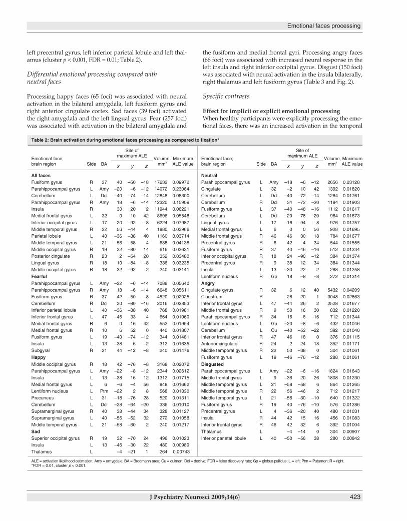
Processing neutral facesThe processing of neutral faces compared with baseline(165 foci), was associated with an increased activation of vis -ual areas (bilateral fusiform gyrus, left lingual gyrus, inferioroccipital gyrus), the cerebellum (bilateral declive), limbic areas (left amygdala and left cingulate gyrus), subcortical areas (right lentiform nucleus), prefrontal regions (left medialfrontal gyrus, right middle frontal gyrus, precentral gyrus)and the left insula (cluster p < 0.001, false discovery rate[FDR] = 0.01; Table 2).
Processing happy facesThe processing of happy faces compared with baseline(112 foci) was associated with an increased activation in the
right middle occipital gyrus, left precuneus, left amygdala,left insula, left medial frontal gyrus, left putamen, left cere-bellum, bilateral supramarginal gyrus and left middle tempo-ral gyrus (cluster p < 0.001, FDR = 0.01; Table 2).
Processing sad facesThe processing of sad faces compared with baseline (37 foci)showed an increased activation in the right superior occipitalgyrus, left insula and left thalamus (cluster p < 0.001,FDR = 0.01; Table 2).
Processing angry facesThe processing of angry faces compared with baseline(77 foci) activated the right cingulate and anterior cingulategyri, the right parahippocampal gyrus, left cerebellum, sub-cortical regions such as the left globus pallidus and the rightclaustrum and prefrontal regions such as the bilateral inferiorfrontal gyrus and the right middle frontal gyrus (clusterp < 0.001, FDR = 0.01; Table 2).
Processing fearful facesDuring the processing of fearful faces compared with base-line (338 foci), there was a significant increase of neural acti-vation in the bilateral amygdala and fusiform gyrus, rightcerebellum, left inferior parietal lobule, left inferior frontaland right medial frontal gyrus (cluster p < 0.001, FDR = 0.01;Table 2).
Processing disgusted facesWhen processing disgusted faces compared with baseline(48 foci), there was an increased neural response in the leftamygdala, fusiform gyrus, bilateral middle temporal gyrus,left middle frontal and right inferior frontal gyri, right insula,
Fusar-Poli et al.
422 J Psychiatry Neurosci 2009;34(6)
Fig. 1: Neurofunctional network subserving processing of humanfaces. The areas in yellow represent activation likelihood estimationneurofunctional maps of brain areas activated during processing ofhuman faces (human faces > fixation, false discovery rate = 0.01,cluster p < 0.001).
Emotional faces processing
J Psychiatry Neurosci 2009;34(6) 423
left precentral gyrus, left inferior parietal lobule and left thal-amus (cluster p < 0.001, FDR = 0.01; Table 2).
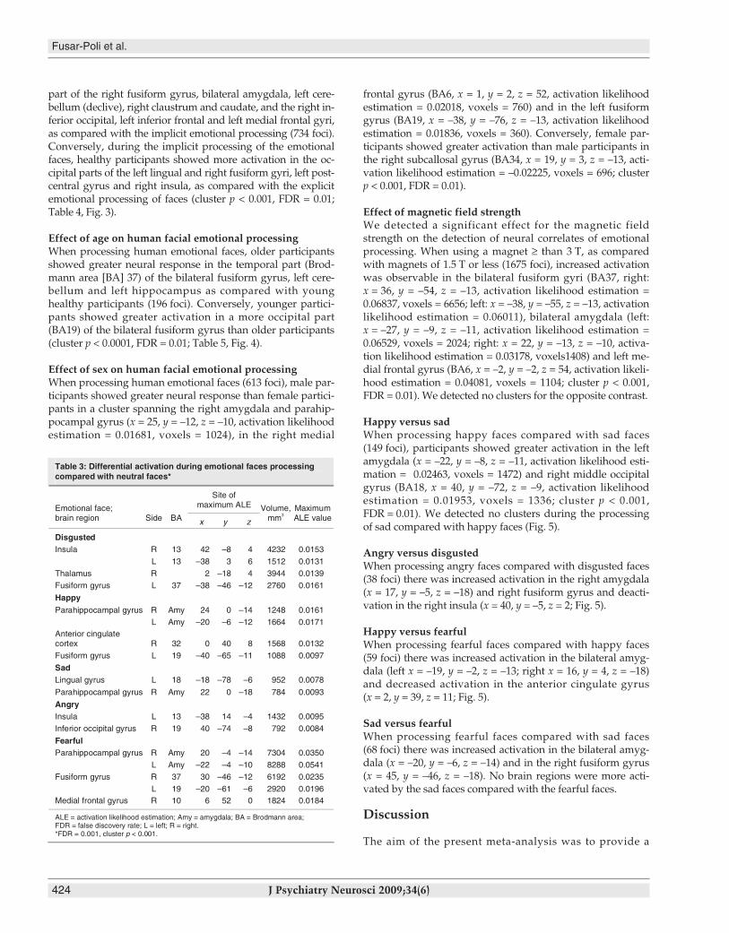
Differential emotional processing compared with neutral faces
Processing happy faces (65 foci) was associated with neuralactivation in the bilateral amygdala, left fusiform gyrus andright anterior cingulate cortex. Sad faces (39 foci) activatedthe right amygdala and the left lingual gyrus. Fear (257 foci)was associated with activation in the bilateral amygdala and
the fusiform and medial frontal gyri. Processing angry faces(66 foci) was associated with increased neural response in theleft insula and right inferior occipital gyrus. Disgust (150 foci)was associated with neural activation in the insula bilaterally,right thalamus and left fusiform gyrus (Table 3 and Fig. 2).
Specific contrasts
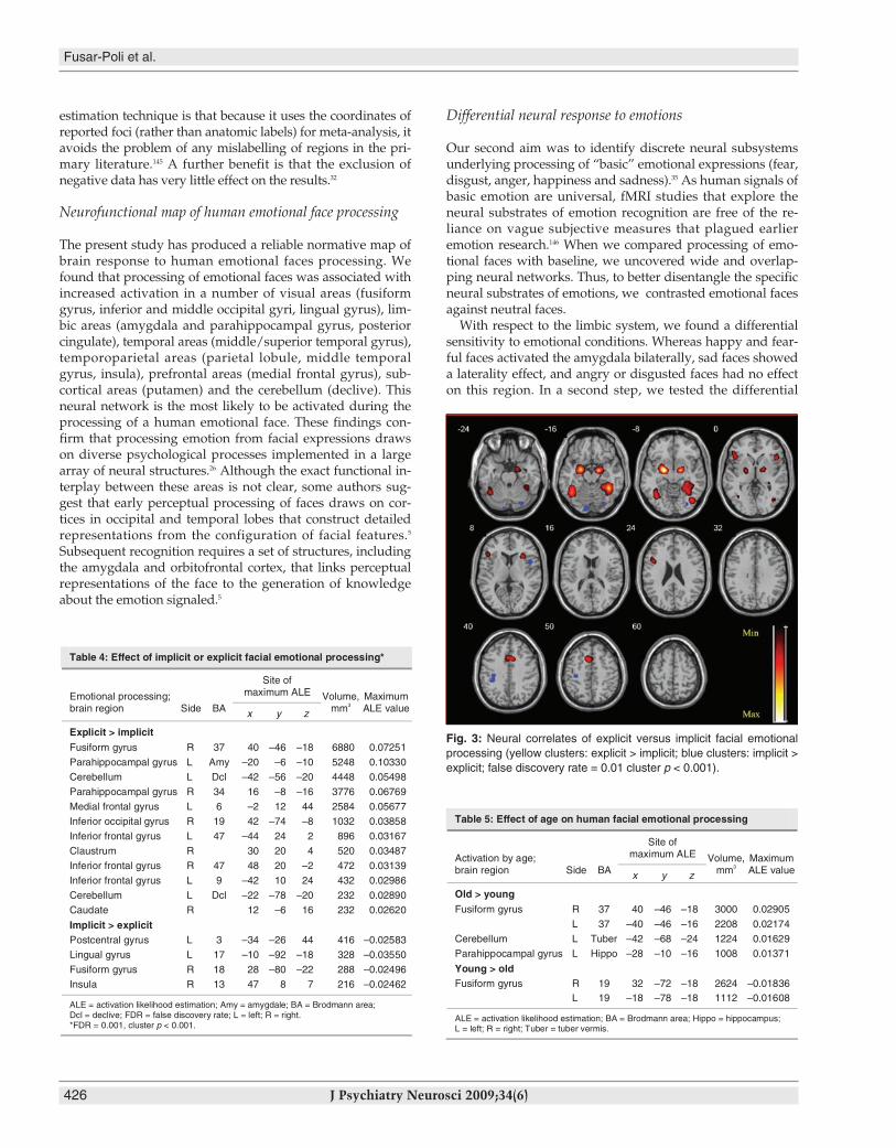
Effect for implicit or explicit emotional processingWhen healthy participants were explicitly processing the emo-tional faces, there was an increased activation in the temporal
Table 2: Brain activation during emotional faces processing as compared to fixation*
Site ofmaximum ALE
Site ofmaximum ALE
Emotional face;brain region Side BA x y z
Volume,mm3
MaximumALE value
Emotional face;brain region Side BA x y z
Volume,mm3
MaximumALE value
All faces NeutralFusiform gyrus R 37 40 –50 –18 17632 0.09972 Parahippocampal gyrus L Amy –18 –6 –12 2656 0.03128
Parahippocampal gyrus L Amy –20 –6 –12 14072 0.23064 Cingulate L 32 –2 10 42 1392 0.01820
Cerebellum L Dcl –40 –74 –14 12848 0.08300 Cerebellum L Dcl –40 –72 –14 1264 0.01761
Parahippocampal gyrus R Amy 18 –6 –14 12320 0.15909 Cerebellum R Dcl 34 –72 –20 1184 0.01903
Insula R 30 20 2 11944 0.06221 Fusiform gyrus L 37 –40 –48 –16 1112 0.01617
Medial frontal gyrus L 32 0 10 42 8696 0.05548 Cerebellum L Dcl –20 –78 –20 984 0.01673
Inferior occipital gyrus L 17 –20 –92 –8 6224 0.07987 Lingual gyrus L 17 –16 –94 –8 976 0.01757
Middle temporal gyrus R 22 56 –44 4 1880 0.03966 Medial frontal gyrus L 6 0 0 56 928 0.01695
Parietal lobule L 40 –36 –38 40 1160 0.03714 Middle frontal gyrus R 46 46 30 18 784 0.01677
Middle temporal gyrus L 21 –56 –58 4 688 0.04138 Precentral gyrus R 6 42 –4 34 544 0.01555
Middle occipital gyrus R 19 32 –80 14 616 0.03631 Fusiform gyrus R 37 40 –46 –16 512 0.01234
Posterior cingulate R 23 2 –54 20 352 0.03480 Inferior occipital gyrus R 18 24 –90 –12 384 0.01374
Lingual gyrus R 18 10 –84 –8 336 0.03235 Precentral gyrus R 9 38 12 34 384 0.01344
Middle occipital gyrus R 18 32 –92 2 240 0.03141 Insula L 13 –30 22 2 288 0.01258
Fearful Lentiform nucleus R Gp 18 –8 –8 272 0.01314
Parahippocampal gyrus L Amy –22 –6 –14 7088 0.05640 AngryParahippocampal gyrus R Amy 18 –6 –14 6648 0.05611 Cingulate gyrus R 32 6 12 40 5432 0.04209
Fusiform gyrus R 37 42 –50 –8 4520 0.02025 Claustrum R 28 20 1 3048 0.02863
Cerebellum R Dcl 30 –80 –16 2016 0.02853 Inferior frontal gyrus L 47 –44 26 2 2528 0.01677
Inferior parietal lobule L 40 –36 –38 40 768 0.01981 Middle frontal gyrus R 9 50 16 30 832 0.01220
Inferior frontal gyrus L 47 –46 33 4 664 0.01960 Parahippocampal gyrus R 34 16 –8 –16 712 0.01344
Medial frontal gyrus R 6 0 16 42 552 0.01954 Lentiform nucleus L Gp –20 –8 –6 432 0.01046
Medial frontal gyrus R 10 6 52 0 440 0.01807 Cerebellum L Cu –40 –52 –22 392 0.01040
Fusiform gyrus L 19 –40 –74 –12 344 0.01481 Inferior frontal gyrus R 47 46 18 0 376 0.01115
Insula L 13 –38 6 –2 312 0.01635 Anterior cingulate R 24 2 24 18 352 0.01171
Subgyral R 21 44 –12 –8 240 0.01476 Middle temporal gyrus R 22 50 –38 0 304 0.01061
Happy Fusiform gyrus L 19 –46 –76 –12 288 0.01061
Middle occipital gyrus R 18 42 –76 –8 3168 0.02072 DisgustedParahippocampal gyrus L Amy –22 –8 –12 2344 0.02612 Parahippocampal gyrus L Amy –22 –6 –16 1824 0.01643
Insula L 13 –38 16 12 1312 0.01715 Middle frontal gyrus L 9 –36 20 26 1808 0.01230
Medial frontal gyrus L 6 –6 –4 56 848 0.01662 Middle temporal gyrus L 21 –58 –58 6 864 0.01265
Lentiform nucleus L Ptm –22 2 8 568 0.01330 Middle temporal gyrus R 22 56 –46 2 712 0.01217
Precuneus L 31 –18 –76 28 520 0.01311 Middle temporal gyrus L 21 –56 –30 –10 640 0.01322
Cerebellum L Dcl –38 –64 –20 336 0.01010 Fusiform gyrus R 19 40 –76 –10 576 0.01286
Supramarginal gyrus R 40 38 –44 34 328 0.01127 Precentral gyrus L 4 –36 –20 40 480 0.01031
Supramarginal gyrus L 40 –56 –52 32 272 0.01058 Insula R 44 42 15 16 456 0.01083
Middle temporal gyrus L 21 –58 –60 2 240 0.01217 Inferior frontal gyrus R 46 42 32 6 392 0.01004
Sad Thalamus L –4 –14 0 304 0.00907
Superior occipital gyrus R 19 32 –70 24 496 0.01023 Inferior parietal lobule L 40 –50 –56 38 280 0.00842
Insula L 13 –46 –30 22 480 0.00989
Thalamus L –4 –21 1 264 0.00743
ALE = activation likelihood estimation; Amy = amygdale; BA = Brodmann area; Cu = culmen; Dcl = declive; FDR = false discovery rate; Gp = globus pallidus; L = left; Ptm = Putamen; R = right.*FDR = 0.01, cluster p < 0.001.
part of the right fusiform gyrus, bilateral amygdala, left cere-bellum (declive), right claustrum and caudate, and the right in-ferior occipital, left inferior frontal and left medial frontal gyri,as compared with the implicit emotional processing (734 foci).Conversely, during the implicit processing of the emotionalfaces, healthy participants showed more activation in the oc-cipital parts of the left lingual and right fusiform gyri, left post-central gyrus and right insula, as compared with the explicitemotional processing of faces (cluster p < 0.001, FDR = 0.01;Table 4, Fig. 3).
Effect of age on human facial emotional processingWhen processing human emotional faces, older participantsshowed greater neural response in the temporal part (Brod-mann area [BA] 37) of the bilateral fusiform gyrus, left cere-bellum and left hippocampus as compared with younghealthy participants (196 foci). Conversely, younger partici-pants showed greater activation in a more occipital part(BA19) of the bilateral fusiform gyrus than older participants(cluster p < 0.0001, FDR = 0.01; Table 5, Fig. 4).
Effect of sex on human facial emotional processingWhen processing human emotional faces (613 foci), male par-ticipants showed greater neural response than female partici-pants in a cluster spanning the right amygdala and parahip-pocampal gyrus (x = 25, y = –12, z = –10, activation likelihoodestimation = 0.01681, voxels = 1024), in the right medial
frontal gyrus (BA6, x = 1, y = 2, z = 52, activation likelihoodestimation = 0.02018, voxels = 760) and in the left fusiformgyrus (BA19, x = –38, y = –76, z = –13, activation likelihoodestimation = 0.01836, voxels = 360). Conversely, female par-ticipants showed greater activation than male participants inthe right subcallosal gyrus (BA34, x = 19, y = 3, z = –13, acti-vation likelihood estimation = –0.02225, voxels = 696; clusterp < 0.001, FDR = 0.01).
Effect of magnetic field strengthWe detected a significant effect for the magnetic fieldstrength on the detection of neural correlates of emotionalprocessing. When using a magnet ≥ than 3 T, as comparedwith magnets of 1.5 T or less (1675 foci), increased activationwas observable in the bilateral fusiform gyri (BA37, right:x = 36, y = –54, z = –13, activation likelihood estimation =0.06837, voxels = 6656; left: x = –38, y = –55, z = –13, activationlikelihood estimation = 0.06011), bilateral amygdala (left:x = –27, y = –9, z = –11, activation likelihood estimation =0.06529, voxels = 2024; right: x = 22, y = –13, z = –10, activa-tion likelihood estimation = 0.03178, voxels1408) and left me-dial frontal gyrus (BA6, x = –2, y = –2, z = 54, activation likeli-hood estimation = 0.04081, voxels = 1104; cluster p < 0.001,FDR = 0.01). We detected no clusters for the opposite contrast.
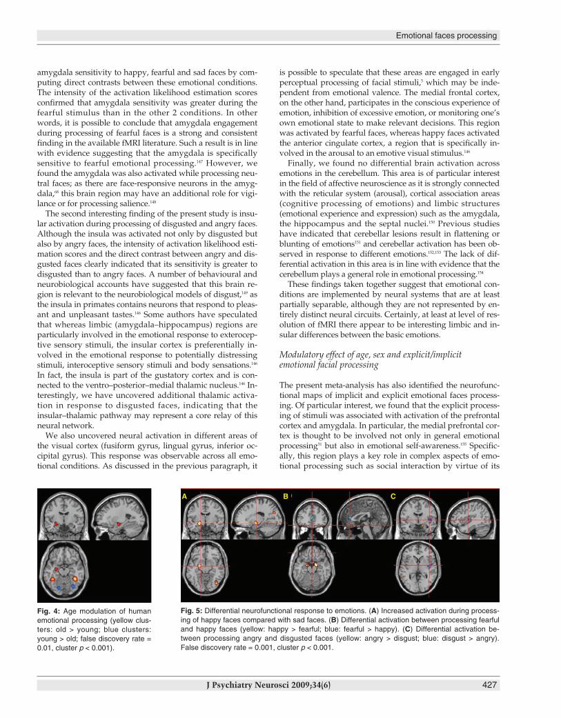
Happy versus sadWhen processing happy faces compared with sad faces(149 foci), participants showed greater activation in the leftamygdala (x = –22, y = –8, z = –11, activation likelihood esti-mation = 0.02463, voxels = 1472) and right middle occipitalgyrus (BA18, x = 40, y = –72, z = –9, activation likelihood estimation = 0.01953, voxels = 1336; cluster p < 0.001,FDR = 0.01). We detected no clusters during the processingof sad compared with happy faces (Fig. 5).
Angry versus disgustedWhen processing angry faces compared with disgusted faces(38 foci) there was increased activation in the right amygdala(x = 17, y = –5, z = –18) and right fusiform gyrus and deacti-vation in the right insula (x = 40, y = –5, z = 2; Fig. 5).
Happy versus fearfulWhen processing fearful faces compared with happy faces(59 foci) there was increased activation in the bilateral amyg-dala (left x = –19, y = –2, z = –13; right x = 16, y = 4, z = –18)and decreased activation in the anterior cingulate gyrus(x = 2, y = 39, z = 11; Fig. 5).
Sad versus fearfulWhen processing fearful faces compared with sad faces(68 foci) there was increased activation in the bilateral amyg-dala (x = –20, y = –6, z = –14) and in the right fusiform gyrus(x = 45, y = –46, z = –18). No brain regions were more acti-vated by the sad faces compared with the fearful faces.
Discussion
The aim of the present meta-analysis was to provide a
Fusar-Poli et al.
424 J Psychiatry Neurosci 2009;34(6)
Table 3: Differential activation during emotional faces processingcompared with neutral faces*
Site ofmaximum ALEEmotional face;
brain region Side BA x y z
Volume,mm3
MaximumALE value
DisgustedInsula R 13 42 –8 4 4232 0.0153
L 13 –38 3 6 1512 0.0131
Thalamus R 2 –18 4 3944 0.0139
Fusiform gyrus L 37 –38 –46 –12 2760 0.0161
HappyParahippocampal gyrus R Amy 24 0 –14 1248 0.0161
L Amy –20 –6 –12 1664 0.0171Anterior cingulatecortex R 32 0 40 8 1568 0.0132
Fusiform gyrus L 19 –40 –65 –11 1088 0.0097
SadLingual gyrus L 18 –18 –78 –6 952 0.0078
Parahippocampal gyrus R Amy 22 0 –18 784 0.0093
AngryInsula L 13 –38 14 –4 1432 0.0095
Inferior occipital gyrus R 19 40 –74 –8 792 0.0084
FearfulParahippocampal gyrus R Amy 20 –4 –14 7304 0.0350
L Amy –22 –4 –10 8288 0.0541
Fusiform gyrus R 37 30 –46 –12 6192 0.0235
L 19 –20 –61 –6 2920 0.0196Medial frontal gyrus R 10 6 52 0 1824 0.0184
ALE = activation likelihood estimation; Amy = amygdala; BA = Brodmann area;FDR = false discovery rate; L = left; R = right.*FDR = 0.001, cluster p < 0.001.
Emotional faces processing
J Psychiatry Neurosci 2009;34(6) 425
neurofunctional map of human emotional face processing inhealthy individuals.
Methodological issues
We adopted a multiple-steps approach with the encompass-ing objective of reducing heterogeneity across fMRI findings.In a first step (study selection), we chose to include fMRI stud-ies only and avoid positron emission tomography studiesgiven the profound methodological differences of these meth-ods. In addition, to control for considerable variation ob-served in the results of imaging experiments addressing evenclosely related experimental paradigms,32 our meta-analysisfocused on a single paradigm (i.e., the presentation of humanfaces and emotional picture) during emotional processing (at-tentive or mnestic studies were not considered). To overcomethe lack of statistical power associated with single fMRI stud-ies, we selected a large sample of 105 studies relating to1600 healthy volunteers and 1785 foci. Although there are nocommunity-accepted criteria for interpreting the activation
likelihood estimation results, for a study of this size if 6 ormore foci contribute to a cluster it is considered very robust,and if 3–5 foci contribute to a cluster, it is acceptable.144 It is notconvincing if only 1 or 2 foci contribute to a cluster. As recentactivation likelihood estimation meta-analyses have beenpublished with samples of 12 studies and fewer than 10 foci,144
our results are particularly robust. We have chosen a function– location statistical approach (voxel-based, activationlikelihood estimation) in place of the standard effect-size metaanalysis, because it is the location, rather than the magnitude,of the effect that is of interest in the present study.32 Specific -ally, activation likelihood estimation assumes that althougheach study reports the specific coordinates of activations, is-sues such as intersubject variability in brain anatomy and dif-ferences in investigators’ labels for anatomic regions may leadto some uncertainty as to the actual locations of these peaks.In fact, one of the difficulties when comparing imaging stud-ies is that there is considerable variability when labelling neu-roanatomical regions, and differences in nomenclature couldobscure findings. An advantage of the activation likelihood
Happy
Sad
Angry
Fearful
Disgusted
Fig. 2: Brain maps of neural activation in response to happy, sad, angry, fearful and disgusted human faces compared with neutral faces (false discovery rate = 0.001, cluster p < 0.001).Amy = amygdala; FG = fusiform gyrus; MFG = medial frontal gyrus.
estimation technique is that because it uses the coordinates ofreported foci (rather than anatomic labels) for meta-analysis, itavoids the problem of any mislabelling of regions in the pri-mary literature.145 A further benefit is that the exclusion ofnegative data has very little effect on the results.32
Neurofunctional map of human emotional face processing
The present study has produced a reliable normative map ofbrain response to human emotional faces processing. Wefound that processing of emotional faces was associated withincreased activation in a number of visual areas (fusiformgyrus, inferior and middle occipital gyri, lingual gyrus), lim-bic areas (amygdala and parahippocampal gyrus, posteriorcingulate), temporal areas (middle/superior temporal gyrus),temporoparietal areas (parietal lobule, middle temporalgyrus, insula), prefrontal areas (medial frontal gyrus), sub-cortical areas (putamen) and the cerebellum (declive). Thisneural network is the most likely to be activated during theprocessing of a human emotional face. These findings con-firm that processing emotion from facial expressions drawson diverse psychological processes implemented in a largearray of neural structures.26 Although the exact functional in-terplay between these areas is not clear, some authors sug-gest that early perceptual processing of faces draws on cor-tices in occipital and temporal lobes that construct detailedrepresentations from the configuration of facial features.5
Subsequent recognition requires a set of structures, includingthe amygdala and orbitofrontal cortex, that links perceptualrepresentations of the face to the generation of knowledgeabout the emotion signaled.5
Differential neural response to emotions
Our second aim was to identify discrete neural subsystemsunderlying processing of “basic” emotional expressions (fear,disgust, anger, happiness and sadness).35 As human signals ofbasic emotion are universal, fMRI studies that explore theneural substrates of emotion recognition are free of the re-liance on vague subjective measures that plagued earlieremotion research.146 When we compared processing of emo-tional faces with baseline, we uncovered wide and overlap-ping neural networks. Thus, to better disentangle the specificneural substrates of emotions, we contrasted emotional facesagainst neutral faces.
With respect to the limbic system, we found a differentialsensitivity to emotional conditions. Whereas happy and fear-ful faces activated the amygdala bilaterally, sad faces showeda laterality effect, and angry or disgusted faces had no effecton this region. In a second step, we tested the differential
Fusar-Poli et al.
426 J Psychiatry Neurosci 2009;34(6)
Fig. 3: Neural correlates of explicit versus implicit facial emotionalprocessing (yellow clusters: explicit > implicit; blue clusters: implicit >explicit; false discovery rate = 0.01 cluster p < 0.001).
Table 4: Effect of implicit or explicit facial emotional processing*
Site ofmaximum ALEEmotional processing;
brain region Side BA x y z
Volume,mm3
MaximumALE value
Explicit > implicitFusiform gyrus R 37 40 –46 –18 6880 0.07251
Parahippocampal gyrus L Amy –20 –6 –10 5248 0.10330
Cerebellum L Dcl –42 –56 –20 4448 0.05498
Parahippocampal gyrus R 34 16 –8 –16 3776 0.06769
Medial frontal gyrus L 6 –2 12 44 2584 0.05677
Inferior occipital gyrus R 19 42 –74 –8 1032 0.03858
Inferior frontal gyrus L 47 –44 24 2 896 0.03167
Claustrum R 30 20 4 520 0.03487
Inferior frontal gyrus R 47 48 20 –2 472 0.03139
Inferior frontal gyrus L 9 –42 10 24 432 0.02986
Cerebellum L Dcl –22 –78 –20 232 0.02890
Caudate R 12 –6 16 232 0.02620
Implicit > explicitPostcentral gyrus L 3 –34 –26 44 416 –0.02583
Lingual gyrus L 17 –10 –92 –18 328 –0.03550
Fusiform gyrus R 18 28 –80 –22 288 –0.02496
Insula R 13 47 8 7 216 –0.02462
ALE = activation likelihood estimation; Amy = amygdale; BA = Brodmann area;Dcl = declive; FDR = false discovery rate; L = left; R = right.*FDR = 0.001, cluster p < 0.001.
Table 5: Effect of age on human facial emotional processing
Site ofmaximum ALEActivation by age;
brain region Side BA x y z
Volume,mm3
MaximumALE value
Old > youngFusiform gyrus R 37 40 –46 –18 3000 0.02905
L 37 –40 –46 –16 2208 0.02174
Cerebellum L Tuber –42 –68 –24 1224 0.01629
Parahippocampal gyrus L Hippo –28 –10 –16 1008 0.01371
Young > oldFusiform gyrus R 19 32 –72 –18 2624 –0.01836
L 19 –18 –78 –18 1112 –0.01608
ALE = activation likelihood estimation; BA = Brodmann area; Hippo = hippocampus;L = left; R = right; Tuber = tuber vermis.
Emotional faces processing
J Psychiatry Neurosci 2009;34(6) 427
amygdala sensitivity to happy, fearful and sad faces by com-puting direct contrasts between these emotional conditions.The intensity of the activation likelihood estimation scoresconfirmed that amygdala sensitivity was greater during thefearful stimulus than in the other 2 conditions. In otherwords, it is possible to conclude that amygdala engagementduring processing of fearful faces is a strong and consistentfinding in the available fMRI literature. Such a result is in linewith evidence suggesting that the amygdala is specificallysensitive to fearful emotional processing.147 However, wefound the amygdala was also activated while processing neu-tral faces; as there are face-responsive neurons in the amyg-dala,68 this brain region may have an additional role for vigi-lance or for processing salience.148
The second interesting finding of the present study is insu-lar activation during processing of disgusted and angry faces.Although the insula was activated not only by disgusted butalso by angry faces, the intensity of activation likelihood esti-mation scores and the direct contrast between angry and dis-gusted faces clearly indicated that its sensitivity is greater todisgusted than to angry faces. A number of behavioural andneurobiological accounts have suggested that this brain re-gion is relevant to the neurobiological models of disgust,149 asthe insula in primates contains neurons that respond to pleas-ant and unpleasant tastes.146 Some authors have speculatedthat whereas limbic (amygdala–hippocampus) regions areparticularly involved in the emotional response to exterocep-tive sensory stimuli, the insular cortex is preferentially in-volved in the emotional response to potentially distressingstimuli, interoceptive sensory stimuli and body sensations.146
In fact, the insula is part of the gustatory cortex and is con-nected to the ventro–posterior–medial thalamic nucleus.146 In-terestingly, we have uncovered additional thalamic activa-tion in response to disgusted faces, indicating that theinsular–thalamic pathway may represent a core relay of thisneural network.
We also uncovered neural activation in different areas ofthe visual cortex (fusiform gyrus, lingual gyrus, inferior oc-cipital gyrus). This response was observable across all emo-tional conditions. As discussed in the previous paragraph, it
is possible to speculate that these areas are engaged in earlyperceptual processing of facial stimuli,5 which may be inde-pendent from emotional valence. The medial frontal cortex,on the other hand, participates in the conscious experience ofemotion, inhibition of excessive emotion, or monitoring one’sown emotional state to make relevant decisions. This regionwas activated by fearful faces, whereas happy faces activatedthe anterior cingulate cortex, a region that is specifically in-volved in the arousal to an emotive visual stimulus.146
Finally, we found no differential brain activation acrossemotions in the cerebellum. This area is of particular interestin the field of affective neuroscience as it is strongly connectedwith the reticular system (arousal), cortical association areas(cognitive processing of emotions) and limbic structures(emotional experience and expression) such as the amygdala,the hippocampus and the septal nuclei.150 Previous studieshave indicated that cerebellar lesions result in flattening orblunting of emotions151 and cerebellar activation has been ob-served in response to different emotions.152,153 The lack of dif-ferential activation in this area is in line with evidence that thecerebellum plays a general role in emotional processing.154
These findings taken together suggest that emotional con-ditions are implemented by neural systems that are at leastpartially separable, although they are not represented by en-tirely distinct neural circuits. Certainly, at least at level of res-olution of fMRI there appear to be interesting limbic and in-sular differences between the basic emotions.
Modulatory effect of age, sex and explicit/implicit emotional facial processing
The present meta-analysis has also identified the neurofunc-tional maps of implicit and explicit emotional faces process-ing. Of particular interest, we found that the explicit process-ing of stimuli was associated with activation of the prefrontalcortex and amygdala. In particular, the medial prefrontal cor-tex is thought to be involved not only in general emotionalprocessing31 but also in emotional self-awareness.155 Specific -ally, this region plays a key role in complex aspects of emo-tional processing such as social interaction by virtue of its
Fig. 4: Age modulation of humanemotional processing (yellow clus-ters: old > young; blue clusters:young > old; false discovery rate =0.01, cluster p < 0.001).
A B C
Fig. 5: Differential neurofunctional response to emotions. (A) Increased activation during process-ing of happy faces compared with sad faces. (B) Differential activation between processing fearfuland happy faces (yellow: happy > fearful; blue: fearful > happy). (C) Differential activation be-tween processing angry and disgusted faces (yellow: angry > disgust; blue: disgust > angry).False discovery rate = 0.001, cluster p < 0.001.
connections with the discrete parts of the temporal lobe andsubcortical structures that control autonomic responses.156
Given the anatomic connections between the medial pre-frontal cortex and the amygdala,157 the coactivation of these2 structures may reflect possible influence of cortical controlon explicit processing. The amygdala–medial prefrontal cir-cuitry has been termed as the emotion generation–regulationcircuit158 and is implicated in attention to threat and inter -pretation of emotional stimuli.159 Previous lesion studies haveconfirmed that insults to these areas result in emotion dysregulation.160
Conversely, the implicit processing was associated with ac-tivation in more ventral regions spanning the inferior pre-frontal cortex and insula. The insular cortex plays a key rolein emotional processing owing to its abundant connectionswith other association and primary sensory areas and its in-volvement in language, vestibular and pain perception (for areview see Nagai et al.161). In particular, insular projections tothe inferior prefrontal cortex and amygdala may convey so-cial information from emotional expressions.22 The anteriorpart of the insular cortex is considered to be a limbic-relatedcortex and part of the interoceptive system, which associates(with a mirror neuron link) external and internal experience.25
This insular subregion provides information about aversivebody states associated with conditional stimuli, signallingthis information to prefrontal brain areas that are critical forthe allocation of attention and the execution of actions.162 Thecognitive control of appraisal and emotional relevance is notmediated by the orbitofrontal cortex alone, but arises fromlarge-scale systems that include the amygdala and the insularcortices.163 In line with such evidence our findings suggestthat the explicit or implicit emotional processing is regulatedby the functional interaction of a network comprising thesebrain regions.
With respect to age, we demonstrated that the neural re-sponse to emotional faces in the fusiform gyrus, the cerebel-lum and the hippocampus is modulated by this factor. Thehippocampus shows extensive connections with extrastriatevisual areas, including the fusiform gyrus. Previous fMRIparadigms have confirmed that brain activity in this area isaffected by age.164 The observed age-related differences couldreflect functional compensation within the neural system in-volved in perception of facial affect or the fact that olderadults process emotional information in a different mannerthan young adults. Finally, we found that male participantsactivated the limbic (amygdala/parahippocampal gyrus) andthe prefrontal cortices more than female participants duringemotional processing; conversely, female participantsshowed greater activation in the right subcallosal gyrus. Pre-vious studies found that male and female participants engagethe amygdala and the prefrontal cortex in different wayswhile passively viewing emotional faces.15
Implications for studies of emotional processing in psychiatric populations
Over the past decade, a number of behavioural findings haveshown that alterations of emotional faces processing play a
role in a range of psychiatric disorders spanning affective dis-eases (e.g., major depression, bipolar disorders, anxiety- related disorders)24 and the psychotic spectrum (e.g., schizo-phrenia,165–167 autism168). For instance, depressed participantsshow a state-related positive bias toward negative emotionalcues (e.g., sad faces) and a bias away from positive emotionalcues (e.g., happy faces).24 These findings are in line with cog-nitive theories of depression, which emphasizes the role ofnegative biases in information processing in the etiology andmaintenance of depression.169 Consistent with these observa-tions, there are data to suggest there may be an analogous,state-related negative recognition bias for negative emotionsin mania.91 On the other hand, patients with schizophreniahave difficulty recognizing the emotion that correspondswith a given facial expression.2,170–172 Early deficit in visual pro-cessing underlying emotion recognition170,173 is a hallmark ofschizophrenia, with consequences for cognitive function,174
social function2 and subjective well-being.171 Specific alter-ations of emotional faces processing of schizophrenic patientsinclude bias for threat-related emotional material, which maybe regarded with increased significance by delusion-proneindividuals, and it is possible that this bias is involved in theformation of delusional beliefs.2,175,176
Facial emotional stimuli may serve as valid tools tappingon neural networks implicated in emotional processing.Thus,30 fMRI has been exponentially employed to address theneurophysiological substrates of impairments in emotionalfaces processing under different psychiatric conditions.177 Al-though many of the brain regions activated during facialemotional processing in healthy participants are also impli-cated in the pathophysiology of psychiatric disorders,24 cur-rent imaging literature indicates contrasting findings and avariable picture8 and is unable to definitively characterizewhat brain region is etiologically associated with each psy-chiatric disease. For this reason, the use of the physiologicmaps provided herein will help to strengthen future studyhypotheses and designs by robustly identifying regions ofnormal activation.
Limitations
There are several limitations to our study. The first is the het-erogeneity of the studies included. Factors such as behav-ioural performance, demographic information, substanceabuse, cognitive function and personality traits, which couldpotentially influence the results, vary across the differentsamples. Although we have attempted to address the effectof age, sex, implicit/explicit emotional processing and inten-sity of magnet, at the present time it is not possible to directlyevaluate the influence of all these factors on the results. Second, our methods did not allow for weighting of the re-sults based on the level of statistical significance reported ineach study. This means that we cannot exactly determine therelative strengths of activation differences. Third, althoughthe quantitative meta-analytic method used herein representsa substantial advance for integrating functional neuroimag-ing data, the method remains subject to the basic limitation ofliterature reviews, in particular the “file drawer” problem.
Fusar-Poli et al.
428 J Psychiatry Neurosci 2009;34(6)
Emotional faces processing
J Psychiatry Neurosci 2009;34(6) 429
However, a benefit of activation likelihood estimation meta-analyses is that the exclusion of negative data has very littleeffect on the results.32 Conversely, it is possible that studiesemploying ROI analysis of brain activation may have influ-enced the resulting activation patterns and potentially biasedthe results. Previous activation likelihood estimation stud-ies144 did not acknowledge such a methodological limitation,including in the same database whole brain and ROI studies.To our knowledge, the present study is the first activationlikelihood estimation meta-analysis to address this point byproviding details of the methodological approach adopted ineach study.
Issues for future research
Functional neuroimaging is currently advancing from thesimple detection and localization of cortical activation to theinvestigation of complex cortical processes and associatedfunctional relations between cortical areas. Such researchquestions can no longer be addressed by the isolated analysisof single experiments alone, but necessitate the consolidationof results across different cognitive tasks and experimentalparadigms. This again makes meta-analyses an increasinglyimportant part in the evaluation of functional imaging re-sults.8 A combination of functional imaging and electrophysi-ological techniques (i.e., fMRI and electroencephalography)may also represent the future research instrument to combinethe high spatial resolution of the first technique with the hightemporal resolution of the second. Finally, although the evi-dence suggests that defined areas of the brain are responsiveto specific facial expressions, recent evidence indicates neur -onal subspecialization in face-specific brain regions for differ-ent components of facial perception such as facial expression,viewing of familiar and unfamiliar faces, and dis criminationof spatial relations of facial features (i.e., eyes, lips or nose).28
Investigations into the neural systems underlying processingof these cues for each of the basic emotions may be helpful tofurther elucidate their neural representation.
Conclusion
The wide neurofunctional network underlying human facesprocessing includes a number of visual, limbic, temporo- parietal, prefrontal and subcortical areas as well as the cere-bellum. Whereas occipital areas and the cerebellum werecommonly activated across different emotions, a discrete re-sponse to valence has been reported for the limbic systemand insular cortex. Although the basic emotions are not rep-resented by entirely distinct neural circuits, they are at leastpartially separable. Sex, age and consciousness modulate theneurophysiological response to human emotional faces.
Acknowledgement: We thank Dr. Angela Laird for her valuable advice with the meta-analytical package.
Competing interests: None declared.
Contributors: Drs. Fusar-Poli, Placentino, Allen, Barale, Perez,McGuire and Politi designed the study. Drs. Fusar-Poli, Carletti,Landi, Abbamonte and Gasparotti acquired the data, which
Drs. Fusar-Poli, Surguladze, Benedetti and McGuire analyzed.Drs. Fusar-Poli, Landi and Perez wrote the article, which all other au-thors reviewed. All authors approved the final version for publication.
References
1. Tate AJ, Fischer H, Leigh AE, et al. Behavioural and neurophysio-logical evidence for face identity and face emotion processing inanimals. Philos Trans R Soc Lond B Biol Sci 2006;361:2155-72.
2. Phillips ML, David AS. Facial processing in schizophrenia anddelusional misidentification: cognitive neuropsychiatric ap-proaches. Schizophr Res 1995;17:109-14.
3. Frank MG, Stennett J. The forced-choice paradigm and the percep-tion of facial expressions of emotion. J Pers Soc Psychol 2001;80:75-85.
4. Grossmann T, Johnson MH. The development of the social brain inhuman infancy. Eur J Neurosci 2007;25:909-19.
5. Adolphs R. Neural systems for recognizing emotion. Curr OpinNeurobiol 2002;12:169-77.
6. Kanwisher N, McDermott J, Chun MM. The fusiform face area: amodule in human extrastriate cortex specialized for face percep-tion. J Neurosci 1997;17:4302-11.
7. Peelen MV, Downing PE. The neural basis of visual body percep-tion. Nat Rev Neurosci 2007;8:636-48.
8. Neumann J, von Cramon DY, Lohmann G. Model-based clusteringof meta-analytic functional imaging data. Hum Brain Mapp2008;29:177-92.
9. Fischer H, Sandblom J, Gavazzeni J, et al. Age-differential patternsof brain activation during perception of angry faces. Neurosci Lett2005;386:99-104.
10. Deeley Q, Daly EM, Azuma R, et al. Changes in male brain re-sponses to emotional faces from adolescence to middle age. Neuroimage 2008;40:389-97.
11. Iidaka T, Okada T, Murata T, et al. Age-related differences in themedial temporal lobe responses to emotional faces as revealed byfMRI. Hippocampus 2002;12:352-62.
12. Pine DS, Grun J, Zarahn E, et al. Cortical brain regions engaged bymasked emotional faces in adolescents and adults: an fMRI study.Emotion 2001;1:137-47.
13. Guyer AE, Monk CS, McClure-Tone EB, et al. A developmental examination of amygdala response to facial expressions. J CognNeurosci 2008;20:1565-82.
14. Schulte-Rüther M, Markowitsch HJ, Shah NJ, et al. Gender differ-ences in brain networks supporting empathy. Neuroimage 2008;42:393-403.
15. McClure EB, Monk CS, Nelson EE, et al. A developmental exam -ination of gender differences in brain engagement during evalua-tion of threat. Biol Psychiatry 2004;55:1047-55.
16. Kranz F, Ishai A. Face perception is modulated by sexual prefer-ence. Curr Biol 2006;16:63-8.
17. Lee TM, Liu HL, Hoosain R, et al. Gender differences in neuralcorrelates of recognition of happy and sad faces in humans as-sessed by functional magnetic resonance imaging. Neurosci Lett2002;333:13-6.
18. Habel U, Windischberger C, Derntl B, et al. Amygdala activationand facial expressions: explicit emotion discrimination versus im-plicit emotion processing. Neuropsychologia 2007;45:2369-77.
19. Gorno-Tempini ML, Pradelli S, Serafini M, et al. Explicit and inci-dental facial expression processing: an fMRI study. Neuroimage2001;14:465-73.
20. Whalen PJ, Rauch SL, Etcoff NL, et al. Masked presentations ofemotional facial expressions modulate amygdala activity withoutexplicit knowledge. J Neurosci 1998;18:411-8.
21. Chen CH, Lennox B, Jacob R, et al. Explicit and implicit facial af-fect recognition in manic and depressed states of bipolar disorder:a functional magnetic resonance imaging study. Biol Psychiatry2006;59:31-9.
22. Critchley H, Daly E, Phillips M, et al. Explicit and implicit neuralmechanisms for processing of social information from facial ex-pressions: a functional magnetic resonance imaging study. HumBrain Mapp 2000;9:93-105.
23. Posamentier MT, Abdi H. Processing faces and facial expressions.Neuropsychol Rev 2003;13:113-43.
24. Leppanen JM. Emotional information processing in mood
Fusar-Poli et al.
430 J Psychiatry Neurosci 2009;34(6)
disorders: a review of behavioral and neuroimaging findings. CurrOpin Psychiatry 2006;19:34-9.
25. Gobbini MI, Haxby JV. Neural systems for recognition of familiarfaces. Neuropsychologia 2007;45:32-41.
26. Vuilleumier P, Pourtois G. Distributed and interactive brain mech-anisms during emotion face perception: evidence from functionalneuroimaging. Neuropsychologia 2007;45:174-94.
27. Pourtois G, Vuilleumier P. Dynamics of emotional effects on spa-tial attention in the human visual cortex. Prog Brain Res 2006;156:67-91.
28. Masella RS, Meister M. The neuroanatomic basis of facial percep-tion and variable facial discrimination ability: implications for orthodontics. Am J Orthod Dentofacial Orthop 2007;132:293-301.
29. Davidson RJ, Irwin W. The functional neuroanatomy of emotionand affective style. Trends Cogn Sci 1999;3:11-21.
30. Haxby JV, Hoffman EA, Gobbini MI. Human neural systems forface recognition and social communication. Biol Psychiatry 2002;51:59-67.
31. Phan KL, Wager T, Taylor SF, et al. Functional neuroanatomy ofemotion: a meta-analysis of emotion activation studies in PET andfMRI. Neuroimage 2002;16:331-48.
32. Murphy FC, Nimmo-Smith I, Lawrence AD. Functional neuro -anatomy of emotions: a meta-analysis. Cogn Affect Behav Neurosci2003;3:207-33.
33. Fox PT, Laird AR, Lancaster JL. Coordinate-based voxel-wisemeta-analysis: dividends of spatial normalization. Report of a vir-tual workshop. Hum Brain Mapp 2005;25:1-5.
34. Wild B, Erb M, Bartels M. Are emotions contagious? Evoked emo-tions while viewing emotionally expressive faces: quality, quan-tity, time course and gender differences. Psychiatry Res 2001;102:109-24.
35. Ekman P, Friesen W. Pictures of facial affect. Palo Alto (CA): Con-sulting Psychologists Press; 1976.
36. Gur RC, Sara R, Hagendoorn M, et al. A method for obtaining 3-dimensional facial expressions and its standardization for use inneurocognitive studies. J Neurosci Methods 2002;115:137-43.
37. Laird AR, Fox PM, Price CJ, et al. ALE meta-analysis: controllingthe false discovery rate and performing statistical contrasts. HumBrain Mapp 2005;25:155-64.
38. Turkeltaub PE, Eden GF, Jones KM, et al. Meta-analysis of thefunctional neuroanatomy of single-word reading: method and val-idation. Neuroimage 2002;16:765-80.
39. Rama P, Courtney SM. Functional topography of working mem-ory for face or voice identity. Neuroimage 2005;24:224-34.
40. Wolfensberger SP, Veltman DJ, Hoogendijk WJ, et al. Amygdalaresponses to emotional faces in twins discordant or concordant forthe risk for anxiety and depression. Neuroimage 2008;41:544-52.
41. Payer DE, Lieberman MD, Monterosso JR, et al. Differences in cor-tical activity between methamphetamine-dependent and healthyindividuals performing a facial affect matching task. Drug AlcoholDepend 2008;93:93-102.
42. De Martino B, Kalisch R, Rees G, et al. Enhanced processing ofthreat stimuli under limited attentional resources. Cereb Cortex2008; 19:127-33.
43. Surguladze SA, Elkin A, Ecker C, et al. Genetic variation in theserotonin transporter modulates neural system-wide response tofearful faces. Genes Brain Behav 2008;7:543-51.
44. Bryant RA, Kemp AH, Felmingham KL, et al. Enhanced amygdalaand medial prefrontal activation during nonconscious processingof fear in posttraumatic stress disorder: an fMRI study. Hum BrainMapp 2008;29:517-23.
45. Seitz RJ, Schafer R, Scherfeld D, et al. Valuating other people’semotional face expression: a combined functional magnetic reso-nance imaging and electroencephalography study. Neuroscience2008;152:713-22.
46. Loughead J, Gur RC, Elliott M, et al. Neural circuitry for accurateidentification of facial emotions. Brain Res 2008;1194:37-44.
47. Miskowiak K, Inkster B, Selvaraj S, et al. Erythropoietin improvesmood and modulates the cognitive and neural processing of emo-tion 3 days post administration. Neuropsychopharmacology 2008; 33:611-8.
48. Hermans EJ, Ramsey NF, van Honk J. Exogenous testosterone en-hances responsiveness to social threat in the neural circuitry of so-cial aggression in humans. Biol Psychiatry 2008;63:263-70.
49. Andersson F, Glaser B, Spiridon M, et al. Impaired activation of
face processing networks revealed by functional magnetic reso-nance imaging in 22q11.2 deletion syndrome. Biol Psychiatry 2008;63: 49-57.
50. Pavuluri MN, O’Connor MM, Harral E, et al. Affective neural cir-cuitry during facial emotion processing in pediatric bipolar disor-der. Biol Psychiatry 2007;62:158-67.
51. Coccaro EF, McCloskey MS, Fitzgerald DA, et al. Amygdala andorbitofrontal reactivity to social threat in individuals with impul-sive aggression. Biol Psychiatry 2007;62:168-78.
52. Hessl D, Rivera S, Koldewyn K, et al. Amygdala dysfunction inmen with the fragile X premutation. Brain 2007;130:404-16.
53. Dannlowski U, Ohrmann P, Bauer J, et al. Amygdala reactivitypredicts automatic negative evaluations for facial emotions. Psychiatry Res 2007;154:13-20.
54. Deeley Q, Daly EM, Surguladze S, et al. An event related func-tional magnetic resonance imaging study of facial emotion pro-cessing in Asperger syndrome. Biol Psychiatry 2007;62:207-17.
55. Salloum JB, Ramchandani VA, Bodurka J, et al. Blunted rostral an-terior cingulate response during a simplified decoding task of neg-ative emotional facial expressions in alcoholic patients. Alcohol ClinExp Res 2007;31:1490-504.
56. Anderson IM, Del-Ben CM, McKie S, et al. Citalopram modulationof neuronal responses to aversive face emotions: a functional MRIstudy. Neuroreport 2007;18:1351-5.
57. Rauch AV, Ohrmann P, Bauer J, et al. Cognitive coping style mod-ulates neural responses to emotional faces in healthy humans: a 3-T FMRI study. Cereb Cortex 2007;17:2526-35.
58. Ashwin C, Baron-Cohen S, Wheelwright S, et al. Differential ac-tivation of the amygdala and the ‘social brain’ during fearfulface-processing in Asperger syndrome. Neuropsychologia 2007;45:2-14.
59. Gur RE, Loughead J, Kohler CG, et al. Limbic activation associatedwith misidentification of fearful faces and flat affect in schizophre-nia. Arch Gen Psychiatry 2007;64:1356-66.
60. Miskowiak K, O’Sullivan U, Harmer CJ. Erythropoietin reducesneural and cognitive processing of fear in human models of anti-depressant drug action. Biol Psychiatry 2007;62:1244-50.
61. Kleinhans NM, Johnson LC, Mahurin R, et al. Increased amygdalaactivation to neutral faces is associated with better face memoryperformance. Neuroreport 2007;18:987-91.
62. Fusar-Poli P, Allen P, Lee F, et al. Modulation of neural responseto happy and sad faces by acute tryptophan depletion. Psychophar-macology (Berl) 2007;193:31-44.
63. Pelphrey KA, Morris JP, McCarthy G, et al. Perception of dynamicchanges in facial affect and identity in autism. Soc Cogn Affect Neurosci 2007;2:140-9.
64. Benuzzi F, Pugnaghi M, Meletti S, et al. Processing the socially rel-evant parts of faces. Brain Res Bull 2007;74:344-56.
65. Lee TW, Josephs O, Dolan RJ, et al. Imitating expressions: emotion-specific neural substrates in facial mimicry. Soc Cogn Affect Neurosci 2006;1:122-35.
66. Williams LM, Liddell BJ, Kemp AH, et al. Amygdala-prefrontaldissociation of subliminal and supraliminal fear. Hum Brain Mapp2006;27:652-61.
67. Bird G, Catmur C, Silani G, et al. Attention does not modulateneural responses to social stimuli in autism spectrum disorders.Neuroimage 2006;31:1614-24.
68. Fitzgerald DA, Angstadt M, Jelsone LM, et al. Beyond threat:amygdala reactivity across multiple expressions of facial affect.Neuroimage 2006;30:1441-8.
69. Grosbras MH, Paus T. Brain networks involved in viewing angryhands or faces. Cereb Cortex 2006;16:1087-96.
70. Somerville LH, Wig GS, Whalen PJ, et al. Dissociable medial tem-poral lobe contributions to social memory. J Cogn Neurosci 2006;18:1253-65.
71. Britton JC, Taylor SF, Sudheimer KD, et al. Facial expressions andcomplex IAPS pictures: common and differential networks. Neuroimage 2006;31:906-19.
72. Holt DJ, Kunkel L, Weiss AP, et al. Increased medial temporal lobeactivation during the passive viewing of emotional and neutral fa-cial expressions in schizophrenia. Schizophr Res 2006;82:153-62.
73. Taylor SE, Eisenberger NI, Saxbe D, et al. Neural responses toemotional stimuli are associated with childhood family stress. BiolPsychiatry 2006;60:296-301.
74. Batut AC, Gounot D, Namer IJ, et al. Neural responses associated
Emotional faces processing
J Psychiatry Neurosci 2009;34(6) 431
with positive and negative emotion processing in patients with leftversus right temporal lobe epilepsy. Epilepsy Behav 2006;9:415-23.
75. Wright CI, Wedig MM, Williams D, et al. Novel fearful faces activate the amygdala in healthy young and elderly adults. Neurobiol Aging 2006;27:361-74.
76. Sambataro F, Dimalta S, Di Giorgio A, et al. Preferential responsesin amygdala and insula during presentation of facial contempt anddisgust. Eur J Neurosci 2006;24:2355-62.
77. Pessoa L, Japee S, Sturman D, et al. Target visibility and visualawareness modulate amygdala responses to fearful faces. CerebCortex 2006;16:366-75.
78. Williams LM, Kemp AH, Felmingham K, et al. Trauma modulatesamygdala and medial prefrontal responses to consciously at-tended fear. Neuroimage 2006;29:347-57.
79. Dapretto M, Davies MS, Pfeifer JH, et al. Understanding emotionsin others: mirror neuron dysfunction in children with autism spec-trum disorders. Nat Neurosci 2006;9:28-30.
80. Wright P, Liu Y. Neutral faces activate the amygdala during iden-tity matching. Neuroimage 2006;29:628-36.
81. Shin LM, Wright CI, Cannistraro PA, et al. A functional magneticresonance imaging study of amygdala and medial prefrontal cor-tex responses to overtly presented fearful faces in posttraumaticstress disorder. Arch Gen Psychiatry 2005;62:273-81.
82. Noesselt T, Driver J, Heinze HJ, et al. Asymmetrical activation inthe human brain during processing of fearful faces. Curr Biol2005;15:424-9.
83. Straube T, Mentzel HJ, Miltner WH. Common and distinct brainactivation to threat and safety signals in social phobia. Neuropsy-chobiology 2005;52:163-8.
84. Williams MA, McGlone F, Abbott DF, et al. Differential amygdalaresponses to happy and fearful facial expressions depend on selec-tive attention. Neuroimage 2005;24:417-25.
85. Strauss MM, Makris N, Aharon I, et al. fMRI of sensitization to an-gry faces. Neuroimage 2005;26:389-413.
86. Cools R, Calder AJ, Lawrence AD, et al. Individual differences inthreat sensitivity predict serotonergic modulation of amygdala re-sponse to fearful faces. Psychopharmacology (Berl) 2005;180:670-9.
87. Habel U, Klein M, Kellermann T, et al. Same or different? Neuralcorrelates of happy and sad mood in healthy males. Neuroimage2005; 26:206-14.
88. Reinders AA, den Boer JA, Buchel C. The robustness of perception.Eur J Neurosci 2005;22:524-30.
89. Moriguchi Y, Ohnishi T, Kawachi T, et al. Specific brain activationin Japanese and Caucasian people to fearful faces. Neuroreport2005; 16:133-6.
90. Cannistraro PA, Wright CI, Wedig MM, et al. Amygdala responsesto human faces in obsessive-compulsive disorder. Biol Psychiatry2004; 56:916-20.
91. Lennox BR, Jacob R, Calder AJ, et al. Behavioural and neurocogni-tive responses to sad facial affect are attenuated in patients withmania. Psychol Med 2004;34:795-802.
92. Singer T, Kiebel SJ, Winston JS, et al. Brain responses to the ac-quired moral status of faces. Neuron 2004;41:653-62.
93. Williams LM, Das P, Harris AW, et al. Dysregulation of arousaland amygdala-prefrontal systems in paranoid schizophrenia. Am JPsychiatry 2004;161:480-9.
94. Phillips ML, Williams LM, Heining M, et al. Differential neural re-sponses to overt and covert presentations of facial expressions offear and disgust. Neuroimage 2004;21:1484-96.
95. Vuilleumier P, Richardson MP, Armony JL, et al. Distant influ-ences of amygdala lesion on visual cortical activation during emo-tional face processing. Nat Neurosci 2004;7:1271-8.
96. Straube T, Kolassa IT, Glauer M, et al. Effect of task conditions onbrain responses to threatening faces in social phobics: an event- related functional magnetic resonance imaging study. Biol Psych -iatry 2004;56:921-30.
97. Glascher J, Tuscher O, Weiller C, et al. Elevated responses to con-stant facial emotions in different faces in the human amygdala: anfMRI study of facial identity and expression. BMC Neurosci2004;5:45.
98. Nomura M, Ohira H, Haneda K, et al. Functional association of theamygdala and ventral prefrontal cortex during cognitive evalua-tion of facial expressions primed by masked angry faces: an event-related fMRI study. Neuroimage 2004;21:352-63.
99. Schroeder U, Hennenlotter A, Erhard P, et al. Functional neu-
roanatomy of perceiving surprised faces. Hum Brain Mapp 2004; 23:181-7.
100. Somerville LH, Kim H, Johnstone T, et al. Human amygdala re-sponses during presentation of happy and neutral faces: correla-tions with state anxiety. Biol Psychiatry 2004;55:897-903.
101. Etkin A, Klemenhagen KC, Dudman JT, et al. Individual differencesin trait anxiety predict the response of the basolateral amygdala tounconsciously processed fearful faces. Neuron 2004;44:1043-55.
102. Bishop SJ, Duncan J, Lawrence AD. State anxiety modulation ofthe amygdala response to unattended threat-related stimuli. J Neurosci 2004;24:10364-8.
103. Surguladze SA, Brammer MJ, Young AW, et al. A preferential in-crease in the extrastriate response to signals of danger. Neuroimage2003;19:1317-28.
104. Killgore WD, Young AD, Femia LA, et al. Cortical and limbic activation during viewing of high- versus low-calorie foods. Neuroimage 2003;19:1381-94.
105. Bradley MM, Sabatinelli D, Lang PJ, et al. Activation of the visualcortex in motivated attention. Behav Neurosci 2003;117:369-80.
106. Yang TT, Menon V, Reid AJ, et al. Amygdalar activation associ-ated with happy facial expressions in adolescents: a 3-T functionalMRI study. J Am Acad Child Adolesc Psychiatry 2003;42:979-85.
107. Wright CI, Martis B, McMullin K, et al. Amygdala and insular re-sponses to emotionally valenced human faces in small animal spe-cific phobia. Biol Psychiatry 2003;54:1067-76.
108. Wicker B, Keysers C, Plailly J, et al. Both of us disgusted in My in-sula: the common neural basis of seeing and feeling disgust. Neuron 2003;40:655-64.
109. Nelson EE, McClure EB, Monk CS, et al. Developmental differ-ences in neuronal engagement during implicit encoding of emo-tional faces: an event-related fMRI study. J Child Psychol Psychiatry2003;44:1015-24.
110. Schwartz CE, Wright CI, Shin LM, et al. Differential amygdalar re-sponse to novel versus newly familiar neutral faces: a functionalMRI probe developed for studying inhibited temperament. BiolPsychiatry 2003;53:854-62.
111. Wright CI, Martis B, Schwartz CE, et al. Novelty responses anddifferential effects of order in the amygdala, substantia innomi-nata, and inferior temporal cortex. Neuroimage 2003;18:660-9.
112. Lange K, Williams LM, Young AW, et al. Task instructions modu-late neural responses to fearful facial expressions. Biol Psychiatry2003;53:226-32.
113. Phillips ML, Gregory LJ, Cullen S, et al. The effect of negative emo-tional context on neural and behavioural responses to oesophagealstimulation. Brain 2003;126:669-84.
114. Wild B, Erb M, Eyb M, et al. Why are smiles contagious? An fMRIstudy of the interaction between perception of facial affect and fa-cial movements. Psychiatry Res 2003;123:17-36.
115. Canli T, Sivers H, Whitfield SL, et al. Amygdala response to happyfaces as a function of extraversion. Science 2002;296:2191.
116. Yang TT, Menon V, Eliez S, et al. Amygdalar activation associatedwith positive and negative facial expressions. Neuroreport 2002;13:1737-41.
117. Gur RE, McGrath C, Chan RM, et al. An fMRI study of facial emo-tion processing in patients with schizophrenia. Am J Psychiatry2002;159:1992-9.
118. Gur RC, Schroeder L, Turner T, et al. Brain activation during facialemotion processing. Neuroimage 2002;16:651-62.
119. Kosaka H, Omori M, Murata T, et al. Differential amygdala re-sponse during facial recognition in patients with schizophrenia: anfMRI study. Schizophr Res 2002;57:87-95.
120. Hariri AR, Tessitore A, Mattay VS, et al. The amygdala response toemotional stimuli: a comparison of faces and scenes. Neuroimage2002;17:317-23.
121. Thomas KM, Drevets WC, Whalen PJ, et al. Amygdala response tofacial expressions in children and adults. Biol Psychiatry 2001;49:309-16.
122. Thomas KM, Drevets WC, Dahl RE, et al. Amygdala response tofearful faces in anxious and depressed children. Arch Gen Psych -iatry 2001;58:1057-63.
123. Williams LM, Phillips ML, Brammer MJ, et al. Arousal dissociatesamygdala and hippocampal fear responses: evidence from simul-taneous fMRI and skin conductance recording. Neuroimage 2001;14:1070-9.
124. Narumoto J, Okada T, Sadato N, et al. Attention to emotion
Fusar-Poli et al.
432 J Psychiatry Neurosci 2009;34(6)
modulates fMRI activity in human right superior temporal sulcus.Brain Res Cogn Brain Res 2001;12:225-31.
125. Vuilleumier P, Armony JL, Driver J, et al. Effects of attention andemotion on face processing in the human brain: an event-relatedfMRI study. Neuron 2001;30:829-41.
126. Iidaka T, Omori M, Murata T, et al. Neural interaction of theamygdala with the prefrontal and temporal cortices in the process-ing of facial expressions as revealed by fMRI. J Cogn Neurosci2001;13:1035-47.
127. Kesler-West ML, Andersen AH, Smith CD, et al. Neural substratesof facial emotion processing using fMRI. Brain Res Cogn Brain Res2001;11:213-26.
128. Whalen PJ, Shin LM, McInerney SC, et al. A functional MRI studyof human amygdala responses to facial expressions of fear versusanger. Emotion 2001;1:70-83.
129. Rauch SL, Whalen PJ, Shin LM, et al. Exaggerated amygdala re-sponse to masked facial stimuli in posttraumatic stress disorder: afunctional MRI study. Biol Psychiatry 2000;47:769-76.
130. Phillips ML, Williams L, Senior C, et al. A differential neural re-sponse to threatening and non-threatening negative facial expres-sions in paranoid and non-paranoid schizophrenics. Psychiatry Res1999;92:11-31.
131. Phillips ML, Bullmore ET, Howard R, et al. Investigation of facialrecognition memory and happy and sad facial expression percep-tion: an fMRI study. Psychiatry Res 1998;83:127-38.
132. Phillips ML, Young AW, Scott SK, et al. Neural responses to facialand vocal expressions of fear and disgust. Proc Biol Sci 1998;265:1809-17.
133. Sprengelmeyer R, Rausch M, Eysel UT, et al. Neural structures as-sociated with recognition of facial expressions of basic emotions.Proc Biol Sci 1998;265:1927-31.
134. Phillips ML, Young AW, Senior C, et al. A specific neural substratefor perceiving facial expressions of disgust. Nature 1997;389:495-8.
135. Breiter HC, Etcoff NL, Whalen PJ, et al. Response and habituationof the human amygdala during visual processing of facial expres-sion. Neuron 1996;17:875-87.
136. Brett M, Christoff K, Cusack R, et al. Using the Talairach atlas withthe MNI space. Neuroimage 2001;13:S85.
137. Lancaster JL, Tordesillas-Gutierrez D, Martinez M, et al. Bias be-tween MNI and Talairach coordinates analyzed using the ICBM-152 brain template. Hum Brain Mapp 2007;28:1194-205.
138. Ellison-Wright I, Glahn DC, Laird AR, et al. The anatomy of first-episode and chronic schizophrenia: an anatomical likelihood esti-mation meta-analysis. Am J Psychiatry 2008;165:1015-23.
139. Chikazoe J, Konishi S, Asari T, et al. Activation of right inferiorfrontal gyrus during response inhibition across response modali-ties. J Cogn Neurosci 2007;19:69-80.
140. Dickstein SG, Bannon K, Castellanos FX, et al. The neural corre-lates of attention deficit hyperactivity disorder: an ALE meta-analysis. J Child Psychol Psychiatry 2006;47:1051-62.
141. Owen AM, McMillan KM, Laird AR, et al. N-back working mem-ory paradigm: a meta-analysis of normative functional neuroimag-ing studies. Hum Brain Mapp 2005;25:46-59.
142. McMillan KM, Laird AR, Witt ST, et al. Self-paced working mem-ory: validation of verbal variations of the n-back paradigm. BrainRes 2007;1139:133-42.
143. Witt ST, Laird AR, Meyerand ME. Functional neuroimaging cor -relates of finger-tapping task variations: an ALE meta-analysis.Neuroimage 2008;42:343-56.
144. Li H, Chan RC, McAlonan GM, et al. Facial emotion processing inschizophrenia: a meta-analysis of functional neuroimaging data.Schizophr Bull 2009 May 20. [Epub ahead of print]
145. Laird AR, McMillan KM, Lancaster JL, et al. A comparison of label-based review and ALE meta-analysis in the Stroop task. HumBrain Mapp 2005;25:6-21.
146. Husted DS, Shapira NA, Goodman WK. The neurocircuitry of obsessive-compulsive disorder and disgust. Prog Neuropsycho -pharmacol Biol Psychiatry 2006;30:389-99.
147. Adolphs R. Fear, faces, and the human amygdala. Curr OpinNeuro biol 2008;18:166-72.
148. Davis M, Whalen PJ. The amygdala: vigilance and emotion. MolPsychiatry 2001;6:13-34.
149. Calder AJ, Keane J, Manes F, et al. Impaired recognition and expe-rience of disgust following brain injury. Nat Neurosci 2000;3:1077-8.
150. Baillieux H, De Smet HJ, Paquier PF, et al. Cerebellar neurocogni-tion: insights into the bottom of the brain. Clin Neurol Neurosurg2008;110:763-73.
151. Schmahmann J. The role of cerebellum in affect and psychosis.J Neurolinguistics 2000;13:189-214.
152. Reiman EM, Lane R, Ahern G, et al. Neuroanatomical correlates ofexternally and internally generated human emotion. Am J Psych -iatry 1997;154:918-25.
153. Sacchetti B, Baldi E, Lorenzini CA, et al. Cerebellar role in fear- conditioning consolidation. Proc Natl Acad Sci U S A 2002;99:8406-11.
154. Turner BM, Paradiso S, Marvel CL, et al. The cerebellum and emo-tional experience. Neuropsychologia 2007;45:1331-41.
155. Scheuerecker J, Frodl T, Koutsouleris N, et al. Cerebral differencesin explicit and implicit emotional processing–an fMRI study. Neuropsychobiology 2007;56:32-9.
156. Rudebeck PH, Bannerman DM, Rushworth MF. The contributionof distinct subregions of the ventromedial frontal cortex to emo-tion, social behavior, and decision making. Cogn Affect Behav Neurosci 2008;8:485-97.
157. Ongur D, Price JL. The organization of networks within the orbitaland medial prefrontal cortex of rats, monkeys and humans. CerebCortex 2000;10:206-19.
158. Ghashghaei HT, Hilgetag CC, Barbas H. Sequence of informationprocessing for emotions based on the anatomic dialogue betweenprefrontal cortex and amygdala. Neuroimage 2007;34:905-23.
159. Bishop SJ. Neurocognitive mechanisms of anxiety: an integrativeaccount. Trends Cogn Sci 2007;11:307-16.
160. Banks SJ, Eddy KT, Angstadt M, et al. Amygdala-frontal connec-tivity during emotion regulation. Soc Cogn Affect Neurosci 2007;2:303-12.
161. Nagai M, Kishi K, Kato S. Insular cortex and neuropsychiatric dis-orders: a review of recent literature. Eur Psychiatry 2007;22:387-94.
162. Paulus MP, Stein MB. An insular view of anxiety. Biol Psychiatry2006; 60:383-7.
163. Bechara A, Damasio H, Damasio AR. Emotion, decision makingand the orbitofrontal cortex. Cereb Cortex 2000;10:295-307.
164. Miller SL, Celone K, DePeau K, et al. Age-related memory impair-ment associated with loss of parietal deactivation but preservedhippocampal activation. Proc Natl Acad Sci U S A 2008;105:2181-6.
165. Edwards J, Jackson HJ, Pattison PE. Emotion recognition via facialexpression and affective prosody in schizophrenia: a methodo -logical review. Clin Psychol Rev 2002;22:789-832.
166. Tremeau F. A review of emotion deficits in schizophrenia. Dialogues Clin Neurosci 2006;8:59-70.
167. Marwick K, Hall J. Social cognition in schizophrenia: a review offace processing. Br Med Bull 2008;88:43-58.
168. Jemel B, Mottron L, Dawson M. Impaired face processing inautism: Fact or artifact? J Autism Dev Disord 2006;36:91-106.
169. Norbury R, Mackay CE, Cowen PJ, et al. Short-term antidepres-sant treatment and facial processing. Functional magnetic reso-nance imaging study. Br J Psychiatry 2007;190:531-2.
170. Sergi MJ, Green MF. Social perception and early visual processingin schizophrenia. Schizophr Res 2003;59:233-41.
171. Tsoi DT, Lee KH, Khokhar WA, et al. Is facial emotion recognitionimpairment in schizophrenia identical for different emotions? Asignal detection analysis. Schizophr Res 2008;99:263-9.
172. Whittaker JF, Deakin JF, Tomenson B. Face processing in schizo-phrenia: defining the deficit. Psychol Med 2001;31:499-507.
173. Sergi MJ, Rassovsky Y, Nuechterlein KH, et al. Social perception asa mediator of the influence of early visual processing on functionalstatus in schizophrenia. Am J Psychiatry 2006;163:448-54.
174. Sachs G, Steger-Wuchse D, Kryspin-Exner I, et al. Facial recogni-tion deficits and cognition in schizophrenia. Schizophr Res2004;68:27-35.
175. Green MJ, Williams LM, Davidson DJ. Processing of threat-relatedaffect is delayed in delusion-prone individuals. Br J Clin Psychol2001;40:157-65.
176. Kline JS, Smith JE, Ellis HC. Paranoid and nonparanoid schizo-phrenic processing of facially displayed affect. J Psychiatr Res1992;26:169-82.
177. Drevets WC, Price JL, Furey ML. Brain structural and functionalabnormalities in mood disorders: implications for neurocircuitrymodels of depression. Brain Struct Funct 2008;213:93-118.


![Abnormalities of intrinsic regional brain activity in first-episode …jpn.ca/wp-content/uploads/2019/12/45-1-55.pdf · high risk], patients with first-episode schizophrenia and pa-tients](https://img.pdfslide.us/doc/110x75/5f69dd57b52f2325534be6d6/abnormalities-of-intrinsic-regional-brain-activity-in-first-episode-jpncawp-contentuploads20191245-1-55pdf.jpg)