Embed Size (px)
Citation preview

Functional and structural reversibility of H-7 effects on the conventional
aqueous outflow pathway in monkeysq
Ilana Sabanaya, Baohe Tianb, B’ann T. Gabeltb, Benjamin Geigera, Paul L. Kaufmanb,*
aDepartment of Molecular Cell Biology, Weizmann Institute of Science, Rehovot, IsraelbDepartment of Ophthalmology and Visual Sciences, Clinical Science Center, University of Wisconsin, F4/328 CSC-3220,
600 Highland Avenue, Madison, WI 53792-3284, USA
Received 26 March 2003; accepted in revised form 10 September 2003
Abstract
To determine the mechanism of H-7-induced outflow resistance decrease, the reversibility of H-7 effects on outflow pathway was studied
physiologically and morphologically in live monkey eyes. Total outflow facility was measured by two-level constant pressure perfusion before
(baseline measurement) and after (post-drug measurement) anterior chamber (AC) exchange with 300 mM H-7 or vehicle in opposite eyes of
eight monkeys. H-7 was then removed by AC exchange with drug-free vehicle in both eyes, followed by a 2·5 hr waiting period, after which
outflow facility was measured again with (Group 2; n ¼ 4) or without (Group 1; n ¼ 4) another preceding drug-free AC exchange. For
morphological study, five monkeys were initially perfused similarly to Group 1 in physiology, but the facility measurement beginning 2·5 hr
after drug removal was either omitted or replaced by gold solution infusion. Following baseline measurement, two of the five monkeys received
H-7 or vehicle in opposite eyes, while three monkeys received H-7 in both eyes 2·5 hr apart, contributing one H-7-treated ‘recovery’ eye and
one H-7-treated ‘acute’ eye. After perfusion, both eyes of all five monkeys were studied by light and electron microscopy. Outflow facility
during post-drug measurement in the H-7-treated eye was significantly increased by two-fold. However, the facility increase was reduced when
measured beginning 2·5 hr after drug removal, with the reduction being greater in Group 1. ‘Recovered’ outflow facility after drug removal
gradually increased again under continuous AC infusion with drug-free vehicle. Morphologically, major changes in and around Schlemm’s
canal (SC) in the H-7-treated ‘acute’ eye included protrusion of the entire inner wall (IW) into SC, relaxation of the IW cells and reorganization
of the IW cytoskeleton. The changes in IW cells and juxtacanalicular region of the H-7-treated ‘recovery’ eye were non-uniform, with areas
resembling the vehicle-treated eye (‘contracted areas’) and areas resembling the H-7-treated ‘acute’ eye (‘relaxed areas’). The average
junction-to-junction distances in the IW cells of the H-7-treated ‘recovery’ eye were intermediate between the vehicle-treated eye and the H-7-
treated ‘acute’ eye. In conclusion, H-7’s effect on outflow facility seems reversible, but AC exchange or continuous infusion with drug-free
vehicle can re-elevate the ‘recovered’ outflow facility. Major morphological changes in the TM immediately after H-7 include IW protrusion,
cellular relaxation and cytoskeleton reorganization. The decrease in ‘relaxed areas’ in the TM, in conjunction with the reversed outflow facility,
2·5 hr after drug removal suggests that cellular relaxation in the TM is the structural basis for H-7-induced increase in outflow facility.
q 2004 Elsevier Ltd. All rights reserved.
Keywords: cytoskeleton; H-7; monkey eye; outflow facility; trabecular meshwork
1. Introduction
The serine-threonine kinase inhibitor H-7 (1-(5-isoqui-
nolinyl-sulfonyl)-2 methylpiperazine) inhibits actomyosin-
driven contractility, probably by inhibiting myosin light
chain kinase or Rho kinase. This leads to deterioration of the
actin microfilament system and perturbation of its membrane
anchorage, and loss of stress fibres and focal contacts in
many types of cultured cells including human trabecular
cells (Birrell et al., 1989; Yu and Gotlieb, 1992; Volberg
et al., 1994; Bershadsky et al., 1996; Gills et al., 1998; Tian
et al., 1998; Liu et al., 2001). H-7 administered intracam-
erally or topically to living cynomolgus monkey eyes
increases outflow facility and decreases intraocular pressure
(IOP) (Tian et al., 1998) by a mechanism independent of the
ciliary muscle, presumably acting directly on the trabecular
0014-4835/$ - see front matter q 2004 Elsevier Ltd. All rights reserved.
DOI:10.1016/j.exer.2003.09.007
Experimental Eye Research 78 (2004) 137–150
www.elsevier.com/locate/yexer
q The University of Wisconsin and the Weizmann Institute of Science
hold a patent related to this manuscript; accordingly, Drs Kaufman (UW)
and Geiger (WIS) have a proprietary interest.* Corresponding author. Dr Paul L. Kaufman, Department of
Ophthalmology and Visual Sciences, Clinical Science Center, University
of Wisconsin, F4/328 CSC-3220, 600 Highland Avenue, Madison, WI
53792-3284, USA.
E-mail address: [email protected] (P.L. Kaufman).

meshwork (TM) (Tian et al., 1999). The H-7-induced
increase in outflow facility is dose-, time- and pressure-
dependent (Tian et al., 1998). Although a small portion of the
effect of H-7 on outflow facility appears to last several
weeks, the major facility increase following H-7 seems
reversible 2–3 hr after drug removal from the anterior
chamber (AC). However, the ‘recovered’ facility again
becomes gradually elevated by continuous AC infusion with
drug-free vehicle (Tian et al., 1998). Morphological study of
the TM in the live monkey eye indicates that H-7 induces
apparent distension of the juxtacanalicular (JXT) and
Schlemm’s canal (SC) areas and substantial increase of
intercellular spaces, accompanied by a dramatic reduction in
intercellular contacts between JXT cells. The tight organiz-
ation of the cytoskeleton is perturbed, the IW cells generally
adopt a flatter, wider ‘relaxed’ appearance after H-7, and
intracamerallly infused gold tracer is distributed much more
broadly and uniformly in the JXT and subcanalicular areas
(Sabanay et al., 2000). Although it is commonly accepted
that the increase in outflow facility after H-7 is related to the
structural changes in the TM, mechanisms for the reversi-
bility and re-elevation of outflow facility after H-7’s removal
are still unknown. However, it seems important to identify
what morphological changes in the TM are the structural
basis for the functional reversibility in such ‘recovered’ eyes,
since it would help to further understand the mechanism of
H-7 effects on resistance in the TM, which might facilitate
the development of specific target-selective cytoskeletal
anti-glaucoma medications. We therefore specifically stu-
died the functional and structural reversibility of the H-7-
induced outflow facility increase and the effect of fluid flow
through the TM on the reversibility.
2. Materials and methods
2.1. Animals and anaesthesia
Thirteen normal cynomolgus monkeys (Macaca fasci-
cularis), weighing 3–5 kg, were studied, eight in physi-
ology protocols and five in morphology protocols. Most
monkeys had undergone prior AC perfusions but not within
the preceding 5–6 weeks; all were free of AC cells and flare
by slit-lamp biomicroscopy. All investigations were in
accordance with University of Wisconsin and NIH guide-
lines, and with the ARVO Statement on the use of animals in
Ophthalmic and Vision Research. Anesthesia was induced
by intramuscular (i.m.) ketamine (10 mg kg21), followed by
i.v. (15 mg kg21) or i.m. (35 mg kg21) pentobarbital-Na.
2.2. Chemicals and drug preparation
H-7 (1-(5-isoquinolinyl-sulfonyl)-2-methylpiperazine)
was obtained from Sigma (St Louis, MO, USA) and stored
at 48C. H-7 solution (300 mM) for perfusions was freshly
prepared in Barany’s solution (Barany, 1964). Cationic
colloidal gold (5 nm particles; 5 £ 1013 particles ml21 in
20% glycerol) and bovine serum albumin (BSA)-conjugated
gold (10 nm particles; 5·7 £ 1012 particles ml21 in 20%
glycerol þ 1% BSA; optical density ¼ 3·13 at 520 nm)
were from British BioCell International Ltd, distributed by
Ted Pella, Inc, Redding, CA, USA. BSA-conjugated gold
(,4 ml) was dialyzed before use against 300 ml £ 3
changes of Barany’s solution. Vehicle þ gold solution
was formulated as 4·26 ml Barany’s þ 240 ml cationized
gold (5 nm particles) þ 3·50 ml dialyzed non-cationized
gold albumin (10 nm particles; optical density ¼ 1·9) or
6·46 ml Barany’s þ 240 ml cationized gold (5 nm
particles) þ 3·30 ml dialyzed non-cationized gold albumin
(10 nm particles; optical density ¼ 2·0), so that the final
solution contained equal concentrations of 5 nm particles of
cationized gold and 10 nm particles of non-cationized gold
conjugated to albumin.
2.3. Outflow facility
Total outflow facility was determined by two-level
constant pressure perfusion of the AC with Barany’s mock
aqueous humour, correcting for the internal resistance of the
perfusion apparatus as appropriate (Barany, 1965). In the
physiology protocols, the perfusion pressures were ,15/
25mmHg as in previous physiology studies (Tian et al.,
1998). In the morphology protocols, the pressures were
,25/35mmHg in both eyes to facilitate flow of gold
particles and fixative into the TM. The AC of both eyes of
the monkey was cannulated with a branched needle with one
branch connected to a reservoir and the other to a pressure
transducer, and an unbranched needle with tubing clamped
off. Following 35 min baseline facility measurement
bilaterally, the clamped tubing from the unbranched needle
was then connected to a syringe containing H-7 or vehicle.
The syringe was placed in a variable speed infusion pump
and the tubing previously connecting one branch of the
branched needle to the reservoir was disconnected and
opened to air as a temporary outflow line. This allowed
infusion of 2 ml of solution through the AC over ,12 min
to exchange the AC contents. IOP was maintained at
,15mmHg during the exchange infusion. The reservoir
was emptied and re-filled with the same solution being
perfused through the eye. After the AC exchange, the
‘temporary outflow’ tubing was reconnected to the reservoir
and the syringe tubing was clamped again, so that the
system was ready to continue the facility measurement.
Physiology protocols. To determine the reversibility of
H-7 effects on outflow facility and the effect of fluid flow on
the reversibility, eight monkeys were studied in these
protocols. Following baseline facility measurement, post-
drug and post-vehicle outflow facility was determined for
55 min (post-drug measurement) beginning 30 min after AC
exchange with 300 mM H-7/vehicle, followed by continuous
infusion of H-7 or vehicle from the reservoir to the AC, in
opposite eyes. H-7 was then removed by AC exchange with
I. Sabanay et al. / Experimental Eye Research 78 (2004) 137–150138

drug-free Barany’s solution in both eyes, after which the
reservoirs were emptied and refilled with drug-free vehicle
and closed for 2·5 hr. Outflow facility was then measured
again (measurement beginning 2·5 hr after drug removal)
with (Group 2; n ¼ 4) or without (Group 1; n ¼ 4) another
preceding AC exchange with drug-free Barany’s solution in
both eyes (Fig. 1). All eight monkeys are apparent H-7
responders (with outflow facility in post-drug measurement
increased by 50% or more after adjustment for baseline and
contralateral control eye washout).
Morphology protocols. To determine the structural basis
for the reversibility of H-7 effects on outflow facility by
light/electron microscopy (LM/EM), five monkeys were
initially perfused as in Group 1/physiology. Following
baseline measurement, two monkeys received H-7 or
vehicle in opposite eyes, each contributing one H-7-treated
‘recovery’ eye and one vehicle-treated ‘recovery’ eye.
However, the outflow facility measurement by two-level
constant pressure perfusion (25/35mmHg) beginning 2·5 hr
after drug removal was replaced by outflow rate measure-
ment at 25mmHg, during which Barany’s þ gold solution
containing 30 ml ml21 cationic gold (5 nm particles) and
438 ml ml21 dialyzed non-cationized gold albumin (10 nm
particles) was allowed to flow into both eyes for 30 or
50 min. Following the gold infusion, the inflow tubing was
switched to reservoirs containing Ito’s fixative that was
allowed to flow into the eyes for 60–90 min at 25mmHg
(Fig. 2(A)). In the other three monkeys, baseline outflow
facility was measured bilaterally as in the two monkeys
above, after which the AC of one eye (the H-7-treated
‘recovery eye’) for each monkey was exchanged with H-7
immediately (H-7 solution contained gold for one animal),
and then post-drug outflow facility of this eye was measured
for 45 min beginning 30 min after the AC exchange. The
drug was then removed from the AC by an additional AC
exchange with drug-free Barany’s solution and the reservoir
for this eye was then shut down for 2·5 hr. Following
baseline facility measurement, the reservoir for the other
eye (the H-7-treated ‘acute eye’) was closed until 1 hr after
the reservoir for the H-7-treated ‘recovery eye’ had been
shut down. Then, the same procedures as for the ‘recovery
eye’ were repeated for the ‘acute eye’ of each monkey, so
that the drug-free AC exchange for the ‘acute eye’ was
finished at the same time as the 2·5 hr recovery period for
the ‘recovery eye’ ended. At this time, both eyes of two of
the three monkeys were immediately fixed simultaneously
with Ito’s fixative for 90 min (Fig. 2(B)). The eyes of the
third monkey were fixed with Ito’s following a preceding
30-min infusion with drug-free Barany’s þ gold solution
containing 24 ml ml21 cationic gold (5 nm particles) and
330 ml ml21 dialyzed non-cationized gold albumin (10 nm
particles) (Fig. 2(C)).
Before perfusions, the femoral artery and vein of each
monkey in the morphology protocols were cannulated.
During fixation with Ito’s solution (beginning 40–45 min
after Ito’s flow into the eye), exsanguinations were
conducted by infusing lactated Ringers solution through
the femoral vein while letting blood flow out of the femoral
artery. Blood pressure in the artery during exsanguinations
was monitored and maintained at the original level by
adjusting the flow of lactated Ringers solution.
2.4. LM and EM of the TM
After fixation, both eyes of the five monkeys in the
morphology protocols were enucleated under supplemental
pentobarbital anaesthesia just before euthanization by
pentobarbital overdose. The anterior segment was quad-
risected and placed in Ito’s fixative and embedded in Epon-
812 (SPI-PON; SPI, West Chester, PA, USA). Specimens
Fig. 1. Effect of fluid flow on reversibility of H-7 effects on outflow facility in monkeys. BL, baseline measurement (35 min); P1, post-drug measurement
(55 min); P2, measurement beginning 2·5 hr after drug removal (45 min); Ex, AC exchange; Res, reservoir. An additional AC exchange with drug-free
Barany’s solution immediately before P2 was conducted in B (Group 2), but not in A (Group 1). Data are mean ^ S.E.M. (l min21 mmHg21 for n animals, each
contributing one H-7- and one vehicle-treated eye.
I. Sabanay et al. / Experimental Eye Research 78 (2004) 137–150 139

for LM were sectioned (2 mm) with an ultramicrotome
(Leica Ultracut-UCT, Vienna, Austria), stained with Epoxy
tissue stain (EMS, Fort Washington, PA), and recorded
using a digital camera (Nikon E800, DXM1200, Tokyo,
Japan). All four quadrants in all eyes were evaluated by LM.
Only regions that were grossly intact were examined by EM,
usually 2–3 quadrants per specimen. For transmission EM,
700 A sections were cut in the ultramicrotome, stained with
uranyl acetate and lead citrate, examined with a trans-
mission electron microscope (model CM12; Philips,
Eindhoven, The Netherlands) and recorded with a SIS
Biocam CCD, 1024 £ 1024 pixel camera (Munster,
Germany). Measurement of junction-to-junction (J–J)
distances of the IW cells was performed using the analysis
software (Soft Imaging System GmbH, Muenster,
Germany), directly on the microscope images. All adherens
junctions were marked manually, and the distance between
marks was used for calculating cell sizes. The quantitative
data of J–J distance of each eye were obtained from ,120
ultrathin sections cut along different regions that were very
well preserved and had excellent contrast on the images.
Other regions were also examined, but not measured (.10
grids for every specimen, in different quarters).
2.5. Statistical analysis
For the physiology protocols, data are presented as
mean ^ S.E.M. for n eyes or animals. Pre- or post-drug
treated vs. contralateral control; post-drug or post-vehicle
vs. ipsilateral baseline; and baseline corrected post-drug
treated vs. control comparisons were made using the two-
tailed paired t-test for ratios vs. 1·0 or differences vs. 0·0.
Physiology data from morphology protocols are presented
per animal.
Fig. 2. Time tracks for morphology protocols. BL, baseline measurement (35 min); Ex, AC exchange; Res, reservoir; OU, both eyes; OD, H-7 ‘recovery’ eye;
OS, H-7 ‘acute’ eye; Veh, vehicle.
I. Sabanay et al. / Experimental Eye Research 78 (2004) 137–150140

3. Results
3.1. Outflow facility/outflow rate
Physiology protocols. After adjustment for baselines and
resistance washout in the contralateral control eye, post-
drug or post-drug-removal outflow facility in the H-7-
treated eye was increased by 119 ^ 40% (n ¼ 4; P , 0·05)
or 44 ^ 44% ðP . 0·3Þ in Group 1 that did not receive an
additional drug-free AC exchange immediately before the
measurement beginning 2·5 hr after drug removal. The last
post-drug facility value in the H-7-treated eye was 0·55 ^
0·15 ml min21 mmHg21 (mean ^ S.E.M; P , 0·05) higher
than that in the contralateral control eye. However, the
difference declined to 0·08 ^ 0·18 ml min21 mmHg21 at
the first facility measurement beginning 2·5 hr after drug
removal, followed by a gradual increase over time due to a
faster facility increase in the H-7-treated eye than that in the
contralateral control eye (Fig. 1(A); Table 1(A)). In Group 2
that received an additional drug-free AC exchange immedi-
ately before the measurement beginning 2·5 hr after drug
removal, post-drug or post-drug-removal outflow facility in
the H-7-treated eye was increased by 159 ^ 51% (n ¼ 4;
P ¼ 0·05) or 119 ^ 19% ðP , 0·01Þ: The last post-drug
facility value in the H-7-treated eye was 0·63 ^
0·09 ml min21 mmHg21 ðP , 0·01Þ higher than that in the
contralateral control eye. However, the difference was still
0·28 ^ 0·07 ml min21 mmHg21 ðP , 0·05Þ at the first
facility measurement beginning at 2·5 hr after drug removal,
and then increased only slightly over time in the H-7-treated
eye, probably because the resistance washout reached
plateau quickly after the drug-free AC exchange (Fig.
1(B); Table 1B). Additionally, outflow facility in the
contralateral control eye in the first four monkeys increased
from 0·40 ^ 0·10 to 0·64 ^ 0·19 ml min21 mmHg21 during
post-drug measurement (resistance washout) and then
returned to 0·41 ^ 0·09 ml min21 mmHg21 at the first
facility measurement beginning 2·5 hr after drug removal
(reversibility of resistance washout), followed by a gradual
increase over time (resistance washout again). The resist-
ance washout and its reversibility in the contralateral control
eye in the second four monkeys were similar to but not as
great as in the first four monkeys (from 0·32 ^ 0·08 to
0·39 ^ 0·12 ml min21 mmHg21 in the post-drug measure-
ment and then to 0·34 ^ 0·09 ml min21 mmHg21 at the
beginning of the measurement after drug removal).
Resistance washout in the measurement beginning 2·5 hr
after drug removal did not occur in the second four animals.
(Table 1; Fig. 1).
Morphology protocols. In the two monkeys that received
H-7 or vehicle in opposite eyes, baseline outflow rate was
similar in both eyes, but post-drug outflow rate in the H-7-
treated eye was substantially higher than that in the
contralateral control eye. However, outflow rate in the H-
7-treated eye during the measurement beginning 2·5 hr after
Table 1
Effect of AC exchange on the reversibility of H-7 effects on outflow facility
H-7 Vehicle H-7/vehicle H-7 2 vehicle
A ðn ¼ 4Þ
Overall 35(BL) or 45 (Rx) min perfusion
BL 0·35 ^ 0·06 0·38 ^ 0·06 0·94 ^ 0·17
RxP1 0·88 ^ 0·15 0·50 ^ 0·14 2·14 ^ 0·70
RxP2 0·81 ^ 0·39 0·56 ^ 0·18 1·49 ^ 0·67
RxP1/BL 2·53 ^ 0·22 1·23 ^ 0·18 2·19 ^ 0·40Y
RxP2/BL 2·28 ^ 1·03 1·42 ^ 0·31 1·44 ^ 0·44
Specific single values
P11st 0·53 ^ 0·11 0·40 ^ 0·10 0·13 ^ 0·11
P1last 1·19 ^ 0·23 0·64 ^ 0·19 0·55 ^ 0·15†
P21st 0·49 ^ 0·19 0·41 ^ 0·09 0·08 ^ 0·18
B ðn ¼ 4Þ
Overall 35(BL) or 45 (Rx) min perfusion
BL 0·35 ^ 0·04 0·36 ^ 0·09 1·14 ^ 0·27
RxP1 0·79 ^ 0·02 0·35 ^ 0·08 2·96 ^ 1·07
RxP2 0·65 ^ 0·05 0·33 ^ 0·09 2·38 ^ 0·46
RxP1/BL 2·36 ^ 0·26 0·97 ^ 0·13 2·59 ^ 0·51*
RxP2/BL 1·96 ^ 0·25 0·89 ^ 0·05 2·19 ^ 0·19‡
Specific single values
P11st 0·46 ^ 0·06 0·32 ^ 0·08 0·15 ^ 0·11
P1last 1·02 ^ 0·03 0·39 ^ 0·12 0·63 ^ 0·09‡
P21st 0·62 ^ 0·10 0·34 ^ 0·09 0·28 ^ 0·07†
Effect of AC exchange with drug-free vehicle on the reversibility of
300 mM H-7 effects on outflow facility in living monkeys. A: Monkeys that
did not receive an additional preceding drug-free AC exchange before the
measurement 2·5 hr after drug removal; B: Monkeys that received an
additional drug-free AC exchange before the measurement 2·5 hr after drug
removal; BL ¼ baseline; Rx ¼ post-drug; P1 ¼ post-drug measurement;
P2 ¼ measurement 2·5 hr after drug removal; RxP1 or RxP2 ¼ post-
drug/vehicle outflow facility in P1 or P2; P11st ¼ first value of P1 outflow
facility; P1last ¼ last value of P1 outflow facility; P21st ¼ first value of P2
outflow facility. Facility data are mean ^ S.E.M. ( ml min21 mmHg21) for n
animals, each contributing one eye receiving H-7 and one receiving vehicle.
Ratios are unitless. *P ¼ 0·05 ,†P , 0·05; ‡P , 0·01 by the two-tailed
paired t-test for differences – 0·0 or ratios – 1·0.
Table 2
Reversibility of H-7 effects on outflow rate in monkeys
Monkey in Fig. 3A Monkey in Fig. 3B
H-7 Vehicle H-7 Vehicle
BL 4·18 6·11 4·39 6·43
P1
Initial 10 min 29·71 7·09 15·53 6·90
After 10 min 43·74 10·61 30·61 14·30
P2
Initial 10 min 7·67 6·54 7·96 5·66
After 10 min 17·17 12·54 11·19 7·17
This table shows the reversibility of H-7 effects on outflow rate in the
two monkeys that received 300 mM H-7 or vehicle in opposite eyes in the
morphology protocol (Fig. 2(A)). Outflow rates ( ml min21) at 25mmHg
during baseline measurement (BL) and post-drug measurement (P1) were
obtained from the two-level constant pressure perfusion (25/35mmHg).
Outflow rate at 2·5 hr after drug removal (P2) was obtained from the
measurement during gold solution infusion at 25mmHg following the 2·5 hr
waiting period (Fig. 3).
I. Sabanay et al. / Experimental Eye Research 78 (2004) 137–150 141

drug removal initially fell to a level similar to that in the
contralateral control eye, and then gradually increased again
over time (Table 2; Fig. 3). In the three monkeys that
received H-7 in both eyes 2·5 hr apart, post-drug facility or
outflow rate in all eyes was substantially increased
compared to ipsilateral baseline (data not shown). Their
facility or outflow rate beginning 2·5 hr after drug removal
was not measured.
3.2. Morphology
LM. As shown in Fig. 4, the trabecular segment of the
pathway was not grossly affected by the addition of H-7 or
its withdrawal. The overall geometry of the pathway was not
affected and the collagen beams retained a normal
appearance. There were, however, some typical changes
in the morphology of the SC. In the H-7-treated ‘acute’ eye,
there was a major protrusion of the IW towards the lumen of
the SC, particularly prominent in the central region of the
canal, and separation of the IW cells from the underlying
JXT cells. In the H-7-treated ‘recovery’ eye, the IW of SC
Fig. 3. Cumulative outflow in the two monkeys that received H-7 or vehicle
in opposite eyes before fixation (n ¼ 1 in panel A and B). BL, baseline
measurement (35 min); P1, post-drug measurement (45 min); P2, measure-
ment beginning 2·5 hr after drug removal (50 min in A; 30 min in B); Ex,
AC exchange; Res, reservoir. Note that the flow during BL or P1 was
obtained from two-level constant pressure perfusion (25/35mmHg), but
only the flow at 25mmHg is displayed. The flow in P2 was obtained from
the measurement during gold solution infusion at 25mmHg. Calculation of
the cumulative flow for each period started from zero.
Fig. 4. Low magnification light microscopic view of the trabecular
meshwork. H-7, H-7-treated ‘acute’ eye; Rev, H-7-treated ‘recovery’ eye;
Control, vehicle-treated eye. The trabecular meshwork and Schlemm’s
canal (SC) remain intact in the H-7-treated ‘acute’ eye and the H-7-treated
‘recovery’ eye. In the H-7-treated ‘acute’ eye, there is a major protrusion of
the inner wall toward the lumen of SC. In the H-7-treated ‘recovery’ eye,
the inner wall is highly non-uniform, with alternating protruding and
retracted regions.
I. Sabanay et al. / Experimental Eye Research 78 (2004) 137–150142

became highly non-uniform, with alternating protruding and
retracted regions (Figs. 4 and 5).
EM. As shown in Figs. 6–9, distribution of gold particles
in the outflow pathway differed between vehicle-treated
eyes, H-7-treated ‘acute’ eyes and H-7-treated ‘recovery’
eyes (see below). However, no observable difference was
found in the distribution of cationized and non-cationized
gold particles in any eye. Both cationized and non-cationized
gold particles tended to bind to the ECM along the outflow
pathway. The vehicle-treated eyes exhibited different
features of the IW cells, including variable thickness, the
occasional presence of giant vacuoles and the presence of
ECM along the sub-endothelial region. JXT cells and their
processes were abundant along the basal aspects of the IW
cells, often directly contacting them (Fig. 6(a)). Higher
magnification revealed sparsely distributed gold particles,
associated with the sub-IW cells (Fig. 6(b)). Within the
cytoplasm, highly ordered arrays of intermediate filaments
were noted (Fig. 6(c)). Overall, these features are similar to
those previously described (Sabanay et al., 2000). In H-7-
treated ‘acute’ eyes, major morphological changes occurred
in the IW cells and JXT region, including protrusion of the
entire IW into the SC (Fig. 7(a)), relaxation of the IW cells
and reorganization of the IW cytoskeleton (Fig. 7(b)),
accompanied by considerable loss of electron-dense ECM
from the JXT region, as previously described (Sabanay et al.,
2000). Cell–cell junctions were retained and gold particles
were abundant throughout the sub-IW space (Fig. 7(b) and
(c)). Examination of the same region in H-7-treated
‘recovery’ eyes revealed a non-uniform morphology along
the IW, with regions that largely resembled those of the
vehicle-treated eye (‘contracted regions’, Fig. 8(a)–(c)),
alternating with ‘relaxed areas’ similar to the IW of the H-7-
treated ‘acute’ eye (Fig. 9(a)–(c)). The two areas were
usually comparable in extent. The ‘contracted areas’ were
usually retracted from the lumen of SC, and contained well-
organized sub-IW spaces with JXT cells bridging between
the SC and the TM beams (Fig. 8(a)). Cell–cell junctions
were retained and sub-IW ECM was abundant (i.e. re-
appearance of ECM deposits), with moderate amounts of
associated gold particles. Occasionally, gold-containing
vesicles were detected close to the lumenal membrane of
the IW cells, suggesting that the gold particles were
transported into the canal by transcytosis through the IW
cells. The cytoskeletal packing within the cytoplasm also
recovered to normal (Fig. 8(b) and (c)). The ‘relaxed areas’
along the IW in H-7-treated ‘recovery’ eyes were usually
associated with general protrusion into the lumen of the SC,
dilation of the sub-IW space and loss of cytoskeletal
organization (Fig. 9(a)–(c)). High concentrations of gold
Fig. 5. High magnification of Fig. 4. Arrows indicate the retracted regions in the H-7-treated ‘recovery’ eye.
I. Sabanay et al. / Experimental Eye Research 78 (2004) 137–150 143

particles were present in these areas, associated with residual
ECM (Fig. 9(b) and (c)). The ECM in the JXT area in the
vehicle-treated eye, the H-7-treated ‘acute’ eye and the H-7-
treated ‘recovery’ eye was commonly organized in discrete
collagen-like fibres.
To further characterize the ‘regional’ organization of the
IW after H-7 withdrawal, we measured the J–J distances
along the IW in the vehicle-treated eye, the H-7 treated
‘acute’ eye and the H-7-treated ‘recovery’ eye (Fig. 10). The
measured J–J distances were naturally variable, according
to the specific region in each cell included in the section.
However, the quantitative data of the vehicle-treated eye,
the H-7-treated ‘acute’ eye or the H-7-treated ‘recovery’ eye
were based on ,120 ultrathin sections cut along different
regions of each eye, and were in line with many repetitions
in different animals (in total at least 5–7 animals for vehicle
and H-7-treated ‘acute’ eyes from this study and a previous
study (Sabanay et al., 2000); two animals for the H-7-treated
‘recovery’ eye from the present study only), significantly
reducing a possible confounding by natural variability in
the J–J distance. Comparison of the vehicle-treated eye to
the H-7-treated ‘acute’ eye revealed a doubling of the
average cell width, in agreement with previous findings
(Fig. 10) (Sabanay et al., 2000). Interestingly, the H-7-
treated ‘recovery’ eye regained a contracted morphology
(e.g. cells # 24–40 of one eye in Fig. 10), flanked by relaxed
areas (e.g. cells 45–55 of the same eye in Fig. 10). The
average J–J distances in the IW of the H-7-treated
‘recovery’ eye were intermediate (e.g. 3·8 ^ 2·7 mm),
between the vehicle-treated eye (e.g. 2·8 ^ 1·7 mm) and
the H-7-treated ‘acute’ eye (e.g. 7·3 ^ 7·2 mm). Although
the averaged J–J distance in the IW of the H-7-treated
‘recovery’ eye varied somewhat in different monkeys or
protocols (Fig. 2), the tendency was always the same; i.e. the
value in the H-7-treated ‘recovery’ eye was always much
smaller than that in the H-7-treated ‘acute’ eye, but slightly
larger than that in the vehicle-treated eye. Additionally the
monkey in Fig. 3(A) had a little bit larger averaged J–J
distance in the H-7-treated ‘recovery’ eye than the monkey
in Fig. 3(B) (4 vs. 3·8 mm), in agreement with the former
Fig. 6. Electron microscopy of the contralateral control eye (vehicle-treated ‘recovery’ eye) for the H-7 ‘recovery’ eye (see Figs. 8 and 9). (a) Low
magnification view of the Schlemm’s canal (SC) and juxtacanalicular (JXT) regions shows a septal structure (SEP) in the lumen of the canal, the presence of
extracellular material (ECM) in the JXT region and the presence of giant vacuoles (GV) in the inner wall cells. (b) Higher power magnification of the JXT
region shows scattered gold particles (small black dots) associated with a loose array of subcanalicular fibers (arrows). MF, microfilaments (arrow). (c) High
power magnification image shows tightly packed arrays of intermediate filaments (IF) in the inner wall endothelial cells (arrow).
I. Sabanay et al. / Experimental Eye Research 78 (2004) 137–150144
having received a longer duration of gold solution infusion
(50 vs. 30 min) and exhibiting a greater outflow rate (Table
2; Fig. 3) before fixation than the latter.
4. Discussion
Similar to our previous study (Tian et al., 1998), the
modified reversibility protocol in the present study has
shown that the H-7-induced increase in outflow facility is
reversible after the drug is removed from the AC for 2·5 hr.
A continuous infusion of drug-free vehicle is able to
re-elevate the ‘recovered’ outflow facility over time. The
AC exchange with drug-free vehicle immediately before the
measurement beginning 2·5 hr after drug removal partially
inhibits the reversibility or re-elevates the ‘recovered’
outflow facility quickly. Both findings suggest that the fluid
flow through the TM may re-open the ‘recovered’ outflow
pathways. Per previous studies (Maepea and Bill, 1989,
1992), the pressure in SC ðPSCÞ of the monkey eye is ,7·6–
14·3mmHg when spontaneous IOP is ,12·2–19·2mmHg.
The relationship between the pressures in the SC and AC is
grossly defined as ‘IOP ¼ 0·73 PSC þ 8·7mmHg’. However,
when IOP is increased stepwise from the spontaneous level
to 30mmHg, the PSC is only slightly increased by
,1·7mmHg. Therefore, the pressure gradient between the
SC and the AC is increased when IOP is increased. IOP
during perfusions (15/25mmHg) or AC exchange
(,15mmHg) is higher than that during the 2·5-hr recovery
period (,7mmHg under pentobarbital anaesthesia (Erick-
son-Lamy et al., 1984), and fluid flow through the AC driven
by the pump during AC exchange produces high-frequency
pressure fluctuation (,1mmHg) that may induce flow pulses
against the surface of the TM. The mechanism for the re-
elevation of outflow facility after the reversibility therefore
could be related to the pressure gradient between the AC and
SC and/or the pressure fluctuation in the AC.
To determine structural changes in the TM during the
reversibility and the re-elevation of outflow facility, we
conducted LM/EM studies on monkeys that underwent
perfusions similar to those in the physiology protocols,
but received a cationized þ non-cationized gold solution
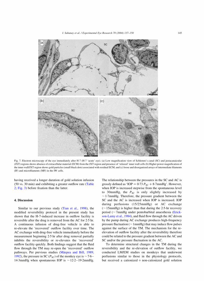
Fig. 7. Electron microscopy of the eye immediately after H-7 (H-7 ‘acute’ eye). (a) Low magnification view of Schlemm’s canal (SC) and juxtacanaicular
(JXT) regions shows absence of extracellular material (ECM) from the JXT region and presence of ‘relaxed’ inner wall cells (b) Higher power magnification of
the inner wall/JXT region shows gold particles (small black dots) associated with residual ECM, and (c) loose and disorganized arrays of intermediate filaments
(IF) and microfilaments (MF) in the IW cells.
I. Sabanay et al. / Experimental Eye Research 78 (2004) 137–150 145

infusion either during the AC exchange or immediately
before the fixation in some protocols. To facilitate flow of
gold particles and fixative into the TM, the perfusion
pressures were also elevated to ,25/35mmHg in both eyes
in the morphology protocols. Although the higher pressures
(25/35mmHg) may induce a stronger resistance washout
and possibly more apparent pressure-dependent changes in
the TM after the drug when compared to the lower pressures
(15/25mmHg) as in physiology protocols, they will not
affect the conclusions based on the difference between eyes
that were perfused at the same pressures. Unlike a previous
study (Ethier and Chan, 2001), no observable difference was
found in the distribution of cationized and non-cationized
gold particles in any eye. Both cationized and non-
cationized gold particles are not merely ‘fluid phase
markers’ but tend to bind to the ECM along the outflow
pathway (Sabanay, et al., 2000), which is of assistance
to the comparison in the TM morphology between
the H-7-treated ‘recovery’ eye and the contralateral
vehicle-treated eye or the contralateral H-7-treated ‘acute’
eye.
As described previously (Sabanay et al., 2000), the IW
cells in the vehicle-treated eye are in a ‘contracted’ state
with highly ordered arrays of intermediate filaments within
the cytoplasm; in the H-7-treated ‘acute’ eye, major
morphological changes in and around SC include protrusion
of the entire IW into the SC, relaxation of the IW cells and
reorganization of the IW cytoskeleton. The overall protru-
sion of the IW into the lumen, which seems to indicate that
the ‘relaxed’ IW after H-7 cannot resist the pressure, may
account, at least in part, for the increase in the area of the
drainage pathway. However, the same region in the H-7-
treated ‘recovery’ eye shows a non-uniform morphology
along the IW, with ‘contracted’ regions similar to those in
Fig. 8. Electron microscopy of H-7 ‘recovery’ eye—A ‘contracted’ region along Schlemm’s canal (SC). (a) Low magnification view of the SC and
juxtacanaicular (JXT) regions shows ‘re-appearance’ of extracellular material (ECM) deposits in the JXT region and presence of ‘contracted’ inner wall cells.
(b) Higher power magnification of the JXT region shows sparse gold particles (small black dots) associated with dense, apparently new ECM deposits. The
ECM-bound gold may be undergoing transcytosis, being transported from the basal to the lumenal aspect of the inner wall cells (arrowhead). (c) High power
magnification image shows tightly packed arrays of intermediate filaments in the inner wall endothelial cells and gold particles in vesicles (V) throughout inner
wall cells (arrow). The arrowhead indicates the transcytosis of gold particles as in (b).
I. Sabanay et al. / Experimental Eye Research 78 (2004) 137–150146

the vehicle-treated eye and ‘relaxed’ regions similar to those
in the H-7-treated ‘acute’ eye. Accordingly, the average J–J
distance in the IW of the H-7-treated ‘recovery’ eye is
intermediate between the vehicle-treated eye and the H-7-
treated ‘acute’ eye. It is not clear what determines the
‘contracted’ regions and the ‘relaxed’ regions in the
recovery stage. However, since our physiology data indicate
that a continuous infusion of drug-free vehicle or AC
exchange with drug-free vehicle immediately before the
measurement beginning 2·5 hr after drug removal is able to
re-elevate the ‘recovered’ outflow facility, different flow
rates or pressures in different regions in the TM during the
recovery period may be responsible for the non-uniform
morphology. Additionally, the ‘contracted’ cells may not
necessarily mean that the cells have completely recovered
from H-7-induced relaxation. These cells may ‘contract’ or
‘retract’ under the lower pressure/fluid flow rate during the
2·5 hr waiting period. However, they may more easily be
extended or expanded again under the higher pressure/fluid
flow rate after the perfusion is restarted than normal cells,
which may explain the progressive re-increase in outflow
facility shortly after re-starting perfusion with drug-free
Barany’s solution. The still ‘relaxed’ endothelial cells in the
TM of the H-7-treated ‘recovery’ eye may be related to the
effect of fluid flow through the TM during gold infusion
immediately before Ito’s fixation, but incomplete recovery
of H-7-induced cellular relaxation in the TM cannot be
excluded. Nevertheless, the re-‘contracted’ endothelial cells
could reduce the area of the drainage surface, and in turn
decrease outflow facility.
In the present, as well as previous (Sabanay et al., 2000)
study, ECM in the region along the basal aspects of the IW
cells of the H-7-treated ‘acute’ eye was markedly reduced,
compared to the vehicle-treated eye. This may indicate
either that H-7 increases the ECM washout from the JXT
region as previously proposed (Sabanay et al., 2000), or that
the IW cells in the H-7-treated ‘acute’ eye ‘balloon’ towards
the canal, leaving behind the ECM deposits or part of them
in more proximal areas of the outflow pathway. However, to
date we cannot state whether the ECM that was localized in
that region was transported to the SC or ‘diluted’ in more
proximal areas of the outflow pathway. Additionally, this
study also revealed some ‘re-appeared’ ECM deposits in the
JXT region of the H-7-treated ‘recovery’ eye. The ‘re-
appeared’ ECM in the H-7-treated ‘recovery’ eye seems
much more dense than the ECM in the vehicle-treated eye.
However, it is also not clear whether the ‘re-appearance’ of
ECM indicates that the ECM ‘diluted’ in more proximal
areas of the outflow pathway by ‘ballooned’ IW cells re-
condenses to its original position when the IW cells
Fig. 9. Electron microscopy of H-7 ‘recovery’ eye—A ‘relaxed’ region along Schlemm’s canal (SC). (a) Low magnification view of the SC and
juxtacanalicular (JXT) regions shows absence of extracellular material (ECM) in the JXT region and presence of ‘relaxed’ inner wall cells (asterisks). (b)
Higher power magnification of the JXT region shows many gold particles (small black dots) associated with residual ECM fibers. (c) High power magnification
image shows loose and disorganized arrays of intermediate filaments in the inner wall endothelial cells. Tight junctions (TJ) are unaffected. CAV, caveoli.
I. Sabanay et al. / Experimental Eye Research 78 (2004) 137–150 147

re-contract, or that new biosynthesis of ECM occurs during
the 2·5 hr waiting period, or both. Further studies are needed
to clarify this issue.
Resistance ‘washout’ refers to the progressive increase in
outflow facility that occurs during prolonged perfusion with
drug-free mock aqueous humour in both in vivo and
enucleated non-human eyes (Kaufman et al., 1988;
Erickson-Lamy et al., 1990). In the present study, we
noted that resistance washout in the contralateral vehicle-
treated eye was also reversible after the 2·5 hr recovery
period. The mechanisms for the reversibility of resistance
‘washout’ are not clear yet. Since wash-away of ECM from
the outflow pathway may be the mechanism of the
resistance ‘washout’ (Johnson et al., 1993; Kee et al.,
1996; Gual et al., 1997), a reasonable hypothesis could
be that the ECM newly synthesized during the 2·5 hr waiting
period may be involved in the reversibility of resistance
‘washout’. However, it is unknown if the ECM biosynthesis
could occur within 2–3 hr. Additionally, recent studies
show that washout may result from a loss of aqueous
Fig. 10. Histograms show the junction-to-junction distance ( (m) in the inner wall of Schlemm’s canal. The variability in cell size is partly attributable to the
variable orientation of the cells as well as to differences in cell contraction. Data in each panel is obtained from one relevant eye. Note the ‘relaxing’ effect of H-
7, with doubling of the average cell width in the H-7-treated ‘acute’ eye vs. the vehicle-treated eye, and the presence of ‘contracted regions’ (e.g. cells #10–40)
and ‘relaxed regions’ (e.g. cells #45–55) along the canal in the H-7-treated ‘recovery’ eye.
I. Sabanay et al. / Experimental Eye Research 78 (2004) 137–150148

funnelling through preferential channels, caused by
separation of the IW cells from the underlying JXT
connective tissue during perfusion (Johnson et al., 1992;
Overby et al., 2002). This suggests that the ECM wash-away
mechanism alone may not be sufficient to explain resistance
‘washout’, and that pressure-induced cellular structural
changes from the perfusion may be also involved. Since
there are some similar TM structural changes in the
perfusion-induced resistance ‘washout’ and the H-7-
induced outflow increase (e.g. ECM washout and separation
of the IW cells from the underlying JXT cells), and since the
effect of H-7 on outflow facility is pressure-dependent, one
might hypothesize that H-7 may simply accelerate the
resistance washout process. However, the key structural
change for H-7 to increase outflow facility is cellular
relaxation, which did not occur during drug-free perfusions.
Taken together, the present data support that the re-
‘contracted’ cells in the TM are the primary structural basis
for the reversibility of the H-7-induced increase in outflow
facility. This indicates that H-7 effects represent transient
alterations in the cellular contractility and cytoskeletal
organization rather than irreversible toxicity, important for a
potential anti-glaucoma medication since safety is a major
consideration. However, the reversibility may also indicate
that the effect of H-7 on outflow facility is short-lived, so
that H-7, as with all currently available anti-glaucoma
medications, may need frequent repetitive administrations
to maintain lower IOP. Nevertheless, H-7’s effect on
outflow facility may last longer in the glaucomatous eye
with elevated IOP, since as the IOP returns toward baseline
in the glaucomatous eye due to reduced concentration of H-
7 in the TM over time, aqueous humour driven by the high
pressure gradient between the AC and SC may reopen the
‘recovered’ outflow pathway, similar to the situation in the
facility measurement beginning 2·5 hr after drug removal in
the current experiments. Additionally, a single intracameral
injection of H-7 in glaucomatous eyes might wash out
abnormal ECM and produce long-lasting ocular hypoten-
sion, as a so-called one-time ‘pharmacologic trabeculoca-
nalotomy’ (Kaufman et al., 1979; Epstein, 1987; Kaufman,
1992). Moreover, recent advances in molecular genetics and
gene transfer technology (Borras et al., 1996; Liu et al.,
1999; Borras et al., 2001; Vittitow et al., 2002) may allow
long-term re-setting of these outflow regulatory mechan-
isms to therapeutic advantage in glaucoma. Thus, one can
imagine over or under-expressing a particular kinase in the
TM cells of the living eye so as to partially inhibit cellular
contractility and ‘loosen’ cell–cell or cell–ECM attach-
ments, thereby permanently increasing outflow facility and
reducing IOP.
Acknowledgements
This study was supported by grants from the US National
Eye Institute (EY02698), the American Health Assistance
Foundation, the Glaucoma Research Foundation, Research
to Prevent Blindness, the Wisconsin Alumni Research
Foundation, and the Ocular Physiology Research and
Education Foundation. B.G. is the incumbent of the
E. Neter Chair in Cell and Tumour Biology. The authors
thank Jennifer Seeman and Julie Kiland for conducting
cannulations of femoral artery and vein in monkeys.
References
Barany, E.H., 1964. Simultaneous measurement of changing intraocular
pressure and outflow facility in the vervet monkey by constant pressure
infusion. Invest. Ophthalmol. 3, 135–143.
Barany, E.H., 1965. Relative importance of autonomic nervous tone and
structure as determinants of outflow resistance in normal monkey eyes
(Cercopithecus ethiops and Macaca irus). In: Rohen, J.W. (Ed.), The
Structure of the Eye, Second Symposium, F.K. Schattauer, Stuttgart, pp.
223–236.
Bershadsky, A., Chausovsky, A., Becker, E., Lyubimova, A., Geiger, B.,
1996. Involvement of microtubules in the control of adhesion-
dependent signal transduction. Curr. Biol. 6, 1279–1289.
Birrell, G.B., Hedberg, K.K., Habliston, D.L., Griffith, O.H., 1989. Protein
kinase C inhibitor H-7 alters the actin cytoskeleton of cultured cells.
J. Cell Physiol. 141, 74–84.
Borras, T., Gabelt, B.T., Klintworth, G.K., Peterson, J.C., Kaufman, P.L.,
2001. Non-invasive observation of repeated adenoviral GFP gene
delivery to the anterior segment of the monkey eye in vivo. J. Gene
Med. 3, 437–449.
Borras, T., Tamm, E.R., Zigler, J.S., 1996. Ocular adenovirus gene transfer
varies in efficiency and inflammatory response. Invest. Ophthalmol.
Vis. Sci. 37, 1282–1293.
Epstein, D.L., 1987. Open angle glaucoma. Why not a cure? Arch.
Ophthalmol. 105, 1187–1188.
Erickson-Lamy, K.A., Kaufman, P.L., McDermott, M.L., France, N.K.,
1984. Comparative anesthetic effects of aqueous humor dynamics in the
cynomolgus monkeys. Arch. Ophthalmol. 102, 1815–1820.
Erickson-Lamy, K., Schroeder, A.M., Bassett-Chu, S., Epstein, D.L.,
1990. Absence of time-dependent facility increase (washout) in the
perfused enucleated human eye. Invest. Ophthalmol. Vis. Sci. 31,
2384–2388.
Ethier, C.R., Chan, D.W.-H., 2001. Cationic ferritin changes outflow
facility in human eyes whereas anionic ferritin does not. Invest.
Ophthalmol. Vis. Sci. 42, 1795–1802.
Gills, J.P., Roberts, B.C., Epstein, D.L., 1998. Microtubule disruption leads
to cellular contraction in human trabecular meshwork cells. Invest.
Ophthalmol. Vis. Sci. 39, 653–658.
Gual, A., Llobet, A., Gilabert, R., Borras, M., Pales, J., Bergamini, M.V.,
Belmonte, C., 1997. Effects of time of storage, albumin, and osmolality
changes on outflow facility (C) of bovine anterior segment in vitro.
Invest. Ophthalmol. Vis. Sci. 38, 2165–2171.
Johnson, M., Gong, H., Freddo, T.F., Ritter, N., Kamm, R., 1993. Serum
proteins and aqueous outflow facility resistance in bovine eyes. Invest.
Ophthalmol. Vis. Sci. 34, 3549–3557.
Johnson, M., Shapiro, A., Ethier, C.R., Kamm, R.D., 1992. Modulation of
outflow resistance by the pores of the inner wall endothelium. Invest.
Ophthalmol. Vis. Sci. 33, 1670–1675.
Kaufman, P.L., 1992. Pharmacologic trabeculocanalotomy. Facilitating
aqueous outflow by assaulting the meshwork cytoskeleton,
junctional complexes, and extracellular matrix. Arch. Ophthalmol.
110, 34–36.
Kaufman, P.L., Svedbergh, B., Lutjen-Drecoll, E., 1979. Medical
trabeculocanalotomy in monkeys with cytochalasin B or EDTA. Ann.
Ophthalmol. 11, 795–796.
I. Sabanay et al. / Experimental Eye Research 78 (2004) 137–150 149

Kaufman, P.L., True-Gabelt, B., Erickson-Lamy, K.A., 1988. Time-
dependence of perfusion outflow facility in the cynomolgus monkey.
Curr. Eye Res. 7, 721–726.
Kee, C., Gabelt, B.T., Gange, S.J., Kaufman, P.L., 1996. Serum effects on
aqueous outflow during anterior chamber perfusion in monkeys. Invest.
Ophthalmol. Vis. Sci. 37, 1840–1848.
Liu, X., Brandt, C.R., Gabelt, B.T., Bryar, P.J., Smith, M.E., Kaufman,
P.L., 1999. Herpes simplex virus mediated gene transfer to primate
ocular tissues. Exp. Eye Res. 69, 385–395.
Liu, X., Cai, S., Glasser, A., Volberg, T., Polansky, J.R., Fauss, D.J.,
Geiger, B., Kaufman, P.L., 2001. Effects of H-7 on cultured human
trabecular meshwork cells. Mol. Vis. 7, 145–153. http://www.molvis.
org/molvis/v7/a21/.
Maepea, O., Bill, A., 1989. The pressures in the episcleral veins.
Schlemm’s canal and the trabecular meshwork in monkeys: effects of
changes in intraocular pressure. Exp. Eye Res. 49, 645–663.
Maepea, O., Bill, A., 1992. Pressures in the juxtacanalicular tissue and
Schlemm’s canal in monkeys. Exp. Eye Res. 54, 879–883.
Overby, D., Gong, H., Qiu, G., Freddo, T., Johnson, M., 2002. The
mechanism of increasing outflow facility during washout in the bovine
eye. Invest. Ophthalmol. Vis. Sci. 43, 3455–3464.
Sabanay, I., Gabelt, B.T., Tian, B., Kaufman, P.L., Geiger, B., 2000. H-7
effects on structure and fluid conductance of monkey trabecular
meshwork. Arch. Ophthalmol. 118, 955–962.
Tian, B., Gabelt, B.T., Peterson, J.A., Kiland, J.A., Kaufman, P.L.,
1999. H-7 increases trabecular facility and facility after ciliary
muscle disinsertion in monkeys. Invest. Ophthalmol. Vis. Sci. 67,
293–295.
Tian, B., Kaufman, P.L., Volberg, T., Gabelt, B.T., Geiger, B., 1998. H-7
disrupts the actin cytoskeleton and increases outflow facility. Arch.
Ophthalmol. 116, 633–643.
Vittitow, J.L., Garg, R., Rowlette, L.L., Epstein, D.L., O’Brien, E.T.,
Borras, T., 2002. Gene transfer of dominant-negative RhoA increases
outflow facility in perfused human anterior segment cultures. Mol. Vis.
8, 32–44.
Volberg, T., Geiger, B., Citi, S., Bershadsky, A.D., 1994. Effect of protein
kinase inhibitor H-7 on the contractility, integrity, and membrane
anchorage of the microfilament system. Cell Motil. Cytoskeleton 29,
321–338.
Yu, J.C., Gotlieb, A.I., 1992. Disruption of endothelial actin microfilaments
by protein kinase C inhibitors. Microvasc. Res. 43, 100–111.
I. Sabanay et al. / Experimental Eye Research 78 (2004) 137–150150