Embed Size (px)
Citation preview

Research ArticleFunctional and Structural Details about the Fabella:What the Important Stabilizer Looks Like in the CentralEuropean Population
Nicole Helene Hauser, Sebastian Hoechel, Mireille Toranelli,Joerg Klaws, and Magdalena Müller-Gerbl
Department of Biomedicine, Musculoskeletal Research, University of Basel, Pestalozzistrasse 20, 4056 Basel, Switzerland
Correspondence should be addressed to Sebastian Hoechel; [email protected]
Received 18 January 2015; Accepted 5 March 2015
Academic Editor: Ilker Ercan
Copyright © 2015 Nicole Helene Hauser et al. This is an open access article distributed under the Creative Commons AttributionLicense, which permits unrestricted use, distribution, and reproduction in any medium, provided the original work is properlycited.
The posterolateral corner of the knee accommodating the fabella complex is of importance in orthopaedic surgery. Unfortunately,there is a lack of data in literature for clinical routine. Therefore, we investigated the fabella’s characteristics, biomechanical nature,and present histologic details. Of special interest were the fabella’s occurrence and position, calcium concentration as long-termload intake indicator, and the histology.Within our analysis, fabellae were found in 30.0% of all datasets, located on the upper part ofthe posterolateral femoral condyle.The region of fabella contact on this condyle showed a significantly lower calcium concentrationthan its surroundings. Histologically, the fabella showed no articular cartilage but a clearly distinguishable fabellofibular ligamentthat consisted of two bundles: one, as already described in literature inserted at the fibular tip, and another part newly describedon the top of the lateral meniscus. In its role of stabilizing the soft tissue structures of the posterolateral knee, the fabella seems toserve as suspension for the ligaments evolving from its base. Even though a joint formation of any kind is unlikely, the presence ofa fabella needs to be kept in mind during knee examination and any surgical procedures.
1. Introduction
The occurrences of patients with knee injuries are constantlyrising. Though injuries of the medial compartment are morefrequent, the consequence of a traumatic distress on thelateral side is more disabling, since the lateral compartmentis subjected to greater force during gait [1, 2]. In addition,possible posterolateral pathology may remain unrecognizedif injuries of the cruciate ligaments mask secondary findingsdue to their extensive symptoms [3, 4]. In its stabilizing func-tion, the posterolateral corner with its complex arrangementof muscles, tendons, and ligaments is of crucial importancefor the physiological function of the knee. It is suggestedthat untreated damage and therefore insufficient support ofthe posterior knee not only prolong the healing process,but also cause postsurgical failure after cruciate ligamentreconstruction [5, 6]. Since the injury occurs following directvarus force in external rotation of the tibia as well as suddenhyperextension of the knee, various case reports describe
traumata after car accidents with frontal impact not only tothe ligamentous structures, but also to the occasional fabellapresent [7–11]. To understand the possible injuries and repairmechanisms of the posterolateral corner, the arrangementand variation of anatomical structures must be considered. Aliterature review of the fabella is confusing, since informationabout the fabella complex is obtained by many differentresearch methods. Unfortunately, the derived results are stillcompared to each other which results in a mix of datawhich is difficult to handle. Despite the fact that informationabout the central European population is negligible [12],various numbers of Chinese and Japanese studies report theoccurrences of the fabella positioned in the lateral head ofthe gastrocnemius muscle and the possible structure of thefabellofibular ligament (FFL) [13, 14].
Tabira et al. report in 2013 the prevalence of the fabella tobe 68.6 percent (%) per knee in the elderly Japanese popula-tion. Includedwere bony and cartilaginous findings evaluated
Hindawi Publishing CorporationBioMed Research InternationalVolume 2015, Article ID 343728, 8 pageshttp://dx.doi.org/10.1155/2015/343728
2 BioMed Research International
by inspection and palpation within the lateral head of thegastrocnemius muscle [15]. On the contrary, Kawashima etal. report their results of 66% bony and cartilaginous fabellaepresent in a similar Japanese study per gastrocnemius headand not per knee [14]. In addition, some studies conducttheir focus on clinical radiographs which mainly take intoconsideration the prevalence of osseous fabellae [16]. Themain confusion arises, when these findings are comparedwith each other and the methods of examination are notclearly stated [17].
Despite the differences in these data, the descriptionof the endochondral ossification and the occurrence andstructure of the FFL is consistent. Running from the base ofthe fabella to the styloid process of the fibular head, it servesas a static stabilizer of the knee, which tenses in full extension[18]. It can be found in up to 80% of humans with a fabellapresent [16, 19, 20].
For the orthopaedic surgeon, this information providesideas about the posterolateral knee complex and helps totackle arising symptoms like pain and swelling in this area,especially if the interaction of the fabella and the postero-lateral femoral condyle (PLFC) is taken into account. Manyresearchers favour the idea of a fabellofemoral joint withtypically associated joint diseases like chondromalacia andosteoarthritis, which can be found in many case reports[7, 11, 21]. In progressive stages, the cartilage of the fabellais described as softened and fibrillated or even completelyabsent. In this case, the bare subchondral bone plate ofthe fabella comes into contact with the femoral condyleand leads to increasing posterolateral pain [22]. The grossanatomy of the proclaimed joint in a healthy state and theformation of the interacting surfaces are difficult to retrievefrom literature.Thedescription of the joint cavitywas done bygelatine-injection, which was not characterized any furtherand documented in black and white pictures in which themarkings hide the important areas. What can clearly beidentified is the distinctive impression on the PLFC causedby the fabella [14, 17]. Histologic images of the fabella andthe surrounding formation lack representation in literature.Most of the presentations are very small and reveal onlyfractions of the posterolateral aspect of the knee. In addition,the images available aremainly printed in black andwhite andare not connected to an overview presentation [17, 20, 23, 24].In these studies, the cartilage formation and the interactionwith the femoral condyle are not sufficiently described anddo not provide conclusive information about the anatomicalarrangement in order to evaluate and understand patientcases better.
Our goal was to (1) determine the incidence and positionof the fabella in a central European population and tobetter estimate clinical appearances. (2) Furthermore, wedescribe the biomechanical impact of the little sesamoid boneon its interaction with the femur in order to determine apossible pressure distribution. (3) Histologic demonstrationwill define the anatomical structures with special attentionto the ligamentous tissue and clarify the formation of theexisting cartilage in order to determine any possible jointformation. The arrangement of the structures of the postero-lateral knee will be shown in its entirety including all bony
surroundings. A stained overview presentation is not yetavailable in literature, since deficient bone decalcification hashampered the production of cuttings that include materialswith different rigidity.
2. Materials and Methods
2.1. Sample Collection. Four hundred knees of 200 Europeanswere included (cadaveric study group-corpses donated toscience and research, 99 men, 101 women; conventionaldatasets generated by computed tomography (CT), extendedknee position).Thedatawas acquired during investigations atthe Institute of Anatomy, University of Basel. The sample ageranged from20 to 104 years (mean: 75.8, SD: 19.43).Histologicprocedures were carried out selectively on five of the mostprominent and representative bony fabella samples.
2.2. Descriptive Quantitative Analysis. We evaluated CT-studies (SOMATOM 16, Siemens, Erlangen, Germany, 120-kilovolt, 180-milliampere-second, axial slices) with a slicethickness of 0.6 millimetres (mm) (only the bony fabellacould be registered with the CT-method).Three-dimensional(3D) reconstructions (ANALYZE 11.0, Biomedical ImagingResource,Mayo Foundation, Rochester, USA; VGStudioMax2.2, Heidelberg, Germany) were orientated in coronal poste-rior view for determination of the location of the fabella. Togain comparable data, a size-independent measurement gridsystemwas applied on the PLFC to allocate the correspondingcoordinates to the centre of the fabella (Figures 1(a)–1(c)).The determined location coordinates of every fabella weresuperimposed on one reconstructed 3D-sample of the PLFCwith respect to the anatomical orientation for final evaluation.
To determine the size of the fabella, the largest diameter(𝑥) was measured (SOMATOM 16, Siemens, Erlangen, Ger-many) in coronal posterior view, regardless of the anatomicalknee-axes. For the second dimension, the largest diameter ofthe corresponding perpendicular orientation (𝑦) was used.
2.3. Joint Impact Analysis. The method of CT-osteoabsorp-tiometry (CT-OAM) for assessment of the density distri-bution was used on the same 3D reconstructions of theconventional datasets from the descriptive analysis describedabove. The 3D reconstructions of the knee were dividedinto datasets of the fabella and the femoral component. Thesubchondral bone plate (SBP) of the PLFC and of the fabellaas the region of interest was arranged in a way to be incoronal frontal view. Using a “maximum intensity projection”the software (ANALYZE 11.0, Biomedical Imaging Resource,Mayo Foundation, Rochester, USA) projected themost densevoxels onto the surface and assigned them colours, wherethe highest density values (>1200 Hounsfield units) wererepresented in black and lower values in red, yellow, green,and blue (in descending order) [25, 26]. In accordance withthe density values displayed, phantom measurements led tothe calculation of the corresponding mineral content as anindication of the long-term load intake [27].
For statistical analysis, themean concentration of calciumhydroxyapatite/Ca
5(PO4)3(OH) (subsequently abbreviated

BioMed Research International 3
(a) (b)
15 10 5 0
5
10
15
20
25
30
(c)
Figure 1: 3D reconstructions for descriptive quantitative analysis. (a) 3D reconstruction of the distal femur, patella, and fabella in sagittalview. (b) 3D reconstruction of (a) positioned in posterior view. (c) Applied coordinate-grid system on the posterolateral femoral condyle fordetermination of fabellar position.
(a) (b)
(c) (d)
Figure 2: Method of CT-osteoabsorptiometry on 3D reconstructions to determine density distribution within the subchondral bone plate.(a) Distal femur in posterior view with fabella arranged in anterior view (subchondral bone plate of articular surface shown). (b) Subchondralbone plates shown with colour-coded density distribution (black, red, yellow, green, and blue = Hounsfield units in descending order in 200unit steps). (c) Fabella in original position on density distribution pattern. (d) Density distribution with marked region of fabella contact onthe posterolateral femoral condyle.
as CAHA) within the SBP of the fabella and the correspond-ing area of contact on the PLFC was evaluated. In addition,the mineral content of the SBP of both posterior femoralcondyles was measured for reference (Figures 2(a)–2(d)).
2.4. Histologic Imaging. For sectioning and staining, the fivelargest fabella samples were dissected and removed with theattached soft tissue structures as well as the correspondingPLFC.
The dissected tissue was treated to dehydration in ethanolstarting with 40% increasing to 100% over a time period of 25days. Afterwards, the initial defatting process was increased
using isopropyl alcohol and chloroform. After completion,the next step included methyl methacrylate (MMA) infil-tration for 3 days at a storage temperature of 4∘ Celsius.Following this step of infiltration and mixing of chemicals,the resulting solutionwas exchanged for pureMMA again forthe final embedding at 4∘ Celsius.The time of polymerizationwas in accordance with the size of the sample and lastedapproximately one month. For all further steps, the hardenedMMA blocks embedding the bone samples were used.
The sectioning in sagittal anatomical orientation wasperformed using a diamond wheel saw with 400-micrometre(𝜇m) saw band thickness. The resulting slices (thickness:
4 BioMed Research International
Table 1: Fabella occurrence in accordance with selected age groups.
Distribution of fabella occurrence
Age group 𝑛∗ Male Female Number of
knees Fabellae present Bilat./Unilat. per𝑁∗∗ Percentage perage group (%)
Relativedistribution
20–29 5 3 2 10 0 0 0.00 0.0030–39 5 4 1 10 2 B-1 1.90 0.3840–49 17 11 6 34 9 B-4; U-1 8.57 0.5050–59 8 4 4 16 2 B-1 1.90 0.2460–69 14 10 4 28 6 B-3 5.71 0.4170–79 38 20 18 76 17 B-8; U-1 16.19 0.4380–89 65 30 35 130 41 B-17; U-7 39.05 0.6090–99 45 17 28 90 28 B-11; U-6 26.67 0.59100–109 3 0 3 6 0 0 0.00 0.00Sum 200 99 101 400 105 B-45; U-15 100.00∗CT-studies of both knees.∗∗B: bilateral; U: unilateral.
600𝜇m) were fixed on white, light-transmissive object hold-ers for further processing. To accomplish optimal stainingconditions, the slides were ground down to 200𝜇m andpolished. Staining methods obtained the following.
(i) Toluidine blue epoxy staining of 3 𝜇m [28], forbasophil structures to acquire different shades of bluewhere calcified cartilage shows the darkest shade,
(ii) Trichrome Masson-Goldner surface staining of 3 𝜇m[29], wheremineralized bone and collagen are stainedgreen, calcified cartilage is stained light green, andmuscle tissue and cytoplasm are stained in differ-ent shades of red. The resulting histologic sliceswere documented for inspection (20.5 : 1 Zoom andFusionOptics Technology Leica M205 C; Canon EOS40D).
2.5. Statistical Analysis. Continuous variables were expressedwith mean, standard deviation, and minimum-maximumvalues where categorical variables were reported as frequencyand related percentage. Independent samples t-test was per-formed between group comparisons. Linear regression anal-ysis was performed for modelling the relationship betweenPLFC, FAS, and ROFC. All age group data were testedfor normalcy and homogeneity using Kolmogorov-Smirnovtests. For the gender distribution analysis, the unpaired two-sample t-test was used.
All statistical analyses were done using RStudio (RStudio:integrated development environment for R, Version 0.96.122,Boston, MA, USA). The significance level for all statisticaltests was set a priori to <0.001.
3. Results
3.1. Descriptive Quantitative Analysis. 30.0% of all CT-studies(each CT-study of one human included the left and rightknee) presented with 105 bony fabellae overall where thebilateral to unilateral occurrence ratio was 3 : 1 (bilateral: 45;unilateral: 15). The relative occurrence showed no significant
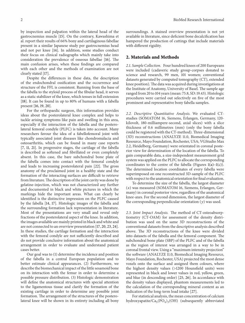
Figure 3: Results of descriptive quantitative analysis. Colour-codedfabella positions on posterolateral femoral condyle (red, yellow, andgreen; in descending order).
(𝑃 > 0.001) difference between the assigned age groups,where fabellae were present (mean occurrence: 23.63%; min:12.5%; max: 31.54%; SD: 6.71%). The data did not reveal anydifference in gender distribution (fabellae in male versusfabellae in female: 1 : 1; 𝑃 = 0.453) (Table 1). The fabella waspositioned invariably over the PLFC and in close relationto its lateral border (Figure 3). The measured sizes of theanalysed fabellae ranged from (𝑥) 4.84mm, (𝑦) 3.63mm to(𝑥) 13.12mm, (𝑦) 11.71mm.
3.2. Joint Impact Analysis. The CAHA concentration of theposteromedial femoral condyle (mean: 461.14mg/mL; min:282.66mg/mL; max: 656.63mg/mL; SD: 112.93mg/mL) wassignificantly (𝑃 < 0.001) higher compared to the PLFC(mean: 402.59mg/mL; min: 260.16mg/mL; max: 577.28mg/mL; SD: 92.82mg/mL), hosting the fabella. The measuredconcentration of the region of fabella contact (ROFC) onthe PLFC was significantly (𝑃 < 0.001) lower (mean:336.77mg/mL; min: 198.23mg/mL; max: 521.98mg/mL; SD:91.20mg/mL) than the mean value measured over the wholePLFC itself.Themineral content of the ROFC, in comparison

BioMed Research International 5
180.0
230.0
280.0
330.0
380.0
430.0
480.0
530.0
200.0 400.0 600.0 800.0
Calc
ium
hyd
roxy
apat
ite co
ncen
trat
ion
ofre
gion
of f
abel
la co
ntac
t
Calcium hydroxyapatite concentration ofposterolateral femoral condyle;
fabellar articular surface
(a)
150
250
350
450
550
650
750
Calc
ium
hyd
roxy
apat
ite co
ncen
trat
ion
Poste
rom
edia
lfe
mor
al co
ndyl
e
fem
oral
cond
yle
Poste
rola
tera
l
Regi
on o
ffa
bella
cont
act
Fabe
llar
artic
ular
surfa
ce
(b)
Figure 4: Results of calcium hydroxyapatite analysis of the subchondral bone plates. (a) Level of calcium hydroxyapatite (mg/mL) of thesubchondral bone plates of the articular surfaces of interest. (b) Dependency of the evaluated data of the region of fabella contact on theposterolateral femoral condyle (𝑦; mg/mL) to the concentration on the mean posterolateral femoral condyle and the fabellar articular surface(𝑥; mg/mL).
to the mineral content on the whole PLFC, showed a meandifference of −16.59% (min: −1.77%; max: −33.53%).
The fabellar articular surface (FAS) had the highestCAHA concentration (mean: 487.09mg/mL;min: 259.29mg/mL; max: 778.15mg/mL; SD: 142.99mg/mL) which was onthe same level as seen in other joints of the human body(Figure 4(a)).
The linear regression (of CAHA concentration) of theROFC is dependent on the PLFC and can be interpreted as
(i) ROFC=0.878×PLFC− 16.843 (𝑅2 =0.80;𝑃 < 0.001).
As for the CAHA concentration of the ROFC which isdependent on the FAS:
(i) ROFC = 0.546 × FAS + 70.79 (𝑅2 = 0.73; 𝑃 < 0.001)(Figure 4(b)).
3.3. Histologic Imaging. Represented on the sagittal sectionsof the posterolateral corner of the knee, the macroscopicimages involve all corresponding bones (Figures 5(A) and5(B)). The fabella was located within the lateral gastrocne-mius tendon. The collagen fibres can be found along theanterior and posterior sides of the fabella, joining again toform the muscle-tendon junction at its base. The concaveimprint on the articular cartilage of the femur induced bythe fabella is formed in the topmost region of the articularcartilage of the PLFC (Figure 5; arrow). The FFL originatingfrom the base of the fabella crosses over the popliteal tendonand is inserted at the styloid process at the tip of the fibula. Asecond bundle of the FFL can be identified which separatesfrom the main bundle and is inserted at the topmost rim ofthe lateral meniscus.
The magnified image (Figure 6) demonstrated differentzones of the femoral articular cartilage with its subsequenttidemark, calcified cartilage, and the SBP.The correspondingsurface of the fabella is composed of collagen fibres originat-ing from the gastrocnemius tendon. Just below these longi-tudinal structures, an unmineralized fibrocartilage followedby tidemark andmineralized fibrocartilage is distinguishable.The medullary cavity within the fabella consists of a clearlydefined trabecular network including osteocytes. The accu-mulation of unmineralized and mineralized fibrocartilage inthe middle part of the anterior side of the fabella demon-strates the beginning of pathologic thickening.
4. Discussion
The frequency of occurrence of the fabella is discussed indifferent ways in literature. While Kawashima et al. [14]report cartilage and bony fabellae to be present in 66% of150 gastrocnemius heads, the comparing paper of Tabiraet al. [15] calculated their 68.6% per knee. Another paper,referring to Kawashima et al., quoted them with 92%, anumber which does not appear in the paper at all [17].The confusion arises from different mathematic procedures,calculated either per person, per knee, or per gastrocnemiushead.The interpretation of the described differences ismainlybased on the state and formation of the fabella, classified aseither bony and cartilaginous or soft and hard [14–17, 20, 24].CT-data will only show bony samples, whereas dissectionmay derive both and enlarge the number of findings [4]. Theso-called commonly known fact that a fabella ossifies at 3years of age confronts the idea of an induced ossification withaging [6, 14]. Following the data of Minowa et al. who found

6 BioMed Research International
∗
a
b cd
ef
h
i
k
(A)
l
g
c
l
a
b
ef
g
(B)
hi
k
d
∗
5mm
Figure 5: Histologic imaging of posterolateral corner of theknee (sagittal section). (A) Toluidine blue staining. (B) TrichromeMasson-Goldner staining. On both (A) and (B): a: gastrocnemiustendon; b: posterolateral femoral condyle; c: femur condyle impres-sion; d: bony fabella; e: collagen fibres of fabellofibular ligament;f: muscle cells of lateral gastrocnemius head; g: femoral articularcartilage; h: lateral meniscus; i: popliteus muscle; k: lateral condyleof tibial plateau; l: fibular head; ∗second bundle of the FFL.
1000 𝜇m
a
b
cd
fg
h
l ki
e
Figure 6: Fusion ofmagnified histological sections (1000𝜇m; Tolui-dine blue staining). a: gastrocnemius tendon; b: femoral articularcartilage; c: medullary cavity of fabella; d: trabecular bone; e: min-eralized fibrocartilage; f: osteocytes; g: tidemark; h: unmineralizedfibrocartilage; i: collagen fibres of the fabellofibular ligament; k:muscle cells with nuclei; l: fibrocyte.
bony fabellae in fetuses already, one has to rethink about theossification timeframe mentioned above. To provide reliabledata of the central European population that adds to thepresent state in literature and describes details of the fabellafor clinicians, we limited our study to CT-recognizable, bonyappearances as they will be the ones discovered in clinical
Figure 7: Density distribution of subchondral bone plates of thefabella and femur.Distribution patternwithmarked region of fabellacontact on the posterolateral femoral condyle.
routine. Our findings are in accordance with the commonlyunderstood 30% of fabellae present [16]. Within our study,the distribution of occurrence proved to be quite consistentregarding the patients’ age. It is for sure that intrinsic geneticfactors as well as extrinsic epigenetic stimuli trigger theossification of this sesamoid bone.One interpretation that it isdue to the aging process is not supported by our data. Reasonsfor the absence of fabellae in the age groups 20–29 and 100–109 will presumably be the limited number of CT-datasets.
All evaluated bony fabella samples were situated withinthe tendon of the lateral gastrocnemius muscle and in closerelation to the lateral border of the PLFC. In contrast tothe literature, we observed the majority located within thesuperior lateral area. Despite the previous reported mainlocation of the fabella the inferior lateral area of the PLFC[14, 15], we only found about 30% of all fabellae located there.
The data of the long-term loading history evaluated withthemethod of CT-OAM revealed surprising results. Since themineralization distribution of the SBP changes in adaptationto the long-term load intake of a joint by CAHA integrationand degradation and correlates directly with its mechanicalstrength, the distribution of themineral content within a jointsurface can be regarded as a reflection of the load intakeover time and represents the loading history [25, 30–32]. Dueto the fact that the interacting parts of the femoral condyleand the fabella are described as a joint, we expected joint-loading to be represented in the density distribution pattern[14]. The PLFC, however, showed to be less mineralized inthe ROFC than the rest of the evaluated area. The contourof the fabella itself is even recognizable as it is coded in adifferent colour resembling a lower mineral content (Figures7 and 2(d)). A joint formation between the correspondingbones does not seem to exist here. In addition, the histologicpictures support these findings with the absence of articularcartilage and a cover of collagen fibres on the fabella. TheSBP of the fabella meanwhile shows a mineral content thatis on a comparable level to other joints [27]. The fact of thelower load uptake of the ROFC leads us to take the knee-biomechanics into account for explanation.Within its fabellacomplex, the sesamoid bone serves as the combined originof the oblique popliteal ligament, arcuate ligament, and theFFL as well as the plantaris muscle. All these structures fix the

BioMed Research International 7
sesamoid bone in its position within the gastrocnemius head.Due to the rollback of the lateral femoral condyle duringknee flexion, the PLFC moves on the tibial plateau over agreater distance than themedial one. From 120∘ of flexion, thelateral femoral condyle moves 23mm in anterior directionuntil −5∘ of extension. On the medial side, the contact pointonly moves 3mm [19]. This kinematic condition producesmore tensile stress on the lateral head than on the medialone which might serve as an extrinsic epigenetic stimulus totrigger the calcification but surely separates the PLFC fromthe fabella and reduces the impact as shown.
The histologic images in their representation of theposterolateral knee complex add to the current available datain literature, since they include a stained overview, as wellas detailed information about the structural compositionof the bony components as well as soft tissue. In additionto the already described imprint of the fabella onto thefemoral articular cartilage, the tendon of the gastrocnemiusmuscle can clearly be identified surrounding the fabella andembedding it. Articular cartilage on the fabella is missing.In addition to the already in literature described FFL (orig-inating from the base of the fabella and being inserted at thestyloid process of the fibula), a second bundle of collagenfibres is present. Also originating at the base, it separates fromthe main FFL and is inserted at the topmost corner of thelateral meniscus (Figures 5(A) and 5(B)) [18]. A posteriorfixation of the lateral meniscus is therefore possible throughthis ligamentous bundle.This constellation, however, forms acapsule-like surrounding of the fabella which might be theexplanation for the observed articular cavity described byKawashima et al. [14].
Possible limitations to this study are seen in the patho-logic alterations of the histologic samples. An osteoarthriticprocess can be found in the middle part of the fabella. Sincethe continuity of the collagen fibres is clearly visible in itsfull extent, we nevertheless regard this information to berepresentative.
5. Concluding Remarks
A fabella is present within the posterolateral knee complexin 30.0% of the European population and needs to bedistinguished from any fracture parts suspected within thisarea. In its function of supporting the soft tissue structures,it imprints on the articular cartilage of the PLFC in closerelation to its lateral border where it is constantly found inCT-datasets if present. Although this close relation createdan imprint on the femoral articular cartilage that provesinteraction between the fabella and the PLFC, the SBP of thefemoral part does not reveal any signs of long-term loadingfrom the fabella in this area. The fabella itself shows no signof articular cartilage. Instead, it is isolated from the femur,just being surrounded by fixating collagen fibres originatingfrom the lateral head of the gastrocnemius muscle. In itsrole of stabilizing soft tissue structures, it seems to serve assuspension for the ligament evolving from its base. Despiteprevious descriptions of this FFL running distally and beinginserted at the styloid process, we clearly identified a second
bundle inserted into the top rim of the lateral meniscus,whichwe assume providesmechanical support and a possibleback-tracking of the lateral meniscus during its slidingmovement on the lateral tibial condyle. Certainly, within thecomplex field of traumatic knee injuries, a distortion withdamage to the lateral meniscus is bound to damage thisligamentous structure as well. Next to the described fabella,the FFL with its second, meniscal attached bundle needs tobe kept in mind during knee examination.
Conflict of Interests
The authors declare that they have no conflict of interests.Furthermore, the authors received no grant or sources offinancial support related to the topic or topics of this paper.
Authors’ Contribution
NicoleHeleneHauser andMagdalenaMuller-Gerbl designedthe study and collected the data. Joerg Klaws and Sebas-tian Hoechel developed the methodology. Mireille Toranellicontributed the histology work. Nicole Helene Hauser andSebastianHoechel wrote the paper.NicoleHeleneHauser andSebastian Hoechel contributed equally to this work.
Acknowledgments
The authors would like to thank Mrs. Christine Muller-Thompson for the linguistic correction and final proof. Fur-thermore, they appreciate the help ofMr. Peter Zimmermannand his kind contribution of ideas towards the histologicstaining as well as Mr. Roger Kurz for his support with therequired preparation.
References
[1] J. A. Nicholas, “Acute and chronic lateral instabilities of theknee: diagnosis, characteristics, and treatment,” in Proceedingsof the AAOS Symposium on Reconstructive Surgery of the Knee,Mosby, St. Louis, Mo, USA, May 1978.
[2] W. Muller, R. Biedert, F. Hefti, R. P. Jakob, U. Munzinger, andH. U. Staubli, “OAK knee evaluation. A new way to assess kneeligament injuries,” Clinical Orthopaedics and Related Research,no. 232, pp. 37–50, 1988.
[3] D. M. Veltri and R. F. Warren, “Posterolateral instability of theknee,”The Journal of Bone and Joint Surgery—AmericanVolume,vol. 76, no. 3, pp. 460–472, 1994.
[4] R. F. Laprade, C. J. Griffith, B. R. Coobs, A. G. Geeslin, S. Johan-sen, and L. Engebretsen, “Improving outcomes for posterolat-eral knee injuries,” Journal of Orthopaedic Research, vol. 32, no.4, pp. 485–491, 2014.
[5] S. J. O’Brien, R. F. Warren, H. Pavlov, R. Panariello, and T.L. Wickiewicz, “Reconstruction of the chronically insufficientanterior cruciate ligament with the central third of the patellarligament,” The Journal of Bone & Joint Surgery—AmericanVolume, vol. 73, no. 2, pp. 278–286, 1991.
[6] R. F. LaPrade, S. Johansen, J. Agel, M. A. Risberg, H. Moksnes,and L. Engebretsen, “Outcomes of an anatomic posterolateralknee reconstruction,” The Journal of Bone and Joint SurgerySeries A, vol. 92, no. 1, pp. 16–22, 2010.

8 BioMed Research International
[7] A. Robertson, S. C. E. Jones, R. Paes, and G. Chakrabarty, “Thefabella: a forgotten source of knee pain?” The Knee, vol. 11, no.3, pp. 243–245, 2004.
[8] F. Franceschi, U. G. Longo, L. Ruzzini et al., “Dislocation ofan enlarged fabella as uncommon cause of knee pain: a casereport,”The Knee, vol. 14, no. 4, pp. 330–332, 2007.
[9] J. Y. Tang, H. Mulcahy, and F. Chew, “High-energy fracture ofthe fabella,” Radiology Case Reports, vol. 5, no. 4, 2010.
[10] G. M. Heideman, K. E. Baynes, A. P. Mautz, M. S. DuBois, andJ. W. Roberts, “Fabella fracture with CT imaging: a case report,”Emergency Radiology, vol. 18, no. 4, pp. 357–361, 2011.
[11] A. R. F. Barreto, F. A. Chagas-Neto,M.D. Crema et al., “Fractureof the fabella: a rare injury in knee trauma,” Case Reports inRadiology, vol. 2012, Article ID 390150, 3 pages, 2012.
[12] O. Raheem, J. Philpott, W. Ryan, and M. O’Brien, “Anatomicalvariations in the anatomy of the posterolateral corner of theknee,” Knee Surgery, Sports Traumatology, Arthroscopy, vol. 15,no. 7, pp. 895–900, 2007.
[13] Z. Dannawi, V. Khanduja, K. K. Vemulapalli, J. Zammit, andM.El-Zebdeh, “Arthroscopic excision of the fabella,”The Journal ofKnee Surgery, vol. 20, no. 4, pp. 299–301, 2007.
[14] T. Kawashima, H. Takeishi, S. Yoshitomi, M. Ito, and H. Sasaki,“Anatomical study of the fabella, fabellar complex and itsclinical implications,” Surgical and Radiologic Anatomy, vol. 29,no. 8, pp. 611–616, 2007.
[15] Y. Tabira, T. Saga, N. Takahashi, K. Watanabe, M. Nakamura,and K.-I. Yamaki, “Influence of a fabella in the gastrocnemiusmuscle on the common fibular nerve in Japanese subjects,”Clinical Anatomy, vol. 26, no. 7, pp. 893–902, 2013.
[16] G. C. Terry and R. F. LaPrade, “The posterolateral aspect of theknee: anatomy and surgical approach,”The American Journal ofSports Medicine, vol. 24, no. 6, pp. 732–739, 1996.
[17] S.-X. Zeng, X.-L. Dong, R.-S. Dang et al., “Anatomic study offabella and its surrounding structures in a Chinese population,”Surgical and Radiologic Anatomy, vol. 34, no. 1, pp. 65–71, 2012.
[18] P. Tyler, A. Datir, and A. Saifuddin, “Magnetic resonance imag-ing of anatomical variations in the knee. Part 1: ligamentous andmusculotendinous,” Skeletal Radiology, vol. 39, no. 12, pp. 1161–1173, 2010.
[19] Y. C. Kim, I. H. Chung, W. K. Yoo, J.-S. Suh, S. J. Kim, andC. I. Park, “Anatomy and magnetic resonance imaging of theposterolateral structures of the knee,” Clinical Anatomy, vol. 10,no. 6, pp. 397–404, 1997.
[20] T. Minowa, G. Murakami, H. Kura, D. Suzuki, S. H. Han, andT. Yamashita, “Does the fabella contribute to the reinforcementof the posterolateral corner of the knee by inducing thedevelopment of associated ligaments?” Journal of OrthopaedicScience, vol. 9, no. 1, pp. 59–65, 2004.
[21] R. Goldenberg and E. L. Wild, “Chondromalacia fabellae,” TheJournal of Bone and Joint Surgery (American Volume), vol. 24,no. 3, pp. 688–690, 1952.
[22] D. S.Weiner and I.Macnab, “The ‘fabella syndrome’: an update,”Journal of Pediatric Orthopaedics, vol. 2, no. 4, pp. 405–408,1982.
[23] J. G. Silva, C. A. A. Chagas, D. F. M. Torres, L. Servidio, A. C.Vilela, andW. A. Chagas, “Morphological analysis of the fabellain Brazilians,” International Journal of Morphology, vol. 28, no.1, pp. 105–110, 2010.
[24] P. Phukubye and O. Oyedele, “The incidence and structureof the fabella in a South African cadaver sample,” ClinicalAnatomy, vol. 24, no. 1, pp. 84–90, 2011.
[25] M. Muller-Gerbl, R. Putz, N. Hodapp, E. Schulte, and B. Wim-mer, “Computed tomography-osteoabsorptiometry for assess-ing the density distribution of subchondral bone as ameasure oflong-term mechanical adaptation in individual joints,” SkeletalRadiology, vol. 18, no. 7, pp. 507–512, 1989.
[26] M. Muller-Gerbl, R. Putz, N. Hodapp, E. Schulte, and B.Wimmer, “Demonstration of subchrondral density patterns byCT osteoabsorptiometry (CT-OAM) for in vivo assessment ofindividual stresses in joints,” Zeitschrift fur Orthopadie und ihreGrenzgebiete, vol. 128, no. 2, pp. 128–133, 1990.
[27] M. Muller-Gerbl, “The subchondral bone plate,” Advances inAnatomy, Embryology, and Cell Biology, vol. 141, pp. 1–134, 1998.
[28] B. F. Trump, E. A. Smuckler, and E. P. Benditt, “A methodfor staining epoxy sections for light microscopy,” Journal ofUltrasructure Research, vol. 5, no. 4, pp. 343–348, 1961.
[29] J. Goldner, “A modification of the masson trichrome techniquefor routine laboratory purposes,” The American Journal ofPathology, vol. 14, no. 2, pp. 237–243, 1938.
[30] F. Pauwels, Gesammelte Abhandlungen zur funktionellenAnatomie des Bewegungsapparates, Springer, Berlin, Germany,1965.
[31] D. R. Carter, T. E. Orr, and D. P. Fyhrie, “Relationships betweenloading history and femoral cancellous bone architecture,”Journal of Biomechanics, vol. 22, no. 3, pp. 231–244, 1989.
[32] S. Hoechel, D. Wirz, and M. Muller-Gerbl, “Density andstrength distribution in the human subchondral bone plate ofthe patella,” International Orthopaedics, vol. 36, no. 9, pp. 1827–1834, 2012.