Embed Size (px)
Citation preview
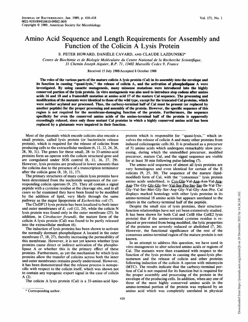
Vol. 171, No. 1
Amino Acid Sequence and Length Requirements for Assembly andFunction of the Colicin A Lysis Protein
S. PETER HOWARD., DANIELE CAVARD, AND CLAUDE LAZDUNSKI*
Centre de Biochimie et de Biologie Moleculaire du Centre National de la Recherche Scientifique,31 Chemin Joseph Aiguier, B.P. 71, 13402 Marseille Cedex 9, France
Received 15 July 1988/Accepted 8 October 1988
The roles of the various parts of the mature colicin A Iysis protein (Cal) in its assembly into the envelope andits function in causing "quasi-lysis," the release of colicin A, and the activation of phospholipase A wereinvestigated. By using cassette mutagenesis, many missense mutations were introduced into the highlyconserved portion of the lysis protein. In vitro mutagenesis was also used to introduce stop codons after aminoacids 16 and 18 and a frameshift mutation at amino acid 17 of the mature Cal sequence. The processing andmodification of the mutants were identical to those of the wild type, except for the truncated Cal proteins, whichwere neither acylated nor processed. Thus, the carboxy-terminal half of Cal must be present (or replaced byanother peptide) for the proper processing and assembly of the protein. However, the specific sequence of this
region is not required for the membrane-damaging function of the protein. Furthermorc, the sequencespecificity for even the conserved amino acids of the amino-terminal half of the protein is apparentlyexceedingly relaxed, since only those mutant Cal proteins in which a highly conserved amino acid has beenreplaced by a glutamate were impaired in their function.
Most of the plasmids which encode colicins also encode asmall protein, called lysis protein (or bacteriocin releaseprotein), which is required for the release of colicins fromproducing cells to the extracellular medium (6, 11, 12, 24, 26,28, 30, 31). The genes for these small, 28- to 33-amino-acidproteins form an operon with the colicin structural gene andare coregulated under SOS control (8, 11, 16, 27, 29).However, lysis proteins are produced in lower amounts thanthe colicins due to the presence of a transcription terminatorafter the colicin gene (8, 10, 11, 17).The primary structures of many colicin lysis proteins have
been determined from the nucleotide sequences of the cor-responding colicin operons (9, 25). They all contain a signalpeptide with a cysteine residue at the cleavage site, and in allcases so far examined they have been found to be modifiedby the addition of lipid, very likely through the samepathway as the major lipoprotein of Escherichia coli (7).The CloDF13 lysis protein has been localized to both inner
and outer membranes of E. coli (11, 24), while the colicin Nlysis protein was found only in the outer membrane (25). Inaddition, in Citrobacter freundii, the mature form of thecolicin A lysis protein (Cal) was found to be partly releasedinto the extracellular medium (6).The induction of lysis proteins has been shown to activate
the normally dormant phospholipase A located in the outermembrane (7, 18, 27), thereby increasing the permeability ofthis membrane. However, it is not yet known whether lysisproteins cause direct or indirect activation of the phospho-lipase A or whether this is the primary effect of theseproteins. Furthermore, as yet the mechanism by which lysisproteins allow the transfer of colicins across both the innerand outer membranes remains poorly understood. However,it has been demonstrated that the release process is nonspe-cific with respect to the colicin itself, which was shown notto contain any topogenic export signal in the case of colicinA (2).The colicin A lysis protein (Cal) is a 33-amino-acid lipo-
* Corresponding author.
protein which is responsible for "quasi-lysis," which in-volves the release of colicin A and many other proteins frominduced colicinogenic cells (6). It is produced as a precursorof 51 amino acids which undergoes remarktzbly slow proc-essing, during which the unmodified precursor, modifiedprecursor, mature Cal, and the sigpal sequence are visiblefor at least 30 min following pulse-labeling (7).The amino acid sequences of almost all lysis proteins are
very homologous and even identical for certain of the Ecolicins (9, 25, 30). The sequence of the mature (lipid-modified) form of Cal, with the "consensus" lysis proteinamino acids underlined, is Cys-Gln-Val-Asn-Asn-Val-ArgA-Thr-Gly-Gly-Gly-Ser-Val-Ser-Pro-Ser-S.r-Ile-Val-Thr-Gly-Val-Ser-Met-Gly-Ser-Asp-Gly-Val-Gly-Asn-Pro. Caldisplays marked homology with other lysis proteins in itsamino-terminal 18 amino acids but appears unrelated to theothers in the carboxy-terminal half of the peptide.
Despite the small size of lysis proteins, their structure-function relationships have not yet been extensively studied.It has been shown for both Cal and CelB (the ColE2 lysisprotein) that if the amino-terminal cysteine residue is re-placed or prevented from being modified, the lytic propertiesof the proteins are severely reduced or abolished (7, 26).However, the functional significance of the rest of theconsensus amino-terminal region of the mature protein is notknown.
In an attempt to address this question, we have used invitro mutagenesis to alter selected amino acids or regions ofCal. The mutants were then examined with respect to thefunction of the lysis protein in causing the quasi-lysis phe-nomenon and the release of colicin and other proteinsfollowing induction of the colicin A operon with mitomycin(MTC). The results indicate that the carboxy-terminal por-tion of Cal is not required for its function but is required forthe proper assembly and processing of the protein in theenvelope of the producing cells. In addition, when any one ofthree of the most highly conserved amino acids in theamino-terminal portion of the protein was replaced by anamino acid which was completely unrelated in physical
410
JOURNAL OF BACTERIOLOGY, Jan. 1989, p. 410-4180021-9193/89/010410-09$02.00/0Copyright © 1989, American Society for Microbiology
Dow
nloa
ded
from
http
s://j
ourn
als.
asm
.org
/jour
nal/j
b on
20
Dec
embe
r 20
21 b
y 14
.36.
241.
162.
REQUIREMENTS FOR LYSIS PROTEIN ASSEMBLY AND FUNCTION
properties, quasi-lysis and the release of colicin A werefound to be unaffected except when the introduced aminoacid carried a negative charge.
MATERIALS AND METHODS
Bacterial strains and plasmids. E. coli K-12 strains W3110and JE5505 lpp pps his proA argE thi gal xyl mtl tsx wereused as the host strains in all experiments involving thesynthesis and release of colicin A. JM105 thi rpsL endAsbc-15 hsdR4 A(lac-pro) (F' traD36 proAB lacIqZAM15)(32), JM105 made mutL by P1 transduction from BMH 71-18mutL (14), M13mpl8 amIV (4), and C600 thr leu thi lacYlsupE44 (1) were used for mutagenesis and recombinant DNAprocedures.The plasmid pAE11 (7), derived from pColA9 (16), was
used as the source of the cal gene for in vitro mutagenesis.Growth conditions. Strains were routinely grown in LB
medium. For radiolabeling of proteins with [35S]methionineor [35S]cysteine, they were grown in M9 medium supple-mented with thiamine (1 ,ug/ml), glycerol (0.4%), requiredamino acids (50 ,ug/ml), and methionine assay medium(0.5%) or cysteine assay medium (0.5%) (Difco Laborato-ries, Detroit, Mich.). The same medium was used for [2-3H]glycerol labeling, except that it contained 0.4% lactate asthe carbon source.MTC was used as the inducer of the colicin A operon at
300 ng/ml, in cultures having an OD600 of 1. All cultures wereincubated at 37°C with good aeration.
Labeling experiments. All labeling was performed after 45min of MTC treatment. Cells were labeled with [35S]me-thionine (45 ixCi/ml, 1,350 Ci/mmol) for 1 min and chasedwith 500 p,g of unlabeled methionine per ml. [35S]cysteinewas added at a concentration of 25 ,uCi/ml (1,200 Ci/mmol)for 2 min in the presence of methionine (25 ,ug/ml) andchased with unlabeled cysteine (250 jig/ml). Lipoproteinswere labeled with [2-3H]glycerol (133 ,uCi/ml, 1 Ci/mmol) for90 min without a chase. After various periods of chase asnoted in the figure legends, 125-,ul samples were precipitatedwith an equal volume of 25% ice-cold trichloroacetic acidand washed with 90% acetone before being solubilized insample buffer in preparation for electrophoresis. For analy-sis of phospholipids, cells were prelabeled by overnightgrowth in LB containing 10 ,uCi of [U-_4C]acetate (57 mCi/mmol) per ml. The cells were then diluted to an OD6o0 of 0.2in fresh medium, grown to an OD6o0 of approximately 1.0,and induced with 300 ng of MTC per ml. After a further 4 hof incubation, the cultures were sampled and their lipidswere extracted as described previously (7). After thin-layerchromatography, the labeled lipids were located by autora-diography, scraped from the plates, and counted in scintil-lation fluid.
Site-directed mutagenesis and recombinant DNA proce-dures. Oligonucleotides (Table 1) were synthesized by phos-phoramidite chemistry on an Applied Biosystems model381A DNA synthesizer. For in vitro M13 mutagenesis (usedto construct pKA with oligomers a and b and pS18 witholigomer c), the SphI-HindIII fragment of pAE11, whichcontains the coding sequence for the mature Cal protein inwhich the SphI site had been previously created (7), wasinserted into M13mpl8 amIV (4, 21). Single-stranded DNA(1 ,ug) was prepared and incubated at 85°C with 10 pmol ofunphosphorylated mutagenic primer and 10 pmol of phos-phorylated amber correction oligonucleotide in 10 p1l of 10mM Tris hydrochloride (pH 8.0)-10 mM MgCl2. After beingcooled to room temperature, the primers were extended for
TABLE 1. Deoxyribonucleotides used for the constructionof cal mutants
Oligo- Sequenceamer
a.5'GTTTCACCCTCATCTATCG-3'b. 5'-CACCCTCGAGTATCGTTACC-3'c .5'-CCCTCGAGTTAATAGTTACCGG-3'd.5'-CCAAGTAAACAATGTCGNAGATACTG-3'e. 5'-TCTNCGACATTGTTTACTTGGCATG-3'f. 5'-GAGGTGGTTCTGTTTCACCC-3'g .5'-TCGAGGGTGAAACAGAACCACCTCCAGTA-3'h. 5'-CCAAGTAAACAATGTCAGGGATACTG-3'i .'-TCCCTGAGATTGTTTACTTGGCATG-3'j .5'-GAGNAGGTTCTGTTTCACCA-3'k. 5'-TCGATGGTGAAACAGAACCTNCTCCAGTA-3'1. 5'-GAGGTGGTTCTCNATCACCA-3'm .5'-TCGATGGTGATNGAGAACCACCTCCAGTA-3'n. 5'-GAGGTGGTTCTGTTTCACCCTAG-3'O. '-TCGACTAGGGTGAAACAGAACCACCTCCAGTA-3'a N, Any nucleotide.
8 h at 10°C with 1 U of DNA polymerase I (Klenowfragment) and 5 U of T4 DNA ligase in the presence of 0.25mM deoxyribonucleoside triphosphates and 0.5 mM ATP.Samples were used to transfect CaCl2-treated JM105 mutLcells, and mutants were identified either by direct analysis ofmini-RF preparations or by hybridization screening with themutagenic oligonucleotide as described previously (4). Afterdideoxy sequencing of a region encompassing the entire calgene, the SphI-HindIII fragment which contained the muta-tion was exchanged with the same fragment of pAE11 orpKA.
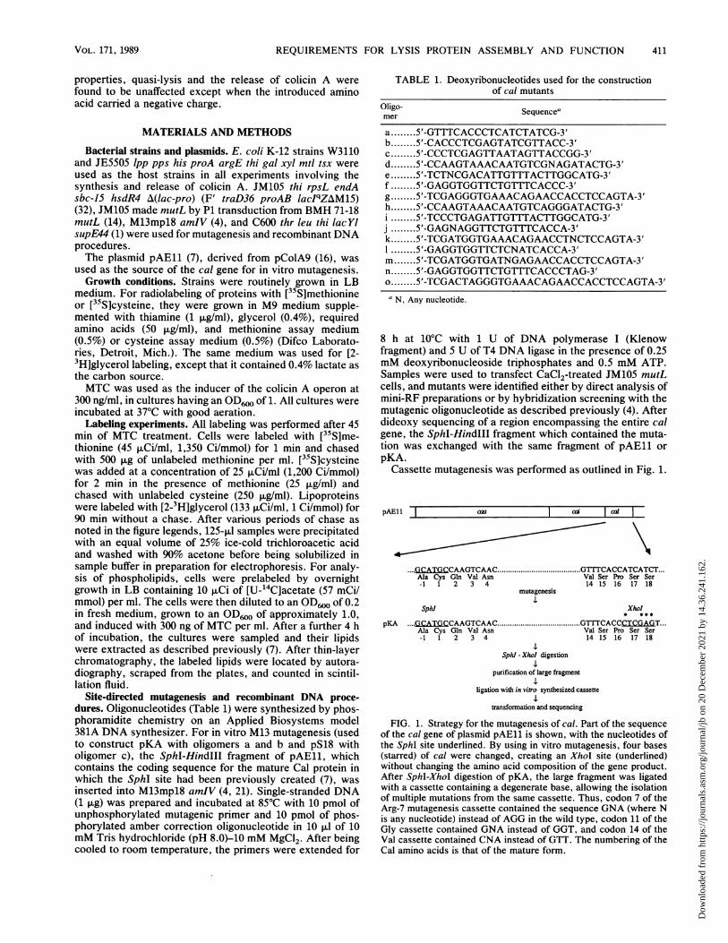
Cassette mutagenesis was performed as outlined in Fig. 1.
pAEll aa I aa
.GCATGCCAAGTCAACAla Cys Gln Val Asn-1 1 2 3 4
.GCVal Ser14 15
I 1I
'ACCATCATCT..r Pro Ser Seri 16 17 18
mutagenesis
Sphl Xhol
pKA ....GCATGCCAAGTCAAC ....................... GTCACCCTCGAGT...Ala Cys Gln Val Asn Val Ser Pro Ser Ser-1 1 2 3 4 14 15 16 17 18
SphI - XhoI digestion
purification of large fragment
ligation with in vitro synthesized cassette
transfornation and sequencing
FIG. 1. Strategy for the mutagenesis of cal. Part of the sequenceof the cal gene of plasmid pAE11 is shown, with the nucleotides ofthe SphI site underlined. By using in vitro mutagenesis, four bases(starred) of cal were changed, creating an XhoI site (underlined)without changing the amino acid composition of the gene product.After SphI-XhoI digestion of pKA, the large fragment was ligatedwith a cassette containing a degenerate base, allowing the isolationof multiple mutations from the same cassette. Thus, codon 7 of theArg-7 mutagenesis cassette contained the sequence GNA (where Nis any nucleotide) instead of AGG in the wild type, codon 11 of theGly cassette contained GNA instead of GGT, and codon 14 of theVal cassette contained CNA instead of GTT. The numbering of theCal amino acids is that of the mature form.
VOL. 171, 1989 411
Dow
nloa
ded
from
http
s://j
ourn
als.
asm
.org
/jour
nal/j
b on
20
Dec
embe
r 20
21 b
y 14
.36.
241.
162.
412 HOWARD ET AL.
The cassettes used to create the various mutations werecomposed of the oligonucleotides listed in Table 1 as fol-lows: oligomers d, e, f, and g for the Arg-7 substitutions;oligomers h, i, j, and k for the Gly-11 substitutions; oligo-mers h, i, 1, and m for the Val-14 substitutions; oligomers h,i, n, and o for S16. In each case, the 4-oligonucleotidecassette (the internal 5'-deoxynucleotides of which had beenphosphorylated with polynucleotide kinase) was allowed toprehybridize in 10 mM MgCl2-10 mM Tris (pH 7.5) afterbeing heated to 80°C. This was then ligated at a 100:1 molarratio to the large SphI-XhoI fragment of pKA, which hadbeen purified from low-melting-point agarose. To create theFS2 mutation, pKA was digested with XhoI, treated with T4DNA polymerase in the presence of deoxyribonucleosidetriphosphates, and religated. This resulted in the duplicationof the T of the XhoI site in pKA, causing a +2 frameshift.For all of the mutants constructed by using plasmids, mini-preparations of the plasmids obtained after transformation ofC600 cells were sequenced to identify the mutations beforebeing transformed into W3110 and resequenced.
General methods. For analysis of proteins released into theculture medium upon induction of the colicin A operon,samples were taken from induced or uninduced cultures andcentrifuged for 1 min at 15,000 x g. Cell pellets andsupernatants were dissolved in electrophoresis samplebuffer. Sodium dodecyl sulfate-polyacrylamide gel electro-phoresis (SDS-PAGE) and SDS-PAGE gels with urea wereelectrophoresed and treated for fluorography as previouslydescribed (7).
Materials. All enzymes were purchased from BoehringerMannheim Biochemicals (Indianapolis, Ind.) and used ac-cording to their recommendations. MTC was purchasedfrom Sigma Chemical Co. (St. Louis, Mo.). Globomycin wasa gift from Mamoru Arai (Sankyo, Tokyo). [35S]methionine,[35S]cysteine, [U-14C]acetate, and [2-3H]glycerol were pur-chased from the Radiochemical Centre, Amersham, UnitedKingdom. All other chemicals used were reagent grade.
RESULTS
Construction of cal gene mutants. An SphI restriction sitehad been previously introduced into the cal gene of pColA9to facilitate site-directed mutagenesis at the processing andmodification site of pro-Cal (7). This led to plasmid pAE11.Here, the construction strategy was to create a cassettecontaining the DNA region encoding the 18-amino-acidconsensus part of Cal to investigate the functional signifi-cance of this region and of the carboxy-terminal sequence ofthe protein.To this end, an XhoI site was first introduced by oligonu-
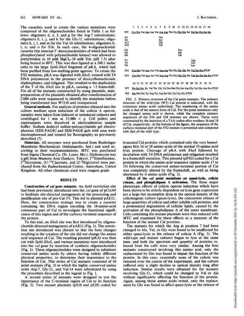
cleotide-directed mutagenesis of pAE11 (Fig. 1). The restric-tion site introduced was chosen so that the base changesresulting in the creation of the site did not change the aminoacid sequence of Cal. The resulting plasmid (pKA) was thencut with SphI-XhoI, and various mutations were introducedinto the cal gene by insertion of synthetic oligonucleotides(Fig. 1). These oligonucleotides were designed to substituteconserved amino acids by others having widely differentphysical properties, to determine their importance to thefunction of Cal. This series of Cal mutants consisted of 10point mutants (Fig. 2) in which the highly conserved aminoacids Arg-7, Gly-11, and Val-14 were substituted by usingthe procedure described in the legend to Fig. 1.A second series of mutants were designed to test the
importance of the C-terminal region of Cal to its function(Fig. 2). Two mutant plasmids (pS16 and pS18) coded for
1 2 3 4 5 6 7 8 9 10 11 12 13 14 15 16 17 18
WT Cys Gln Val An Asn Val ArMAs Thr Gly Gly Gly Ser Yal Ser Pro Ser Sr
A2A6AllA32
G4G6G8
viV3V8
GluAlaGlyVal
GluAlaVal
ArgGlnLeu
S16 CysGn Val AD.Asn Val ArgAM Thlr Gly Gly Ser Val Ser ProSTOP
S18 Cys Gln Val Asn Asn Val Arg Asp Thr Gly Gly Gly Ser Val Ser Pro Ser SerSTOP
16 17 18 19 20 21 22 23 24 25 26 27 28 29 30 31 32 33
WT Pro SerHSerIe Val Thr Gly Val Ser Met Gly Ser Asp Gly Val Gly Asn Pro STOP
FS2 Em Phe Glu Tyr Arg Tyr Arg Ser Glu Tyr Gly Glu STOP
FIG. 2. Primary structure of the Cal point mutants. The primarystructure of the wild-type (WT) Cal protein is indicated, with theconsensus amino acids underlined. The numbering of the aminoacids is that of the mature form of Cal. For the point mutations onlythe changed amino acid is shown, while the complete primarysequences of the S16 and S18 mutants are shown. These wereconstructed by the insertion of a TAA codon after residues 16 and 18of Cal, respectively. At the bottom of the figure, the sequence of thecarboxy-terminal part of the FS2 mutant is presented and comparedwith that of the wild type.
truncated Cal proteins which contained only the very homol-ogous first 16 or 18 amino acids of the normal 33-amino-acidlysis protein. Cleavage of pKA with XhoI followed byincubation with T4 DNA polymerase and religation resultedin a frameshift mutation. This plasmid (pFS2) coded for a Calprotein in which the amino acid sequence (amino acids 17 to33) following the conserved amino-terminal portion of Calwas completely altered by the frameshift, as well as beingshortened by 6 amino acids (Fig. 2).
Effect of the cal point mutations on quasi-lysis, colicinrelease, and phospholipase A activation. The three majorphenotypic effects of colicin operon induction which havebeen shown to be strictly dependent on lysis gene expressionare a large but incomplete drop in the optical density of thecolicinogenic culture (quasi-lysis), the concurrent release oflarge quantities of colicin and other soluble cell proteins, anda pronounced degradation of cellular lipids, caused by theactivation of the phospholipase A of the outer membrane.Cells containing the mutant plasmids were thus induced withMTC and examined for these effects as a measure of thefunction of the mutant Cal proteins.The mutants for which the Arg at position 7 had been
changed to Ala, Val, or Gly were found to be unaffected foreither quasi-lysis or the release of colicin A (Fig. 3). Thewild-type and mutant cultures began to lyse at the sametime, and both the spectrum and quantity of proteins re-leased from the cells were very similar. Among the fourmutants constructed involving this amino acid, only thereplacement by Glu was found to impair the function of theprotein. In this case, essentially none of the colicin wasreleased over the course of the experiment, and the culturesuffered only a slight decline in optical density long afterinduction. Similar results were obtained for the mutantsinvolving Gly-11, which could be changed to Val or Alawithout significantly affecting the function of the protein.Again, among those amino acids tested, only the replace-ment by Glu was found to affect quasi-lysis or the release of
J. BACTERIOL.
Dow
nloa
ded
from
http
s://j
ourn
als.
asm
.org
/jour
nal/j
b on
20
Dec
embe
r 20
21 b
y 14
.36.
241.
162.
REQUIREMENTS FOR LYSIS PROTEIN ASSEMBLY AND FUNCTION
ARG 7 mutants
1 2 3 4 5Time (hours)
6
VAL14 mutants
0
WT GLY ALA GLU VALC s c s c s c s c s
.... ..
therelease of co...cin A.(Fig. 3)
a-
the colicin, and in this case, the effect was less pronouncedthan for the replacement of Arg-7 by Glu (Fig. 3). The aminoacid Val-14 could be changed to Gln, Arg, or Leu withoutaffecting the fupction of Cal in causing either quasi-lysis orthe release of colicin A (Fig. 3).The lipid composition of cells containing the various
plasmids was evaluated after [14C]acetate labeling followedby induction with MTC. As shown in Table 2, induction of
TABLE 2. Effect of Cal and mutant Cal inductionon lipid composition
% of radioactivity recovered after thin-layerStrain chromatography'
Lyso PG PE DPG FFA NL
W3110(pColA9),N.I.b 0.4 7.1 90.7 0.4 0.4 1.1W3110(pColA9) 8.1 1.1 35.8 0.8 51.6 2.6W3110(pA1l) 8.5 2.9 47.4 0.9 37.8 2.5W3110(pVl) 7.7 2.9 46.4 1.2 38.8 3.0W3110(pA2) 1.4 5.8 83.4 0.7 5.7 3.0W3110(pG4) 1.7 7.7 79.6 0.7 6.2 4.2W3110(pS16) 1.0 6.5 86.8 0.6 2.7 2.4W3110(pS18) 1.2 7.0 87.3 0.5 2.8 1.3W3110(pFS2) 8.8 1.9 45.7 1.0 39.7 2.9
a Lyso, Lysophosopholipids; PG, phosphatidylglycerol; PE, phosphatidyl-ethanolamine; D)PG, diphosphatidylglycerol; FFA, free fatty acids; NL,neutral lipids.
b N.I., Not induced. All other strains were induced with MTC (300 ng/ml)for 4 h before harvesting and lipid extraction.
GLY11 mutants
3 4 5 6 7 0 1 2 3 4 5 6 7ne (hours) Time (hours)
FIG. 3. Effect of Cal point mutations on quasi-lysis and colicin Arelease. W3110 cells containing the point mutant plasmids weregrown in LB medium and induced with MTC at the time indicatedwith the arrows (top). The OD6w of the cultures was measured atintervals and plotted against the time of incubation at 37°C. After 3h of induction, samples of the four Arg-7 mutants were centrifuged,and quantities of the cells (C) and culture supernatants (S) repre-senting equal amounts of the original culture were analyzed bySDS-PAGE. The Coomassie blue-stained gel is presented (bottom).The standards in the right lane are (from top to bottom) phosphory-lase A (94 kDa), bovine serum albumin (68 kDa), ovalbumin (43kDa), carbonic anhydrase (30 kDa), soybean trypsin inhibitor (21kDa), and lysozyme (14 kDa). The position of colicin A is indicatedby an arrow. WT, Wild type.
the wild-type cal gene in W3110(pColA9) cells resulted in adramatic decrease in the levels of phosphatidylethanolamineand phosphatidylglycerol and concurrent large increases inthe amount of lysophospholipids and of free fatty acids.Induction of W3110 cells carrying cal point mutant plasmidssuch as pVl or pAll provoked the same effects as observedfor the wild type, whereas those containing pA2 and pG4, inwhich a glutamate residue had been introduced, did not(Table 2). In these cells there were only slight increases inthe quantities of lysophospholipids and free fatty acids. Theresults for these strains resembled those previously obtainedfor cal mutants containing alterations at the lipid modifica-tion site, which also display a reduction in the extent ofquasi-lysis and release of colicin A (7).
Modffication and processing of the cal point mutants. Wehave previously observed that Cal is a slowly processedlipoprotein for which the unmodified precursor, lipid-mod-ified precursor, mature form, and signal sequence can beidentified on fluorograms after electrophoresis of [35S]methi-onine-pulse-labeled cells (7). The various forms of Cal mi-grate very close or similarly to the various forms of the majorlipoprotein Lpp.To clearly demonstrate the migration of the various Cal
forms in our gel system, we compared the synthesis of Cal inboth lpp+ and Ipp strains carrying pColA9. As shown in Fig.4a and c, after pulse-labeling with [35S]methionine and 30min of chase, the Cal mature form and its signal peptide wereclearly detected in MTC-treated cells. The Cal mature formmigrated underneath the major lipoprotein (missing in the
10
EcoC]0
0
0
0.1*
MTC
ALAW.T.VAL
t ~~~~GLY
MTC
a.NARGW.T.
MTC
t sW~~~~.T.VAL
ALA
VOL. 171, 1989 413
1
Dow
nloa
ded
from
http
s://j
ourn
als.
asm
.org
/jour
nal/j
b on
20
Dec
embe
r 20
21 b
y 14
.36.
241.
162.
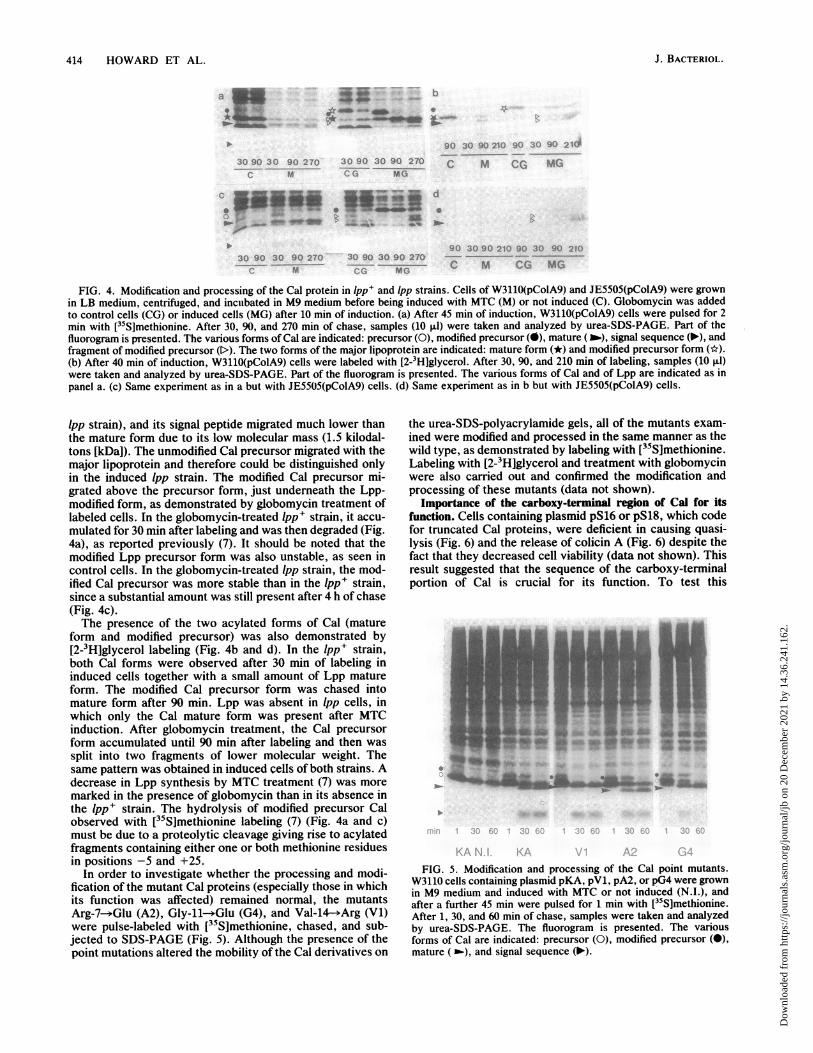
414 HOWARD ET AL.
a-
30 90 30 90 270C M
*_ _
90 30 90 210 900-;. 2 I1:30 90 30 90 270 C: M c MGSECO MG
30 90 30 90 270M
30 90 30 90 270Ct MG
90 30 90 210 90 30 90 210
C M CG MG
FIG. 4. Modification and processing of the Cal protein in lpp+ and Ipp strains. Cells of W3110(pColA9) and JE5505(pColA9) were grownin LB medium, centrifuged, and incubated in M9 medium before being induced with MTC (M) or not induced (C). Globomycin was addedto control cells (CG) or induced cells (MG) after 10 min of induction. (a) After 45 min of induction, W3110(pColA9) cells were pulsed for 2min with [35S]methionine. After 30, 90, and 270 min of chase, samples (10 ,ul) were taken and analyzed by urea-SDS-PAGE. Part of thefluorogram is presented. The various forms of Cal are indicated: precursor (0), modified precursor (X), mature (b.), signal sequence (-'), andfragment of modified precursor (D). The two forms of the major lipoprotein are indicated: mature form (*) and modified precursor form (*).(b) After 40 min of induction, W3110(pColA9) cells were labeled with [2-3Hlglycerol. After 30, 90, and 210 min of labeling, samples (10 ,ul)were taken and analyzed by urea-SDS-PAGE. Part of the fluorogram is presented. The various forms of Cal and of Lpp are indicated as inpanel a. (c) Same experiment as in a but with JE5505(pColA9) cells. (d) Same experiment as in b but with JE5505(pColA9) cells.
lpp strain), and its signal peptide migrated much lower thanthe mature form due to its low molecular mass (1.5 kilodal-tons [kDaJ). The unmodified Cal precursor migrated with themajor lipoprotein and therefore could be distinguished onlyin the induced Ipp strain. The modified Cal precursor mi-grated above the precursor form, just underneath the Lpp-modified form, as demonstrated by globomycin treatment oflabeled cells. In the globomycin-treated lpp+ strsin, it accu-mulated for 30 min after labeling and was then degraded (Fig.4a), as reported previously (7). It should be noted that themodified Lpp precursor form was also unstable, as seen incontrol cells. In the globomycin-treated lpp strain, the mod-ified Cal precursor was more stable than in the lpp+ strain,since a substantial amount was still present after 4 h of chase(Fig. 4c).The presence of the two acylated forms of Cal (mature
form and modified precursor) was also demonstrated by[2-3H]glycerol labeling (Fig. 4b and d). In the lpp+ strain,both Cal forms were observed after 30 min of labeling ininduced cells together with a small amount of Lpp matureform. The modified Cal precursor form was chased intomature form after 90 min. Lpp was absent in lpp cells, inwhich only the Cal mature form was present after MTCinduction. After globomycin treatment, the Cal precursorform accumulated until 90 min after labeling and then was
split into two fragments of lower molecular weight. Thesame pattern was obtained in induced celIs of both strains. Adecrease in Lpp synthesis by MTC treatment (7) was more
marked in the presence of globomycin than in its absence inthe lpp+ strain. The hydrolysis of modified precursor Calobserved with [35S]methionine labeling (7) (Fig. 4a and c)must be due to a proteolytic cleavage giving rise to acylatedfragments containing either one or both methionine residuesin positions -5 and +25.
In order to investigate whether the processing and modi-fication of the mutant Cal proteins (especially those in whichits function was affected) remained normal, the mutantsArg-7--Glu (A2), Gly-11-Glu (G4), and Val-14--*Arg (V1)were pulse-labeled with [35S]methionine, chased, and sub-jected to SDS-PAGE (Fig. 5). Although the presence of thepoint mutations altered the mobility of the Cal derivatives on
the urea-SDS-polyacrylamide gels, all of the mutants exam-ined were modified and processed in the same manner as thewild type, as demonstrated by labeling with [35S]methionine.Labeling with [2-3H]glycerol and treatment with globomycinwere also carried out and confirmed the modification andprocessing of these mutants (data not shown).
Importance of the carboxy-terminal region of Cal for itsfunction. Cells containing plasmid pS16 or pS18, which codefor truncated Cal proteins, were deficient in causing quasi-lysis (Fig. 6) and the release of colicin A (Fig. 6) despite thefact that they decreased cell viability (data not shown). Thisresult suggested that the sequence of the carboxy-terminalportion of Cal is crucial for its function. To test this
_ S _~~~~*4~414''1b^^ 3
FIG. 5. Modification and processing of the Cal point mutants.W3110 cells containing plasmid pKA, pV1, pA2, or pG4 were grownin M9 medium and induced with MTC or not induced (N.I.), andafter a further 45 min were pulsed for 1 min with [5S]methionine.After 1, 30, and 60 min of chase, samples were taken and analyzedby urea-SDS-PAGE. The fluorogram is presented. The variousforms of Cal are indicated: precursor (0), modified precursor (0),mature (S Mo),and signal sequence(m-).
J. BACTERIOL.
C d*w elf0 0
... -MOP-' .W" -Pmp. *w -.,. - Oft'* 0-
.1001,
Dow
nloa
ded
from
http
s://j
ourn
als.
asm
.org
/jour
nal/j
b on
20
Dec
embe
r 20
21 b
y 14
.36.
241.
162.
REQUIREMENTS FOR LYSIS PROTEIN ASSEMBLY AND FUNCTION
10
EC)CDCO0
0.1
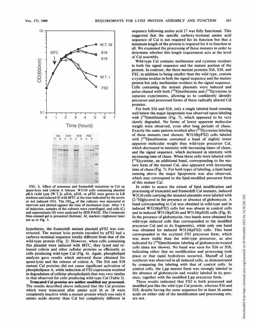
FIG. 6. Equasi-lysis a
pKA (wild tmedium andor not inducintervals ancof induction,and supernalblue-stainedare as in Fig
hypothesisstructed. Tcarboxy-teiwild-type pthis plasmileased colicells produanalysis gzquasi-lysismutant Cal
phospholipin degradatto that obsTruncate
The resultswhich wercompletelyamino acid
sequence following amino acid 17 was fully functional. Thissuggested that the specific carboxy-terminal amino acidsequence of Cal is not required for its function but that a
W.T. Ni minimum length of the protein is required for it to function atall. We examined the processing of these mutants in order to
MTC S16 determine whether this length requirement acts at the level
S18 of Cal assembly.Wild-type Cal contains methionine and cysteine residues
in both the signal sequence and the mature portion of theprotein. In contrast, the three mutant proteins S16, S18, andFS2, in addition to being smaller than the wild type, contain
W.T. a cysteine residue in both the signal sequence and the matureprotein but only methionine residues in the signal sequence.
FS2 Cells containing the mutant plasmids were induced andpulse-chased with both [35S]methionine and [35S]cysteine inseparate experiments, allowing us to confidently identifyprecursor and processed forms of these radically altered Calproteins.For both S16 and S18, only a single labeled band running
, .,X,--l--T--- , , ---, , ,well below the major lipoprotein was observed upon labeling0 1 2 3 4 5 6 7 with [35S]methionine (Fig. 7), which appeared to be very
slowly degraded. No forms of lower apparent molecularTime (hours) weight were observed, even after long periods of chase.
Exactly the same pattern resulted after [35S]cysteine labelingKA S16 S18 FS2 of these mutants (not shown). W3110(pFS2) cells labeled
C s c s c s c s with [35S]methionine contained a band of slightly lowerapparent molecular weight than wild-type precursor Cal,
Ws*. _ _ _ . _r:-which decreased in intensity with increasing times of chase,
and the signal sequence, which increased in intensity with-._wincreasing time of chase. When these cells were labeled with
[35S]cysteine, an additional band, corresponding to the ma-ture form of the mutant Cal, also appeared with increasingtime of chase (Fig. 7). For both types of labeling, a faint bandrunning above the major lipoprotein was also observed,which may correspond to the lipid-modified precursor formof this mutant Cal.
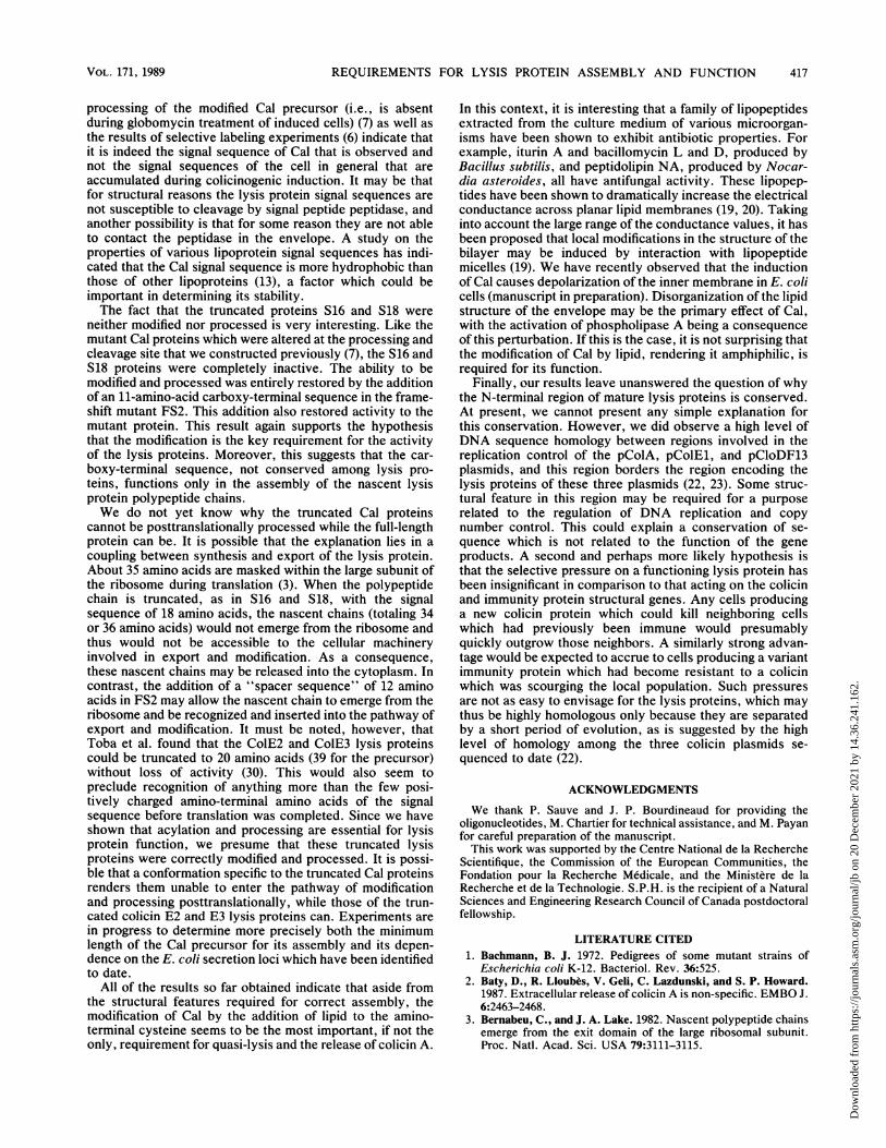
>ffect of nonsense and frameshift mutations in Cal on In order to assess the extent of lipid modification andmnd colicin A release. W3110 cells containing plasmid processing of truncated and frameshift Cal mutants, inducedtype [W.T.]), pS16, pS18, or pFS2 were grown in LB W3110 cells carrying the mutated plasmids were labeled withinduced (I) with MTC at the time indicated by the arrow 3-ed (NI). The ODwo of the cultures was measured at [2-3H]glycerol in the presence or absence of globomycin. AI plotted against the time of incubation (top). After 3 h band corresponding to Cal was obtained in wild-type and insamples of the cultures were centrifuged, and cells (C) induced W3110(pFS2) cells but was absent in control cells
tants (S) were analyzed by SDS-PAGE. The Coomassie and in induced W3110(pS16) and W3110(pS18) cells (Fig. 8).gel is presented (bottom). Mr markers (rightmost lane) In the presence of globomycin, two bands were obtained for3. wild-type induced cells that corresponded to the modified
precursor Cal and to its fragment(s), while only one bandthe frameshift mutant plasmid pFS2 was con- was obtained for induced W3110(pFS2) cells. This band
'he mutant lysis protein encoded by pFS2 had a corresponded to the acylated FS2 precursor form, whichrminal sequence totally different from that of the was more stable than the wild-type precursor, as also3rotein (Fig. 2). However, when cells containing
wa mor stbl tha th widtp prcro,sas)rotein(Fg.2)Hweerwencelsg indicated by [5S]methionine labeling of globomycin-treatedid were induced with MTC, they lysed and re- cells (data not shown). No band was seen for S16 or S18,Icin and other cellular proteins as efficiently asicing wild-type Cal (Fig. 6). Again, phospholipid indlcating either that no modlfication and processing tookave results which mirrored those obtained for place or that rapid hydrolysis occurred. Shutoff of Lppand the release of colicin A. The S16 and S18 synthesis was observed in all induced cells, as demonstrated
I proteins did not cause significant activation of by comparing the labeling with that of control cells. Inase A, while induction of FS2 expression resulted control cells, the Lpp mature form was strongly labeled inion of cellular phospholipids that was very similar the absence of globomycin and weakly labeled in its pres-erved for cells producing wild-type Cal (Table 2). ence, together with the modified Lpp precursor form.,d Cal proteins are neither modified nor processed. These results indicated that FS2 is both processed andi described above indicated that the Cal proteins modified just like the wild-type Cal protein, whereas S16 andre truncated after amino acid 16 or 18 were S18, despite having the same sequence for at least 16 aminoinactive while a mutant protein which was only 6 acids on either side of the modification and processing site,
Is shorter than Cal but completely different in are not.
VOL. 171, 1989 415
_-_-_- __-____ _rJ_________
Dow
nloa
ded
from
http
s://j
ourn
als.
asm
.org
/jour
nal/j
b on
20
Dec
embe
r 20
21 b
y 14
.36.
241.
162.
416 HOWARD ET AL.
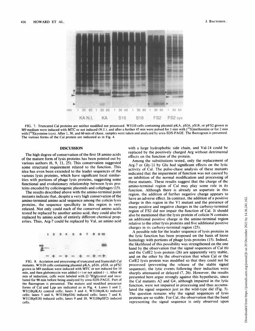
S
FIG. 7. Truncated Cal proteins are neither modified nor processed. W3110 cells containing plasmid pKA, pS16, pS18, or pFS2 grown inM9 medium were induced with MTC or not induced (N.I.), and after a further 45 min were pulsed for 1 min with [35S]methionine or for 2 minwith [35S]cysteine (cys). After 1, 30, and 60 min of chase, samples were taken and analyzed by urea-SDS-PAGE. The fluorogram is presented.The various formns of the Cal protein are indicated as in Fig. 4.
DISCUSSION
The high degree of conservation of the first 18 amino acidsof the mature form of lysis proteins has been pointed out byvarious authors (6, 9, 11, 25). This conservation suggestedsome structural requirement related to the function. Thisidea has even been extended to the leader sequences of thevarious lysis proteins, which have significant local similar-ities with portions of phage lysis proteins, suggesting some
functional and evolutionary relationship between lysis pro-
teins encoded by colicinogenic plasmids and coliphages (15).The results described above with the amino-terminal point
mutants indicate that, despite a very high conservation of theamino-terminal amino acid sequence among the colicin lysisproteins, the sequence specificity in this region is very
relaxed. Not only could each of the conserved amino acidstested be replaced by another amino acid, they could also bereplaced by amino acids of entirely different chemical prop-
erties. Thus, Arg-7 could be replaced by Val, an amino acid
1 2 3 4 5 6 7 8 9 10
++ _
+_+
FIG. 8. Acylation and processing of truncated and frameshift Calmutants. W3110 cells containing plasmid pKA, pS16, pS18, or pFS2grown in M9 medium were induced with MTC or not induced for 10min, and then globomycin was added (+) or not added (-). After 40min of induction, cells were labeled with [2-3H]glycerol and incu-bated for 90 min before being analyzed by urea-SDS-PAGE. Part ofthe fluorogram is presented. The mature and modified precursorforms of Cal and Lpp are indicated as in Fig. 4. Lanes 1 and 2,W3110(pKA) control cells; lanes 3 and 4, W3110(pKA) inducedcells; lanes 5 and 6, W3110(pS16) induced cells; lanes 7 and 8,W3110(pS18) induced cells; lanes 9 and 10, W3110(pSF2) inducedcells.
with a large hydrophobic side chain, and Val-14 could bereplaced by the positively charged Arg without detrimentaleffects on the function of the protein.Among the substitutions tested, only the replacement of
Arg-7 or Gly-11 by Glu had significant effects on the lyticactivity of Cal. The pulse-chase analysis of these mutantsindicated that the impairment of function was not caused byan inhibition of the normal modification and processing ofthese mutants. These results suggest that the charge of theamino-terminal region of Cal may play some role in itsfunction. Although there is already an aspartate in thisregion, the addition of further negative charge appears tohave an adverse effect. In contrast, the addition of a positivecharge in this region in the Vl mutant and the presence ofmany positive and negative charges in the carboxy-terminalregion of FS2 did not impair the function of Cal. It shouldalso be mentioned that the lysis protein of colicin N containsan additional positive charge in the amino-terminal regionrelative to the other lysis proteins and five additional positivecharges in its carboxy-terminal region (25).A possible role for the leader sequence of lysis proteins in
the lytic function has been proposed on the basis of loosehomology with portions of phage lysis proteins (15). Indeed,the likelihood of this possibility was strengthened on the onehand by the observation that the signal sequences of Cal (6)and the ColE2 lysis protein (26) are apparently very stable,and on the other by the observation that when Cal or theColE2 lysis protein was modified so that they could not beprocessed (preventing the release of the stable signalsequence), the lytic events following their induction weresharply attenuated or delayed (7, 26). However, the resultspresented here argue strongly against this hypothesis, sincethe Cal mutants A2 and G4, although impaired in the lyticfunction, were not impaired in processing and thus accumu-lated the signal sequence just as the wild-type did (Fig. 5).The question remains why the signal sequences of lysisproteins are so stable. For Cal, the observation that the bandrepresenting the signal sequence is only observed upon
J. BACTERIOL.
" 4ift't-
*IWI
to 'I.". +mt. p. %,.
Dow
nloa
ded
from
http
s://j
ourn
als.
asm
.org
/jour
nal/j
b on
20
Dec
embe
r 20
21 b
y 14
.36.
241.
162.
REQUIREMENTS FOR LYSIS PROTEIN ASSEMBLY AND FUNCTION
processing of the modified Cal precursor (i.e., is absentduring globomycin treatment of induced cells) (7) as well asthe results of selective labeling experiments (6) indicate thatit is indeed the signal sequence of Cal that is observed andnot the signal sequences of the cell in general that areaccumulated during colicinogenic induction. It may be thatfor structural reasons the lysis protein signal sequences arenot susceptible to cleavage by signal peptide peptidase, andanother possibility is that for some reason they are not ableto contact the peptidase in the envelope. A study on theproperties of various lipoprotein signal sequences has indi-cated that the Cal signal sequence is more hydrophobic thanthose of other lipoproteins (13), a factor which could beimportant in determining its stability.The fact that the truncated proteins S16 and S18 were
neither modified nor processed is very interesting. Like themutant Cal proteins which were altered at the processing andcleavage site that we constructed previously (7), the S16 andS18 proteins were completely inactive. The ability to bemodified and processed was entirely restored by the additionof an 11-amino-acid carboxy-terminal sequence in the frame-shift mutant FS2. This addition also restored activity to themutant protein. This result again supports the hypothesisthat the modification is the key requirement for the activityof the lysis proteins. Moreover, this suggests that the car-boxy-terminal sequence, not conserved among lysis pro-teins, functions only in the assembly of the nascent lysisprotein polypeptide chains.We do not yet know why the truncated Cal proteins
cannot be posttranslationally processed while the full-lengthprotein can be. It is possible that the explanation lies in acoupling between synthesis and export of the lysis protein.About 35 amino acids are masked within the large subunit ofthe ribosome during translation (3). When the polypeptidechain is truncated, as in S16 and S18, with the signalsequence of 18 amino acids, the nascent chains (totaling 34or 36 amino acids) would not emerge from the ribosome andthus would not be accessible to the cellular machineryinvolved in export and modification. As a consequence,these nascent chains may be released into the cytoplasm. Incontrast, the addition of a "spacer sequence" of 12 aminoacids in FS2 may allow the nascent chain to emerge from theribosome and be recognized and inserted into the pathway ofexport and modification. It must be noted, however, thatToba et al. found that the ColE2 and ColE3 lysis proteinscould be truncated to 20 amino acids (39 for the precursor)without loss of activity (30). This would also seem topreclude recognition of anything more than the few posi-tively charged amino-terminal amino acids of the signalsequence before translation was completed. Since we haveshown that acylation and processing are essential for lysisprotein function, we presume that these truncated lysisproteins were correctly modified and processed. It is possi-ble that a conformation specific to the truncated Cal proteinsrenders them unable to enter the pathway of modificationand processing posttranslationally, while those of the trun-cated colicin E2 and E3 lysis proteins can. Experiments arein progress to determine more precisely both the minimumlength of the Cal precursor for its assembly and its depen-dence on the E. coli secretion loci which have been identifiedto date.
All of the results so far obtained indicate that aside fromthe structural features required for correct assembly, themodification of Cal by the addition of lipid to the amino-terminal cysteine seems to be the most important, if not theonly, requirement for quasi-lysis and the release of colicin A.
In this context, it is interesting that a family of lipopeptidesextracted from the culture medium of various microorgan-isms have been shown to exhibit antibiotic properties. Forexample, iturin A and bacillomycin L and D, produced byBacillus subtilis, and peptidolipin NA, produced by Nocar-dia asteroides, all have antifungal activity. These lipopep-tides have been shown to dramatically increase the electricalconductance across planar lipid membranes (19, 20). Takinginto account the large range of the conductance values, it hasbeen proposed that local modifications in the structure of thebilayer may be induced by interaction with lipopeptidemicelles (19). We have recently observed that the inductionof Cal causes depolarization of the inner membrane in E. colicells (manuscript in preparation). Disorganization of the lipidstructure of the envelope may be the primary effect of Cal,with the activation of phospholipase A being a consequenceof this perturbation. If this is the case, it is not surprising thatthe modification of Cal by lipid, rendering it amphiphilic, isrequired for its function.
Finally, our results leave unanswered the question of whythe N-terminal region of mature lysis proteins is conserved.At present, we cannot present any simple explanation forthis conservation. However, we did observe a high level ofDNA sequence homology between regions involved in thereplication control of the pColA, pColEl, and pCloDF13plasmids, and this region borders the region encoding thelysis proteins of these three plasmids (22, 23). Some struc-tural feature in this region may be required for a purposerelated to the regulation of DNA replication and copynumber control. This could explain a conservation of se-quence which is not related to the function of the geneproducts. A second and perhaps more likely hypothesis isthat the selective pressure on a functioning lysis protein hasbeen insignificant in comparison to that acting on the colicinand immunity protein structural genes. Any cells producinga new colicin protein which could kill neighboring cellswhich had previously been immune would presumablyquickly outgrow those neighbors. A similarly strong advan-tage would be expected to accrue to cells producing a variantimmunity protein which had become resistant to a colicinwhich was scourging the local population. Such pressuresare not as easy to envisage for the lysis proteins, which maythus be highly homologous only because they are separatedby a short period of evolution, as is suggested by the highlevel of homology among the three colicin plasmids se-quenced to date (22).
ACKNOWLEDGMENTS
We thank P. Sauve and J. P. Bourdineaud for providing theoligonucleotides, M. Chartier for technical assistance, and M. Payanfor careful preparation of the manuscript.
This work was supported by the Centre National de la RechercheScientifique, the Commission of the European Communities, theFondation pour la Recherche Medicale, and the Ministere de laRecherche et de la Technologie. S.P.H. is the recipient of a NaturalSciences and Engineering Research Council of Canada postdoctoralfellowship.
LITERATURE CITED1. Bachmann, B. J. 1972. Pedigrees of some mutant strains of
Escherichia coli K-12. Bacteriol. Rev. 36:525.2. Baty, D., R. Lloubes, V. Geli, C. Lazdunski, and S. P. Howard.
1987. Extracellular release of colicin A is non-specific. EMBO J.6:2463-2468.
3. Bernabeu, C., and J. A. Lake. 1982. Nascent polypeptide chainsemerge from the exit domain of the large ribosomal subunit.Proc. Natl. Acad. Sci. USA 79:3111-3115.
VOL. 171, 1989 417
Dow
nloa
ded
from
http
s://j
ourn
als.
asm
.org
/jour
nal/j
b on
20
Dec
embe
r 20
21 b
y 14
.36.
241.
162.
418 HOWARD ET AL.
4. Carter, P., H. Bedouele, and G. Winter. 1985. Improved oligo-nucleotide site-directed mutagenesis using M13 vectors. Nu-cleic Acids Res. 13:4431- 4443.
5. Cavard, D., V. Crozel, J. P. Gorvel, F. Pattus, D. Baty, and C.Lazdunski. 1986. A molecular, genetic and immunological ap-proach to the functioning of colicin A, a pore-forming protein. J.Mol. Biol. 187:449-459.
6. Cavard, D., R. Lloul*s, J. Morion, M. Chartier, and C. Laz-dunski. 1985. Lysis protein encoded by plasmid ColA-CA31.Gene sequence and export. Mol. Gen. Genet. 199:95-100.
7. Cavard, D., D. Baty, S. P. Howard, H. M. Verhelj, and C.Lazdunski. 1987. Lipoprotein nature of the colicin A lysisprotein: effect of amino acid substitutions at the site of modifi-cation and processing. J. Bacteriol. 169:2187-2194.
8. Cole, S. T., B. Saint-Joanes, and A. P. Pugsley. 1985. Molecularcharacterisation of the colicin E2 operon and identification of itsproducts. Mol. Gpn. Genet. 196:465-472.
9. de Graaf, F. K., and B. Oudega. 1986. Production and release ofcloacin DF13 and related colicins. Curr. Top. Microbiol. Immu-nol. 125:183-205.
10. Ebina, Y., and A. Nakazawa. 1983. Cyclic AMP-dependentinitiation and p-dependent termination of colicin El gene tran-scription. J. Biol. Chem. 258:7072-7078.
11. Hakkaart, M. J. J., E. Veltkamp, and H. J. J. Ntkamp. 1981.Protein H encoded by plasmid CloDF13 involved in lysis of thebacterial host. I. Localisation of the gene and identification andsubcellular localisation of the gene H product. Mol. Gen. Genet.183:318-325.
12. Jakes, K., and N. D. Zinder. 1984. Plasmid ColE3 specifies alysis protein. J. Bacteriol. 157:582-590.
13. Klein, P., R. I. Somorjai, and P. Lau. 1988. Distinctive proper-ties of signal sequences from bacterial lipoproteins. ProteinEng. 2:15-20.
14. Kramer, B., W. Kramer, and H. J. Fritz. 1984. Differentbase-base mismatches are corrected with different efficienciesby the methyl-directed DNA mismatch-repair system of E. coli.Cell 38:879-887.
15. Lau, P. C., M. A. Hefford, and P. Klein. 1987. Structuralrelatedness of lysis proteins from colicinogenic plasmids andicosahedral coliphages. Mol. Biol. Evol. 4:544-556.
16. LloubAs, R., D. Baty, and C. Lazdunski. 1986. The promoters ofthe genes for colicin production, release and immunity in theColA plasmid: effects of convergent transcription and LexAprotein. Nucleic Acids Res. 14:2621-2636.
17. Lloul*s, R., D. Baty, and C. Lazdunski. 1988. Transcriptionalterminators in the caa-cal operon and cai gene. Nucleic AcidsRes. 16:3739-3749.
18. Luirink, J., C. van der Sande, J. Touaen, E. Veltkamp, F. K.de Graaf, and B. Oudeg. 1986. Effects of divalent cations and ofphospholipase A activity on excretion of cloacin DF13 and lysisof host cells. J. Gen. Microbiol. 132:825-834.
19. Maget-Dana, R., F. Heitz, M. Ptak, F. Peypoux, and M. Gui-nand. 1985. Bacteria lipopeptides induce ion-conducting poresin planar bilayers. Biochem. Biophys. Res. Commun. 129:965-971.
20. Maget-Dana, R., M. Ptak, F. Peypoux, and G. Michel. 1985.Pore-forming properties of iturin A, a lipopeptide antibiotic.Biochim. Biophys. Acta 815:405-409.
21. Messing, J. 1983. New M13 vectors for cloning. MethodsEnzymol. 101:20-78.
22. Morlon, J., M. Chartier, M. Bidaud, and C. Lazunski. 1988.The complete nucleotide sequence of the colicinogenic plasmidColA. High extent of homology with ColEl. Mol. Gen. Genet.211:231-243.
23. Morlon, J., R. Lloubbs, S. Varenne, M. Chartier, and C.Lazdunski. 1983. Complete nucleotide sequence of the struc-tural gene for colicin A, a gene translated at non-uniform rate. J.Mol. Biol. 170:271-285.
24. Oudega, B., A. Ykema, F. Stegehuis, and F. K. de Graaf. 1984.Detection and subcellular localization of mature protein Hinvolved in excretion of cloacin DF13. FEMS Microbiol. Lett.22:101-108.
25. Pugsley, A. 1988. The immunity and lysis genes of ColN plasmidpCHAP4. Mol. Gen. Genet. 211:335-341.
26. Pugley, A. P., and S. T. Cole. 1987. An unmodified form of theColE2 lysis protein, an envelope lipoprotein, retains reducedability to promote colicin E2 release and lysis ofproducing cells.J. Gen. Microbiol. 133:2411-2420.
27. Pugsley, A. P., and M. Schwartz. 1983. Expression of a gene ina 400-base-pair fragment of colicin plasmid ColE2-P9 is suffi-cient to cause host cell lysis. J. Bacteriol. 156:109-114.
28. Pugsley, A. P., and M. Schwartz. 1984. Colicin E2 release: lysis,leakage or secretion? Possible role of a phospholipase. EMBOJ. 3:2393-2397.
29. Sablk, J. F., J. L. Suit, and S. E. Luria. 1983. cea-kil operon ofthe ColEl plasmid. J. Bacteriol. 153:1479-1485.
30. Toba, M., H. Masaki, and T. Ohta. 1986. Primary structures ofthe ColE2-P9 and ColE3-CA38 lysis genes. J. Biochem. 99:591-596.
31. Watson, R. J., P. C. K. Lau, T. Vernet, and L. P. Visentin. 1986.Characterization and nucleotide sequence of a colicin-releasegene in the hic region of plasmid CoIE3-CA38. Gene 42:351-355.
32. Yanisch-Perron, C., J. Vieira, and J. Messing. 1985. ImprovedM13 phage cloning vectors and host strains: nucleotide se-quence of the M13mpl8 and pUC19 vectors. Gene 33:103-119.
J. BACTERIOL.
Dow
nloa
ded
from
http
s://j
ourn
als.
asm
.org
/jour
nal/j
b on
20
Dec
embe
r 20
21 b
y 14
.36.
241.
162.