Embed Size (px)
Citation preview

Function of Interstitial Cells of Cajal in the RabbitPortal Vein
Maksym I. Harhun, Dmitri V. Gordienko, Oleksandr V. Povstyan, Ray F. Moss, Thomas B. Bolton
Abstract—Interstitial cells of Cajal (ICCs) were identified in the intact fixed media of the rabbit portal vein (RPV) usingc-kit staining. The following experiments were performed using single cells preparations of the enzyme-dispersedvessel. Surviving contacts between the processes of single ICCs and the bodies of smooth muscle cells (SMCs) wereobserved in electron micrographs and by confocal microscopy. Spontaneous rhythmical [Ca2�]i oscillations wereobserved in ICCs after loading with the calcium indicator fluo-3 and were associated with depolarizations of the ICCsrecorded by tight-seal patch pipette. To investigate signal transmission from ICCs to SMCs in dispersed cell pairs, orwithin small surviving fragments of the ICC network, an ICC was stimulated under voltage-clamp, while changes in[Ca2�]i in the stimulated cell as well as in a closely adjacent SMCs or ICCs were monitored using fast x-y confocalimaging of fluo-3 fluorescence. After stimulation of an single voltage-clamped ICC by a depolarizing step similar induration to depolarizations associated with spontaneous [Ca2�]i oscillations, a depolarization and transient elevation of[Ca2�]i was observed in a closely adjacent SMCs after a delay of up to 4 seconds. In contrast, signal transmission fromICC to ICC was much faster, the delay being less than 200 ms. These results suggest that the an ICC may, in additionto generating an electrical signal (such as a slow wave) and thereby acting as a pacemaker for vascular SMCs of RPV,also releases some unknown diffusible substance, which depolarizes the SMCs. (Circ Res. 2004;95:619-626.)
Key Words: interstitial cells of Cajal � rabbit portal vein � calcium waves � pacemaker activity� vascular smooth muscle
Recently we have demonstrated c-kit–positive cells in thewall of the rabbit portal vein (RPV).1 After enzymatic
dispersion of the RPV as well as guinea pig mesentericarteries,2 cells closely resembling morphologically the inter-stitial cells of Cajal (ICCs) described in other tissues3–5 wereobserved; others have identified such cells in lymphaticvessels6 and bladder.7 ICCs have been widely described in thegastrointestinal tract and in urethral smooth muscle layers andare considered to play a pacemaker role in these tissues4,5 bygenerating rhythmical changes in membrane potential, slowwaves, which are suggested to be transmitted to SMCs.8,9
ICCs observed in the gastrointestinal tract express the antigenc-kit, have long processes, a high ability to accumulatemethylene blue, and an inability to contract. They form gapjunctions with SMCs and may serve to mediate signaltransmission from nerves to SMCs.10,11
In enzyme-dissociated cells of RPV contacts between theprocesses of ICCs and the bodies of SMCs, as well as smallfragments of the ICC network, were observed. This observa-tion prompted us to test whether any signal could be trans-mitted from one cell to another within these multicellularstructures. As calcium is involved as a second messenger in
the variety of intracellular signaling pathways and is themajor controller of SMCs tension, we used imaging of thefluorescence signal from a pair or small group of intercon-nected cells preloaded with the calcium-sensitive dye, fluo-3AM, combined with simultaneous electrophysiological record-ing from two cells to detect if there was any signal transmis-sion between them. We found that signal transmission fromICC to ICC appeared to be much faster than that from ICC toSMC, whereas spontaneous rhythmical [Ca2�]i waves in ICCswere associated with depolarization of the cell membrane.These observations suggest that the ICC network may act asa pacemaker for SMCs. A preliminary account of some of thiswork has previously been reported in abstract form.12
Materials and MethodsCell IsolationA section of RPV �20 mm long, upstream from the anastomosis ofits right and left branches, was removed from male New Zealandrabbits (2 to 3.5 kg, 42 animals) immediately after they had beenkilled by an overdose of pentobarbitone injected into the ear vein asapproved under Schedule 1 of the UK Animals (Scientific Proce-dures) Act 1986. Enzyme dispersion of the cells in the vessel wallwas performed as previously described.1 All experiments were
Original received December 18, 2003; resubmission received July 26, 2004; accepted August 12, 2004.From the Pharmacology and Clinical Pharmacology (M.I.H., D.V.G., O.V.P., T.B.B.) and Image Resource Facility (R.F.M.), Department of Basic
Medical Sciences, St George’s Hospital Medical School, Cranmer Terrace, London, UK; and Department of Nerve-Muscle Physiology (M.I.H., D.V.G.,O.V.P.), A.A. Bogomoletz Institute of Physiology, Ukraine.
Correspondence to Dr M.I. Harhun, Pharmacology and Clinical Pharmacology Basic Medical Sciences Department, St George’s Hospital MedicalSchool, Cranmer Terrace, London SW17 0RE, UK. E-mail [email protected]
© 2004 American Heart Association, Inc.
Circulation Research is available at http://www.circresaha.org DOI: 10.1161/01.RES.0000143014.04535.a3
619
by guest on July 20, 2018http://circres.ahajournals.org/
Dow
nloaded from

performed at room temperature (22 to 24°C) within 6 hours ofenzyme dispersion.
ImmunohistochemistryTo visualize the distribution of ICCs in the wall of RPV, we used theacetone-fixation protocol previously described by Ordog et al.13 Thebinding of rat monoclonal antibodies, raised against c-kit protein inmouse (RDI) was visualized with Alexa Fluor 488–conjugatedchicken anti-rat IgG (Molecular Probes Inc).
Electron MicroscopyThe cells were fixed in glutaraldehyde in cacodylate buffer, post-fixed in 1% osmium tetroxide, dehydrated in graded alcohols, andmounted on aluminum stubs. For viewing under an electron micro-scope, Zeiss EM 940 SEM, specimens were shadowed with a thinlayer of evaporated gold that gives the secondary electron image seenon the monitor.
Patch-Clamp RecordingExperiments were performed using amphotericin B (Sigma)perforated-patch tight-seal recording under voltage- or current-clampconditions. The electrical signals were recorded using an Axopatch200A patch-clamp amplifier or Multiclamp 700A for double-patchexperiments (both Axon Instruments). Voltage protocols were gen-erated and electrical signals were digitized at 1-kHz using a Digidata
1200 or 1322A hosted by a PC running pClamp 6.0 or 8.2 software(Axon Instruments). The cells were bathed in solution of thefollowing composition (in mmol/L): KCl 6, NaCl 120, MgCl2 1.2,CaCl2 2.5, D-glucose 12, and HEPES 10; pH was adjusted to 7.3 withNaOH. The pipette solution contained the following (in mmol/L):KCl 85, KH2PO4 30, MgSO4 5, EGTA 1, HEPES 10 (pH wasadjusted to 7.3 with KOH), and 200 �g/mL amphotericin B.
Confocal MicroscopyCells were loaded with the Ca2� sensitive indicator fluo-3AM(Molecular Probes Inc.) as previously described.14 An Axiovert100M inverted microscope attached to an LSM 510 laser-scanningunit (Zeiss) was used for x-y time-series or line-scan fluorescenceconfocal imaging. The acquisition and analysis of the data wereperformed as described elsewhere.1,14,15
StatisticsWhere appropriate, data are presented as mean�SEM for number ofthe cells (n) analyzed.
ResultsTo identify the location of ICCs in the wall of the RPV, fixedsegments of the RPV were incubated with anti–c-kit antibod-ies followed by staining with Alexa Fluor 488–conjugated
Figure 1. C-kit–positive cells within thewall of the RPV. Subendothelial (A) and(B) deeper intramuscular (up to �70 �mfrom the endothelium) layers of c-kit–positive cells were detected in the wall ofthe vessel. Vessel was opened longitudi-nally and viewed perpendicular to thewall of the vessel. In all panels, the longi-tudinal axis of the vessel is vertical. Con-siderable variations in the density ofICCs were detected in both layers withinthe same tissue preparation. Note thedifference in the density of subendothe-lial ICCs in the two different areas(C and D).
620 Circulation Research September 17, 2004
by guest on July 20, 2018http://circres.ahajournals.org/
Dow
nloaded from
IgG. Confocal x-y imaging of Alexa Fluor 488 fluorescencerevealed c-kit–positive cells at two locations: subendothelial(Figure 1A) and in a deeper intramuscular location (up to70 �m from the endothelium; Figure 1B), which is consistentwith our previous finding on living preparations of the RPV.1
We found that c-kit–positive cells were distributed in the wallof RPV at different densities in both subendothelial andintramuscular layers. In some regions, ICCs appeared at highdensity (up to 150 cell per mm2) (Figure 1C), whereas in theothers only a few ICCs were observed (Figure 1D). Suchvariability in the distribution of ICC along the vein isconsistent with heterogeneity of spontaneous contractile ac-tivity of different areas of the vein wall, which was frequentlyobserved on dissection of vein fragments (see alsoHermsmeyer).16
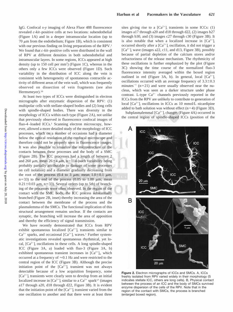
At least two types of ICCs were distinguished in electronmicrographs after enzymatic dispersion of the RPV: (1)multipolar cells with stellate-shaped bodies and (2) long cellswith spindle-shaped bodies. There was diversity in themorphology of ICCs within each type (Figure 2A), not unlikethat previously observed in fluorescence confocal images offluo-4–loaded ICCs.1 Scanning electron microscopy, how-ever, allowed a more detailed study of the morphology of ICCprocesses, which on a number of occasions had a diameterbelow the optical resolution of the confocal microscope andtherefore could not be properly seen in fluorescence images.It was also possible to visualize the microstructure of thecontacts between these processes and the body of a SMC(Figure 2B). The ICC processes had a length of between 2and 260 �m, mean 26�4 �m, n�114 (such variability beingprobably partially attributable to damage of some processeson cell isolation) and a diameter gradually decreasing fromthe root of the process (0.4 to 3 �m; mean 1.0�0.1 �m,n�33) to the end of the process (0.05 to 1.00 �m; mean0.21�0.03 �m, n�33). Several orders (up to 5th) of branch-ing of the processes were often observed. In the region of thecontact with the SMC body, the ICC process substantiallybranched (Figure 2B, inset) thereby increasing the area of thecontact between the membrane of the process and theplasmalemma of the SMCs. The functional implication of thisstructural arrangement remains unclear. If the contacts aresynaptic, the branching will increase the area of appositionand thereby the efficiency of signal transmission.
We have recently demonstrated that ICCs from RPVexhibit spontaneous localized [Ca2�]i transients similar toCa2� sparks, and occasional [Ca2�]i waves.1 Further system-atic investigations revealed spontaneous rhythmical, yet lo-cal, [Ca2�]i oscillations in these cells. A long spindle-shapedICC (Figure 3A, a) loaded with fluo-3 (Figure 3A, b)exhibited spontaneous transient increases in [Ca2�]i, whichoccurred at a frequency of �0.1 Hz and were restricted to thecentral region of the ICC (Figure 3B). Although the preciseinitiation point of the [Ca2�]i transient was not alwaysdetectable because of a low acquisition frequency, some[Ca2�]i transients were clearly seen to develop from an initiallocalized increase in [Ca2�]i similar to a Ca2� spark17 (imagesa17 through a20, d18 through d22, Figure 3B). It is evidentthat the initiation point of the [Ca2�]i transient varied from theone oscillation to another and that there were at least three
sites giving rise to a [Ca2�]i transients in some ICCs: (1)images a17 through a20 and d18 through d22, (2) images b27through b30, and (3) images c27 through c30 (Figure 3B). Itis also notable that when a localized increase in [Ca2�]i
occurred shortly after a [Ca2�]i oscillation, it did not trigger a[Ca2�]i wave (images a22, c11, and d13, Figure 3B), possiblybecause of partial depletion of the calcium stores and/orrefractoriness of the release mechanism. The rhythmicity ofthese oscillations is further emphasized by the plot (Figure3C) showing the time course of the normalized fluo-3fluorescence intensity averaged within the boxed regionoutlined in red (Figure 3A, b). In general, local [Ca2�]i
oscillations occurred with an average frequency of 3.3�0.3minutes�1 (n�21) and were usually observed near the nu-cleus, which was seen as a darker structure under phasecontrast. L-type Ca2� channels previously reported in theICCs from the RPV are unlikely to contribute to generation oflocal [Ca2�]i oscillations in ICCs as 10 mmol/L nicardipineadded to bath solution was without effect (n�4) (Figure 3D).
Subplasmalemmal [Ca2�]i changes (Figure 4A) occurred inthe central region of spindle-shaped ICCs (position of the
Figure 2. Electron micrographs of ICCs and SMCs. A, ICCsfreshly isolated from RPV varied widely in their morphology (Sindicates stellate ICC, others are long cells). B, Physical contactbetween the process of an ICC and the body of SMCs survivedenzyme dispersion of the cells of the RPV. Note that in theregion of the contact with SMCs, the process is branched(enlarged boxed region).
Harhun et al Pacemakers in the Vasculature 621
by guest on July 20, 2018http://circres.ahajournals.org/
Dow
nloaded from

scan line is depicted by yellow line on the transmitted lightimage, Figure 4B) and were associated with spontaneousdepolarizations of the cell (plot in Figure 4B). The line-scanimage at 667 Hz revealed that the rise in [Ca2�]i was initiatedat a single site (red arrow, Figure 4A) and spread as a [Ca2�]i
wave within a restricted region (see also Figure 3B) extend-ing along about half of the scan line. This is further empha-sized by the time-course plots (Figure 4C) of the normalizedflorescence signal (F/F0) at two positions along the scan line,depicted by red and green bars in the line-scan image (Figure4A), by red and green arrows on the transmitted light image(Figure 4B), and shown in corresponding colors. It is notablethat only a large amplitude [Ca2�]i transient (initiated at a sitedepicted by red bars and red arrow, Figure 4A, and red arrow,Figure 4B) was associated with membrane depolarization(Figure 4B), whereas a long-lasting small-amplitude increasein [Ca2�]i in an adjacent region (top part of the line-scanimage, Figure 4A, and green plot, Figure 4C) was withouteffect on membrane potential.
However, can these excitatory signals be transmitted to anadjacent SMC? To test this, ICC-SMC pairs that survivedenzymatic dispersion were preloaded with the Ca2�-sensitiveindicator fluo-3 (Figure 5A). The ICC of surviving ICC-SMCpair was stimulated by voltage steps (from �60 mV to �30mV by 30 mV increments) similar in duration to spontaneousdepolarizations associated with [Ca2�]i oscillations (Figure4B), whereas fast x-y confocal imaging of fluo-3 fluorescencein both cells was performed (Figure 5B through 5E). Images15 to 38 of these 48 images are shown in the galleries (panelsa in Figure 5B through 5E). The corresponding trace ofwhole-cell current through the ICC membrane is shown inpanels b in Figure 5B through 5E. The gallery shown inpanels c in Figure 5B through 5E is formed by the fluo-3fluorescence images taken from two boxed areas (1 and 2,Figure 5A, b) located in (1) ICCs and (2) SMCs. Thefluorescence intensity in the images was normalized to theaverage fluorescence intensity in control (before voltage stepwas applied) and color coded as indicated by the bar (F/F0).
Figure 3. Rhythmical [Ca2�]i oscillations in a singleICCs freshly isolated from the RPV. A, (a) Trans-mitted light image of spontaneously active ICCand (b) fluorescent confocal image of the samecell after loading with fluo3. B, 150 sequential flu-orescent confocal x-y images acquired at 1 Hzfrom the white boxed region (box 1) of the ICC.Fluorescence intensity was normalized to theaverage fluorescence intensity in 55 images show-ing the most uniform and least intense fluores-cence among the time-series and color coded asindicated by bar F/F0. Note that spontaneousrhythmical [Ca2�]i oscillations occurred at �0.1 Hz.C, Time course plot of normalized fluorescencesignal averaged within the small boxed region inthe central part of the cell (Ab, box 2, outlined inred). D, Rhythmical [Ca2�]i oscillations in anothersingle ICCs were not blocked by addition of10 �mol/L nicardipine.
622 Circulation Research September 17, 2004
by guest on July 20, 2018http://circres.ahajournals.org/
Dow
nloaded from

The time course plots of normalized fluorescence intensityaveraged within each box (outlined in green and black, Figure5A, b) are shown in the corresponding color in panel d(Figure 5B through 5E). When no depolarizing pulse wasapplied (Figure 5B), no change in the fluorescence signal wasobserved in both cells. Depolarization of the ICC to �30 mVevoked a [Ca2�]i transient in the ICC with no [Ca2�]i changein the adjacent SMC (Figure 5C). When the ICC wasdepolarized to 0 mV (Figure 5D), the rise in [Ca2�]i in theICC was followed by a delayed (up to 4 seconds) rise of[Ca2�]i in the SMC. The rise of [Ca2�]i in the ICC caused bythe voltage step to �30 mV was followed by a rise in [Ca2�]i
in the adjacent SMC that occurred with a shorter delay, had alarger magnitude, and revealed more than one peak (Figure5E). (The data shown in Figure 5B through 5E are alsoavailable as video clips in online data supplement at http://circres.ahajournals.org). Altogether, 20 ICC-SMC pairs wereexamined, and in 6 cases, stimulation of the ICC resulted ina [Ca2�]i elevation in an adjacent SMC that occurred with anaverage delay of 1.8�0.6 seconds (n�5) when the membranepotential was stepped from �60 mV to 0 mV.
To investigate changes in the membrane potential in theadjacent SMC during stimulation of the ICC in the ICC-SMCpair, we performed double patch experiments where ICC wasstimulated under voltage clamp by 5-second step from �60 to0 mV, and SMC membrane potential was recorded undercurrent clamp (Figure 6). In two of five successful recordings,small (up to 20 mV) delayed depolarizations of themembrane-adjacent SMCs were observed while ICCs werestimulated.
In some cases, small “patches” of the intact ICC network(comprising 2 to 4 ICCs) also survived enzymatic dispersionof the RPV. This allowed the dynamics of signal transmissionwithin the ICC network to be monitored; the same approachas described in Figure 5 was used (ie, one ICC was stimulatedwhile the change in the fluo-3 fluorescence in all intercon-nected ICCs was imaged). In the example shown in Figure 7
(see video clip in online data supplement), one stellate-shapedICC was voltage-clamped using tight-seal perforated-patchtechnique after the cell network was preloaded with fluo-3 AM
(Figure 7A). The ICC was stimulated by a 3-second voltagestep from a holding potential of �60 to 0 mV. The voltageprotocol was synchronized with the x-y imaging protocol(Figure 7B). When the voltage protocol with its correspond-ing current trace (Figure 7C, a) was related to the galleries(Figure 7C, b), each formed by a series of 30 images from thethree boxed regions from three contiguous cells (outlined bygreen, magenta, and blue, Figure 7A, b) and the correspond-ing time-course plots of the self-ratio fluorescence (F/F0)averaged within each box (Figure 7C, c), it became evidentthat, on stimulation, [Ca2�]i increased in less than 200 ms inall three ICCs. Thus, signal transmission within ICC networkappeared to be much faster than from ICC to SMC.
DiscussionBecause the ICCs in rabbit portal vein were c-kit positive, itseems possible that they have similar functions to those ofICCs in gut, namely, as pacemakers of electrical, and so thecontractile activity, and as intermediaries between nerve andmuscle. The present experiments do not address the latterpossibility but do provide some information about communi-cation between ICCs and SMCs.
Using confocal imaging of fluo-3 fluorescence, we ob-served for the first time in single ICCs freshly isolated fromRPV rhythmical local [Ca2�]i oscillations (Figure 3). Theaverage frequency of these oscillations (3.3�0.3 minutes�1
and allowing for a maximum of 20% change in frequency for1°C rise in temperature18) was similar to the frequency ofspontaneous depolarizations recorded with microelectrodefrom the pacemaker regions of multicellular preparations ofRPV (8 minutes�1 at 38°C). Combining confocal fluorescenceimaging with monitoring of the cell membrane potentialrevealed that [Ca2�]i oscillations in ICC were coupled tomembrane depolarization (Figure 4) and may underlie slow
Figure 4. Spontaneous depolarizationsassociated with local [Ca2�]i oscillationsin a single ICC. A, Confocal line-scanimage obtained from current-clampedfluo-3 loaded ICC. Position of the scanline is shown in the transmitted lightimage (depicted by yellow line in theinsert in B). Line-scanning was per-formed at 667 Hz. Fluorescence intensitywas normalized to the average fluores-cence intensity during the first 100 linesand color-coded as indicated by bar(F/F0). B, Corresponding trace of mem-brane potential from the same cell. C,Time course of the normalized fluores-cence intensity at two sites (depicted bygreen and red bars on the line-scanimage, A, and by green and red arrowsin the transmitted light image, B) areplotted in corresponding colors.
Harhun et al Pacemakers in the Vasculature 623
by guest on July 20, 2018http://circres.ahajournals.org/
Dow
nloaded from

waves previously demonstrated with microelectrode tech-nique.18 This coupling may occur through activation ofCa2�-dependent membrane ion channels, either chloride1,19 orcationic20 channels similar to those previously described inSMCs from RPV.21,22 In cultured ICCs from the murine smallintestine, it was proposed that a Ca2�-inhibited cationicconductance may contribute to the pacemaker current andgeneration of electrical slow waves.20 In urethral ICCs,unitary currents that could contribute to pacemaker activityhad properties similar to spontaneous transient Ca2�-dependent inward chloride currents23 and were shown to beactivated by IP3R-mediated Ca2� release from intracellularstores.19
The presence of spontaneous depolarizations associatedwith increases in [Ca2�]i is consistent with the ICCs in rabbitportal vein having a pacemaker function. This is in keepingwith the spontaneous mechanical and electrical activity thatthis vessel normally shows. It is well known that spontaneousactivity of the RPV is myogenic18,24 and the presence of the
multiple pacemakers in its wall is quite possible.24 Microelec-trode recordings at different positions along the RPV18 andasynchronous contractile activity of the different fragments ofthe vein wall16 suggest the existence of specialized pace-maker regions. In the present study, by screening regions ofRPV immunostained with anti–c-kit antibodies, we demon-strated that the density of the ICC network in both subendo-thelial and intramuscular layers varied widely along the wallof the vessel (Figure 1), which provides a histological basisfor the existence of pacemaker regions. This may suggest thatin the vasculature, especially in blood vessels with spontane-ous contractile and electrical activity, an ICC network mayserve as a pacemaker.
Further evidence was provided by our experiments onsmall clusters of linked ICCs. Depolarization of one ICCspread rapidly in less than 200 ms to other ICCs in the cluster.Thus, an ICC network in the wall of the RPV could besynchronized by such electrical connections and the ICCnetwork initiate depolarization of adjacent SMCs linked tothem by low resistance pathways.
Figure 5. Signal transmission from ICCs to SMCs.A, (a) Superimposition of a transmitted light imageon a fluo-3 fluorescent confocal image of a ICC-SMC pair and (b) normalized fluorescence confocalimage (F/F0) of this pair with two boxed (1, greenbox in ICC; 2, black box in SMC) regions of inter-est. ICC was voltage-clamped at �60 mV. Four x-ytime series imaging protocols each comprising 48frames acquired at 5 Hz were performed sequen-tially, whereas the ICC membrane potential wasstepped to different levels (between �60 and �30mV by 30 mV increments, B through E, respec-tively). In B through E, panel a shows 24 sequentialconfocal images taken as a part of the x-y timeseries protocol (see earlier), panel b shows thevoltage protocol (bottom) and corresponding cur-rent record (top), panel c is the corresponding gal-lery formed by sequentially aligning from left toright 24 fluorescence confocal images of the twoboxed regions of interest (Ab; labeled 1 in ICC and2 in SMC) numbered, respectively, on the righthand side of the gallery, panel d is the correspond-ing plot of the time course of the normalized fluo-rescence (F/F0) averaged within each of tworegions of interest (Ab, boxes 1 and 2). Note, thatdepolarization of the ICC membrane to 0 mV or to�30 mV caused a delayed rise in [Ca2�]i in theadjacent SMC. Also note, that the latency of theSMC response decreased with an increase of theamplitude of the voltage step applied to the ICC.
624 Circulation Research September 17, 2004
by guest on July 20, 2018http://circres.ahajournals.org/
Dow
nloaded from

The difficult experiments involving simultaneous record-ing of electrical activity from adjacent ICCs and SMCsrevealed that, on occasions, depolarization of the ICCs couldresult in a delayed depolarization of the associated SMC(Figure 7). This did not seem to involve an electrotonicspread of depolarization from the ICC to the SMC via thelong processes and their “feet” in contact with the SMC(Figure 2) because no obvious depolarization was initiated inthe SMC at the beginning of the step depolarization of the
ICC. In any case, at present, there is no evidence available asto whether the feet form low resistance gap junctions with theSMCs. Also, it should be borne in mind that in the intacttissue the cell bodies of ICCs are generally closely applied tothose of SMCs and likely form gap junctions with them;therefore, additional gap junctions formed at the end ofprocesses may not be necessary, although they may stilloccur. Rather, that the delay before SMC depolarizationoccurred suggests that some paracrine or vasoactive diffus-ible substance may start to be released from the ICC ondepolarization, and this acts, after a short delay caused bydiffusion, or to allow a sufficient concentration to build up, toproduce depolarization of the SMC in turn. Presumably thesubstance can persist after the end of ICC depolarization andgive rise to further changes in [Ca2�]i (Figure 5E, d) andlikely membrane potential (Figure 7) in the adjacent SMCs.There is no information on the identity of the substanceinvolved and identification, given the technically demandingexperiments involved, will be difficult to obtain. Whether thishypothetical substance is released from the cell body anddiffuses to the SMCs or is released from the processes is notknown. Thus ICCs, in addition to an electrical link to SMCs,also release a diffusible substance that depolarizes them.
We considered some possible sources of artifact. Stimula-tion of the SMC directly by a current (similar to thatgenerated by the ICC in response to the voltage steps from
Figure 6. Simultaneous tight-seal recording from both an ICCand SMC of an ICC-SMC pair. Voltage protocol (A) and corre-sponding current (B) recorded from stimulated ICC under volt-age clamp. C, Membrane potential of the SMC recorded syn-chronously under current clamp. Note the delayeddepolarization of the SMC during stimulation of ICC.
Figure 7. Signal transmission within ICC net-work. A, (a) Superimposition of a transmittedlight image on a fluo-3 fluorescent confocalimage of a small “patch” of the ICC networkand (b) corresponding normalized confocal fluo-rescence image (F/F0) with three boxed (greenin stimulated cell, magenta and blue in adjacentcells) regions of interest. One of the ICCs (seeposition of the patch pipette in Aa) wasvoltage-clamped at �60 mV using perforated-patch tight-seal technique. Voltage protocol(step to 0 mV) was synchronized with the x-ytime series imaging protocol comprising of 48frames acquired at 5 Hz. B, 40 sequential con-focal images taken as part of an x-y time seriesprotocol (see earlier). C, Panel a shows voltageprotocol (bottom) and corresponding currentrecord (atop), panel b is the corresponding gal-lery formed by the sequential aligning from leftto right of 30 fluorescence confocal images ofthe three boxed regions of interest (green,magenta and blue outlines in Ab: 1 in stimu-lated ICC, 2 and 3 in two adjacent ICCs) num-bered respectively on the left hand side of thegallery. Panel c is the corresponding plot of thetime course of the normalized fluorescence(F/F0) averaged within each of the three regionsof interest (green, magenta, and blue outlines inAb) and shown in the corresponding color.Note that on stimulation of one ICC, [Ca2�]iincreased in all three interconnected ICCs inless than 200 ms.
Harhun et al Pacemakers in the Vasculature 625
by guest on July 20, 2018http://circres.ahajournals.org/
Dow
nloaded from

�60 mV to 0 to 30 mV) applied through the free-tip of apatch pipette within less than 1 �m of the SMC surface(compare to the distance between SMC and ICCs, Figure 5)or even touching but not sealed to the cell, had no effect on[Ca2�]i in the stimulated cell (n�10, unpublished data, 2004).However, stimulation of the ICC under voltage-clamp led tothe transmission of the signal to the SMC and activatedvoltage-gated K� current through the ICC membrane (panelsb, Figure 5D and 5E), ie, an extrusion of K� from the ICCinto the extracellular media. This will cause a transientincrease in the extracellular K� concentration ([K�]o) close tothe ICC, which could potentially be sensed by the SMC. Wetherefore estimated to what extent [K�]o could increase in the“inner” space between ICCs and SMCs during this process.The upper limit could be calculated from the assumption thatK� released into the “inner” volume between the ICC andneighboring SMC does not immediately diffuse into the“outer” volume and is accumulated in the “inner” volumeduring the voltage step. The “inner” volume between ICC andSMC (Figure 5) was calculated as a product of the areabetween these two cells measured from transmitted lightimage (�1764 �m2; Figure 5A, a) and the cell thickness(�4 �m) and was found to be �7 pL. The amount of K�
extruded from the ICC into the inner volume through K�
channels before [Ca2�]i in adjacent SMCs started to rise wascalculated from the integral of the whole-cell K� current. Thetotal charge carried by K� through the IC membrane over thisperiod was �0.8�10�9 Q, what corresponds to 0.008 pmolK� extruded from the cell. Even if a quarter of this K� wasreleased into the inner volume and did not diffuse from it intothe outer volume, it would cause only 0.3 mmol/L increase in[K�]o in the inner volume by the time the SMC started torespond. In the real situation, K� is diffusing freely into theouter volume and an increase in [K�]o in the inner volumewould be therefore substantially smaller. Thus, it seemshighly unlikely that K� current generated by an ICC inresponse to voltage step can stimulate a neighboring SMCeither by extracellular current spread (see earlier) or throughan increase of [K�]o in the vicinity of the SMC.
In summary, this study reports spontaneous [Ca2�]i oscil-lations associated with depolarizations of the ICC membranein a vascular tissue and provides for the first time a directdemonstration of the signal transmission from ICC to SMC.These findings suggest that the ICC network may serve as apacemaker in this rhythmically contracting vessel, generatingand transmitting low frequency electrical signals to theadjacent SMCs. In addition there was evidence for someunknown paracrine or vasoactive substance which was re-leased from the ICC on depolarization causing depolarizationof SMCs.
AcknowledgmentsThis work was supported by Wellcome Trust Grants 042293,060659, and 064786 and by The British Heart Foundation Pro-gramme Grant RG/99001.
References1. Povstyan OV, Gordienko DV, Harhun MI, Bolton TB. Identification of
interstitial cells of Cajal in the rabbit portal vein. Cell Calcium. 2003;33:223–239.
2. Pucovsky V, Moss RF, Bolton TB. Non-contractile cells with thin pro-cesses resembling interstitial cells of Cajal found in the wall of guinea-pigmesenteric arteries. J Physiol. 2003;552:119–133.
3. Langton P, Ward SM, Carl A, Norell MA, Sanders KM. Spontaneouselectrical activity of interstitial cells of Cajal isolated from canineproximal colon. Proc Natl Acad Sci U S A. 1989;86:7280–7284.
4. Sanders KM. A case for interstitial cells of Cajal as pacemakers andmediators of neurotransmission in the gastrointestinal tract. Gastroen-terology. 1996;111:492–515.
5. Sergeant GP, Hollywood MA, McCloskey KD, Thornbury KD, McHaleNG. Specialised pacemaking cells in the rabbit urethra. J Physiol.2000;526 Pt 2:359–366.
6. McCloskey KD, Hollywood MA, Thornbury KD, Ward SM, McHale NG.Kit-like immunopositive cells in sheep mesenteric lymphatic vessels. CellTissue Res. 2002;310:77–84.
7. McCloskey KD, Gurney AM. Kit positive cells in the guinea pig bladder.J Urol. 2002;168:832–836.
8. Barajas-Lopez C, Berezin I, Daniel EE, Huizinga JD. Pacemaker activityrecorded in interstitial cells of Cajal of the gastrointestinal tract. Am JPhysiol. 1989;257:C830–C835.
9. Lee JC, Thuneberg L, Berezin I, Huizinga JD. Generation of slow wavesin membrane potential is an intrinsic property of interstitial cells of Cajal.Am J Physiol. 1999;277:G409–G423.
10. Daniel EE, Wang YF. Gap junctions in intestinal smooth muscle andinterstitial cells of Cajal. Microsc Res Tech. 1999;47:309–320.
11. Daniel EE, Thomas J, Ramnarain M, Bowes TJ, Jury J. Do gap junctionscouple interstitial cells of Cajal pacing and neurotransmission to gastro-intestinal smooth muscle? Neurogastroenterol Motil. 2001;13:297–307.
12. Harhun MI, Gordienko DV, Povstyan OV, Bolton T. B. Intercommuni-cation between interstitial cells (ICs) and smooth muscle cells (SMCs)from rabbit portal vein? Biophys J. 2003;84:105a.
13. Ordog T, Ward SM, Sanders KM. Interstitial cells of cajal generateelectrical slow waves in the murine stomach. J Physiol. 1999;518:257–269.
14. Gordienko DV, Greenwood IA, Bolton TB. Direct visualization of sar-coplasmic reticulum regions discharging Ca2� sparks in vascularmyocytes. Cell Calcium. 2001;29:13–28.
15. Gordienko DV, Bolton TB. Crosstalk between ryanodine receptors andIP3 receptors as a factor shaping spontaneous Ca2� -release events inrabbit portal vein myocytes. J Physiol. 2002;542:743–762.
16. Hermsmeyer K. Multiple pacemaker sites in spontaneously activevascular muscle. Circ Res. 1973;33:244–251.
17. Cheng H, Lederer WJ, Cannell MB. Calcium sparks: elementary eventsunderlying excitation-contraction coupling in heart muscle. Science.1993;262:740–744.
18. Holman ME, Kasby CB, Suthers MB, Wilson JA. Some properties of thesmooth muscle of rabbit portal vein. J Physiol. 1968;196:111–132.
19. Sergeant GP, Hollywood MA, McCloskey KD, McHale NG,Thornbury KD. Role of IP3 in modulation of spontaneous activity inpacemaker cells of rabbit urethra. Am J Physiol Cell Physiol. 2001;280:C1349 –C1356.
20. Koh SD, Jun JY, Kim TW, Sanders KM. A Ca2�-inhibited non-selectivecation conductance contributes to pacemaker currents in mouse interstitialcell of Cajal. J Physiol. 2002;540:803–814.
21. Greenwood IA, Helliwell RM, Large WA. Modulation of Ca2�-activatedCl- currents in rabbit portal vein smooth muscle by an inhibitor ofmitochondrial Ca2� uptake. J Physiol. 1997;505:53–64.
22. Albert AP, Large WA. A Ca2�-permeable non-selective cation channelactivated by depletion of internal Ca2� stores in single rabbit portal veinmyocytes. J Physiol. 2002;538:717–728.
23. Helliwell RM, Large WA. Effect of temperature on spontaneous Ca2�-activated Cl� currents in rabbit portal vein cells. Pflugers Arch. 1995;431:28–31.
24. Sutter MC. The mesenteric-portal vein in research. Pharmacol Rev. 1990;42:287–325.
626 Circulation Research September 17, 2004
by guest on July 20, 2018http://circres.ahajournals.org/
Dow
nloaded from

BoltonMaksym I. Harhun, Dmitri V. Gordienko, Oleksandr V. Povstyan, Ray F. Moss and Thomas B.
Function of Interstitial Cells of Cajal in the Rabbit Portal Vein
Print ISSN: 0009-7330. Online ISSN: 1524-4571 Copyright © 2004 American Heart Association, Inc. All rights reserved.is published by the American Heart Association, 7272 Greenville Avenue, Dallas, TX 75231Circulation Research
published online August 26, 2004;Circ Res.
http://circres.ahajournals.org/content/early/2004/08/26/01.RES.0000143014.04535.a3.citationWorld Wide Web at:
The online version of this article, along with updated information and services, is located on the
http://circres.ahajournals.org/content/suppl/2004/09/07/95.6.619.DC1Data Supplement (unedited) at:
http://circres.ahajournals.org//subscriptions/
is online at: Circulation Research Information about subscribing to Subscriptions:
http://www.lww.com/reprints Information about reprints can be found online at: Reprints:
document. Permissions and Rights Question and Answer about this process is available in the
located, click Request Permissions in the middle column of the Web page under Services. Further informationEditorial Office. Once the online version of the published article for which permission is being requested is
can be obtained via RightsLink, a service of the Copyright Clearance Center, not theCirculation Researchin Requests for permissions to reproduce figures, tables, or portions of articles originally publishedPermissions:
by guest on July 20, 2018http://circres.ahajournals.org/
Dow
nloaded from


![[Cajal] - Advice for a Young Investigator](https://img.pdfslide.us/doc/110x75/5468b723af795992368b5d19/cajal-advice-for-a-young-investigator.jpg)