Embed Size (px)
Citation preview
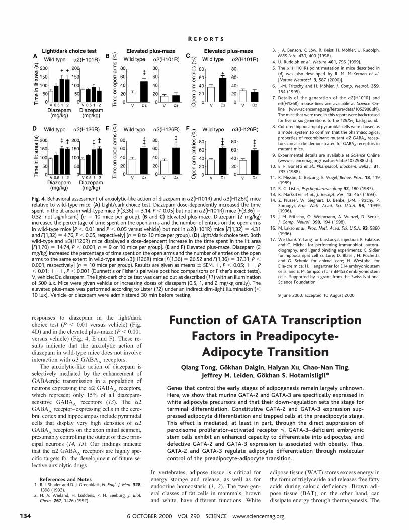
responses to diazepam in the light/darkchoice test (P , 0.01 versus vehicle) (Fig.4D) and in the elevated plus-maze (P , 0.001versus vehicle) (Fig. 4, E and F). These re-sults indicate that the anxiolytic action ofdiazepam in wild-type mice does not involveinteraction with a3 GABAA receptors.
The anxiolytic-like action of diazepam isselectively mediated by the enhancement ofGABAergic transmission in a population ofneurons expressing the a2 GABAA receptors,which represent only 15% of all diazepam-sensitive GABAA receptors (13). The a2GABAA receptor–expressing cells in the cere-bral cortex and hippocampus include pyramidalcells that display very high densities of a2GABAA receptors on the axon initial segment,presumably controlling the output of these prin-cipal neurons (14, 15). Our findings indicatethat the a2 GABAA receptors are highly spe-cific targets for the development of future se-lective anxiolytic drugs.
References and Notes1. R. I. Shader and D. J. Greenblatt, N. Engl. J. Med. 328,
1398 (1993).2. H. A. Wieland, H. Luddens, P. H. Seeburg, J. Biol.
Chem. 267, 1426 (1992).
3. J. A. Benson, K. Low, R. Keist, H. Mohler, U. Rudolph,FEBS Lett. 431, 400 (1998).
4. U. Rudolph et al., Nature 401, 796 (1999).5. The a1(H101R) point mutation in mice described in
(4) was also developed by R. M. McKernan et al.[Nature Neurosci. 3, 587 (2000)].
6. J.-M. Fritschy and H. Mohler, J. Comp. Neurol. 359,154 (1995).
7. Details of the generation of the a2(H101R) anda3(H126R) mouse lines are available at Science On-line (www.sciencemag.org/feature/data/1052988.shl).The mice that were used in this report were backcrossedfor five or six generations to the 129/SvJ background.
8. Cultured hippocampal pyramidal cells were chosen asa model system to confirm that the pharmacologicalproperties of recombinant mutant a2 GABAA recep-tors can also be demonstrated for GABAA receptors inmutant mice.
9. Experimental details are available at Science Online(www.sciencemag.org/feature/data/1052988.shl).
10. E. P. Bonetti et al., Pharmacol. Biochem. Behav. 31,733 (1988).
11. R. Misslin, C. Belzung, E. Vogel, Behav. Proc. 18, 119(1989).
12. R. G. Lister, Psychopharmacology 92, 180 (1987).13. R. Marksitzer et al., J. Recept. Res. 13, 467 (1993).14. Z. Nusser, W. Sieghart, D. Benke, J.-M. Fritschy, P.
Somogyi, Proc. Natl. Acad. Sci. U.S.A. 93, 11939(1996).
15. J.-M. Fritschy, O. Weinmann, A. Wenzel, D. Benke,J. Comp. Neurol. 390, 194 (1998).
16. M. Lakso et al., Proc. Natl. Acad. Sci. U.S.A. 93, 5860(1996).
17. We thank Y. Lang for blastocyst injection; P. Fakitsasand C. Michel for performing immunoblot, autora-diography, and ligand binding experiments; C. Sidlerfor hippocampal cell culture; D. Blaser, H. Pochetti,and G. Schmid for animal care; H. Westphal forEIIa-cre mice; H. Hengartner for E14 embryonic stemcells; and E. M. Simpson for mEMS32 embryonic stemcells. Supported by a grant from the Swiss NationalScience Foundation.
9 June 2000; accepted 10 August 2000
Function of GATA TranscriptionFactors in Preadipocyte-
Adipocyte TransitionQiang Tong, Gokhan Dalgin, Haiyan Xu, Chao-Nan Ting,
Jeffrey M. Leiden, Gokhan S. Hotamisligil*
Genes that control the early stages of adipogenesis remain largely unknown.Here, we show that murine GATA-2 and GATA-3 are specifically expressed inwhite adipocyte precursors and that their down-regulation sets the stage forterminal differentiation. Constitutive GATA-2 and GATA-3 expression sup-pressed adipocyte differentiation and trapped cells at the preadipocyte stage.This effect is mediated, at least in part, through the direct suppression ofperoxisome proliferator–activated receptor g. GATA-3–deficient embryonicstem cells exhibit an enhanced capacity to differentiate into adipocytes, anddefective GATA-2 and GATA-3 expression is associated with obesity. Thus,GATA-2 and GATA-3 regulate adipocyte differentiation through molecularcontrol of the preadipocyte-adipocyte transition.
In vertebrates, adipose tissue is critical forenergy storage and release, as well as forendocrine homeostasis (1, 2). The two gen-eral classes of fat cells in mammals, brownand white, have different functions. White
adipose tissue (WAT) stores excess energy inthe form of triglyceride and releases free fattyacids during caloric deficiency. Brown adi-pose tissue (BAT), on the other hand, candissipate energy through thermogenesis. The
Fig. 4. Behavioral assessment of anxiolytic-like action of diazepam in a2(H101R) and a3(H126R) micerelative to wild-type mice. (A) Light/dark choice test. Diazepam dose-dependently increased the timespent in the lit area in wild-type mice [F(3,36) 5 3.14, P , 0.05] but not in a2(H101R) mice [F(3,36) 50.32, not significant] (n 5 10 mice per group). (B and C) Elevated plus-maze. Diazepam (2 mg/kg)increased the percentage of time spent on the open arms and the number of entries on the open armsin wild-type mice (P , 0.01 and P , 0.05 versus vehicle) but not in a2(H101R) mice [F(1,32) 5 4.31and F(1,32) 5 4.76, P , 0.05, respectively] (n 5 8 to 10 mice per group). (D) Light/dark choice test. Bothwild-type and a3(H126R) mice displayed a dose-dependent increase in the time spent in the lit area[F(1,70) 5 14.74, P , 0.001, n 5 9 or 10 mice per group]. (E and F) Elevated plus-maze. Diazepam (2mg/kg) increased the percentage of time spent on the open arms and the number of entries on the openarms to the same extent in wild-type and a3(H126R) mice [F(1,36) 5 26.52 and F(1,36) 5 37.31, P ,0.001, respectively] (n 5 10 mice per group). Results are given as means 6 SEM. 1, P , 0.05; 11, P, 0.01; 111, P , 0.001 (Dunnett’s or Fisher’s pairwise post hoc comparisons or Fisher’s exact tests).V, vehicle; Dz, diazepam. The light-dark choice test was carried out as described (11) with an illuminationof 500 lux. Mice were given vehicle or increasing doses of diazepam (0.5, 1, and 2 mg/kg orally). Theelevated plus-maze was performed according to Lister (12) under an indirect dim-light illumination (,10 lux). Vehicle or diazepam were administered 30 min before testing.
R E P O R T S
6 OCTOBER 2000 VOL 290 SCIENCE www.sciencemag.org134
coordinated action of the peroxisome prolif-erator–activated receptor (PPAR) g (3) andthe CCAAT/enhancer binding protein (C/EBP) family of transcription factors (4) reg-ulates the adipocyte differentiation program.Subsequent to C/EBPb and C/EBPd expres-sion during differentiation of adipocytes,C/EBPa and PPARg production is stimulated(5). There is a positive feedback loop betweenPPARg and C/EBPa; both factors induce theexpression of the other (6). This synergy drivesthe expression of a complex gene program thatis necessary for the generation and maintenanceof the adipogenic phenotype (1, 2). However,little is known about the commitment of pluri-
potent stem cells into adipogenic lineages andthe genes that control the transition from pre-adipocytes to adipocytes. To identify factorscritical at these early stages, we examinedwhether the genes necessary for the formationof the Drosophila melanogaster fat body, ahomolog of mammalian adipose tissue and liv-er, are conserved in mammals.
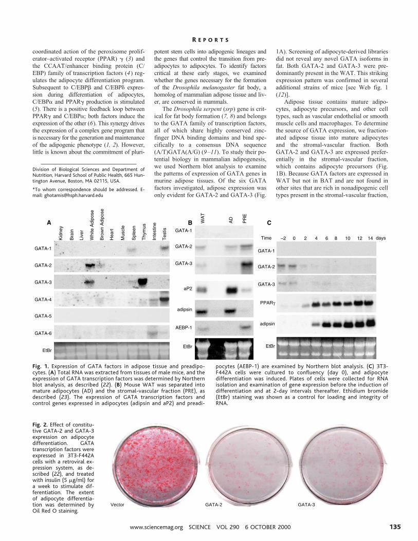
The Drosophila serpent (srp) gene is crit-ical for fat body formation (7, 8) and belongsto the GATA family of transcription factors,all of which share highly conserved zinc-finger DNA binding domains and bind spe-cifically to a consensus DNA sequence(A/T)GATA(A/G) (9–11). To study their po-tential biology in mammalian adipogenesis,we used Northern blot analysis to examinethe patterns of expression of GATA genes inmurine adipose tissues. Of the six GATAfactors investigated, adipose expression wasonly evident for GATA-2 and GATA-3 (Fig.
1A). Screening of adipocyte-derived librariesdid not reveal any novel GATA isoforms infat. Both GATA-2 and GATA-3 were pre-dominantly present in the WAT. This strikingexpression pattern was confirmed in severaladditional strains of mice [see Web fig. 1(12)].
Adipose tissue contains mature adipo-cytes, adipocyte precursors, and other celltypes, such as vascular endothelial or smoothmuscle cells and macrophages. To determinethe source of GATA expression, we fraction-ated adipose tissue into mature adipocytesand the stromal-vascular fraction. BothGATA-2 and GATA-3 are expressed prefer-entially in the stromal-vascular fraction,which contains adipocyte precursors (Fig.1B). Because GATA factors are expressed inWAT but not in BAT and are not found inother sites that are rich in nonadipogenic celltypes present in the stromal-vascular fraction,
Division of Biological Sciences and Department ofNutrition, Harvard School of Public Health, 665 Hun-tington Avenue, Boston, MA 02115, USA.
*To whom correspondence should be addressed. E-mail: [email protected]
Fig. 1. Expression of GATA factors in adipose tissue and preadipo-cytes. (A) Total RNA was extracted from tissues of male mice, and theexpression of GATA transcription factors was determined by Northernblot analysis, as described (22). (B) Mouse WAT was separated intomature adipocytes (AD) and the stromal-vascular fraction (PRE), asdescribed (23). The expression of GATA transcription factors andcontrol genes expressed in adipocytes (adipsin and aP2) and preadi-
pocytes (AEBP-1) are examined by Northern blot analysis. (C) 3T3-F442A cells were cultured to confluency (day 0), and adipocytedifferentiation was induced. Plates of cells were collected for RNAisolation and examination of gene expression before the induction ofdifferentiation and at 2-day intervals thereafter. Ethidium bromide(EtBr) staining was shown as a control for loading and integrity ofRNA.
GATA-3GATA-2Vector
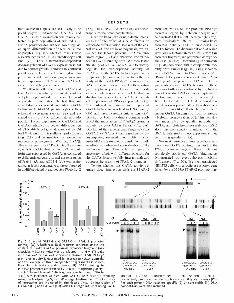
Fig. 2. Effect of constitu-tive GATA-2 and GATA-3expression on adipocytedifferentiation. GATAtranscription factors wereexpressed in 3T3-F442Acells with a retroviral ex-pression system, as de-scribed (22), and treatedwith insulin (5 mg/ml) fora week to stimulate dif-ferentiation. The extentof adipocyte differentia-tion was determined byOil Red O staining.
R E P O R T S
www.sciencemag.org SCIENCE VOL 290 6 OCTOBER 2000 135
their source in adipose tissue is likely to bepreadipocytes. Furthermore, GATA-2 andGATA-3 mRNA expression was readily de-tected in pure populations of cultured 3T3-F442A preadipocytes but was down-regulat-ed upon differentiation of these cells intoadipocytes (Fig. 1C). Identical results werealso obtained in the 3T3-L1 preadipocyte cellline (13). This differentiation-dependentdown-regulation of GATA expression is notdue to contact growth inhibition in confluentpreadipocytes, because cells cultured in non-permissive conditions for adipogenesis main-tained expression of GATA-2 and GATA-3,even after reaching confluence.
We then hypothesized that GATA-2 andGATA-3 are potential preadipocyte markersand play important roles in the regulation ofadipocyte differentiation. To test this, weconstitutively expressed individual GATAfactors in 3T3-F442A preadipocytes with aretroviral expression system (14) and as-sessed their ability to differentiate into adi-pocytes. Forced expression of GATA-2 andGATA-3 inhibited adipocyte differentiationof 3T3-F442A cells, as determined by OilRed O staining of intracellular lipid droplets(Fig. 2A) and examination of molecularmarkers of adipogenesis [Web fig. 2 (12)].The expression of PPARg, Glut4, the adipo-cyte fatty acid binding protein aP2, and ad-ipsin was suppressed by GATA, as comparedto differentiated controls, and the expressionof Pref-1 (15) and AEBP-1 (16) was main-tained at levels comparable to those observedin undifferentiated preadipocytes [Web fig. 2
(12)]. Thus, the GATA-expressing cells weretrapped at the preadipocyte stage.
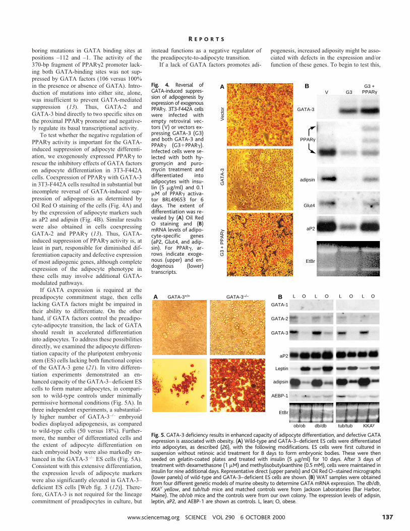
Next, we began exploring potential mech-anisms of the effect of GATA factors onadipocyte differentiation. Because of the cen-tral role of PPARg in adipogenesis, we ex-amined the 0.6-kb proximal PPARg2 pro-moter region (17) and observed several po-tential GATA binding sites. We then testedthe ability of GATA-2 or GATA-3 to directlyregulate the transcriptional activity ofPPARg2. Both GATA factors significantlysuppressed (approximately fivefold) the ac-tivity of the 0.6-kb PPARg2 promoter (Fig.3A). In the same experimental setting, estro-gen receptor response element–driven lucif-erase activity was enhanced by GATA-2, in-dicating the specificity of the GATA-mediat-ed suppression of PPARg2 promoter (13).The carboxyl and amino zinc fingers ofGATA factors are involved in DNA binding(18) and protein-protein interactions (19).Deletion of both zinc-finger domains abol-ished the suppression of PPARg2 promoteractivity by both GATA factors (Fig. 3A).Deletion of the carboxyl zinc finger of eitherGATA-2 or GATA-3 also significantly butincompletely reversed their ability to sup-press PPARg2 promoter. A similar but small-er effect was observed upon deletion of theamino zinc finger. Thus, both zinc fingers arenecessary, albeit with different potency, forthe GATA factors to fully interact with andsuppress the activity of PPARg2 promoter.
To test whether this GATA activity re-quires direct interaction with the PPARg2
promoter, we studied the proximal PPARg2promoter region by deletion analysis anddemonstrated that a 370–base pair (bp) frag-ment (nucleotides –361 to 19) retains basalpromoter activity and is suppressed byGATA factors. To determine if and at whichsites GATA factors interact directly with thispromoter fragment, we performed deoxyribo-nuclease (DNase) I footprinting experiments(Fig. 3B) combined with elecrophoretic mo-bility shift assays (Fig. 3C) using recombi-nant GATA-2 and GATA-3 proteins (20).DNase I footprinting revealed two GATAbinding sites at positions –112 and –1. Se-quence-dependent GATA binding to thesesites was further demonstrated by the forma-tion of specific DNA-protein complexes inelectrophoretic mobility shift assays (Fig.3C). The formation of GATA protein-DNAcomplexes was prevented by the addition of aspecific competitor DNA fragment withknown GATA binding site from the mousea1-globin promoter (Fig. 3C). This complexwas supershifted by specific antibodies toGATA, and glutathione S-transferase (GST)alone had no capacity to interact with theDNA targets used in these experiments, thusconfirming specificity (13).
We next introduced point mutations intothese two GATA binding sites within the370-bp promoter region. These mutationscompletely abolished GATA binding, asdemonstrated by electrophoretic mobilityshift assays (Fig. 3C). We then transfectedNIH 3T3 cells with a luciferase reporter genedriven by the 370-bp PPARg2 promoter har-
Fig. 3. Effect of GATA-2 and GATA-3 on PPARg2 promoteractivity. (A) A luciferase (luc) reporter construct under thecontrol of 0.6-kb PPARg2 proximal promoter fragment (nu-cleotides 2603 to 162) was transfected into NIH 3T3 cellswith GATA-2 or GATA-3 expression plasmids (24). PPARg2promoter activity is expressed in relation to vector controls,and the average of three independent experiments is shown.Error bars indicate standard error. (B) GATA binding toPPARg2 promoter determined by DNase I footprinting analy-sis. A 32P– end labeled DNA fragment (nucleotides 2264 to162) was incubated at 25°C with GST–GATA-2 fusion protein byusing the Footprinting System (Promega, Madison, WI). The two sitesof interaction are indicated by the dotted lines. (C) Interaction ofGATA-2 (G2) and GATA-3 (G3) with DNA fragments containing GATA
sites at 2112 and 21 (nucleotides 2119 to 285 and 222 to 19,respectively), as shown by electrophoretic mobility shift assays (25).For each protein-DNA reaction, specific (S) or nonspecific (N) DNAcompetitors were also included.
R E P O R T S
6 OCTOBER 2000 VOL 290 SCIENCE www.sciencemag.org136
boring mutations in GATA binding sites atpositions –112 and –1. The activity of the370-bp fragment of PPARg2 promoter lack-ing both GATA-binding sites was not sup-pressed by GATA factors (106 versus 100%in the presence or absence of GATA). Intro-duction of mutations into either site, alone,was insufficient to prevent GATA-mediatedsuppression (13). Thus, GATA-2 andGATA-3 bind directly to two specific sites onthe proximal PPARg promoter and negative-ly regulate its basal transcriptional activity.
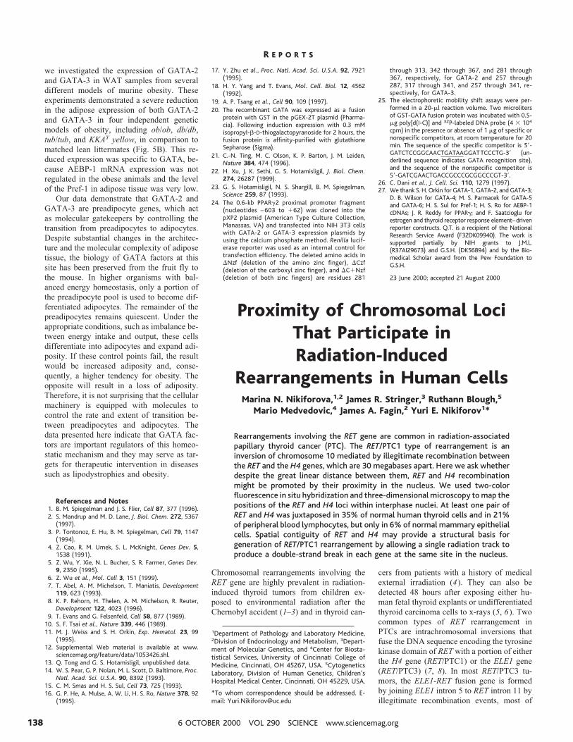
To test whether the negative regulation ofPPARg activity is important for the GATA-induced suppression of adipocyte differenti-ation, we exogenously expressed PPARg torescue the inhibitory effects of GATA factorson adipocyte differentiation in 3T3-F442Acells. Coexpression of PPARg with GATA-3in 3T3-F442A cells resulted in substantial butincomplete reversal of GATA-induced sup-pression of adipogenesis as determined byOil Red O staining of the cells (Fig. 4A) andby the expression of adipocyte markers suchas aP2 and adipsin (Fig. 4B). Similar resultswere also obtained in cells coexpressingGATA-2 and PPARg (13). Thus, GATA-induced suppression of PPARg activity is, atleast in part, responsible for diminished dif-ferentiation capacity and defective expressionof most adipogenic genes, although completeexpression of the adipocyte phenotype inthese cells may involve additional GATA-modulated pathways.
If GATA expression is required at thepreadipocyte commitment stage, then cellslacking GATA factors might be impaired intheir ability to differentiate. On the otherhand, if GATA factors control the preadipo-cyte-adipocyte transition, the lack of GATAshould result in accelerated differentiationinto adipocytes. To address these possibilitiesdirectly, we examined the adipocyte differen-tiation capacity of the pluripotent embryonicstem (ES) cells lacking both functional copiesof the GATA-3 gene (21). In vitro differen-tiation experiments demonstrated an en-hanced capacity of the GATA-3–deficient EScells to form mature adipocytes, in compari-son to wild-type controls under minimallypermissive hormonal conditions (Fig. 5A). Inthree independent experiments, a substantial-ly higher number of GATA-3–/– embryoidbodies displayed adipogenesis, as comparedto wild-type cells (50 versus 18%). Further-more, the number of differentiated cells andthe extent of adipocyte differentiation oneach embryoid body were also markedly en-hanced in the GATA-3–/– ES cells (Fig. 5A).Consistent with this extensive differentiation,the expression levels of adipocyte markerswere also significantly elevated in GATA-3–deficient ES cells [Web fig. 3 (12)]. There-fore, GATA-3 is not required for the lineagecommitment of preadipocytes in culture, but
instead functions as a negative regulator ofthe preadipocyte-to-adipocyte transition.
If a lack of GATA factors promotes adi-
pogenesis, increased adiposity might be asso-ciated with defects in the expression and/orfunction of these genes. To begin to test this,
Vec
tor
GA
TA
-3G
3 +
PP
AR
γ
A BV G3
G3 +PPARγ
adipsin
GATA-3
PPARγ
Glut4
aP2
EtBr
Fig. 4. Reversal ofGATA-induced suppres-sion of adipogenesis byexpression of exogenousPPARg. 3T3-F442A cellswere infected withempty retroviral vec-tors (V) or vectors ex-pressing GATA-3 (G3)and both GATA-3 andPPARg (G31PPARg).Infected cells were se-lected with both hy-gromycin and puro-mycin treatment anddifferentiated intoadipocytes with insu-lin (5 mg/ml) and 0.1mM of PPARg activa-tor BRL49653 for 6days. The extent ofdifferentiation was re-vealed by (A) Oil RedO staining and (B)mRNA levels of adipo-cyte-specific genes(aP2, Glut4, and adip-sin). For PPARg, ar-rows indicate exoge-nous (upper) and en-dogenous (lower)transcripts.
GATA-3–/–GATA-3+/+ L L L LO O O O
KKAyob/ob db/db tub/tub
GATA-2
GATA-3
GATA-1
adipsin
Leptin
EtBr
aP2
AEBP-1
BA
Fig. 5. GATA-3 deficiency results in enhanced capacity of adipocyte differentiation, and defective GATAexpression is associated with obesity. (A) Wild-type and GATA-3–deficient ES cells were differentiatedinto adipocytes, as described (26), with the following modifications. ES cells were first cultured insuspension without retinoic acid treatment for 8 days to form embryonic bodies. These were thenseeded on gelatin-coated plates and treated with insulin (5 mg/ml) for 10 days. After 3 days oftreatment with dexamethasone (1 mM) and methylisobutylxanthine (0.5 mM), cells were maintained ininsulin for nine additional days. Representative direct (upper panels) and Oil Red O–stained micrographs(lower panels) of wild-type and GATA-3–deficient ES cells are shown. (B) WAT samples were obtainedfrom four different genetic models of murine obesity to determine GATA mRNA expression. The db/db,KKAY yellow, and tub/tub mice and matched controls were from Jackson Laboratories (Bar Harbor,Maine). The ob/ob mice and the controls were from our own colony. The expression levels of adipsin,leptin, aP2, and AEBP-1 are shown as controls. L, lean; O, obese.
R E P O R T S
www.sciencemag.org SCIENCE VOL 290 6 OCTOBER 2000 137
we investigated the expression of GATA-2and GATA-3 in WAT samples from severaldifferent models of murine obesity. Theseexperiments demonstrated a severe reductionin the adipose expression of both GATA-2and GATA-3 in four independent geneticmodels of obesity, including ob/ob, db/db,tub/tub, and KKAY yellow, in comparison tomatched lean littermates (Fig. 5B). This re-duced expression was specific to GATA, be-cause AEBP-1 mRNA expression was notregulated in the obese animals and the levelof the Pref-1 in adipose tissue was very low.
Our data demonstrate that GATA-2 andGATA-3 are preadipocyte genes, which actas molecular gatekeepers by controlling thetransition from preadipocytes to adipocytes.Despite substantial changes in the architec-ture and the molecular complexity of adiposetissue, the biology of GATA factors at thissite has been preserved from the fruit fly tothe mouse. In higher organisms with bal-anced energy homeostasis, only a portion ofthe preadipocyte pool is used to become dif-ferentiated adipocytes. The remainder of thepreadipocytes remains quiescent. Under theappropriate conditions, such as imbalance be-tween energy intake and output, these cellsdifferentiate into adipocytes and expand adi-posity. If these control points fail, the resultwould be increased adiposity and, conse-quently, a higher tendency for obesity. Theopposite will result in a loss of adiposity.Therefore, it is not surprising that the cellularmachinery is equipped with molecules tocontrol the rate and extent of transition be-tween preadipocytes and adipocytes. Thedata presented here indicate that GATA fac-tors are important regulators of this homeo-static mechanism and they may serve as tar-gets for therapeutic intervention in diseasessuch as lipodystrophies and obesity.
References and Notes1. B. M. Spiegelman and J. S. Flier, Cell 87, 377 (1996).2. S. Mandrup and M. D. Lane, J. Biol. Chem. 272, 5367
(1997).3. P. Tontonoz, E. Hu, B. M. Spiegelman, Cell 79, 1147
(1994).4. Z. Cao, R. M. Umek, S. L. McKnight, Genes Dev. 5,
1538 (1991).5. Z. Wu, Y. Xie, N. L. Bucher, S. R. Farmer, Genes Dev.
9, 2350 (1995).6. Z. Wu et al., Mol. Cell 3, 151 (1999).7. T. Abel, A. M. Michelson, T. Maniatis, Development
119, 623 (1993).8. K. P. Rehorn, H. Thelen, A. M. Michelson, R. Reuter,
Development 122, 4023 (1996).9. T. Evans and G. Felsenfeld, Cell 58, 877 (1989).
10. S. F. Tsai et al., Nature 339, 446 (1989).11. M. J. Weiss and S. H. Orkin, Exp. Hematol. 23, 99
(1995).12. Supplemental Web material is available at www.
sciencemag.org/feature/data/1053426.shl.13. Q. Tong and G. S. Hotamisligil, unpublished data.14. W. S. Pear, G. P. Nolan, M. L. Scott, D. Baltimore, Proc.
Natl. Acad. Sci. U.S.A. 90, 8392 (1993).15. C. M. Smas and H. S. Sul, Cell 73, 725 (1993).16. G. P. He, A. Mulse, A. W. Li, H. S. Ro, Nature 378, 92
(1995).
17. Y. Zhu et al., Proc. Natl. Acad. Sci. U.S.A. 92, 7921(1995).
18. H. Y. Yang and T. Evans, Mol. Cell. Biol. 12, 4562(1992).
19. A. P. Tsang et al., Cell 90, 109 (1997).20. The recombinant GATA was expressed as a fusion
protein with GST in the pGEX-2T plasmid (Pharma-cia). Following induction expression with 0.3 mMisopropyl-b-D-thiogalactopyranoside for 2 hours, thefusion protein is affinity-purified with glutathioneSepharose (Sigma).
21. C.-N. Ting, M. C. Olson, K. P. Barton, J. M. Leiden,Nature 384, 474 (1996).
22. H. Xu, J. K. Sethi, G. S. Hotamisligil, J. Biol. Chem.274, 26287 (1999).
23. G. S. Hotamisligil, N. S. Shargill, B. M. Spiegelman,Science 259, 87 (1993).
24. The 0.6-kb PPARg2 proximal promoter fragment(nucleotides – 603 to 162) was cloned into thepXP2 plasmid (American Type Culture Collection,Manassas, VA) and transfected into NIH 3T3 cellswith GATA-2 or GATA-3 expression plasmids byusing the calcium phosphate method. Renilla lucif-erase reporter was used as an internal control fortransfection efficiency. The deleted amino acids inDNzf (deletion of the amino zinc finger), DCzf(deletion of the carboxyl zinc finger), and DC1Nzf(deletion of both zinc fingers) are residues 281
through 313, 342 through 367, and 281 through367, respectively, for GATA-2 and 257 through287, 317 through 341, and 257 through 341, re-spectively, for GATA-3.
25. The electrophoretic mobility shift assays were per-formed in a 20-ml reaction volume. Two microlitersof GST-GATA fusion protein was incubated with 0.5-mg poly[d(I-C)] and 32P-labeled DNA probe (4 3 104
cpm) in the presence or absence of 1 mg of specific ornonspecific competitors, at room temperature for 20min. The sequence of the specific competitor is 59-GATCTCCGGCAACTGATAAGGATTCCCTG-39 (un-derlined sequence indicates GATA recognition site),and the sequence of the nonspecific competitor is59-GATCGAACTGACCGCCCGCGGCCCGT-39.
26. C. Dani et al., J. Cell. Sci. 110, 1279 (1997).27. We thank S. H. Orkin for GATA-1, GATA-2, and GATA-3;
D. B. Wilson for GATA-4; M. S. Parmacek for GATA-5and GATA-6; H. S. Sul for Pref-1; H. S. Ro for AEBP-1cDNAs; J. R. Reddy for PPARg; and F. Saatcioglu forestrogen and thyroid receptor response element–drivenreporter constructs. Q.T. is a recipient of the NationalResearch Service Award (F32DK09940). The work issupported partially by NIH grants to J.M.L.(R37AI29673) and G.S.H. (DK56894) and by the Bio-medical Scholar award from the Pew Foundation toG.S.H.
23 June 2000; accepted 21 August 2000
Proximity of Chromosomal LociThat Participate inRadiation-Induced
Rearrangements in Human CellsMarina N. Nikiforova,1,2 James R. Stringer,3 Ruthann Blough,5
Mario Medvedovic,4 James A. Fagin,2 Yuri E. Nikiforov1*
Rearrangements involving the RET gene are common in radiation-associatedpapillary thyroid cancer (PTC). The RET/PTC1 type of rearrangement is aninversion of chromosome 10 mediated by illegitimate recombination betweenthe RET and the H4 genes, which are 30 megabases apart. Here we ask whetherdespite the great linear distance between them, RET and H4 recombinationmight be promoted by their proximity in the nucleus. We used two-colorfluorescence in situ hybridization and three-dimensional microscopy to map thepositions of the RET and H4 loci within interphase nuclei. At least one pair ofRET and H4 was juxtaposed in 35% of normal human thyroid cells and in 21%of peripheral blood lymphocytes, but only in 6% of normal mammary epithelialcells. Spatial contiguity of RET and H4 may provide a structural basis forgeneration of RET/PTC1 rearrangement by allowing a single radiation track toproduce a double-strand break in each gene at the same site in the nucleus.
Chromosomal rearrangements involving theRET gene are highly prevalent in radiation-induced thyroid tumors from children ex-posed to environmental radiation after theChernobyl accident (1–3) and in thyroid can-
cers from patients with a history of medicalexternal irradiation (4). They can also bedetected 48 hours after exposing either hu-man fetal thyroid explants or undifferentiatedthyroid carcinoma cells to x-rays (5, 6). Twocommon types of RET rearrangement inPTCs are intrachromosomal inversions thatfuse the DNA sequence encoding the tyrosinekinase domain of RET with a portion of eitherthe H4 gene (RET/PTC1) or the ELE1 gene(RET/PTC3) (7, 8). In most RET/PTC3 tu-mors, the ELE1-RET fusion gene is formedby joining ELE1 intron 5 to RET intron 11 byillegitimate recombination events, most of
1Department of Pathology and Laboratory Medicine,2Division of Endocrinology and Metabolism, 3Depart-ment of Molecular Genetics, and 4Center for Biosta-tistical Services, University of Cincinnati College ofMedicine, Cincinnati, OH 45267, USA. 5CytogeneticsLaboratory, Division of Human Genetics, Children’sHospital Medical Center, Cincinnati, OH 45229, USA.
*To whom correspondence should be addressed. E-mail: [email protected]
R E P O R T S
6 OCTOBER 2000 VOL 290 SCIENCE www.sciencemag.org138