Embed Size (px)
Citation preview
RESEARCH ARTICLE
Hyperactivity and attention deficits inmicewith decreased levels ofstress-inducible phosphoprotein 1 (STIP1)Flavio H. Beraldo1,*, Anu Thomas1, Benjamin Kolisnyk1,2, Pedro H. Hirata1, Xavier De Jaeger1,Amanda C. Martyn1, Jue Fan1, Daniela F. Goncalves1, Matthew F. Cowan1, Talal Masood1,2,Vilma R. Martins3, Robert Gros1,4, Vania F. Prado1,2,4,5,* and Marco A. M. Prado1,2,4,5,*
ABSTRACTStress-inducible phosphoprotein I (STIP1, STI1 or HOP) is a co-chaperone intermediating Hsp70/Hsp90 exchange of client proteins,but it can also be secreted to trigger prion protein-mediated neuronalsignaling. Some mothers of children with autism spectrum disorders(ASD) present antibodies against certain brain proteins, includingantibodies against STIP1. Maternal antibodies can cross the fetusblood-brain barrier during pregnancy, suggesting the possibility thatthey can interfere with STIP1 levels and, presumably, functions.However, it is currently unknown whether abnormal levels of STIP1have any impact in ASD-related behavior. Here, we used mice withreduced (50%) or increased STIP1 levels (fivefold) to test for potentialASD-like phenotypes. We found that increased STIP1 regulates theabundance of Hsp70 and Hsp90, whereas reduced STIP1 does notaffect Hsp70, Hsp90 or the prion protein. Interestingly, BACtransgenic mice presenting fivefold more STIP1 show no majorphenotype when examined in a series of behavioral tasks, includinglocomotor activity, elevated plus maze, Morris water maze and five-choice serial reaction time task (5-CSRTT). In contrast, mice withreduced STIP1 levels are hyperactive and have attentional deficits onthe 5-CSRTT, but exhibit normal performance for the other tasks. Weconclude that reduced STIP1 levels can contribute to phenotypesrelated to ASD. However, future experiments are needed to definewhether it is decreased chaperone capacity or impaired prion proteinsignaling that contributes to these phenotypes.
KEY WORDS: Touchscreen, Autism, ASD, Stress-induciblephosphoprotein 1, Attention deficits, Mouse model, BAC
INTRODUCTIONIn autism spectrum disorders (ASD), alterations in genetic varianceand neurodevelopmental are both thought to contribute tophenotype heterogeneity. Womb environment and autoimmuneresponses have been proposed to contribute to the complex
behavioral alterations observed in ASD, which include, but arenot limited to, abnormal socialization and communication andstereotyped behavior (Brimberg et al., 2013; Goldani et al., 2014).Several distinct groups have investigated the existence of antibodiesagainst fetal brain tissue in mothers of ASD children (Bauman et al.,2013; Braunschweig et al., 2012b, 2013; Dalton et al., 2003;Nordahl et al., 2013). Passive transfer of maternal anti-brainantibodies to pregnant experimental animal models (including mice,rats and non-human primates) has shown that their offspringdevelop a number of endophenotypes that resemble phenotypes inASD (Bauman et al., 2013; Braunschweig et al., 2012b; Daltonet al., 2003). Indeed, a recent study indicated that the prevalence ofantibodies against fetal brain proteins is increased fourfold inmothers of an ASD child compared with control groups (Brimberget al., 2013). Proteomics analysis has identified six brain proteins astargets for ASD antibodies, including lactate dehydrogenase A andB (LDH), cypin, stress-inducible phosphoprotein protein1 (STIP1),collapsine response mediator proteins 1 and 2 (CRMP1, CRMP2)and Y-box-binding protein (YBX1) (Braunschweig et al., 2013).Interestingly, injection of maternal antibodies that recognize LDH,STIP1 and CRMP1 in developing mouse embryos causes anincrease in cortical neural precursor proliferation and corticalneuron volume, with consequent increase in brain size and weight(Martinez-Cerdeno et al., 2014). These phenotypes are consistentwith the notion that the presence of maternal autoantibodies canaffect neuronal development.
STIP1, also known as heat-shock organizing protein (Hop) orSTI1, is a co-chaperone that interacts concomitantly with heat-shock proteins 70 and 90 (Hsp70 and HsP90) (Abbas-Terki et al.,2002; Chen et al., 1996; Nicolet and Craig, 1989; Picard, 2002;Smith et al., 1993). The chaperone machinery is thought to provide abuffer for cells to respond to environmental challenges; disturbanceof Hsp70/90 chaperone activity decreases cellular resilience tostress (Chen et al., 2015; Hashimoto-Torii et al., 2014; Taipale et al.,2010, 2014). The absence of STIP1 in mice has importantconsequences for development, including increased apoptosis,DNA damage and death (Beraldo et al., 2013). These phenotypesare rescued by transgenic BAC expression of STIP1 (Beraldo et al.,2013).
In addition to its intracellular role as a co-chaperone, STIP1 is alsosecreted by a variety of cells (Erlich et al., 2007; Eustace and Jay,2004; Hajj et al., 2013; Lima et al., 2007; Wang et al., 2010) viaextracellular vesicles (Hajj et al., 2013). Extracellular STIP1 cansignal via the prion protein (PrPC) to produce a myriad of effectsrelated to brain development (Beraldo et al., 2010, 2013; Caetanoet al., 2008; Lopes et al., 2005; Soares et al., 2013). Here, we usedStip1 heterozygous mice (STI1−/+ mice), as well as miceoverexpresssing four- to fivefold more STIP1 (STI1TGA mice), toinvestigate the consequences of alteration of STIP1 levels in vivo.Received 30 July 2015; Accepted 4 September 2015
1Robarts Research Institute, The University of Western Ontario, London, OntarioN6A5B7, Canada. 2Program in Neuroscience, The University of Western Ontario,London, Ontario N6A5B7, Canada. 3Department of Molecular and Cell Biology,International Research Center, A.C. Camargo Cancer Center and National Institutefor Translational Neuroscience Research Center, Sao Paulo, SP 01508-010, Brazil.4Department of Physiology and Pharmacology, The University of Western Ontario,London, Ontario N6A5B7, Canada. 5Department of Anatomy and Cell Biology, TheUniversity of Western Ontario, London, Ontario N6A5B7, Canada.
*Authors for correspondence ([email protected]; [email protected];[email protected])
This is an Open Access article distributed under the terms of the Creative Commons AttributionLicense (http://creativecommons.org/licenses/by/3.0), which permits unrestricted use,distribution and reproduction in any medium provided that the original work is properly attributed.
1457
© 2015. Published by The Company of Biologists Ltd | Disease Models & Mechanisms (2015) 8, 1457-1466 doi:10.1242/dmm.022525
Disea
seModels&Mechan
isms
We report that decreased, but not increased, STIP1 levels affectattention and cause hyperactivity in mice, two phenotypes that arerelated to ASD-like phenotypes. Our results suggest that interferencewith STIP1 functions, which presumably occur in the presence ofSTIP1 antibodies, has the potential to contribute to ASD-likephenotypes.
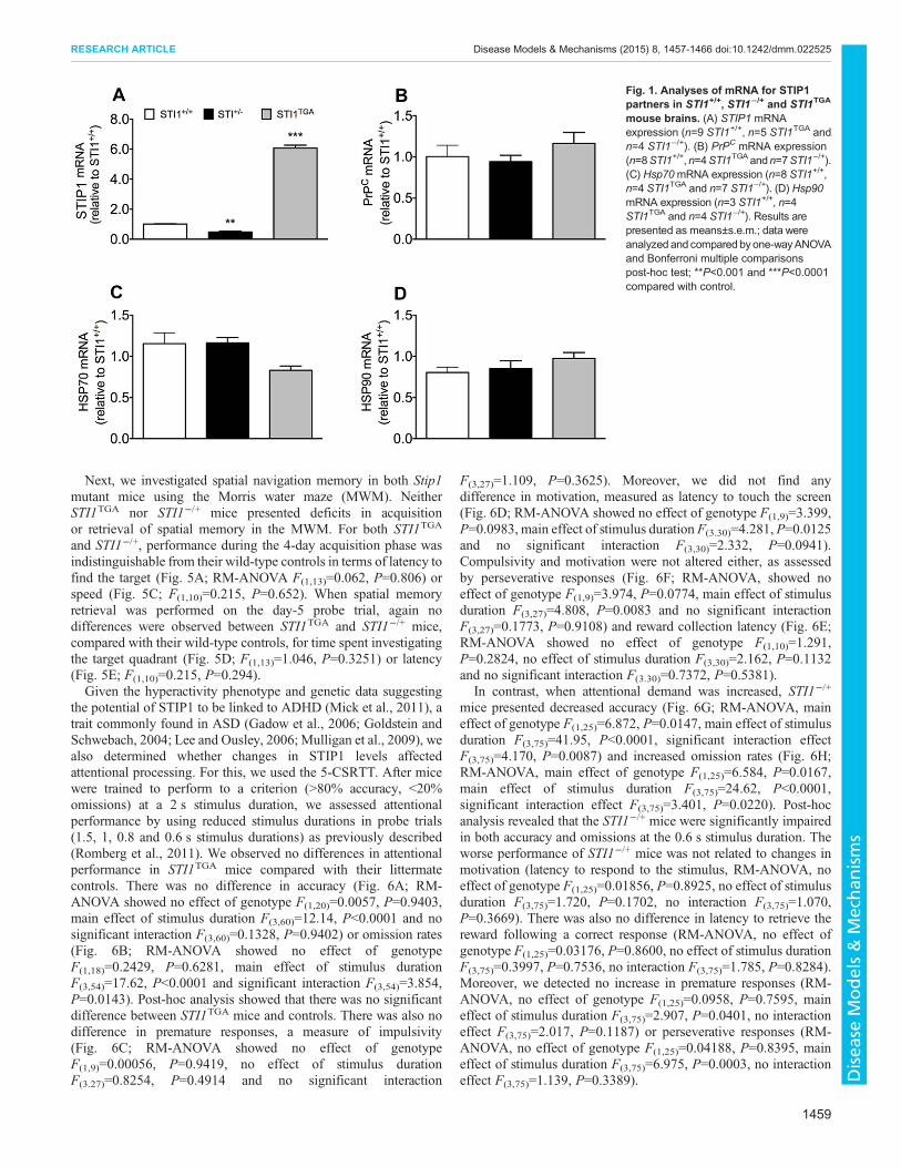
RESULTSWe initially confirmed previous data to show that STI1−/+ micepresent 50% of STIP1mRNA levels in their brain, whereas STI1TGA
mice express almost sixfold more mRNA (Fig. 1A; one-wayANOVA; revealed main effect of genotype F(2.15)=8.521,P<0.0001). In contrast, mRNA levels of known STIP1 interactionpartners PrPC (Fig. 1B; one-way ANOVA F(2.16)=1.475, P=0.2580),Hsp70 (Fig. 1C; one-way ANOVA F(2.16)=0.301, P=0.744)and Hsp90 (Fig. 1D; one-way ANOVA F(2.8)=1.249, P=0.337)were not altered in the brain of the two lines, compared withcontrol mice.Protein levels for STIP1 followed mRNA levels for both STI1TGA
(Fig. 2A; t(15)=4.721, P=0.003) and STI1−/+ (Fig. 2B; t(14)=6.433,
P<0.0001). PrPC protein levels were not different from controls inboth lines (Fig. 2C,D; t(10)=1.049, P=0.391 and t(13)=1.128,P=0.279, respectively). Interestingly, levels of Hsp70 weredecreased by 50% in STI1TGA brains (Fig. 2E; t(7)=5.846,P=0.0006), whereas no change in Hsp70 levels was detected inSTI1−/+ mice (Fig. 2F; t(7)=0.123, P=0.9051), compared withcontrols. Additionally, Hsp90 levels detected with a pan Hsp90antibody were doubled in STI1TGA brains (Fig. 2G; t(22)=4.618,P=0.0001) but not changed in STI1−/+ brains (Fig. 2H; t(10)=0.308,P=0.7639), compared with controls. We then evaluated expressionlevels of Hsp90α (inducible form) and Hsp90β (constitutive form)in the brains of STI1TGA mice and observed that both forms weresignificantly increased (Fig. 2I,J; t(22)=4.618, P=0.0016 andt(16)=5.954, P<0.0001, respectively).
Spontaneous locomotor activity in a new environment canprovide information on neuropsychiatric phenotypes in miceassociated with genetic mutations. The increased number of Stip1copies, with concomitant overexpression of Hsp90 and decreasedexpression of Hsp70 in STI1TGA mice did not seem to have anymajor impact on spontaneous locomotion (Fig. 3A,B; t(29)=1.140,P=0.942) or time spent in the center of the box, which providesinsight on anxiety-like behavior (Fig. 3C; t(29)=1.236, P=0.8669).In contrast, locomotor activity and total locomotion in a newenvironment were increased in STI1−/+ mice (Fig. 3D,E;t(44)=1.879, P=0.0078). However, STI1−/+ mice did not showincreased anxiety-like behavior, as determined by the time spent inthe center of the box (Fig. 3F; t(40)=1.221, P=0.341). We alsoexamined another cohort of STI1−/+ mice using automatedmetabolic cages. In this experiment, which mimics the home cageenvironment, STI1−/+ mice again showed hyperactivity during theday and night periods, considering both total activity (Fig. 3G;t(14)=2.558, P=0.0228 and t(14)=2.230, P=0.0426) and ambulatoryactivity (Fig. 3H; t(14)=2.420, P=0.00297 and t(14)=2.230,P=0.0426). Given this increased motor activity, STI1−/+ mice alsodemonstrated less sleep time (periods of inactivity) (Fig. 3I;t(14)=3949, P=0.0015 and t(14)=2.724, P=0.0165). Also, STI1
−/+
mice showed increased consumption of O2 during the light and darkcycle (Fig. 3J; t(14)=2.464, P=0.027 and t(14)=2.169, P=0.047) andCO2 production during the dark cycle, but not in the light cycle(Fig. 3K; t(14)=2.307, P=0.036 and t(14)=1.360, P=0.195). Nodifferences were observed in other parameters such as respiratoryratio (Fig. 3L; t(14)=0.4455, P=0.6627 and t(14)=0.459, P=0.653),food consumption (Fig. 3M; t(14)=0.5216, P=0.6101 andt(14)=0.6134, P=0.5494), water consumption (Fig. 3N;t(14)=1.801, P=0.0933 and t(14)=0.2752, P=0.7872), and heatproduction (Fig. 3O; t(14)=1.014, P=0.3276 and t(14)=0.1935,P=0.8494) comparing STI1−/+ to STI1+/+ mice for both cycles(light and dark).
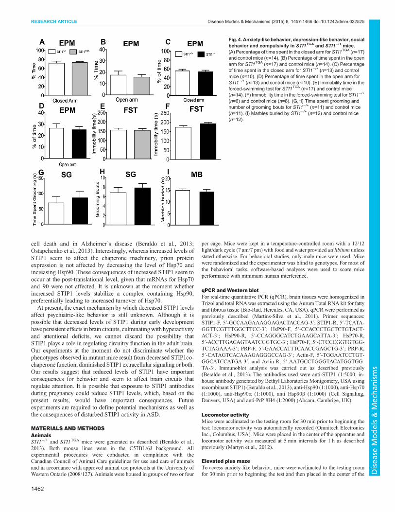
In order to test for other neuropsychiatric-like behaviors as a resultof altered STIP1 levels we tested both STITGA and STI1−/+ mice foranxiety-like behavior (Fig. 4A-D) and depression-like behavior(Fig. 4E,F). Given the hyperactivity of STI1−/+ mice, we alsodecided to investigate whether they had alterations in compulsive-like behavior, assessed by measurement of self-grooming andmarble burying (Fig. 4G-I). There was no difference in the behaviorof either STITGA (Fig. 4A,B,E) or STI1−/+ (Fig. 4C,D,F-I) micecompared with control mice in all these behavioral tasks: time spentin the open arm (Fig. 4C; t19=0.310, P=0.7590), time spent in theclosed arm (Fig. 4D; t(19)=0.3730, P=0.7133), forced swim test(Fig. 4F; t(12)=1.184, P=0.2594), grooming bouts (Fig. 4H;t(20)=0.7848, P=0.4418), time grooming (Fig. 4G; t(20)=0.6072,P=0.5505) and marble burying (Fig. 4I; t(21)=0.4956, P=0.6253).
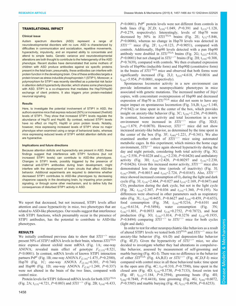
TRANSLATIONAL IMPACT
Clinical issue
Autism spectrum disorders (ASD) represent a range ofneurodevelopmental disorders with no cure. ASD is characterized bydifficulties in communication and socialization, repetitive movements,hyperactivity, impulsivity, and an impaired ability to concentrate andattend to simple tasks. Genetic variance and neurodevelopmentalalterations are both thought to contribute to the heterogeneity of the ASDphenotype. Recent studies have demonstrated that some mothers ofchildren with ASD produce antibodies against six specific proteinspresent in the fetal brain; presumably, these antibodies can interfere withprotein function in the developing brain. One of these antibodies targets aprotein known as stress inducible phosphoprotein 1 (STIP1). Moreover, apolymorphism for STIP1 was recently identified as a potential risk factorin attention deficit hyperactivity disorder, which shares some phenotypeswith ASD. STIP1 is a co-chaperone that mediates the Hsp70/Hsp90exchange of client proteins. It also triggers prion protein-mediatedneuronal signaling.
Results
Here, to investigate the potential involvement of STIP1 in ASD, theauthors examinemice that express reduced (50%) or increased (fivefold)levels of STIP1. They show that increased STIP1 levels regulate theabundance of Hsp70 and Hsp90. By contrast, reduced STIP1 levelshave no effect on Hsp70, Hsp90 or prion protein levels. Notably,however, mice expressing increased levels of STIP1 show no majorphenotype when examined using a range of behavioral tasks, whereasmice expressing reduced levels of STIP1 exhibit attention deficits andare hyperactive.
Implications and future directions
Because attention deficits and hyperactivity are present in ASD, thesefindings suggest that interference with STIP1 functions (but notincreased STIP1 levels) can contribute to ASD-like phenotypes.Changes in STIP1 levels, possibly triggered by the presence ofmaternal anti-STIP1 antibodies during brain development, mightinterfere with the development of brain circuits that affect ASD-likebehavior. Additional experiments are required to determine whetherdecreased STIP1 contributes to ASD-like phenotypes by decreasingchaperone capacity in the developing brain, by impairing prion proteinsignaling, or through some other mechanism, and to define fully theconsequences of disturbed STIP1 activity in ASD.
1458
RESEARCH ARTICLE Disease Models & Mechanisms (2015) 8, 1457-1466 doi:10.1242/dmm.022525
Disea
seModels&Mechan
isms
Next, we investigated spatial navigation memory in both Stip1mutant mice using the Morris water maze (MWM). NeitherSTI1TGA nor STI1−/+ mice presented deficits in acquisitionor retrieval of spatial memory in the MWM. For both STI1TGA
and STI1−/+, performance during the 4-day acquisition phase wasindistinguishable from their wild-type controls in terms of latency tofind the target (Fig. 5A; RM-ANOVA F(1,13)=0.062, P=0.806) orspeed (Fig. 5C; F(1,10)=0.215, P=0.652). When spatial memoryretrieval was performed on the day-5 probe trial, again nodifferences were observed between STI1TGA and STI1−/+ mice,compared with their wild-type controls, for time spent investigatingthe target quadrant (Fig. 5D; F(1,13)=1.046, P=0.3251) or latency(Fig. 5E; F(1,10)=0.215, P=0.294).Given the hyperactivity phenotype and genetic data suggesting
the potential of STIP1 to be linked to ADHD (Mick et al., 2011), atrait commonly found in ASD (Gadow et al., 2006; Goldstein andSchwebach, 2004; Lee and Ousley, 2006; Mulligan et al., 2009), wealso determined whether changes in STIP1 levels affectedattentional processing. For this, we used the 5-CSRTT. After micewere trained to perform to a criterion (>80% accuracy, <20%omissions) at a 2 s stimulus duration, we assessed attentionalperformance by using reduced stimulus durations in probe trials(1.5, 1, 0.8 and 0.6 s stimulus durations) as previously described(Romberg et al., 2011). We observed no differences in attentionalperformance in STI1TGA mice compared with their littermatecontrols. There was no difference in accuracy (Fig. 6A; RM-ANOVA showed no effect of genotype F(1,20)=0.0057, P=0.9403,main effect of stimulus duration F(3,60)=12.14, P<0.0001 and nosignificant interaction F(3,60)=0.1328, P=0.9402) or omission rates(Fig. 6B; RM-ANOVA showed no effect of genotypeF(1,18)=0.2429, P=0.6281, main effect of stimulus durationF(3,54)=17.62, P<0.0001 and significant interaction F(3,54)=3.854,P=0.0143). Post-hoc analysis showed that there was no significantdifference between STI1TGA mice and controls. There was also nodifference in premature responses, a measure of impulsivity(Fig. 6C; RM-ANOVA showed no effect of genotypeF(1,9)=0.00056, P=0.9419, no effect of stimulus durationF(3.27)=0.8254, P=0.4914 and no significant interaction
F(3,27)=1.109, P=0.3625). Moreover, we did not find anydifference in motivation, measured as latency to touch the screen(Fig. 6D; RM-ANOVA showed no effect of genotype F(1,9)=3.399,P=0.0983, main effect of stimulus duration F(3.30)=4.281, P=0.0125and no significant interaction F(3,30)=2.332, P=0.0941).Compulsivity and motivation were not altered either, as assessedby perseverative responses (Fig. 6F; RM-ANOVA, showed noeffect of genotype F(1,9)=3.974, P=0.0774, main effect of stimulusduration F(3,27)=4.808, P=0.0083 and no significant interactionF(3,27)=0.1773, P=0.9108) and reward collection latency (Fig. 6E;RM-ANOVA showed no effect of genotype F(1,10)=1.291,P=0.2824, no effect of stimulus duration F(3,30)=2.162, P=0.1132and no significant interaction F(3.30)=0.7372, P=0.5381).
In contrast, when attentional demand was increased, STI1−/+
mice presented decreased accuracy (Fig. 6G; RM-ANOVA, maineffect of genotype F(1,25)=6.872, P=0.0147, main effect of stimulusduration F(3,75)=41.95, P<0.0001, significant interaction effectF(3,75)=4.170, P=0.0087) and increased omission rates (Fig. 6H;RM-ANOVA, main effect of genotype F(1,25)=6.584, P=0.0167,main effect of stimulus duration F(3,75)=24.62, P<0.0001,significant interaction effect F(3,75)=3.401, P=0.0220). Post-hocanalysis revealed that the STI1−/+ mice were significantly impairedin both accuracy and omissions at the 0.6 s stimulus duration. Theworse performance of STI1−/+ mice was not related to changes inmotivation (latency to respond to the stimulus, RM-ANOVA, noeffect of genotype F(1,25)=0.01856, P=0.8925, no effect of stimulusduration F(3,75)=1.720, P=0.1702, no interaction F(3,75)=1.070,P=0.3669). There was also no difference in latency to retrieve thereward following a correct response (RM-ANOVA, no effect ofgenotype F(1,25)=0.03176, P=0.8600, no effect of stimulus durationF(3,75)=0.3997, P=0.7536, no interaction F(3,75)=1.785, P=0.8284).Moreover, we detected no increase in premature responses (RM-ANOVA, no effect of genotype F(1,25)=0.0958, P=0.7595, maineffect of stimulus duration F(3,75)=2.907, P=0.0401, no interactioneffect F(3,75)=2.017, P=0.1187) or perseverative responses (RM-ANOVA, no effect of genotype F(1,25)=0.04188, P=0.8395, maineffect of stimulus duration F(3,75)=6.975, P=0.0003, no interactioneffect F(3,75)=1.139, P=0.3389).
Fig. 1. Analyses of mRNA for STIP1partners in STI1+/+, STI1−/+ and STI1TGA
mouse brains. (A) STIP1 mRNAexpression (n=9 STI1+/+, n=5 STI1TGA andn=4 STI1−/+). (B) PrPC mRNA expression(n=8STI1+/+,n=4STI1TGAandn=7STI1−/+).(C) Hsp70mRNA expression (n=8 STI1+/+,n=4 STI1TGA and n=7 STI1−/+). (D) Hsp90mRNA expression (n=3 STI1+/+, n=4STI1TGA and n=4 STI1−/+). Results arepresented as means±s.e.m.; data wereanalyzed and compared byone-wayANOVAand Bonferroni multiple comparisonspost-hoc test; **P<0.001 and ***P<0.0001compared with control.
1459
RESEARCH ARTICLE Disease Models & Mechanisms (2015) 8, 1457-1466 doi:10.1242/dmm.022525
Disea
seModels&Mechan
isms

DISCUSSIONThe present experiments tested whether alterations in STIP1 levelshave consequences for psychiatric-like behaviors in mice. Ourresults suggest that decreased, but not increased, STIP1 levels causesignificant behavioral alterations in mice. Spatial learning andmemory, as well as anxiety and depression-like behavior do notseem to be affected by reduced STIP1 levels. However, mutant micedeficient for STIP1 are hyperactive and present attention deficits.STIP1 has recently emerged as a protein of potential interest in
ASD and endophenotypes related to ASD. Maternal autoantibodiesagainst STIP1 have been identified in mothers of children with ASD(Braunschweig et al., 2013). Moreover, recent global-wideassociation study (GWAS) analysis identified a polymorphism inSTIP1 (the human gene coding for STIP1/HOP) as a potential riskfactor in a population of individuals diagnosed with attention-deficitdisorder (Mick et al., 2011), a co-morbidity often associated withASD (Brimberg et al., 2013; Goldani et al., 2014). The consequencesof this polymorphism for STIP1 expression is unknown, but thepresence of autoantibodies against STIP1 might affect expressionlevels of the protein, given that antibodies can penetrate the bloodbrain barrier of the fetus during pregnancy (Braunschweig et al.,2012a; Diamond et al., 2009; Fox et al., 2012; Zhang et al., 2012).Indeed, maternal antibodies that recognize STIP1 and other targetswhen injected in pregnant rodents or developing pups can lead tooffspring with abnormal neurons and behaviors that relate to ASD
(Braunschweig et al., 2012b; Camacho et al., 2014). To a degree,STI1−/+ mice model this early developmental deficit in STIP1 levels.However, in STI1−/+ mice STIP1 expression is persistently decreasedthrough life, which could also have important consequences for thephenotypes described.
STIP1 is a modular protein containing several tetratricopeptide(TRP) repeat domains and aspartate-proline (DP) reach domains(Taipale et al., 2010). TRP1 and TRP2B can interact with Hsp70(Flom et al., 2007; Scheufler et al., 2000), whereas TPR2A is requiredfor interactionwithHsp90 (Flom et al., 2007, 2006). Hsp90 activity isregulated by STIP1 and previous work has shown that in mice noother co-chaperone can replace STIP1 (Beraldo et al., 2013). Recentexperiments have indicated that the chaperone machinery, activatedby the transcription factor heat shock factor 1 (HSF1), is responsiblefor preventing damaging effects from environmental factors in thedeveloping brain (Hashimoto-Torii et al., 2014). Indeed, thechaperone machinery can buffer many stresses at the cellular leveland, therefore, it is not surprising that functional changes in itscomponents have physiological consequences.
In addition to its intracellular chaperone function, STIP1 is alsosecreted by a myriad of cells, including astrocytes via anextracellular vesicle population, which includes exosomes (Hajjet al., 2013). Extracellular STIP1 also mediates importantphysiological responses in the brain. Acting as a trophic factor toengage PrPC to signal in neurons, it regulates neuritogenesis and
Fig. 2. Analyses of protein levels for STIP1 partners in STI1+/+, STI−/+ and STI1TGAmouse brains. (A,B) STIP1 expression in STI1TGA (n=9 STI1+/+ and n=8STI1TGA) and STI1−/+ mice (n=8 STI1+/+ and n=8 STI1−/+). (C,D) PrPC expression in STI1TGA (n=6 STI1+/+ and n=6 STI1TGA) and STI1−/+ mice (n=6 STI1+/+
and n=9 STI1−/+). (E,F) Hsp70 expression in STI1TGA (n=5 STI1+/+ and n=4 STI1TGA) and STI1−/+ mice (n=5 STI1+/+ and n=4 STI1−/+). (G,H) HSP90 expressionin STI1TGA (n=10 STI1+/+ and n=14 STI1TGA) and STI1−/+ mice (n=6 STI1+/+ and n=6 STI1−/+). (I,J) Hsp90β (n=10 STI1+/+ and n=8 STI1TGA) and Hsp90α (n=5STI1+/+ and n=4 STI1TGA) in STI1TGA mice. Results are presented as means±s.e.m.; data were analyzed and compared by Student’s t-test; *P<0.05 and***P<0.0001 compared with control.
1460
RESEARCH ARTICLE Disease Models & Mechanisms (2015) 8, 1457-1466 doi:10.1242/dmm.022525
Disea
seModels&Mechan
isms

neuronal survival (Beraldo et al., 2010; Lopes et al., 2005; Roffeet al., 2010). STIP1 has a role in functional recovery in stroke(Beraldo et al., 2013; Lee et al., 2013). Moreover, STIP1 alsomodulates toxicity of Aβ peptides in models of Alzheimer’s disease(Brehme et al., 2014; Ostapchenko et al., 2013).It is remarkable that mice with increased levels of STIP1 (up to
almost fivefold) do not present any major behavioral alteration. In
the extensive evaluation of cognitive phenotypes in this study,which included anxiety and depression-like behaviors, spatialmemory and attention, we showed that STI1TGA mice perform aswell as littermate controls. These results suggest that strategies toincrease STIP1 levels should not cause toxicity with consequencesfor brain functions. This is important, given that increased STIP1levels might be protective against insults such as stroke-mediated
Fig. 3. Locomotor activity in STI1TGA and STI1−/+mice andmetabolic analyses in STI1−/+mice. (A) Horizontal locomotor activity in an open-field for STI1TGA
(n=14) and STI+/+ control mice (n=14). (B) Cumulative 1 h locomotion for STI1TGA (n=14) and STI+/+ control mice (n=14). (C) Time spent in the center of thelocomotion boxes for STI1TGA (n=14) and STI+/+ control mice (n=14). (D) Horizontal locomotor activity in an open-field for STI1−/+ (n=8) and STI+/+ control mice(n=8). (E) Cumulative 1 h locomotion for STI1−/+ (n=22) and STI+/+ control mice (n=24). (F) Time spent in the center of the locomotion boxes for STI1−/+ (n=22)and STI+/+ control mice (n=24). (G) Total activity in metabolic cages for STI1−/+ (n=8) and STI+/+ control mice (n=8). (H) Ambulatory activity in metabolic cagesfor STI1−/+ (n=8) and STI+/+ control mice (n=8). (I) Sleep time for STI1−/+ (n=8) and STI+/+ control mice (n=8). (J) VO2 for STI1
−/+ (n=8) and STI+/+ control mice(n=8). (K) VCO2 for STI1
−/+ (n=8) and STI+/+ control mice (n=8). (L) Respiratory exchange ratio for STI1−/+ (n=8) and STI+/+ control mice (n=8). (M) Foodconsumption for STI1−/+ (n=8) and STI+/+ control mice (n=8). (N) Water consumption for STI1−/+ (n=8) and STI+/+ control mice (n=8). (O) Energy expenditure forSTI1−/+ (n=8) and STI+/+ control mice (n=8). Results are presented as means±s.e.m.; data were analyzed and compared by Student’s t-test; *P<0.05 comparedwith control.
1461
RESEARCH ARTICLE Disease Models & Mechanisms (2015) 8, 1457-1466 doi:10.1242/dmm.022525
Disea
seModels&Mechan
isms
cell death and in Alzheimer’s disease (Beraldo et al., 2013;Ostapchenko et al., 2013). Interestingly, whereas increased levels ofSTIP1 seem to affect the chaperone machinery, prion proteinexpression is not affected by decreasing the level of Hsp70 andincreasing Hsp90. These consequences of increased STIP1 seem tooccur at the post-translational level, given that mRNAs for Hsp70and 90 were not affected. It is unknown at the moment whetherincreased STIP1 levels stabilize a complex containing Hsp90,preferentially leading to increased turnover of Hsp70.At present, the exact mechanism by which decreased STIP1 levels
affect psychiatric-like behavior is still unknown. Although it ispossible that decreased levels of STIP1 during early developmenthave persistent effects in brain circuits, culminatingwith hyperactivityand attentional deficits, we cannot discard the possibility thatSTIP1 plays a role in regulating circuitry function in the adult brain.Our experiments at the moment do not discriminate whether thephenotypes observed in mutant mice result from decreased STIP1co-chaperone function, diminished STIP1 extracellular signaling or both.Our results suggest that reduced levels of STIP1 have importantconsequences for behavior and seem to affect brain circuits thatregulate attention. It is possible that exposure to STIP1 antibodiesduring pregnancy could reduce STIP1 levels, which, based on thepresent results, would have important consequences. Futureexperiments are required to define potential mechanisms as well asthe consequences of disturbed STIP1 activity in ASD.
MATERIALS AND METHODSAnimalsSTI1−/+ and STI1TGA mice were generated as described (Beraldo et al.,2013). Both mouse lines were in the C57BL/6J background. Allexperimental procedures were conducted in compliance with theCanadian Council of Animal Care guidelines for use and care of animalsand in accordance with approved animal use protocols at the University ofWestern Ontario (2008/127). Animals were housed in groups of two or four
per cage. Mice were kept in a temperature-controlled room with a 12/12light/dark cycle (7 am/7 pm) with food and water provided ad libitum unlessstated otherwise. For behavioral studies, only male mice were used. Micewere randomized and the experimenter was blind to genotypes. For most ofthe behavioral tasks, software-based analyses were used to score miceperformance with minimum human interference.
qPCR and Western blotFor real-time quantitative PCR (qPCR), brain tissues were homogenized inTrizol and total RNAwas extracted using the Aurum Total RNA kit for fattyand fibrous tissue (Bio-Rad, Hercules, CA, USA). qPCR were performed aspreviously described (Martins-Silva et al., 2011). Primer sequences:STIP1-F, 5′-GCCAAGAAAGGAGACTACCAG-3′; STIP1-R, 5′-TCATA-GGTTCGTTTGGCTTCC-3′; HsP90-F, 5′-CCACCCTGCTCTGTACT-ACT-3′; HsP90-R, 5′-CCAGGGCATCTGAAGCATTA-3′; HsP70-R,5′-ACCTTGACAGTAATCGGTGC-3′; HsP70-F, 5′-CTCCCGGTGTGG-TCTAGAAA-3′; PRP-F, 5′-GAACCATTTCAACCGAGCTG-3′; PRP-R,5′-CATAGTCACAAAGAGGGCCAG-3′; Actin-F, 5′-TGGAATCCTGT-GGCATCCATGA-3′; and Actin-R, 5′-AATGCCTGGGTACATGGTGG-TA-3′. Immunoblot analysis was carried out as described previously(Beraldo et al., 2013). The antibodies used were anti-STIP1 (1:5000, in-house antibody generated by Bethyl Laboratories Montgomery, USA usingrecombinant STIP1) (Beraldo et al., 2013), anti-Hsp90 (1:1000), anti-Hsp70(1:1000), anti-Hsp90α (1:1000), anti Hsp90β (1:1000) (Cell Signaling,Danvers, USA) and anti-PrP 8H4 (1:2000) (Abcam, Cambrige, UK).
Locomotor activityMice were acclimated to the testing room for 30 min prior to beginning thetest; locomotor activity was automatically recorded (Omnitech ElectronicsInc., Columbus, USA). Mice were placed in the center of the apparatus andlocomotor activity was measured at 5 min intervals for 1 h as describedpreviously (Martyn et al., 2012).
Elevated plus mazeTo access anxiety-like behavior, mice were acclimated to the testing roomfor 30 min prior to beginning the test and then placed in the center of the
Fig. 4. Anxiety-like behavior, depression-like behavior, socialbehavior and compulsivity in STI1TGA and STI1−/+ mice.(A) Percentage of time spent in the closed arm for STI1TGA (n=17)and control mice (n=14). (B) Percentage of time spent in the openarm for STI1TGA (n=17) and control mice (n=14). (C) Percentageof time spent in the closed arm for STI1−/+ (n=13) and controlmice (n=10). (D) Percentage of time spent in the open arm forSTI1−/+ (n=13) and control mice (n=10). (E) Immobility time in theforced-swimming test for STI1TGA (n=17) and control mice(n=14). (F) Immobility time in the forced-swimming test forSTI1−/+
(n=6) and control mice (n=8). (G,H) Time spent grooming andnumber of grooming bouts for STI1−/+ (n=11) and control mice(n=11). (I) Marbles buried by STI1−/+ (n=12) and control mice(n=12).
1462
RESEARCH ARTICLE Disease Models & Mechanisms (2015) 8, 1457-1466 doi:10.1242/dmm.022525
Disea
seModels&Mechan
isms

elevated plus maze (Med Associates Inc., St Albans, USA). The activity wasrecorded and videos were analyzed using ANY-maze software (StoeltingCo., USA) to determine the amount of time spent in the closed and opensections of the maze.
Forced swimming testDepressive-like behavior was assessed by a forced swim test (FST) asdescribed previously (Martyn et al., 2012). Briefly, mice were placed in a 2 lbeaker containing 1.7 l of water at 25-27°C for 6 min. Experimental sessionswere recorded and immobility time was evaluated using ANY-MazeSoftware (Stoelting Co., USA). Data obtained from the last 4 min of testingwere used for the analysis.
Morris water mazeThe spatial version of Morris water maze (MWM) was conducted asdescribed previously (Kolisnyk et al., 2013; Martyn et al., 2012; Vorhees andWilliams, 2006). Briefly, the task was performed in a 1.5-m diameter/1-mdeep pool filled with water at 25°C. Spatial cues, 40×40 cm boards containing
black symbols (vertical and horizontal stripes, triangles, squares and circles),were placed on the walls distributed around the pool and the platform wassubmerged 1 cm below the surface of the water. Mice were submitted to fourtraining trials a day (90 s each) for four consecutive days with a 15 minintertrial interval. On day 5, memory was assessed by a single 60 s trial onwhich the platform was removed and the time spent in the target quadrant wasevaluated.All the experimental sessionswere recorded and analyzed using theANY-Maze Software.
Five-choice serial reaction time taskThe five-choice serial reaction time task (5-CSRTT) was used toevaluate attention in mice as described previously (Kolisnyk et al., 2013;Romberg et al., 2011). Mice were trained in the 5-CSRTT in automatedBussey–SaksidaTouch screen systems (Campden Instruments Limited,Loughborough, EN) and the data collected using ABET II Touch softwareV.2.18 (Lafayette Instruments, Lafayette, USA). Mice were submitted to apre-training program, which consisted of first habituating the mouse to thetesting chamber with the lights off for 10 min. The next day, the mouse was
Fig. 5. Spatial memory in STI1TGA and STI1−/+ mice. Forthe tests, n=14 STI1+/+ and 14 STI1TGA mice were used totest spatial memory in STI1TGA mice and n=11 STI1+/+ and11 STI1−/+ for STI1−/+ mice. (A) Latency to find the platform.(B) Distance traveled. (C) Speed for STI1TGA mice.(D) Percentage time spent by STI1TGA mice and controls intarget quadrant (T) and in opposite (O), right (R) and left (L)quadrants was measured on day 5 in a 60 s probe trial withthe platform removed. (E) Latency to find the platform.(F) Distance traveled. (G) Speed for STI1−/+ mice.(H) Percentage time spent by STI1−/+ mice and controls ineach quadrant was measured on day 5 in a 60 s probetrial with the platform removed. Results are presented asmeans±s.e.m.; data were analyzed and compared bytwo-way ANOVA; ***P<0.001 and ****P<0.0001 comparedwith time spent in target quadrant.
1463
RESEARCH ARTICLE Disease Models & Mechanisms (2015) 8, 1457-1466 doi:10.1242/dmm.022525
Disea
seModels&Mechan
isms

put in the chamber with the lights off for 20 min. After two days ofhabituation with no reward been offered, the reward tray was primed with11% fat strawberry milkshake (Nielson - Saputo Dairy Products) and a tonewas played when themouse entered the reward tray. This was repeated for thenext 2 days for 40 min sessions. Whenever the mouse returned to the rewardtray, the reward was offered and paired with a tone (phase I). The followingtraining phase consisted in pairing the reward with the presentation of arandom stimulus (flash of light in one of the five windows), which isremoved after 30 s. At this phase, if the mouse touched the screen when thestimulus was displayed, it received a reward. This cyclewas repeated until themouse completed 30 trials or 60 min timeout (phase II). At phase III of thetraining, the stimulus was displayed randomly in one of the five windows.The mouse had to touch the window where the stimulus was displayed toreceive the reward paired with a tone. Similar to phase II, this cycle wasrepeated until themouse completed 30 trials or 60 min timeout. The next step(phase IV) was identical to phase III except by the fact that the mouse had topoke its nose into the reward trail to initiate the task. This process wasrepeated in the last phase of the pre-training (phase V); however, if themouse
touched an incorrect screen, it received a 5 s timeout and the light in thechamber was turned on. After the mouse had finished pre-training andreached criterion at 4 s and 2 s stimulus duration (80% accuracy, 20%omission for three consecutive days), mice were probed for attention deficitsfollowing probe trial schedules: each mouse was tested over two sessions at1.5, 1.0, 0.8 and 0.6 s stimulus duration (the order of the probe trial sessionswas randomized and the groups counterbalanced). Between each differentstimulus duration, each mouse was returned to a 2 s stimulus for twoconsecutive sessions. Number of trials to criterion, accuracy, omission,reward collection latency and perseverative response were analyzed.
Metabolic assessmentsOxygen consumption, carbon dioxide production, respiratory exchangeratio (RER), carbon dioxide production, water and food intake and physicalactivity were simultaneously measured for adult STI1+/+ and STI1+/− miceby using the Comprehensive Lab Animal Monitoring System (CLAMS)interfaced with Oxymax Software (Columbus Instruments, Columbus, OH,USA) as previously described in detail (Guzman et al., 2013; Kolisnyk et al.,
Fig. 6. Five-choice serial reaction time task used tomeasure attention inSTI1TGA andSTI1−/+. For the tests, n=10STI1+/+ and 10STI1TGAmicewere used totest attention in STI1TGA mice and n=13 STI1+/+ and 13 STI1−/+ for STI1−/+ mice. (A) Accuracy during probe trial sessions. (B) Rate of omission. (C) Prematureresponses. (D) Response latency. (E) Reward collection latency. (F) Perseverative responses for STI1TGA mice. (G) Accuracy during probe trial sessions.(H) Rate of omission. (I) Premature responses. (J) Response latency. (K) Reward collection latency. (L) Perseverative response for STI1−/+ mice. Results arepresented as means±s.e.m.; data were analyzed and compared by RM-ANOVA; *P<0.05, **P<0.001 compared with control.
1464
RESEARCH ARTICLE Disease Models & Mechanisms (2015) 8, 1457-1466 doi:10.1242/dmm.022525
Disea
seModels&Mechan
isms

2013). Briefly, mice were individually housed in the metabolic chamberswith ad libitum access to water and food. Following a 16-h habituationperiod, all measurements were obtained every 10 min for 24 h (12 h light/12 h dark).
Marble burying taskA marble burying task was used to assess repetitive and anxiety-likebehavior as previously described (Deacon, 2006).
Assessment of self-groomingSelf-grooming was assessed to evaluate repetitive behavior, as previouslydescribed (McFarlane et al., 2008). Briefly, each mouse was placedindividually in a clean, empty, cage and given a 10 min habituation period,after which the mice were filmed for another 10 min. Cumulative time spentgrooming and number of grooming bouts were counted by an experimenterblinded to the genotypes of the mice.
Statistical analysesData are presented as mean±s.e.m. Statistical analyses were performed usingSigmaStat 3.5 software. Student’s t-test was used to compare twoexperimental groups and for comparison of several experimental groups,two-way ANOVA or two-way repeated-measures ANOVA were used asrequired. Tukey’s post hoc comparison was used when required.
Competing interestsThe authors declare no competing or financial interests.
Author contributionsF.H.B., M.A.M.P., V.F.P., and R.G. conceived and designed experiments. F.H.B.,A.T., B.K., P.H.H., R.G., X.D.J., A.C.M., J.F., D.F.G., M.F.C. and T.M. performed theexperiments. V.R.M. contributed with specific reagents. F.H.B., A.T., B.K., A.C.M.,V.F.P., R.G., V.R.M. and M.A.M.P. analyzed the data. F.H.B., V.F.P. and M.A.M.P.wrote the paper.
FundingThis work was supported by the Canadian Institute of Health Research (MOP136930,MOP 126000 andMOP 89919; M.A.M.P. and V.F.P.), Canadian Foundationfor Innovation (M.A.M.P., V.F.P. and R.G.) and Fundaça ̃o de Amparo a Pesquisa doEstado de Sa ̃o Paulo, Brazil (FAPESP-2009/14027-2; V.R.M.).
ReferencesAbbas-Terki, T., Briand, P.-A., Donze, O. and Picard, D. (2002). The Hsp90 co-chaperones Cdc37 and Sti1 interact physically and genetically. Biol. Chem. 383,1335-1342.
Bauman, M. D., Iosif, A.-M., Ashwood, P., Braunschweig, D., Lee, A.,Schumann, C. M., Van de Water, J. and Amaral, D. G. (2013). Maternalantibodies from mothers of children with autism alter brain growth and socialbehavior development in the rhesus monkey. Transl. Psychiatry 3, e278.
Beraldo, F. H., Arantes, C. P., Santos, T. G., Queiroz, N. G. T., Young, K., Rylett,R. J., Markus, R. P., Prado, M. A. M. and Martins, V. R. (2010). Role of alpha7nicotinic acetylcholine receptor in calcium signaling induced by prion proteininteraction with stress-inducible protein 1. J. Biol. Chem. 285, 36542-36550.
Beraldo, F. H., Soares, I. N., Goncalves, D. F., Fan, J., Thomas, A. A., Santos,T. G., Mohammad, A. H., Roffe, M., Calder, M. D., Nikolova, S. et al. (2013).Stress-inducible phosphoprotein 1 has unique cochaperone activity duringdevelopment and regulates cellular response to ischemia via the prion protein.FASEB J. 27, 3594-3607.
Braunschweig, D., Duncanson, P., Boyce, R., Hansen, R., Ashwood, P.,Pessah, I. N., Hertz-Picciotto, I. and Van de Water, J. (2012a). Behavioralcorrelates of maternal antibody status among children with autism. J. Autism Dev.Disord. 42, 1435-1445.
Braunschweig, D., Golub, M. S., Koenig, C. M., Qi, L., Pessah, I. N., Van deWater, J. and Berman, R. F. (2012b). Maternal autism-associated IgG antibodiesdelay development and produce anxiety in a mouse gestational transfer model.J. Neuroimmunol. 252, 56-65.
Braunschweig, D., Krakowiak, P., Duncanson, P., Boyce, R., Hansen, R. L.,Ashwood, P., Hertz-Picciotto, I., Pessah, I. N. and Van de Water, J. (2013).Autism-specific maternal autoantibodies recognize critical proteins in developingbrain. Transl. Psychiatry 3, e277.
Brehme, M., Voisine, C., Rolland, T., Wachi, S., Soper, J. H., Zhu, Y., Orton, K.,Villella, A., Garza, D., Vidal, M. et al. (2014). A chaperome subnetworksafeguards proteostasis in aging and neurodegenerative disease. Cell Rep. 9,1135-1150.
Brimberg, L., Sadiq, A., Gregersen, P. K. and Diamond, B. (2013). Brain-reactiveIgG correlates with autoimmunity in mothers of a child with an autism spectrumdisorder. Mol. Psychiatry 18, 1171-1177.
Caetano, F. A., Lopes, M. H., Hajj, G. N. M., Machado, C. F., Pinto Arantes, C.,Magalhaes, A. C., Vieira, M. d. P. B., Americo, T. A., Massensini, A. R., Priola,S. A. et al. (2008). Endocytosis of prion protein is required for ERK1/2 signalinginduced by stress-inducible protein 1. J. Neurosci. 28, 6691-6702.
Camacho, J., Jones, K., Miller, E., Ariza, J., Noctor, S., Van de Water, J. andMartinez-Cerdeno, V. (2014). Embryonic intraventricular exposure to autism-specific maternal autoantibodies produces alterations in autistic-like stereotypicalbehaviors in offspring mice. Behav. Brain Res. 266, 46-51.
Chen, S., Prapapanich, V., Rimerman, R. A., Honore, B. and Smith, D. F. (1996).Interactions of p60, a mediator of progesterone receptor assembly, with heatshock proteins hsp90 and hsp70. Mol. Endocrinol. 10, 682-693.
Chen, X., Zhao, C., Li, X., Wang, T., Li, Y., Cao, C., Ding, Y., Dong, M., Finci, L.,Wang, J.-H. et al. (2015). Terazosin activates Pgk1 and Hsp90 to promote stressresistance. Nat. Chem. Biol. 11, 19-25.
Dalton, P., Deacon, R., Blamire, A., Pike, M., McKinlay, I., Stein, J., Styles, P. andVincent, A. (2003). Maternal neuronal antibodies associated with autism and alanguage disorder. Ann. Neurol. 53, 533-537.
Deacon, R. M. J. (2006). Digging and marble burying in mice: simple methods for invivo identification of biological impacts. Nat. Protoc. 1, 122-124.
Diamond, B., Huerta, P. T., Mina-Osorio, P., Kowal, C. and Volpe, B. T. (2009).Losing your nerves? Maybe it’s the antibodies. Nat. Rev. Immunol. 9, 449-456.
Erlich, R. B., Kahn, S. A., Lima, F. R. S., Muras, A. G., Martins, R. A. P., Linden,R., Chiarini, L. B., Martins, V. R. and Moura Neto, V. (2007). STI1 promotesglioma proliferation through MAPK and PI3K pathways. Glia 55, 1690-1698.
Eustace, B. K. and Jay, D. G. (2004). Extracellular roles for the molecularchaperone, hsp90. Cell Cycle 3, 1096-1098.
Flom, G., Weekes, J., Williams, J. J. and Johnson, J. L. (2006). Effect of mutationof the tetratricopeptide repeat and asparatate-proline 2 domains of Sti1 on Hsp90signaling and interaction in Saccharomyces cerevisiae. Genetics 172, 41-51.
Flom,G., Behal, R. H., Rosen, L., Cole, D. G. and Johnson, J. L. (2007). Definitionof the minimal fragments of Sti1 required for dimerization, interaction with Hsp70and Hsp90 and in vivo functions. Biochem. J. 404, 159-167.
Fox, E., Amaral, D. and Van de Water, J. (2012). Maternal and fetal antibrainantibodies in development and disease. Dev. Neurobiol. 72, 1327-1334.
Gadow, K. D., DeVincent, C. J. and Pomeroy, J. (2006). ADHD symptom subtypesin children with pervasive developmental disorder. J. Autism Dev. Disord. 36,271-283.
Goldani, A. A. S., Downs, S. R., Widjaja, F., Lawton, B. and Hendren, R. L.(2014). Biomarkers in autism. Front. Psychiatry 5, 100.
Goldstein, S. and Schwebach, A. J. (2004). The comorbidity of PervasiveDevelopmental Disorder and Attention Deficit Hyperactivity Disorder: results of aretrospective chart review. J. Autism Dev. Disord. 34, 329-339.
Guzman, M. S., De Jaeger, X., Drangova, M., Prado, M. A. M., Gros, R. andPrado, V. F. (2013). Micewith selective elimination of striatal acetylcholine releaseare lean, show altered energy homeostasis and changed sleep/wake cycle.J. Neurochem. 124, 658-669.
Hajj, G. N. M., Arantes, C. P., Dias, M. V. S., Roffe, M., Costa-Silva, B., Lopes,M. H., Porto-Carreiro, I., Rabachini, T., Lima, F. R., Beraldo, F. H. et al. (2013).The unconventional secretion of stress-inducible protein 1 by a heterogeneouspopulation of extracellular vesicles. Cell. Mol. Life Sci. 70, 3211-3227.
Hashimoto-Torii, K., Torii, M., Fujimoto, M., Nakai, A., El Fatimy, R., Mezger, V.,Ju, M. J., Ishii, S., Chao, S.-H., Brennand, K. J. et al. (2014). Roles of heat shockfactor 1 in neuronal response to fetal environmental risks and its relevance to braindisorders. Neuron 82, 560-572.
Kolisnyk, B., Guzman, M. S., Raulic, S., Fan, J., Magalhaes, A. C., Feng, G.,Gros, R., Prado, V. F. and Prado, M. A. M. (2013). ChAT-ChR2-EYFPmice haveenhanced motor endurance but show deficits in attention and several additionalcognitive domains. J. Neurosci. 33, 10427-10438.
Lee, D. O. and Ousley, O. Y. (2006). Attention-deficit hyperactivity disordersymptoms in a clinic sample of children and adolescents with pervasivedevelopmental disorders. J. Child Adolesc. Psychopharmacol. 16, 737-746.
Lee, S.-D., Lai, T. W., Lin, S.-Z., Lin, C.-H., Hsu, Y.-H., Li, C.-Y., Wang, H.-J., Lee,W., Su, C.-Y., Yu, Y.-L. et al. (2013). Role of stress-inducible protein-1 inrecruitment of bone marrow derived cells into the ischemic brains. EMBO Mol.Med. 5, 1227-1246.
Lima, F. R. S., Arantes, C. P., Muras, A. G., Nomizo, R., Brentani, R. R. andMartins, V. R. (2007). Cellular prion protein expression in astrocytes modulatesneuronal survival and differentiation. J. Neurochem. 103, 2164-2176.
Lopes, M. H., Hajj, G. N. M., Muras, A. G., Mancini, G. L., Castro, R. M. P. S.,Ribeiro, K. C. B., Brentani, R. R., Linden, R. and Martins, V. R. (2005).Interaction of cellular prion and stress-inducible protein 1 promotesneuritogenesis and neuroprotection by distinct signaling pathways. J. Neurosci.25, 11330-11339.
Martinez-Cerdeno, V., Camacho, J., Fox, E., Miller, E., Ariza, J., Kienzle, D.,Plank, K., Noctor, S. C. and Van de Water, J. (2014). Prenatal exposure toautism-specific maternal autoantibodies alters proliferation of cortical neural
1465
RESEARCH ARTICLE Disease Models & Mechanisms (2015) 8, 1457-1466 doi:10.1242/dmm.022525
Disea
seModels&Mechan
isms

precursor cells, enlarges brain, and increases neuronal size in adult animals.Cereb. Cortex. [Epub ahead of print] doi:10.1093/cercor/bhu291.
Martins-Silva, C., De Jaeger, X., Guzman, M. S., Lima, R. D. F., Santos, M. S.,Kushmerick, C., Gomez, M. V., Caron, M. G., Prado, M. A. M. and Prado, V. F.(2011). Novel strains of mice deficient for the vesicular acetylcholine transporter:insights on transcriptional regulation and control of locomotor behavior. PLoSONE 6, e17611.
Martyn, A. C., De Jaeger, X., Magalhaes, A. C., Kesarwani, R., Goncalves, D. F.,Raulic, S., Guzman, M. S., Jackson, M. F., Izquierdo, I., Macdonald, J. F. et al.(2012). Elimination of the vesicular acetylcholine transporter in the forebraincauses hyperactivity and deficits in spatial memory and long-term potentiation.Proc. Natl. Acad. Sci. USA 109, 17651-17656.
McFarlane, H. G., Kusek, G. K., Yang, M., Phoenix, J. L., Bolivar, V. J. andCrawley, J. N. (2008). Autism-like behavioral phenotypes in BTBR T+tf/J mice.Genes Brain Behav. 7, 152-163.
Mick, E., McGough, J., Loo, S., Doyle, A. E., Wozniak, J., Wilens, T. E., Smalley,S., McCracken, J., Biederman, J. and Faraone, S. V. (2011). Genome-wideassociation study of the child behavior checklist dysregulation profile. J. Am. Acad.Child Adolesc. Psychiatry 50, 807-817.e8.
Mulligan, A., Anney, R. J. L., O’Regan, M., Chen, W., Butler, L., Fitzgerald, M.,Buitelaar, J., Steinhausen, H.-C., Rothenberger, A., Minderaa, R. et al. (2009).Autism symptoms in Attention-Deficit/Hyperactivity Disorder: a familial trait whichcorrelates with conduct, oppositional defiant, language and motor disorders.J. Autism Dev. Disord. 39, 197-209.
Nicolet, C. M. and Craig, E. A. (1989). Isolation and characterization of STI1, astress-inducible gene from Saccharomyces cerevisiae. Mol. Cell. Biol. 9,3638-3646.
Nordahl, C.W., Braunschweig, D., Iosif, A.-M., Lee, A., Rogers, S., Ashwood, P.,Amaral, D. G. and Van de Water, J. (2013). Maternal autoantibodies areassociated with abnormal brain enlargement in a subgroup of children with autismspectrum disorder. Brain Behav. Immun. 30, 61-65.
Ostapchenko, V. G., Beraldo, F. H., Mohammad, A. H., Xie, Y.-F., Hirata, P. H. F.,Magalhaes, A. C., Lamour, G., Li, H., Maciejewski, A., Belrose, J. C. et al.(2013). The prion protein ligand, stress-inducible phosphoprotein 1, regulatesamyloid-beta oligomer toxicity. J. Neurosci. 33, 16552-16564.
Picard, D. (2002). Heat-shock protein 90, a chaperone for folding and regulation.Cell. Mol. Life Sci. 59, 1640-1648.
Roffe, M., Beraldo, F. H., Bester, R., Nunziante, M., Bach, C., Mancini, G., Gilch,S., Vorberg, I., Castilho, B. A., Martins, V. R. et al. (2010). Prion proteininteraction with stress-inducible protein 1 enhances neuronal protein synthesis viamTOR. Proc. Natl. Acad. Sci. USA 107, 13147-13152.
Romberg, C., Mattson, M. P., Mughal, M. R., Bussey, T. J. and Saksida, L. M.(2011). Impaired attention in the 3xTgAD mouse model of Alzheimer’s disease:rescue by donepezil (Aricept). J. Neurosci. 31, 3500-3507.
Scheufler, C., Brinker, A., Bourenkov, G., Pegoraro, S., Moroder, L., Bartunik,H., Hartl, F. U. and Moarefi, I. (2000). Structure of TPR domain-peptidecomplexes: critical elements in the assembly of the Hsp70-Hsp90multichaperonemachine. Cell 101, 199-210.
Smith, D. F., Sullivan, W. P., Marion, T. N., Zaitsu, K., Madden, B., McCormick,D. J. and Toft, D. O. (1993). Identification of a 60-kilodalton stress-related protein,p60, which interacts with hsp90 and hsp70. Mol. Cell. Biol. 13, 869-876.
Soares, I. N., Caetano, F. A., Pinder, J., Rodrigues, B. R., Beraldo, F. H.,Ostapchenko, V. G., Durette, C., Pereira, G. S., Lopes, M. H., Queiroz-Hazarbassanov, N. et al. (2013). Regulation of stress-inducible phosphoprotein1 nuclear retention by protein inhibitor of activated STAT PIAS1. Mol. CellProteomics 12, 3253-3270.
Taipale, M., Jarosz, D. F. and Lindquist, S. (2010). HSP90 at the hub of proteinhomeostasis: emerging mechanistic insights. Nat. Rev. Mol. Cell Biol. 11,515-528.
Taipale, M., Tucker, G., Peng, J., Krykbaeva, I., Lin, Z.-Y., Larsen, B., Choi, H.,Berger, B., Gingras, A.-C. and Lindquist, S. (2014). A quantitative chaperoneinteraction network reveals the architecture of cellular protein homeostasispathways. Cell 158, 434-448.
Vorhees, C. V. and Williams, M. T. (2006). Morris water maze: procedures forassessing spatial and related forms of learning and memory. Nat. Protoc. 1,848-858.
Wang, T.-H., Chao, A., Tsai, C.-L., Chang, C.-L., Chen, S.-H., Lee, Y.-S., Chen, J.-K., Lin, Y.-J., Chang, P.-Y., Wang, C.-J. et al. (2010). Stress-inducedphosphoprotein 1 as a secreted biomarker for human ovarian cancer promotescancer cell proliferation. Mol. Cell Proteomics 9, 1873-1884.
Zhang, Y., Bolivar, V. J. and Lawrence, D. A. (2012). Developmental exposure tomercury chloride does not impair social behavior of C57BL/6×BTBR F(1) mice.J. Immunotoxicol. 9, 401-410.
1466
RESEARCH ARTICLE Disease Models & Mechanisms (2015) 8, 1457-1466 doi:10.1242/dmm.022525
Disea
seModels&Mechan
isms