Embed Size (px)
Citation preview
416 pISSN 0301-2875, eISSN 2005-3789
Introduction
Peri-implant disease was first introduced in 1987, and was defined as ‘a site-specific infection which yields many features in common with chronic periodontitis.’1 It was classified into peri-implant mu-cositis and peri-implantitis in 1992 and has been revised up to date.2 Nowadays, peri-implant mucositis is considered a state of ‘reversible inflammatory reactions in the soft tissues surrounding a functioning implant’, and peri-implantitis is defined as a state of ‘inflammatory reactions with loss of supporting bone in the tissues surrounding a functioning implant’.2 Of the two, peri-implantitis is a disease that can severely affect an existing implant.1
There are 6 main signs and symptoms of peri-implantitis. (1) Bleeding on probing is always present, (2) marginal tissue may be swollen or red, (3) increase in probing depth with loss of attachment,
(4) pain in case of severe bone loss, (5) mobility, and (6) loss of bone levels around implants in radiographic evaluation.1,3 Due to osseo-integration at the apex, loss of bone can be present without signs of mobility, and hence, an implant without mobility does not necessarily mean that it is disease-free.4,5 Peri-implantitis can be caused by many factors. Bacterial colonization is mainly the primary cause, and while it is controversial, mechanical overload can also be a contributing factor.6,7 History of periodontitis is also known to elevate the risk fac-tors as reported by Karoussis et al.4; 28.6% of patients with history of chronic periodontitis were affected while only 5.8% were affected in periodontally healthy subjects.
In a recent systematic review, Lee et al.8 reported a peri-implantitis prevalence of 19.8% at patient level, and 9.3% at implant level. Ac-cording to this review, almost 20% of patients are at risk of having peri-implantitis, and as the number of patients with dental implants
https://doi.org/10.4047/jkap.2019.57.4.416CASE REpORT
Peri-implantitis 환자에서의 전악 재수복 증례
최낙현 � 조영은 � 박은진*
이화여자대학교 의과대학 치과보철학교실
Full mouth rehabilitation in a patient with peri-implantitis: A case report
Nak-Hyun Choi, Young-Eun Cho, Eun-Jin Park*Department of Prosthodontics, School of Medicine, Ewha Womans University, Seoul, Republic of Korea
Peri-implantitis appears in almost 20% of patients who received implant treatment, and increase in its number is inevitable as time goes by. Although it can be treated by both non-surgical and surgical procedures, in cases which include severe bone loss, explantation and rehabilitation may be necessary. Careful treatment planning and consider-ations to prevent recurrent peri-implantitis should be taken into account. In the following case presented, a patient with chronic periodontitis and peri-implantitis was success-fully rehabilitated after removal of several implants. Extraction and explantation of multiple teeth and implants were followed by full mouth reconstruction with fixed implant prostheses on the mandible and implant retained overdenture on the maxilla. Surgical and prosthetic measures to prevent recurrent peri-implantitis were taken into consider-ation. (J Korean Acad Prosthodont 2019;57:416-24)
Keywords: Peri-implantitis; Full mouth rehabilitation; Zirconia; Computer-aided design and computer-aided manufacturing (CAD/CAM); Implant removal
*Corresponding Author: Eun-Jin ParkDepartment of Prosthodontics, School of Medicine, Ewha Womans University, Ewha Womans University Mokdong Hospital, 1071 Anyangcheon-ro, Yangcheon-gu, Seoul, Republic of Korea+82 (0)2 2650 5042: e-mail, [email protected] history: Received June 11, 2019 / Last Revision July 25, 2019 / Accepted August 1, 2019
2019 The Korean Academy of ProsthodonticsThis is an Open Access article distributed under the terms of the Creative Commons Attribution Non-Commercial License (http://creativecommons.org/licenses/by-nc/4.0) which permits unrestricted non-commercial use, distribution, and reproduction in any medium, provided the original work is properly cited.
c
cc
대한치과보철학회지 57권 4호, 2019년 10월 417
최낙현�조영은�박은진 Peri-implantitis 환자에서의 전악 재수복 증례
increases, subsequent increase in the incidence of peri-implant infec-tions is inevitable. With the numbers increasing, more attention is drawn to preventing and treating these infections. Most treatment methods are focused on periodontal and surgical means.9 Meanwhile, preventing such infections is a more multi-disciplinary process, in which prosthodontist plays a major role.10 In the prosthodontic point of view, one should select materials less prone to bacterial coloniza-tion and create them in a cleansable shape. This includes precise marginal fit, cleansable interproximal contours, smooth emergence profile and avoiding over-contoured form.5,10 This case report focuses on a successfully rehabilitated peri-implantitis patient, raising aware-ness of peri-implantitis while discussing the role of prosthodontist in creating a perio-friendly prosthesis.
Case Report
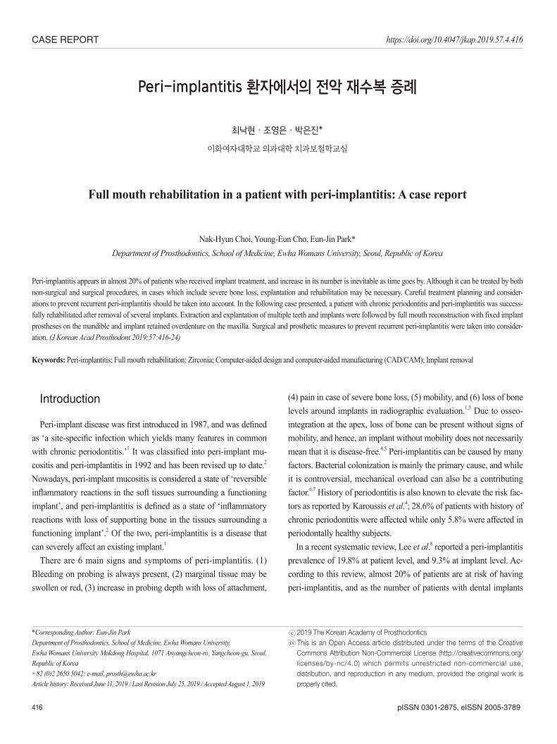
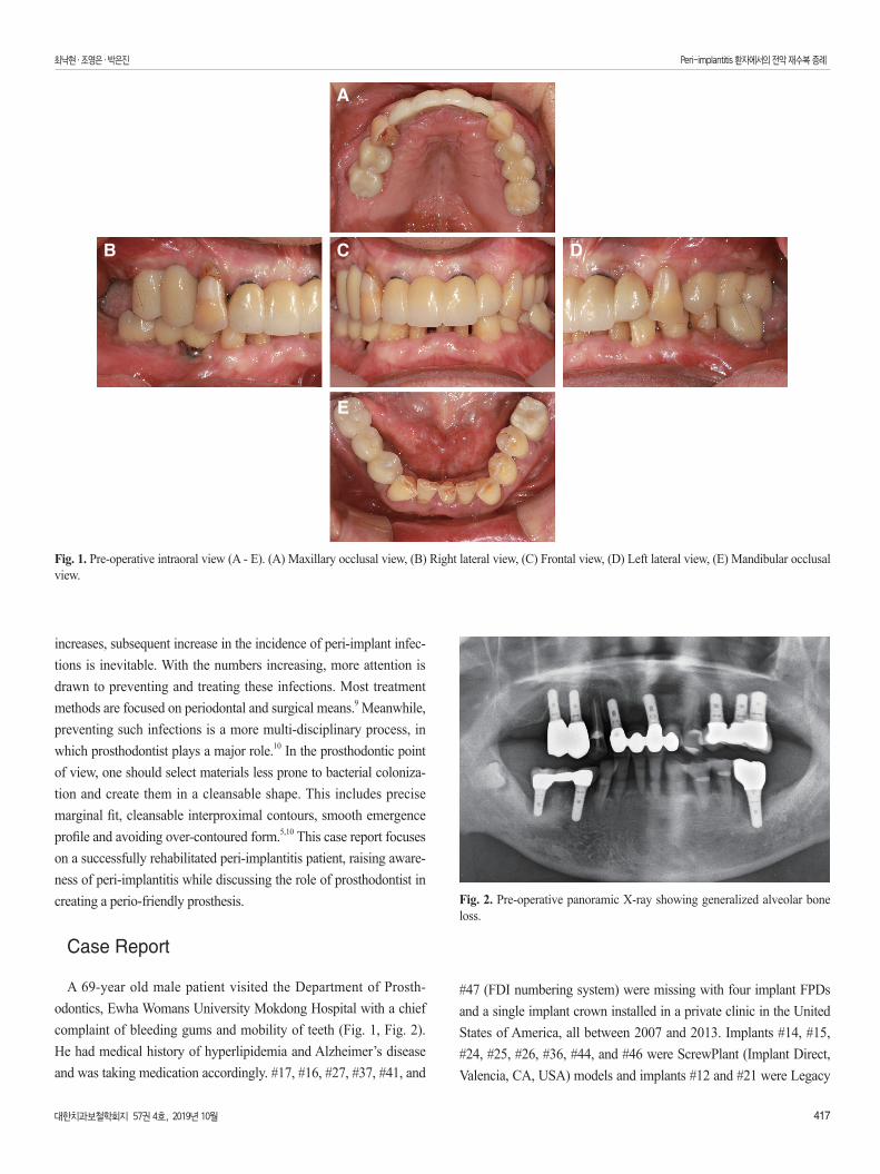
A 69-year old male patient visited the Department of Prosth-odontics, Ewha Womans University Mokdong Hospital with a chief complaint of bleeding gums and mobility of teeth (Fig. 1, Fig. 2). He had medical history of hyperlipidemia and Alzheimer’s disease and was taking medication accordingly. #17, #16, #27, #37, #41, and
#47 (FDI numbering system) were missing with four implant FPDs and a single implant crown installed in a private clinic in the United States of America, all between 2007 and 2013. Implants #14, #15, #24, #25, #26, #36, #44, and #46 were ScrewPlant (Implant Direct, Valencia, CA, USA) models and implants #12 and #21 were Legacy
Fig. 1. Pre-operative intraoral view (A - E). (A) Maxillary occlusal view, (B) Right lateral view, (C) Frontal view, (D) Left lateral view, (E) Mandibular occlusal view.
A
CB D
E
Fig. 2. Pre-operative panoramic X-ray showing generalized alveolar bone loss.
418 대한치과보철학회지 57권 4호, 2019년 10월
최낙현�조영은�박은진 Peri-implantitis 환자에서의 전악 재수복 증례
Implant (Implant Direct, Valencia, CA, USA) models, all of which are hydroxyapatite-coated implants. Implants #24, #25, #26, #36, and #46 showed full probing depth along with pus discharging from severely swollen gingiva. Implant #15 and #45 also showed a deep probing depth of 7-8 mm. Cantilevered maxillary anterior implant FPD revealed ill-fitting margins with palatal porcelain chipping. Lower anterior teeth, #13, #34, and #35 showed severe mobility. Occlusion was also unfavorable due to generalized attrition, deep overbite, and reverse Curve of Spee. Facial examination along with intraoral inspections showed well-developed masseters, leading to a strong bite force. Generalized chronic periodontitis and localized peri-implantitis along with unfavorable occlusion was diagnosed.
From the observations, four main key points for the treatment were considered. Removal of hopeless teeth and implants, strategic use of remaining implants for the new restoration, remodeling of oc-clusion, and prevention of attrition due to strong bite force. As the patient wanted new implant prostheses instead of conventional den-tures, implant installation after removing the diseased implants were planned. To minimize the risk of recurrent peri-implantitis, attention in designing restorations that are favorable to periodontal tissues was necessary.
A treatment plan was developed after collecting the patient’s needs; his first priority was fixed restorations with minimum number of surgeries. However, due to the high economic cost of fixed im-plants, the patient was willing to try a removable restoration as long as the retention is favorable. Fixed restorations with 6 new implants were planned for the mandible, while an overdenture (OVD) on remaining implants and tooth was planned for the maxilla. The op-tion of fixed implant restorations on the maxilla were ruled-out due to the large defect on the left maxilla, as extensive rehabilitation was expected.
Extraction of all natural teeth except #23, #33, and #43, along with removal of implants #15, #24, #25, #26, #36, #44, and #46 were planned, following the explantation criteria by Lang et al.5 The re-maining implants #12, #14, and #21 on the maxilla were to provide
support and retention for the overdenture, along with a metal cap on #23. No additional implants were planned for the maxilla. For the mandible, 6 new implants (implants #32, #34, #36, #42, #44, and #46) would be restored with implant fixed partial dentures (FPDs) with single crowns on #33 and #43. Botulinum toxin injections to reduce masseteric force and selection of materials less prone to attri-tion were additionally considered.
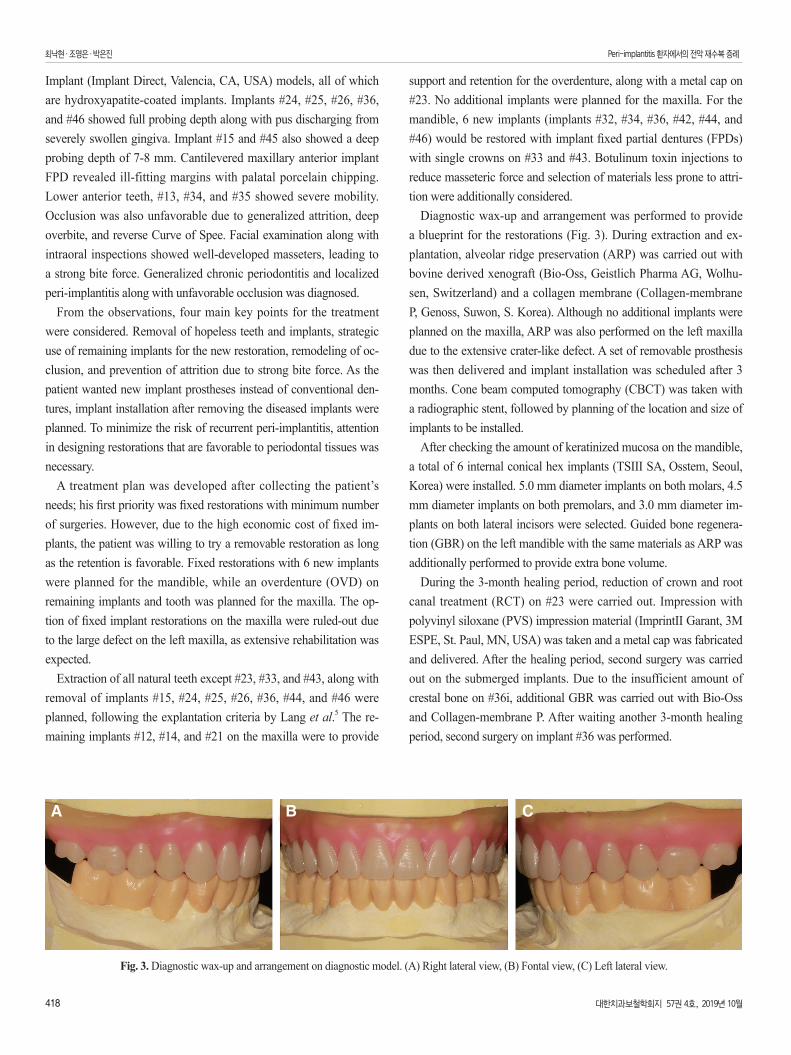
Diagnostic wax-up and arrangement was performed to provide a blueprint for the restorations (Fig. 3). During extraction and ex-plantation, alveolar ridge preservation (ARP) was carried out with bovine derived xenograft (Bio-Oss, Geistlich Pharma AG, Wolhu-sen, Switzerland) and a collagen membrane (Collagen-membrane P, Genoss, Suwon, S. Korea). Although no additional implants were planned on the maxilla, ARP was also performed on the left maxilla due to the extensive crater-like defect. A set of removable prosthesis was then delivered and implant installation was scheduled after 3 months. Cone beam computed tomography (CBCT) was taken with a radiographic stent, followed by planning of the location and size of implants to be installed.
After checking the amount of keratinized mucosa on the mandible, a total of 6 internal conical hex implants (TSIII SA, Osstem, Seoul, Korea) were installed. 5.0 mm diameter implants on both molars, 4.5 mm diameter implants on both premolars, and 3.0 mm diameter im-plants on both lateral incisors were selected. Guided bone regenera-tion (GBR) on the left mandible with the same materials as ARP was additionally performed to provide extra bone volume.
During the 3-month healing period, reduction of crown and root canal treatment (RCT) on #23 were carried out. Impression with polyvinyl siloxane (PVS) impression material (ImprintII Garant, 3M ESPE, St. Paul, MN, USA) was taken and a metal cap was fabricated and delivered. After the healing period, second surgery was carried out on the submerged implants. Due to the insufficient amount of crestal bone on #36i, additional GBR was carried out with Bio-Oss and Collagen-membrane P. After waiting another 3-month healing period, second surgery on implant #36 was performed.
A CB
Fig. 3. Diagnostic wax-up and arrangement on diagnostic model. (A) Right lateral view, (B) Fontal view, (C) Left lateral view.
대한치과보철학회지 57권 4호, 2019년 10월 419
최낙현�조영은�박은진 Peri-implantitis 환자에서의 전악 재수복 증례
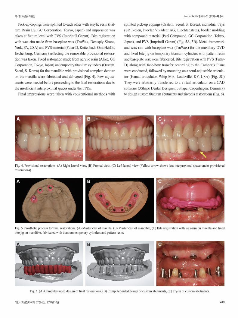
Pick-up copings were splinted to each other with acrylic resin (Pat-tern Resin LS, GC Corporation, Tokyo, Japan) and impression was taken at fixture level with PVS (ImprintII Garant). Bite registration with wax-rim made from baseplate wax (TruWax, Dentsply Sirona, York, PA, USA) and PVS material (Futar-D, Kettenbach GmbH&Co, Eschenburg, Germany) reflecting the removable provisional restora-tion was taken. Fixed restoration made from acrylic resin (Alike, GC Corporation, Tokyo, Japan) on temporary titanium cylinders (Osstem, Seoul, S. Korea) for the mandible with provisional complete denture on the maxilla were fabricated and delivered (Fig. 4). Few adjust-ments were needed before proceeding to the final restorations due to the insufficient interproximal spaces under the FPDs.
Final impressions were taken with conventional methods with
splinted pick-up copings (Osstem, Seoul, S. Korea), individual trays (SR Ivolen, Ivoclar Vivadent AG, Liechtenstein), border molding with compound material (Peri Compound, GC Corporation, Tokyo, Japan), and PVS (ImprintII Garant) (Fig. 5A, 5B). Metal framework and wax-rim with baseplate wax (TruWax) for the maxillary OVD and fixed bite jig on temporary titanium cylinders with pattern resin and baseplate wax were fabricated. Bite registration with PVS (Futar-D) along with face-bow transfer according to the Camper’s Plane were conducted, followed by mounting on a semi-adjustable articula-tor (Hanau articulator, Whip Mix, Louisville, KY, USA) (Fig. 5C) They were arbitrarily transferred to a virtual articulator on a CAD software (3Shape Dental Designer, 3Shape, Copenhagen, Denmark) to design custom titanium abutments and zirconia restorations (Fig. 6).
A CB
Fig. 5. Prosthetic process for final restorations. (A) Master cast of maxilla, (B) Master cast of mandible, (C) Bite registration with wax-rim on maxilla and fixed bite jig on mandible, fabricated with titanium temporary cylinders and pattern resin.
A CB
Fig. 4. Provisional restorations. (A) Right lateral view, (B) Frontal view, (C) Left lateral view (Yellow arrow shows less interproximal space under provisional restorations).
A CB
Fig. 6. (A) Computer-aided design of final restorations, (B) Computer-aided design of custom abutments, (C) Try-in of custom abutments.
420 대한치과보철학회지 57권 4호, 2019년 10월
최낙현�조영은�박은진 Peri-implantitis 환자에서의 전악 재수복 증례
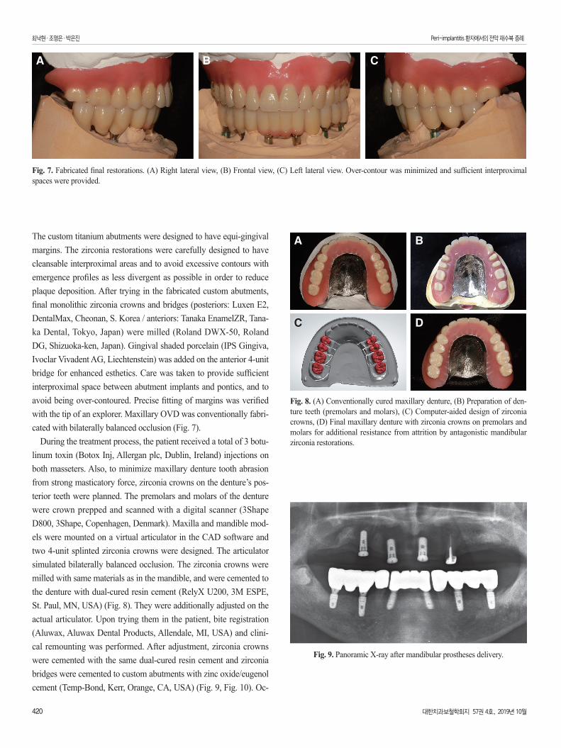
The custom titanium abutments were designed to have equi-gingival margins. The zirconia restorations were carefully designed to have cleansable interproximal areas and to avoid excessive contours with emergence profiles as less divergent as possible in order to reduce plaque deposition. After trying in the fabricated custom abutments, final monolithic zirconia crowns and bridges (posteriors: Luxen E2, DentalMax, Cheonan, S. Korea / anteriors: Tanaka EnamelZR, Tana-ka Dental, Tokyo, Japan) were milled (Roland DWX-50, Roland DG, Shizuoka-ken, Japan). Gingival shaded porcelain (IPS Gingiva, Ivoclar Vivadent AG, Liechtenstein) was added on the anterior 4-unit bridge for enhanced esthetics. Care was taken to provide sufficient interproximal space between abutment implants and pontics, and to avoid being over-contoured. Precise fitting of margins was verified with the tip of an explorer. Maxillary OVD was conventionally fabri-cated with bilaterally balanced occlusion (Fig. 7).
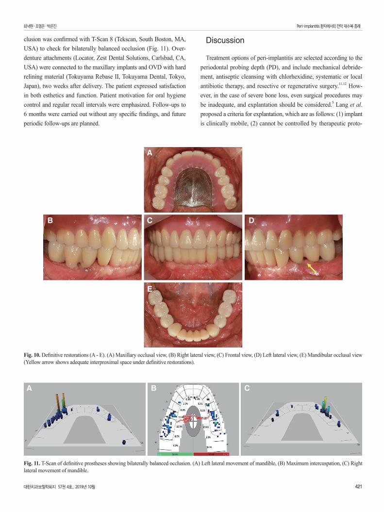
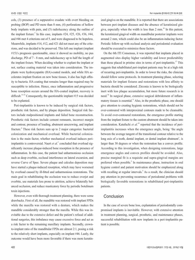
During the treatment process, the patient received a total of 3 botu-linum toxin (Botox Inj, Allergan plc, Dublin, Ireland) injections on both masseters. Also, to minimize maxillary denture tooth abrasion from strong masticatory force, zirconia crowns on the denture’s pos-terior teeth were planned. The premolars and molars of the denture were crown prepped and scanned with a digital scanner (3Shape D800, 3Shape, Copenhagen, Denmark). Maxilla and mandible mod-els were mounted on a virtual articulator in the CAD software and two 4-unit splinted zirconia crowns were designed. The articulator simulated bilaterally balanced occlusion. The zirconia crowns were milled with same materials as in the mandible, and were cemented to the denture with dual-cured resin cement (RelyX U200, 3M ESPE, St. Paul, MN, USA) (Fig. 8). They were additionally adjusted on the actual articulator. Upon trying them in the patient, bite registration (Aluwax, Aluwax Dental Products, Allendale, MI, USA) and clini-cal remounting was performed. After adjustment, zirconia crowns were cemented with the same dual-cured resin cement and zirconia bridges were cemented to custom abutments with zinc oxide/eugenol cement (Temp-Bond, Kerr, Orange, CA, USA) (Fig. 9, Fig. 10). Oc-
Fig. 8. (A) Conventionally cured maxillary denture, (B) Preparation of den-ture teeth (premolars and molars), (C) Computer-aided design of zirconia crowns, (D) Final maxillary denture with zirconia crowns on premolars and molars for additional resistance from attrition by antagonistic mandibular zirconia restorations.
A B
C D
Fig. 9. Panoramic X-ray after mandibular prostheses delivery.
A CB
Fig. 7. Fabricated final restorations. (A) Right lateral view, (B) Frontal view, (C) Left lateral view. Over-contour was minimized and sufficient interproximal spaces were provided.
대한치과보철학회지 57권 4호, 2019년 10월 421
최낙현�조영은�박은진 Peri-implantitis 환자에서의 전악 재수복 증례
clusion was confirmed with T-Scan 8 (Tekscan, South Boston, MA, USA) to check for bilaterally balanced occlusion (Fig. 11). Over-denture attachments (Locator, Zest Dental Solutions, Carlsbad, CA, USA) were connected to the maxillary implants and OVD with hard relining material (Tokuyama Rebase II, Tokuyama Dental, Tokyo, Japan), two weeks after delivery. The patient expressed satisfaction in both esthetics and function. Patient motivation for oral hygiene control and regular recall intervals were emphasized. Follow-ups to 6 months were carried out without any specific findings, and future periodic follow-ups are planned.
Discussion
Treatment options of peri-implantitis are selected according to the periodontal probing depth (PD), and include mechanical debride-ment, antiseptic cleansing with chlorhexidine, systematic or local antibiotic therapy, and resective or regenerative surgery.11,12 How-ever, in the case of severe bone loss, even surgical procedures may be inadequate, and explantation should be considered.5 Lang et al. proposed a criteria for explantation, which are as follows: (1) implant is clinically mobile, (2) cannot be controlled by therapeutic proto-
Fig. 10. Definitive restorations (A - E). (A) Maxillary occlusal view, (B) Right lateral view, (C) Frontal view, (D) Left lateral view, (E) Mandibular occlusal view (Yellow arrow shows adequate interproximal space under definitive restorations).
A
CB D
E
A CB
Fig. 11. T-Scan of definitive prostheses showing bilaterally balanced occlusion. (A) Left lateral movement of mandible, (B) Maximum intercuspation, (C) Right lateral movement of mandible.
422 대한치과보철학회지 57권 4호, 2019년 10월
최낙현�조영은�박은진 Peri-implantitis 환자에서의 전악 재수복 증례
cols, (3) presence of a suppurative exudate with overt bleeding on probing (BOP) and PD more than 8 mm, (4) perforations of hollow body implants with pain, and (5) radiolucency along the outline of the implant fixture.5 In this case, implants #24, #25, #26, #36, #44, and #46 met 4 criterions out of 5, and were diagnosed to be hopeless. Meanwhile, implants #14, #12, and #21 did not meet any of the crite-rions, and was decided to be preserved. This left one implant (implant #15)’s prognosis questionable, since it showed no mobility, no pus discharge, PD of 7 - 8 mm, and radiolucency up to half the length of the implant fixture. When deciding whether to explant the implant or not, surface coating material was taken into consideration. The im-plants were hydroxyapatite (HA)-coated models, and while HA ac-celerates implant fixation on new bone tissues, it also has high affin-ity to bacteria. HA coating that remains longer than required may be susceptible to infection. Hence, once inflammation and progressive bone resorption occurs around the HA-coated implant, recovery is difficult.13-15 Consequently, the questionable implant #15 was decided to be explanted.
Peri-implantitis is known to be induced by surgical risk factors, prosthetic risk factors, and by plaque deposition. Surgical risk fac-tors include malpositioned implants and failed bone reconstruction. Prosthetic risk factors include cement remnants, incorrect margin and contour, presence of loading, abutment unscrewing, and implant fracture.6 These risk factors sum up to 2 major categories: bacterial colonization and mechanical overload. While bacterial coloniza-tion is the main factor, whether mechanical overload induces peri-implantitis is controversial. Naert et al.7 concluded that overload sig-nificantly increase plaque-induced bone resorption in the presence of inflammation. In this case, the patient had unharmonious occlusion such as deep overbite, occlusal interference on lateral excursion, and reverse Curve of Spee. Severe plaque and calculus deposition may have started a plaque-induced resorption, which may have worsened by overload caused by ill-fitted and unharmonious restorations. The main goal in rehabilitating the occlusion was to reduce overjet and overbite, use materials less prone to attrition, achieve bilaterally bal-anced occlusion, and reduce masticatory force by periodic botulinum toxin injections.
However, even with thorough treatment planning, there were some drawbacks. First of all, the mandible was restored with implant FPDs while the maxilla was restored with a denture, which makes the mandible considerably stronger than the maxilla. While this was in-evitable due to the extensive defect and the patient’s refusal of addi-tional surgeries, this imbalance may cause excessive force and act as a risk factor to the remaining maxillary implants. Secondly, crown-to-implant ratio of the mandibular FPDs are almost 2:1, posing a risk to the relatively short implants, especially on implant #46. Lastly, the outcome would have been more favorable if there was more keratin-
ized gingiva on the mandible. It is reported that there are associations between peri-implant diseases and the absence of keratinized gin-giva, especially when the width is less than 2 mm.16 In this patient, the keratinized gingival width on mandibular posterior implants were around 2 mm, which could also be an inflammation-inducing factor. Periodic follow-up with occlusal analysis and periodontal evaluation should be executed to minimize these factors.
On the 6th ITI Consensus, it was reported that implants placed in augmented sites display higher variability and lower predictability than those placed in pristine sites in terms of peri-implantitis.17 This suggests that rehabilitated peri-implant patients may be at a high risk of recurring peri-implantitis. In order to lower the risks, the clinician should follow some protocols. In treatment planning phase, selecting materials for implant fixtures and restorations with less affinity to bacteria should be considered. Zirconia is known to be biologically inert with less plaque accumulation, but more future research is in need.18 In surgical phase, extensive surgical debridement of inflam-matory tissues is essential.11 Also, in the prosthetic phase, one should give attention to creating hygienic restorations, which should not be over-contoured with interproximal contours made to be cleansable. To avoid over-contoured restorations, the emergence profile starting from the implant fixture to the custom abutment should be taken into consideration. Katafuchi et al.19 reported that the incidence of peri-implantitis increases when the emergence angle, being ‘the angle between the average tangent of the transitional contour relative to the long axis of a tooth, dental implant, or dental implant abutment’, is larger than 30 degrees or when the restoration has a convex profile. According to this investigation, when designing restorations, large emergence angles and convex profiles should be avoided. Also, precise marginal fit is a requisite and supra-gingival margins are preferred when possible.5 In maintenance phase, instruction in oral hygiene control and patient motivation should be emphasized along with recalling at regular intervals.5 As a result, the clinician should pay attention in preventing recurrence of periodontal problems with biologically favorable restorations in rehabilitated peri-implantitis patients.
Conclusion
In the case of severe bone loss, explantation of periodontally com-promised implants is inevitable. However, with extensive attention in treatment planning, surgical, prosthetic, and maintenance phases, successful rehabilitation with new implants in a peri-implantitis pa-tient is possible.
대한치과보철학회지 57권 4호, 2019년 10월 423
최낙현�조영은�박은진 Peri-implantitis 환자에서의 전악 재수복 증례
ORCID
Nak-Hyun Choi https://orcid.org/0000-0002-8199-1664Young-Eun Cho https://orcid.org/0000-0003-0341-2150Eun-Jin Park https://orcid.org/0000-0001-6383-449X
References
1. Mombelli A, Müller N, Cionca N. The epidemiology of peri-implantitis. Clin Oral Implants Res 2012;23:67-76.
2. Lindhe J, Meyle J; Group D of European Workshop on Peri-odontology. Peri-implant diseases: Consensus Report of the Sixth European Workshop on Periodontology. J Clin Peri-odontol 2008;35:282-5.
3. Heitz-Mayfield LJ. Peri-implant diseases: diagnosis and risk indicators. J Clin Periodontol 2008;35:292-304.
4. Karoussis IK, Salvi GE, Heitz-Mayfield LJ, Brägger U, Häm-merle CH, Lang NP. Long-term implant prognosis in patients with and without a history of chronic periodontitis: a 10-year prospective cohort study of the ITI Dental Implant System. Clin Oral Implants Res 2003;14:329-39.
5. Lang NP, Wilson TG, Corbet EF. Biological complications with dental implants: their prevention, diagnosis and treat-ment. Clin Oral Implants Res 2000;11:146-55.
6. Tallarico M, Canullo L, Wang HL, Cochran DL, Meloni SM. Classification systems for peri-implantitis: A narrative review with a proposal of a new evidence-based etiology codifica-tion. Int J Oral Maxillofac Implants 2018;33:871-9.
7. Naert I, Duyck J, Vandamme K. Occlusal overload and bone/implant loss. Clin Oral Implants Res 2012;23:95-107.
8. Lee CT, Huang YW, Zhu L, Weltman R. Prevalences of peri-implantitis and peri-implant mucositis: systematic review and meta-analysis. J Dent 2017;62:1-12.
9. Kotsovilis S, Karoussis IK, Trianti M, Fourmousis I. Therapy of peri-implantitis: a systematic review. J Clin Periodontol 2008;35:621-9.
10. Jepsen S, Berglundh T, Genco R, Aass AM, Demirel K, Derks J, Figuero E, Giovannoli JL, Goldstein M, Lambert F, Ortiz-Vigon A, Polyzois I, Salvi GE, Schwarz F, Serino G, Tomasi C, Zitzmann NU. Primary prevention of peri-implantitis: manag-ing peri-implant mucositis. J Clin Periodontol 2015;42:S152-7.
11. Schou S, Berglundh T, Lang NP. Surgical treatment of peri-implantitis. Int J Oral Maxillofac Implants 2004;19:140-9.
12. Mombelli A, Lang NP. The diagnosis and treatment of peri-implantitis evidence for a microbial cause of peri-implant infections. Periodontology 2000;17:63-76.
13. Ding L, Zhang P, Wang X, Kasugai S. A doxycycline-treated hydroxyapatite implant surface attenuates the progression of peri-implantitis: A radiographic and histological study in mice. Clin Implant Dent Relat Res 2019;21:154-9.
14. Becker W, Becker BE, Newman MG, Nyman S. Clinical and microbiologic findings that may contribute to dental implant failure. Int J Oral Maxillofac Implants 1990;5:31-8.
15. Ozeki K, Okuyama Y, Fukui Y, Aoki H. Bone response to titanium implants coated with thin sputtered HA film subject to hydrothermal treatment and implanted in the canine man-dible. Biomed Mater Eng 2006;16:243-51.
16. Roos-Jansåker AM, Renvert H, Lindahl C, Renvert S. Nine- to fourteen-year follow-up of implant treatment. Part III: fac-tors associated with peri-implant lesions. J Clin Periodontol 2006;33:296-301.
17. Salvi GE, Monje A, Tomasi C. Long-term biological com-plications of dental implants placed either in pristine or in augmented sites: A systematic review and meta-analysis. Clin Oral Implants Res 2018;29:294-310.
18. Bremer F, Grade S, Kohorst P, Stiesch M. In vivo biofilm formation on different dental ceramics. Quintessence Int 2011;42:565-74.
19. Katafuchi M, Weinstein BF, Leroux BG, Chen YW, Daubert DM. Restoration contour is a risk indicator for peri-implanti-tis: A cross-sectional radiographic analysis. J Clin Periodontol 2018;45:225-32.
424 대한치과보철학회지 57권 4호, 2019년 10월
CASE REpORT
Peri-implantitis 환자에서의 전악 재수복 증례
최낙현 � 조영은 � 박은진*
이화여자대학교 의과대학 치과보철학교실
임플란트 주위염(peri-implantitis)은 임플란트 치료를 받은 환자의 20%가량에서 발병하며, 시간이 지날수록 유병률이 증가한다. 경도 또는 중등도의 임플란트 주위염은 비수술적 또는 수술적 방법을 통해 치료될 수 있으나 심한 치조골 흡수를 동반한 경우 임플란트 제거(explanation)를 동반한 재건이 요구된다. 임플란트 주위염은 재발 가능성이 높아 치료계획 단계에서부터 임플란트 주위염의 재발을 방지하기 위한 고려가 필요하다. 본 증례는 임플란트 주위염과 만성치주염으로 진단된 환자의 증례로, 다수의 임플란트 제거 및 다수 치아 발치 후 상악은 임플란트 유지 피개의치, 하악은 임플란트 고정성 보철로 전악 수복하였다. 임플란트 주위염의 재발을 예방하기 위한 수술적, 보철적 방법을 통해 성공적으로 수복하였기에 이를 보고하고자 한다. (대한치과보철학회지 2019;57:416-24)
주요단어: 임플란트 주위염; 전악 수복; 지르코니아; Computer-aided design and computer-aided manufacturing (CAD/CAM); 임플란트 제거
*교신저자: 박은진07985 서울 양천구 안양천로 1071 이화여자대학교 의과대학 치과보철학교실02 2650 5042: e-mail, [email protected]원고접수일: 2019년 6월 11일 / 원고최종수정일: 2019년 7월 25일 / 원고채택일: 2019년 8월 1일
2019 대한치과보철학회이 글은 크리에이티브 커먼즈 코리아 저작자표시-비영리 4.0 대한민국 라이선스에 따라 이용하실 수 있습니다.
c
cc