Embed Size (px)
Citation preview

© 2002 Nature Publishing Group346 | MAY 2002 | VOLUME 2 www.nature.com/reviews/immunol
R E V I E W S
mechanisms of antibody-mediated immunity. Once thecomplement system is activated, a chain of reactions thatinvolves proteolysis and assembly occurs, which results incleavage of the third complement component (C3). Thecascade that leads to the cleavage of C3 is called the acti-vation pathway. It is followed by the lytic pathway, duringwhich the membrane-attack complex (MAC) is formed.There are three types of activation pathway: the classical,lectin and alternative pathways (FIG. 1). The classicalpathway is activated by antibody–antigen complexes,whereas the other two pathways — the lectin and alter-native pathways — function in innate immune defence.The lectin pathway involves carbohydrate recognitionby pattern-recognition receptors, such as mannose-binding lectin (MBL) and ficolins, and the subsequentactivation of associated unique enzymes that are knownas MBL-associated serine proteases (MASPs). The alter-native pathway is initiated by the covalent binding of asmall amount of C3 to hydroxyl or amine groups on thecell-surface molecules of microorganisms, and it doesnot involve specific recognition molecules. This pathwayalso functions to amplify the activation of C3 (amplifi-cation loop). Activation of the complement systempromotes three main biological activities: opsonizationof pathogens; chemotaxis and activation of leukocytes;and direct killing of pathogens. Recently, accumulating
Immunity to infection is mediated by two systems — theacquired (or adaptive) immune system and the innate(or natural) immune system. Acquired immunity aroseearly in vertebrate evolution, at some point between thedivergence of the cyclostomes (lamprey) and cartilagi-nous fishes (sharks). The innate immune system is anevolutionarily ancient form of immunity and offers themain resistance to microbial pathogens within the firstminutes, hours or days of an infection1. Innate immu-nity was thought originally to be a non-specific immuneresponse that was characterized by phagocytosis.However, innate immunity has considerable specificityand is able to discriminate between pathogens and self,as well as between classes of pathogen. The recognitionof pathogens is mediated by a set of pattern-recognitionreceptors that bind conserved pathogen-associated mol-ecular patterns (PAMPs) that are shared by broad classesof microorganism. This recognition successfully defendsinvertebrates and vertebrates against infection2.
Complement was first described in the 1890s as aheat-labile protein in serum that ‘complemented’ heat-stable antibodies in the killing of bacteria. Now, thecomplement system, which consists of more than 30plasma and cell-surface proteins, is known to be a highlysophisticated host-defence system that is engaged bothby innate immunity and as one of the main effector
Department of Biochemistry,Fukushima MedicalUniversity School ofMedicine,1 Hikarigao-ka, Fukushima960-1295, Japan.e-mail: [email protected]:10.1038/nri800
EVOLUTION OF THELECTIN–COMPLEMENT PATHWAYAND ITS ROLE IN INNATE IMMUNITYTeizo Fujita
Discrimination between self and non-self by lectins (carbohydrate-binding proteins) is a strategyof innate immunity that is found in both vertebrates and invertebrates. In vertebrates, immunerecognition mediated by ficolins (lectins that consist of a fibrinogen-like and a collagen-likedomain), as well as by mannose-binding lectins, triggers the activation of the complementsystem, which results in the activation of novel serine proteases. The presence of a similar lectin-based complement system in ascidians, our closest invertebrate relatives, indicates that thecomplement system probably had a pivotal role in innate immunity before the evolution of anadaptive immune system in jawed vertebrates.
© 2002 Nature Publishing GroupNATURE REVIEWS | IMMUNOLOGY VOLUME 2 | MAY 2002 | 347
R E V I E W S
C-TYPE LECTIN
A calcium-dependent animallectin that is a carbohydrate-binding protein. The bindingactivity of a C-type lectin isbased on the structure of thecarbohydrate-recognitiondomain, which is highlyconserved among this family.Calcium is essential not only forthe carbohydrate binding itself,but also for the structuralmaintenance of this domain.
COLLECTIN
A C-type lectin that has acollagen-like domain. Onegroup, the secreted lectins,consists of mannose-bindinglectin, bovine conglutinin andcollectin 43 in blood, and thetwo mucosal-associatedproteins surfactant proteins Aand D. The other group consistsof the newly discovered, non-secreted-type collectin liver-1and membrane-type collectinplacenta-1.
Recognition molecules in complement activationIn the classical pathway, C1q, a subcomponent of thefirst complement component (C1), recognizes the Fcregion of immunoglobulins that are bound to antigen5.C1q has an unusual modular structure that consists ofsix globular heads, each of which is connected by astrand to a central fibril-like region that is composed ofcollagen-like, triple-helical structures. Mannose-bindinglectin and ficolins, which are the moieties that mediaterecognition in the lectin pathway, have a similar overallstructure to C1q (FIG. 1).
Mannose-binding lectin is a C-TYPE LECTIN6–8 that has acrucial role in the first line of host defence9. The impor-tance of this molecule is underlined by several clinicalstudies that link MBL-deficiency with increased suscep-tibility to various infectious diseases10–13. MBL belongsto the COLLECTIN family of proteins, which consist of acollagen-like domain and carbohydrate-recognitiondomain (CRD)14. Three polypeptides fold together to
evidence has shown that the complement system alsoacts as an adjuvant by enhancing and directing the adap-tive immune response, and can function in the disposalof apoptotic cells3,4.
In this review, I focus on the elucidation of the lectinpathway, the discovery of which was one of the mostoutstanding in recent complement research. Lectins arenow simply defined as proteins that specifically bind car-bohydrates. In animals, soluble lectins function asweapons against pathogens by aggregating and opsoniz-ing them. These are primitive strategies of innate immu-nity that are found in both invertebrates and vertebrates.Evolutionary pressure, however, has afforded lectins themore powerful ability to activate the complement sys-tem, thereby effectively eliminating pathogens from thehost. The identification of several components of thelectin pathway in ascidians, our closest invertebrate rela-tives, should provide new insights into the complementsystem, particularly in terms of its evolution.
C1r
C1s
Bb
BbB
C2a
C1q
MBL/ficolin
Immunoglobulin
Bacterialantigen
Carbohydrate
Bacterial surface Bacterial surface
Bacterial surface
C3
C2
C4
C3
C3
C3
C3(H2O) C3(H2O)
C3b
C3a
C3bC3b
C3b
C3a
C3a
C3b C3b
C3a
C4b
C2a
C4b
BD
D
MASP2
MASP3
MASP1
sMAP
c Alternative pathway and amplification loop
a Classical pathway b Lectin pathway
?
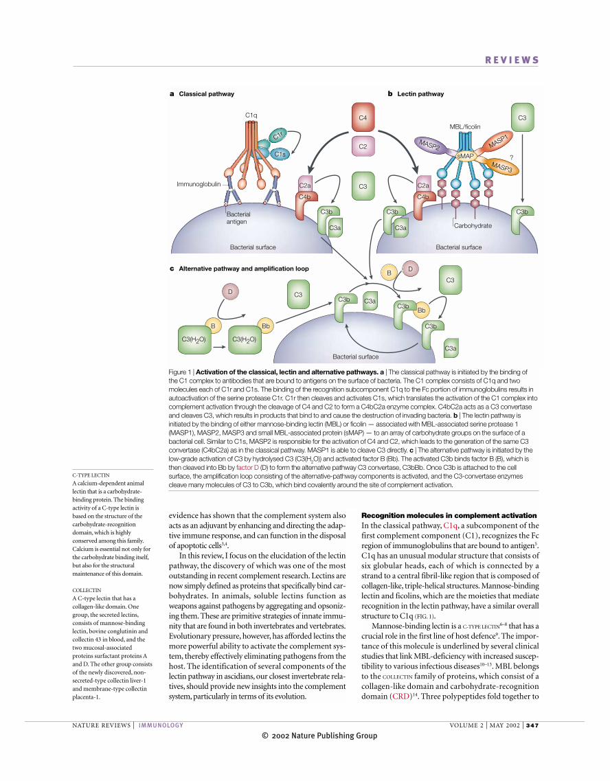
Figure 1 | Activation of the classical, lectin and alternative pathways. a | The classical pathway is initiated by the binding of the C1 complex to antibodies that are bound to antigens on the surface of bacteria. The C1 complex consists of C1q and twomolecules each of C1r and C1s. The binding of the recognition subcomponent C1q to the Fc portion of immunoglobulins results inautoactivation of the serine protease C1r. C1r then cleaves and activates C1s, which translates the activation of the C1 complex intocomplement activation through the cleavage of C4 and C2 to form a C4bC2a enzyme complex. C4bC2a acts as a C3 convertaseand cleaves C3, which results in products that bind to and cause the destruction of invading bacteria. b | The lectin pathway isinitiated by the binding of either mannose-binding lectin (MBL) or ficolin — associated with MBL-associated serine protease 1(MASP1), MASP2, MASP3 and small MBL-associated protein (sMAP) — to an array of carbohydrate groups on the surface of abacterial cell. Similar to C1s, MASP2 is responsible for the activation of C4 and C2, which leads to the generation of the same C3convertase (C4bC2a) as in the classical pathway. MASP1 is able to cleave C3 directly. c | The alternative pathway is initiated by thelow-grade activation of C3 by hydrolysed C3 (C3(H2O)) and activated factor B (Bb). The activated C3b binds factor B (B), which isthen cleaved into Bb by factor D (D) to form the alternative pathway C3 convertase, C3bBb. Once C3b is attached to the cellsurface, the amplification loop consisting of the alternative-pathway components is activated, and the C3-convertase enzymescleave many molecules of C3 to C3b, which bind covalently around the site of complement activation.
© 2002 Nature Publishing Group348 | MAY 2002 | VOLUME 2 www.nature.com/reviews/immunol
R E V I E W S
Ficolins, like MBL, are a group of proteins that contain a collagen-like stem structure. Unlike MBL,however, they have a fibrinogen-like domain, which issimilar to fibrinogen β- and γ-chains21 (FIG. 2). They wereidentified originally as transforming growth factor-β1(TGF-β1)-binding proteins on pig uterus membranes22.Since then, ficolins have been identified in mammals,including human23–28, rodents29, pig22,30 and hedgehog31,and, recently, in ascidians32 (see below and FIG. 3). Serumficolins are lectins that have a common binding speci-ficity for GlcNAc24,28–30. In human serum, two types of ficolin, known as L-ficolin/P35 (ficolin L)24,25 and H-ficolin (Hakata antigen)29,33, have been identified, andboth of them have lectin activity. Another ficolin,known as M-ficolin25,34 or P35-related protein26 — themessenger RNA of which is found in leukocytes andlung — is not considered to be a serum protein.Recently, it has been reported that L-ficolin/P35 and H-ficolin activate the lectin–complement pathway inassociation with MASPs21,35. The functions of the fibrinogen-like domain of the ficolins are not fullyunderstood; however, recent work has shown that thefibrinogen-like domain of several lectins has a similarfunction to the CRD of C-type lectins, which indicatesthat ficolins might also function as pattern-recognitionreceptors to discriminate pathogens from self36,37.
Serine proteases of the lectin pathwayMASPs, a new member of the serine-protease superfam-ily, are proteolytic enzymes that are responsible for acti-vation of the lectin pathway38–41. The MBL–MASPs com-plex was first described as a complement-dependentbactericidal factor in mouse42,43. MBL and ficolins havebeen found to be associated with MASP1 and MASP2,and a non-protease, small MBL-associated protein(sMAP or MAp19; a truncated form of MASP2)44,45.Recently, a third MASP (MASP3), which is generated byalternative splicing of MASP1, was reported to be asso-ciated with MBL15. The overall structure of MASPsresembles that of the two proteolytic components of thefirst factor in the classical complement pathway, C1rand C1s39–41 (FIG. 4).
As illustrated in FIGURE. 1, activation of the classicalpathway is triggered by binding of the recognition sub-component C1q to an antibody, which, in turn, is trans-lated into activation of the serine proteases C1r and C1s.Likewise, binding of the lectin-pathway recognitionmolecules (that is, MBL or ficolins) to microbial carbo-hydrates activates the lectin-pathway-specific serineproteases, MASPs46,47. The detailed molecular events,however, remain to be clarified. MASP2 is the enzymecomponent that — like C1s in the classical pathway —cleaves the complement components C4 and C2 toform the C3 convertase C4bC2a, which is common toboth the lectin- and classical-pathway activationroutes41,48. This result has been confirmed by the func-tional analysis of recombinant MASP2 (REFS 49–52). Bycontrast, MASP1 is able to cleave C3 directly15,53,54,which results in activation of the alternative pathway53.The functions of MASP3 and sMAP are presentlyunknown. MASP1, MASP2, MASP3 and sMAP are
form the structural subunit and 3–6 of these subunitsjoin to form human MBL, which has an apparent mole-cular mass of ~300–650 kDa (REF. 15) (FIG. 2). Through itsCRD, MBL binds carbohydrates with 3- and 4-hydroxylgroups in the pyranose ring in the presence of calcium16.So, prominent ligands for MBL are mannose and N-acetyl-glucosamine (GlcNAc), whereas carbohydratesthat do not fit this steric requirement — for example,galactose and sialic acid, which usually decorate themammalian glycoproteins — have undetectable affinityfor MBL17. This steric specificity of MBL, along with dif-ferences in the spatial organization of its ligands, enablesthe specific recognition of carbohydrates on pathogenicmicroorganisms, including bacteria, fungi, parasiticprotozoans and viruses, and avoids the recognition ofnon-infectious self 18. MBL has been characterized inmammals, chickens19 and carp20 (FIG. 3).
Collagen-like domain Fibrinogen-like domain
Ficolin
MBL
CRD
CRDCollagen-like domain
Collagen-likedomain
Cysteine-richregion
Fibrinogen-like domainCollagen-likedomain
Neck region
MBL
Ficolin
35 kDa
32 kDa
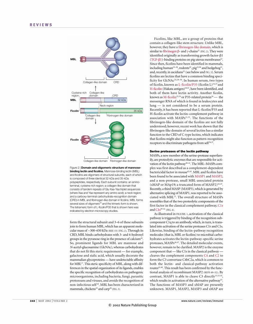
Figure 2 | Domain and oligomeric structure of mannose-binding lectin and ficolins. Mannose-binding lectin (MBL)and ficolins are oligomers of structural subunits, each of whichis composed of three identical 32-kDa and 35-kDapolypeptides, respectively. Each subunit contains: an amino-terminal, cysteine-rich region; a collagen-like domain thatconsists of tandem repeats of Gly-Xaa-Yaa triplet sequences(where Xaa and Yaa represent any amino acid); a neck region;and a carboxy-terminal carbohydrate-recognition domain(CRD) in MBL and fibrinogen-like domain in ficolins. MBL formsseveral sizes of oligomers15 and the trimeric form is shown. The tetrameric form of L-ficolin/P35 that is shown here wasindicated by electron microscopy studies.
© 2002 Nature Publishing GroupNATURE REVIEWS | IMMUNOLOGY VOLUME 2 | MAY 2002 | 349
R E V I E W S
encoded by two genes; sMAP is a truncated form ofMASP2 (REFS 44,45), and MASP3 is produced from theMASP1 gene by alternative splicing15. The MASP1 genehas an H-chain-encoding region that is common toMASP1 and MASP3, which is followed by tandemrepeats of protease-domain-encoding regions that arespecific to MASP3 and MASP1 (FIG. 5).
Molecular evolution of the MASP familyHomologues of the MASP family have been clonedfrom various vertebrate and invertebrate species. Onthe basis of the genomic organization of the serine-protease domains55 and their deduced protein structure,the MASP family can be divided into two phylogeneticlineages — TCN-type and AGY-type lineages56. TheTCN-type lineage, which includes MASP1, has a TCNcodon (where N denotes A, G, C or T) that encodes the active-site serine, the presence of a histidine-loopdisulphide bridge and split exons. By contrast, theAGY-type lineage, which includes MASP2, MASP3,C1r and C1s, is characterized by an AGY codon (whereY denotes C or T) that encodes the active-site serine,the absence of a histidine loop and a single exon56. TheTCN-type MASPs have been observed in ascidians,Xenopus and mammals, whereas AGY-type MASPshave been observed in lamprey, shark, carp, Xenopusand mammals (FIG. 3).
Lectin pathway
Acquired immunity
Classical pathway
C3 and factor B
MBL
Ficolins
MASPs
MASP3
MASP1
MASP2
Sea urchin
Ascidian
Lamprey
Shark
Carp
Frog
Snake
Chicken
Mammals
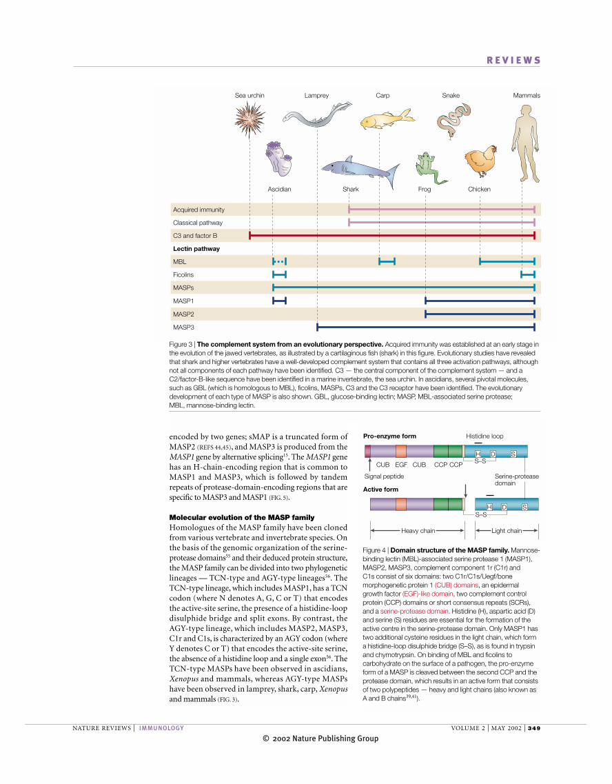
Figure 3 | The complement system from an evolutionary perspective. Acquired immunity was established at an early stage inthe evolution of the jawed vertebrates, as illustrated by a cartilaginous fish (shark) in this figure. Evolutionary studies have revealedthat shark and higher vertebrates have a well-developed complement system that contains all three activation pathways, althoughnot all components of each pathway have been identified. C3 — the central component of the complement system — and aC2/factor-B-like sequence have been identified in a marine invertebrate, the sea urchin. In ascidians, several pivotal molecules,such as GBL (which is homologous to MBL), ficolins, MASPs, C3 and the C3 receptor have been identified. The evolutionarydevelopment of each type of MASP is also shown. GBL, glucose-binding lectin; MASP, MBL-associated serine protease; MBL, mannose-binding lectin.
S–S
Heavy chain Light chain
CCP
Serine-proteasedomain
Pro-enzyme form Histidine loop
H D S
CCPCUB EGF CUBS–S
Active form
Signal peptide
H D S
H D S
Figure 4 | Domain structure of the MASP family. Mannose-binding lectin (MBL)-associated serine protease 1 (MASP1),MASP2, MASP3, complement component 1r (C1r) and C1s consist of six domains: two C1r/C1s/Uegf/bonemorphogenetic protein 1 (CUB) domains, an epidermalgrowth factor (EGF)-like domain, two complement controlprotein (CCP) domains or short consensus repeats (SCRs),and a serine-protease domain. Histidine (H), aspartic acid (D)and serine (S) residues are essential for the formation of theactive centre in the serine-protease domain. Only MASP1 hastwo additional cysteine residues in the light chain, which forma histidine-loop disulphide bridge (S–S), as is found in trypsinand chymotrypsin. On binding of MBL and ficolins tocarbohydrate on the surface of a pathogen, the pro-enzymeform of a MASP is cleaved between the second CCP and theprotease domain, which results in an active form that consistsof two polypeptides — heavy and light chains (also known asA and B chains39,41).

© 2002 Nature Publishing Group350 | MAY 2002 | VOLUME 2 www.nature.com/reviews/immunol
R E V I E W S
Putative original complement systemThe origin of the complement system can be tracedback at least as far as echinoderms, because C3, thekey component of the complement system, and aC2/factor-B-like sequence have been identified in seaurchin58–61. Sea squirts — ascidians that were previouslyknown as tunicates (phylum, Chordata; subphylum,Urochordata) — occupy a pivotal intermediary posi-tion between invertebrates and vertebrates. Therefore,studies of the host-defence mechanisms of ascidianscould provide us with important information about theevolution of a primitive innate immune system in verte-brates. Halocynthia roretzi is a large solitary ascidian thatis native to the coastal waters of Japan. So far, two lectinsthat correspond to mammalian MBL62 and ficolins32,two MASPs63, C3 (REF. 64), a C2/factor-B-like sequence61
and C3 receptor65 have been identified in ascidians (FIG. 3).Although several lectins have been reported in ascid-
ians — including collectin-type lectin in Stylea plicata66,C-type lectin in the colonial ascidian Clavelina picta67
and MBL-like lectin in sea cucumber (the holothurianCucumaria japonica)68 — and are thought to representMBL homologues in invertebrates, their completestructure has not been elucidated. Recently, we havepurified an MBL-like 36-kDa lectin from ascidianplasma62. The purified protein binds specifically toglucose, but not to mannose or GlcNAc, and so wasdesignated glucose-binding lectin (GBL). Sequenceanalysis of GBL complementary DNA revealed that the carboxy-terminal half of the ascidian lectin contains aCRD that is homologous to that of C-type lectin, but itlacks a collagen-like domain, which is present in mam-malian MBLs. Although the structure and bindingspecificity of GBL is different from that of mammalianMBL, GBL associates with two ascidian MASPs, andGBL–MASPs complexes activate ascidian C3 in thesame manner as the human MBL–MASP1 complexactivates human C3. So, it seems probable that GBLmight have evolved as an early prototype of MBL andhas acquired the broad binding specificity for carbohy-drates and the collagen structure that is characteristic ofMBL during evolution. In addition, we have isolatedascidian ficolins that have short collagen-like sequencesand fibrinogen-like domains32. Although it is unknown,at present, whether these ficolins associate with MASPsand activate complement, these observations indicatethat ficolins, as well as GBL, act as the recognition mole-cules of the primitive ascidian complement system in asimilar manner to the mammalian lectin pathway.
C3 has been identified as the main opsonic factor inascidian plasma64, and a C3 receptor has also been iden-tified on ascidian haemocytes as the homologue ofmammalian complement receptor type 3 or type 4 (CR3or CR4)65. Antibodies that are specific for GBL62, C3(REF. 64) and C3 receptor65 completely inhibited thephagocytosis of yeast in ascidians, which indicates thatcomplement-mediated phagocytosis is a central part ofthe physiological function of their primitive complementsystem. In addition, yeast that were treated with purifiedGBL–MASPs complex and C3 were phagocytosed to agreater extent than untreated yeast by haemocytes62.
From an evolutionary point of view, it has been sug-gested that the AGY-type MASPs diverged from theTCN-type MASPs before the emergence of primitivevertebrates. It is possible that an ancient TCN-type genewas converted to the MASP1/3 gene by the insertion of aprocessed, intron-less serine-protease domain56 (AGY-type) (FIG. 5). The MASP-family genes, including thosethat encode MASP2, C1r and C1s, are believed to origi-nate from a common ancestral MASP1/3-like gene. Therecent identification of a C1r/C1s-like sequence in thebony fish carp, in conjunction with phylogenetic analy-sis, implies that the divergence of the gene that encodesC1r/C1s might precede that of MASP2. The absence ofthe phylogenetically older TCN-type MASPs in primi-tive vertebrates, such as lamprey, cartilaginous fishesand bony fishes, can only be explained by the loss ofTCN-type encoding exons in each lineage57.
Accumulating data confirm that MASP3 is present inall of the vertebrates that have been analysed so far. Thisindicates that MASP3 might have a fundamental physio-logical role, although no definitive substrate for MASP3has been identified among the complement components.The presence of the three types of MASP indicates thatthe lectin pathway is present and similarly composed inhigher vertebrates, including amphibians and humans.
AGY
Human MASP2H-chain
TCNAGY
Human MASP1/3 H-chain
Lamprey MASP (MASP3)
H-chainAGY
Ascidian MASPa Ascidian MASPb
TCN
MASP3
H-chainTCNH-chain
MASP1
Figure 5 | Gene organization of the MASP family. The ascidian MASPa and MASPb geneseach consist of heavy (H)-chain-encoding and TCN-type light (L)-chain-encoding regions. Bycontrast, vertebrate MASP genes, for example in lamprey, have the H-chain-encoding region andan AGY-type L-chain-encoding region. The human MASP1/3 gene has a unique structure, in thatit also has the downstream TCN-type L-chain-encoding region. Human MASP1 (TCN-type) andMASP3 (AGY-type) have a common H-chain and distinct L-chains, which are produced from theMASP1/3 gene by alternative splicing. A comparison of gene structure between the ascidiangenes and vertebrate genes indicates that the exon that encodes AGY-type L-chain might haveinserted into the prototype gene by retroposition before the emergence of primitive vertebrates.The MASP-family genes, including MASP2, which is shown in this figure, and C1r and C1s, havesimilar organization, and are believed to be derived from a common ancestral MASP1/3-like geneby gene-duplication events. MASP, mannose-binding-lectin-associated serine protease.

© 2002 Nature Publishing GroupNATURE REVIEWS | IMMUNOLOGY VOLUME 2 | MAY 2002 | 351
R E V I E W S
The classical and lytic pathways of the complementsystem seem to have emerged at the cartilaginous-fishesstage, coincident with the emergence of adaptive immu-nity61. The complement system of lamprey — the mostprimitive vertebrate — also lacks the classical and lyticpathways, which indicates that lamprey probably have asimilar complement system to ascidians. Although themolecular composition of the lectin pathway in carti-laginous and bony fishes has not been fully clarified, theC1r and C1s components of C1 are clearly derived fromthe MASP lineage, and C1q is closely related to MBL orficolins by the substitution of antibody-recognitiondomains for the CRDs or fibrinogen-like domain. Froman evolutionary point of view, the primitive lectin path-way in innate immunity seems to have developed intothe more sophisticated, multifunctional complementsystem of the classical pathway through gene duplica-tion, to serve as an effector system of acquired immunity(FIG. 6).A strong link between the innate immune systemsof invertebrates and acquired immunity in vertebrates is,therefore, established.
As no opsonization was observed in the absence ofGBL62 in ascidians, and the alternative pathway wasfound to be responsible for the activation of serum C3 byyeast cell wall (zymosan), without the involvement of anyrecognition molecules69, the alternative pathway mightnot have emerged in the ascidian lineage. However, thepossibility of a simple role for C2/factor-B-like protein asan amplifier of C3 deposition can not be excluded com-pletely. The sophisticated mechanisms of the alternativepathway to recognize a broad spectrum of pathogensdeveloped more recently.
PerspectivesThe identification and functional characterization of thelectin-based activation mechanisms of the complementsystem have provided new insights into the role of com-plement in innate immunity, which enables molecularpatterns that specifically characterize microorganisms tobe detected. Two human ficolins, L-ficolin/P35 and H-ficolin, as well as MBL, associate with MASPs andactivate the lectin–complement pathway. However, therole of ficolins in innate immune defence remains to beclarified, by carrying out pathogen-binding analysesand constructing gene-targeted animal models.Although a new member of the MASP family, MASP3,and the molecular composition of the MBL–MASPscomplex have been identified15, the activation mecha-nisms of the lectin pathway — in particular those thatare specific to each MASP — remain to be elucidated.MBL and L-ficolin/P35, as well as C1q, act as opsoninsand enhance phagocytosis24,70, but, so far, no opsonicreceptor that recognizes the collagenous region of thesemolecules has been identified47.
An ancient lectin-based complement system in ascid-ians reveals that the primitive complement system is oneof the most highly organized innate immune systems ininvertebrates. The origin of the complement system ismuch more ancient than that of adaptive immunity,which is only found in jawed vertebrates. In this respect,it will be of particular interest to solve the molecular
These observations indicate that a lectin–protease(lectin–MASP) complex, C3 and its receptor might havedeveloped as the minimal ancestral components of a primordial complement system in the ascidian lineage, asshown in FIGURE 6. Therefore, the ascidian complementsystem, which has similar mechanisms of activation andfunction to the mammalian system, has remainedunchanged since its appearance at least 600 million yearsago, well before the emergence of adaptive immunity.
Ab
Ab
C5–C9(lytic pathway)
The complement system from cartilaginous fishes to mammals
Lectin pathway(innate immunity)
Classical pathway(acquired immunity)
Ancient lectin-based complement system (ascidian and lamprey)
C3 receptorAscidian and lamprey
Phagocytosis
Phagocytosis
Phagocytosis
C3 receptor
C3 receptor
C5–C9(lytic pathway)
C3
Lectin
Lectin
BacteriumMASP
C3b
C3b
Bacterium BacteriumMASP
C3
C4
C3b
Bacterium Bacterium
C3
C4
C1q
C1r C1s
C2
C2
Figure 6 | Putative model of an ancient lectin-based complement system and its evolution.The lectin–protease (lectin–MASP) complex, C3 and C3 receptor are probably the minimalancestral components of the primordial complement system, which functioned in an opsonicmanner and appeared in the ascidian lineage. The complement system of lamprey (the mostprimitive vertebrate) lacks the classical and lytic pathways, and so, lamprey seem to have a similarcomplement system to ascidians. Therefore, the complement system developed dramatically at anearly stage of vertebrate evolution into a sophisticated, multifunctional system. Gene-duplicationevents seem to have been important in this process and several sets of homologous complementcomponents are noted, such as MBL and C1q, MASPs and C1r/C1s, C2 and factor B, and C4and C3. Ab, antibody; MASP, MBL-associated serine protease; MBL, mannose-binding lectin.

© 2002 Nature Publishing Group352 | MAY 2002 | VOLUME 2 www.nature.com/reviews/immunol
R E V I E W S
by non-specific activation, elucidation of the function ofC2/factor-B-like protein in sea urchin, ascidians andlamprey will provide interesting data.
architecture of complement in the jawless fish, lamprey.In addition, as the alternative pathway was thought pre-viously to be an ancient mechanism that is characterized
1. Hoffmann, J. A., Kafatos, F. C., Janeway, C. A. & Ezekowitz,R. A. Phylogenetic perspectives in innate immunity. Science284, 1313–1318 (1999).
2. Medzhitov, R. & Janeway, C. Jr. Innate immune recognition:mechanisms and pathways. Immunol. Rev. 173, 89–97(2000).
3. Walport, M. J. Complement. First of two parts. N. Engl. J.Med. 344, 1058–1066 (2001).
4. Walport, M. J. Complement. Second of two parts. N. Engl.J. Med. 344, 1140–1144 (2001).References 3 and 4 provide an excellent review of thecomplement system. They focus on recent advancesin the role of complement in disease, and theinteraction between innate and adaptive immunity.
5. Kishore, U. & Reid, K. B. C1q: structure, function andreceptors. Immunopharmacology 49, 159–170 (2000).
6. Mizuno, Y., Kozutsumi, Y., Kawasaki, T. & Yamashina, I.Isolation and characterization of a mannan-binding proteinfrom rat liver. J. Biol. Chem. 256, 4247–4252 (1981).
7. Drickamer, K., Dordal, M. S. & Reynolds, L. Mannose-binding proteins isolated from rat liver contain carbohydrate-recognition domains linked to collagenous tails. Completeprimary structures and homology with pulmonary surfactantapoprotein. J. Biol. Chem. 261, 6878–6887 (1986).
8. Ezekowitz, R. A., Day, L. E. & Herman, G. A. A humanmannose-binding protein is an acute-phase reactant thatshares sequence homology with other vertebrate lectins. J. Exp. Med. 167, 1034–1046 (1988).
9. Turner, M. W. Mannose-binding lectin: the pluripotentmolecule of the innate immune system. Immunol. Today 17,532–540 (1996).
10. Super, M., Thiel, S., Lu, J., Levinsky, R. J. & Turner, M. W.Association of low levels of mannan-binding protein with acommon defect of opsonisation. Lancet 2, 1236–1239(1989).
11. Summerfield, J. A. et al. Mannose-binding protein genemutations associated with unusual and severe infections inadults. Lancet 345, 886–889 (1995).
12. Neth, O. et al. Mannose-binding lectin binds to a range ofclinically relevant microorganisms and promotescomplement deposition. Infect. Immun. 68, 688–693 (2000).
13. Jack, D. L., Klein, N. J. & Turner, M. W. Mannose-bindinglectin: targeting the microbial world for complement attackand opsonophagocytosis. Immunol. Rev. 180, 86–99(2001).
14. Holmskov, U., Malhotra, R., Sim, R. B. & Jensenius, J. C.Collectins: collagenous C-type lectins of the innate immunedefense system. Immunol. Today 15, 67–74 (1994).
15. Dahl, M. R. et al. MASP-3 and its association with distinctcomplexes of the mannan-binding lectin complement-activation pathway. Immunity 15, 127–315 (2001).This study shows that human MBL is associated witha third serine protease termed MASP3, which isgenerated through alternative splicing of theMASP1/3 gene. Another important finding is thatdifferent MBL oligomers have distinct MASPcomposition and biological activities, as shown by theassociation of MASP1, sMAP and C3 activation withsmaller MBL oligomers.
16. Weis, W. I., Drickamer, K. & Hendrickson, W. A. Structure ofa C-type mannose-binding protein complexed with anoligosaccharide. Nature 360, 127–134 (1992).
17. Drickamer, K. Engineering galactose-binding activity into aC-type mannose-binding protein. Nature 360, 183–186(1992).
18. Sheriff, S., Chang, C. Y. & Ezekowitz, R. A. Humanmannose-binding protein carbohydrate-recognition domaintrimerizes through a triple α-helical coiled-coil. Nature Struct.Biol. 1, 789–794 (1994).
19. Laursen, S. B. & Nielsen, O. L. Mannan-binding lectin (MBL)in chickens: molecular and functional aspects. Dev. Comp.Immunol. 24, 85–101 (2000).
20. Vitved, L. et al. The homologue of mannose-binding lectinin the carp family Cyprinidae is expressed at high level inspleen, and the deduced primary structure predictsaffinity for galactose. Immunogenetics 51, 955–964(2000).
21. Matsushita, M. & Fujita, T. Ficolins and the lectincomplement pathway. Immunol. Rev. 180, 78–85 (2001).
22. Ichijo, H. et al. Molecular cloning and characterization officolin, a multimeric protein with fibrinogen- and collagen-likedomains. J. Biol. Chem. 268, 14505–14513 (1993).
23. Edgar, P. F. Hucolin, a new corticosteroid-binding proteinfrom human plasma with structural similarities to ficolins,transforming growth factor-β-1-binding proteins. FEBS Lett.375, 159–161 (1995).
24. Matsushita, M. et al. A novel human serum lectin withcollagen- and fibrinogen-like domains that functions as anopsonin. J. Biol. Chem. 271, 2448–2454 (1996).
25. Lu, J., Tay, P. N., Kon, O. L. & Reid, K. B. Human ficolin:cDNA cloning, demonstration of peripheral-bloodleucocytes as the major site of synthesis and assignment ofthe gene to chromosome 9. Biochem. J. 313, 473–478(1996).
26. Endo, Y., Sato, Y., Matsushita, M. & Fujita, T. Cloning andcharacterization of the human lectin P35 gene and itsrelated gene. Genomics 36, 515–521 (1996).
27. Harumiya, S. et al. Characterization of ficolins as novelelastin-binding proteins and molecular cloning of humanficolin-1. J. Biochem. (Tokyo) 120, 745–751 (1996).
28. Sugimoto, R. et al. Cloning and characterization of theHakata antigen, a member of the ficolin/opsonin p35 lectinfamily. J. Biol. Chem. 273, 20721–20727 (1998).
29. Fujimori, Y. et al. Molecular cloning and characterization ofmouse ficolin-A. Biochem. Biophys. Res. Commun. 244,796–800 (1998).
30. Ohashi, T. & Erickson, H. P. Two oligomeric forms of plasmaficolin have differential lectin activity. J. Biol. Chem. 272,14220–14226 (1997).
31. Omori-Satoh, T., Yamakawa, Y. & Mebs, D. Theantihemorrhagic factor, erinacin, from the Europeanhedgehog (Erinaceus europaeus), a metalloproteaseinhibitor of large molecular size possessing ficolin/opsoninP35 lectin domains. Toxicon 38, 1561–1580 (2000).
32. Kenjo, A. et al. Cloning and characterization of novel ficolinsfrom the solitary ascidian, Halocynthia roretzi. J. Biol. Chem.276, 19959–19965 (2001).The first description of ficolins in invertebrates asGlcNAc-binding lectins, which are homologues ofmammalian ficolins. The presence of ficolins ininvertebrates indicates their crucial role in innateimmunity.
33. Akaiwa, M. et al. Hakata antigen, a new member of theficolin/opsonin p35 family, is a novel human lectin secretedinto bronchus/alveolus and bile. J. Histochem. Cytochem.47, 777–786 (1999).
34. Teh, C., Le, Y., Lee, S. H. & Lu, J. M-ficolin is expressed onmonocytes and is a lectin binding to N-acetyl-D-glucosamineand mediates monocyte adhesion and phagocytosis ofEscherichia coli. Immunology 101, 225–232 (2000).
35. Matsushita, M., Endo, Y. & Fujita, T. Cutting edge:complement-activating complex of ficolin and mannose-binding-lectin-associated serine protease. J. Immunol. 164,2281–2284 (2000).This paper describes L-ficolin/P35 as another lectinthat is able to mediate complement activation throughassociation with MASPs.
36. Gokudan, S. et al. Horseshoe crab acetyl-group-recognizinglectins involved in innate immunity are structurally related tofibrinogen. Proc. Natl Acad. Sci. USA 96, 10086–10091(1999).
37. Kairies, N. et al. The 2.0 Å crystal structure of tachylectin 5Aprovides evidence for the common origin of the innateimmunity and the blood coagulation systems. Proc. NatlAcad. Sci. USA 98, 13519–13524 (2001).
38. Matsushita, M. & Fujita, T. Activation of the classicalcomplement pathway by mannose-binding protein inassociation with a novel C1s-like serine protease. J. Exp.Med. 176, 1497–1502 (1992).
39. Sato, T., Endo, Y., Matsushita, M. & Fujita, T. Molecularcharacterization of a novel serine protease involved inactivation of the complement system by mannose-bindingprotein. Int. Immunol. 6, 665–669 (1994).
40. Takada, F., Takayama, Y., Hatsuse, H. & Kawakami, M. A new member of the C1s family of complement proteinsfound in a bactericidal factor, Ra-reactive factor, in humanserum. Biochem. Biophys. Res. Commun. 196, 1003–1009(1993).
41. Thiel, S. et al. A second serine protease associated withmannan-binding lectin that activates complement. Nature386, 506–510 (1997).
42. Ji, Y. H. et al. Activation of the C4 and C2 components ofcomplement by a proteinase in serum bactericidal factor,Ra-reactive factor. J. Immunol. 150, 571–578 (1993).
43. Matsushita, M., Takahashi, A., Hatsuse, H., Kawakami, M. &Fujita, T. Human mannose-binding protein is identical to acomponent of Ra-reactive factor. Biochem. Biophys. Res.Commun. 183, 645–651 (1992).
44. Takahashi, M., Endo, Y., Fujita, T. & Matsushita, M. A truncated form of mannose-binding lectin-associatedserine protease (MASP)-2 expressed by alternativepolyadenylation is a component of the lectin complementpathway. Int. Immunol. 11, 859–863 (1999).
45. Stover, C. M., Schwaeble, W. J., Lynch, N. J., Thiel, S. &Speicher, M. R. Assignment of the gene encoding mannan-binding-lectin-associated serine protease 2 (MASP2) tohuman chromosome 1p36.3→p36.2 by in situ hybridizationand somatic-cell hybrid analysis. Cytogenet. Cell Genet. 84,148–149 (1999).
46. Thiel, S. et al. Interaction of C1q and mannan-binding lectin(MBL) with C1r, C1s, MBL-associated serine proteases 1and 2, and the MBL-associated protein MAp19. J. Immunol.165, 878–887 (2000).
47. Gadjeva, M., Thiel, S. & Jensenius, J. C. The mannan-binding-lectin pathway of the innate immune response. Curr.Opin. Immunol. 13, 74–78 (2001).
48. Matsushita, M., Thiel, S., Jensenius, J. C., Terai, I. & Fujita, T.Proteolytic activities of two types of mannose-binding-lectin-associated serine protease. J. Immunol. 165, 2637–2642(2000).
49. Vorup-Jensen, T. et al. Distinct pathways of mannan-bindinglectin (MBL)- and C1-complex autoactivation revealed byreconstitution of MBL with recombinant MBL-associatedserine protease 2. J. Immunol. 165, 2093–2100 (2000).
50. Wallis, R. & Dodd, R. B. Interaction of mannose-bindingprotein with associated serine proteases: effects of naturallyoccurring mutations. J. Biol. Chem. 275, 30962–30969(2000).
51. Chen, C. B. & Wallis, R. Stoichiometry of complexesbetween mannose-binding protein and its associated serineproteases. Defining functional units for complementactivation. J. Biol. Chem. 276, 25894–25902 (2001).
52. Thielens, N. M. et al. Interaction properties of humanmannan-binding lectin (MBL)-associated serine proteases 1and 2, MBL-associated protein 19 and MBL. J. Immunol.166, 5068–5077 (2001).References 49–52 describe the molecular interactionsbetween MASP1, MASP2 and sMAP/MAp19 inassociation with MBL.
53. Matsushita, M. & Fujita, T. Cleavage of the third componentof complement (C3) by mannose-binding-protein-associatedserine protease (MASP) with subsequent complementactivation. Immunobiology 194, 443–448 (1995).
54. Rossi, V. et al. Substrate specificities of recombinantmannan-binding-lectin-associated serine proteases 1 and 2.J. Biol. Chem. 276, 40880–40887 (2001).
55. Endo, Y., Sato, T., Matsushita, M. & Fujita, T. Exon structureof the gene encoding the human mannose-binding-protein-associated serine protease light chain: comparison withcomplement C1r and C1s genes. Int. Immunol.8, 1355–1358 (1996).
56. Endo, Y. et al. Two lineages of mannose-binding-lectin-associated serine protease (MASP) in vertebrates. J. Immunol. 161, 4924–4930 (1998).
57. Matsushita, M., Endo, Y., Nonaka, M. & Fujita, T.Complement-related serine proteases in tunicates andvertebrates. Curr. Opin. Immunol. 10, 29–35 (1998).
58. Smith, L. C., Chang, L., Britten, R. J. & Davidson, E. H. Seaurchin genes expressed in activated coelomocytes areidentified by expressed sequence tags. Complementhomologues and other putative immune response genessuggest immune system homology within thedeuterostomes. J. Immunol. 156, 593–602 (1996).
59. Smith, L. C., Shih, C. S. & Dachenhausen, S. G.Coelomocytes express SpBf, a homologue of factor B, thesecond component in the sea urchin complement system.J. Immunol. 161, 6784–6793 (1998).

© 2002 Nature Publishing GroupNATURE REVIEWS | IMMUNOLOGY VOLUME 2 | MAY 2002 | 353
R E V I E W S
60. Smith, L. C., Clow, L. A. & Terwilliger, D. P. The ancestralcomplement system in sea urchins. Immunol. Rev. 180,16–34 (2001).
61. Nonaka, M. Evolution of the complement system. Curr.Opin. Immunol. 13, 69–73 (2001).An excellent, recent review of the molecular evolutionof the complement system.
62. Sekine, H. et al. An ancient lectin-dependent complementsystem in an ascidian: novel lectin isolated from the plasmaof the solitary ascidian, Halocynthia roretzi. J. Immunol. 167,4504–4510 (2001).This study describes the presence of glucose-bindinglectin (GBL) in the plasma of the solitary ascidian,which has a carbohydrate-recognition domain butlacks a collagen-like domain. The important finding isthat the GBL–MASPs complex activates C3, leading toC3-dependent phagocytosis, which indicates that theprimitive complement system consists of thelectin–proteases complex and C3.
63. Ji, X., Azumi, K., Sasaki, M. & Nonaka, M. Ancient origin ofthe complement lectin pathway revealed by molecularcloning of mannan-binding-protein-associated serineprotease from a urochordate, the Japanese ascidian,Halocynthia roretzi. Proc. Natl Acad. Sci. USA 94,6340–6345 (1997).
64. Nonaka, M. et al. Opsonic complement component C3 inthe solitary ascidian, Halocynthia roretzi. J. Immunol. 162,387–391 (1999).
65. Miyazawa, S., Azumi, K. & Nonaka, M. Cloning andcharacterization of integrin α-subunits from the solitaryascidian, Halocynthia roretzi. J. Immunol. 166, 1710–1715(2001).This study describes the presence of C3 receptor inthe ascidian — which is homologous to human C3receptor, CR3 or CR4 — and that this receptor isinvolved in the phagocytosis of yeast by ascidianhaemocytes.
66. Nair, S. V. et al. A collectin-like protein from tunicates. Comp.Biochem. Physiol. B Biochem. Mol. Biol. 125, 279–289(2000).
67. Vasta, G. R., Quesenberry, M., Ahmed, H. & O’Leary, N. C-type lectins and galectins mediate innate and adaptiveimmune functions: their roles in the complement activationpathway. Dev. Comp. Immunol. 23, 401–420 (1999).
68. Bulgakov, A. A. et al. Isolation and properties of a mannan-binding lectin from the coelomic fluid of the holothurianCucumaria japonica. Biochemistry (Moscow) 65, 933–939(2000).
69. Horstmann, R. D., Pangburn, M. K. & Muller-Eberhard, H. J.Species specificity of recognition by the alternative
pathway of complement. J. Immunol. 134, 1101–1104(1985).
70. Kuhlman, M., Joiner, K. & Ezekowitz, R. A. The humanmannose-binding protein functions as an opsonin. J. Exp.Med. 169, 1733–1745 (1989).
AcknowledgementsThis work was supported by grants from the Ministry of Education,Culture, Sport, Science and Technology, and the Japanese Societyfor the Promotion of Science. I thank Y. Endo, M. Matsushita and M. Takahashi for helpful discussions and sharing of unpublished data.
Online links
DATABASESThe following terms in this article are linked online to:InterPro: http://www.ebi.ac.uk/interpro/CUB domain | CRD | EGF-like domain | fibrinogen-like domain |serine-protease domainLocusLink: http://www.ncbi.nlm.nih.gov/LocusLink/C1q | C1r | C1s | C2 | C3 | C4 | factor B | factor D | fibrinogen-β |fibrinogen-γ | ficolins | H-ficolin | L-ficolin/P35 | M-ficolin | MAC |MASP1 | MASP2 | MBL | TGF-β1Access to this interactive links box is free online.