Embed Size (px)
Citation preview

ILASS Americas, 25th Annual Conference on Liquid Atomization and Spray Systems, Pittsburgh, PA, May 2013
Fuel Jet in Cross-flow: Experimental Study of Fuel Concentration and Temperature
Distributions at Elevated Temperature of the Crossing Flow using Planar Laser-
Induced Phosphorescence
Z. P. Tan*, E. Lubarsky
*, O. Bibik, D. Shcherbik and B. T. Zinn
School of Aerospace Engineering
Georgia Institute of Technology
Atlanta, Georgia 30332-0150 USA
Abstract
This paper describes an experimental investigation of concentration- and temperature-distributions across a
spray from injection of liquid Jet-A into cross-flowing air (JICF) at elevated temperatures encountered in
modern gas turbines. Fuel was injected at room temperature from a 0.671mm dia. orifice (flow coefficient
𝐶𝐷 = 0.683) on the wall of a rectangular air channel (25.4×31.75mm), where air temperature was 𝑇 ≤ 600𝐾.
Thus, Jet-A may exist in liquid or gaseous phases within the spray-pattern, and local temperature of fuel may
exceed 𝑇 > 300𝑜𝐶 at which conventional jet-fuel fluorescence used for concentration-measurements becomes
ineffective. Therefore, a new Planar Laser-Induced Phosphorescence (PLIP) technique to measure local
concentrations in all phases in the expected temperature range was developed and implemented, along with Mie-
scattering for droplets-detection. In PLIP, liquid Jet-A is uniformly-seeded with micron-size phosphor before
being injected into cross-flow; the resulting spray will be illuminated by planar laser (355nm), and subsequently
give off phosphorescence and Mie-scattering signals. The phosphorescence signals will have many bands with
intensities that are overall proportional to concentration, but differently sensitive to temperature. Thus, intensity
ratios between two bands (collected by separate cameras) will be used to determine local temperatures, and
corrected for local Jet-A concentrations. Initially, nano-sized Dysprosium-based phosphor (Dy:YAG) was
investigated for PLIP, but bench-top tests showed that it had weak phosphorescence not good enough for CCD
cameras, even though temperature-sensitivity was good. Therefore, the phosphor of choice was switched to
Europium-based YVO4:Eu, which the same bench-top tests showed had much higher emission intensities.
Initial in-rig spray-test at elevated temperature confirmed that YVO4:Eu phosphorescence in spray was visible
to a camera without intensifier, even at short exposures suitable for PLIP. Next, a three-camera imaging-system
(required for PLIP/Mie-based technique) is to be applied to rig-tests for detailed spray-characterization
(concentration/temperature-distributions and phases), which will cover spray-pattern of ~70×30 jet-diameters
(along and across the spray, respectively).
* Corresponding authors: [email protected], [email protected]

ILASS Americas, 25th Annual Conference on Liquid Atomization and Spray Systems, Pittsburgh, PA, May 2013
1
Introduction
In support of future gas-turbine
development, the study of spray characteristics
(including column break-up point, spray trajectory,
gas/liquid interface, concentration distribution and
temperature) when liquid fuel (Jet-A) is injected as
a jet into high-temperature (T) cross-flowing air
(JICF) is a very important overarching goal for
researchers. The high-T cross-flow simulates
increasing combustor temperatures in modern gas
turbines, where significant vaporization of fuel can
occur prior to combustion. This paper details the
development of a new optical diagnostic technique
specially suited for such high-T multi-phase flow
JICF studies.
In the past, characterization of spatial
distributions of liquid- and gaseous-phase fuel
using a combination of Jet-A planar laser-induced
fluorescence (PLIF) and Mie-scattering imaging
techniques have been attempted for high-T JICF.
PLIF relies on exciting liquid and gaseous Jet-A by
266nm or 355nm laser-sheet; the fluorescence
emission given off during relaxation allows a CCD
camera to image both phases of the fuel. Mie-
scattering is based on the scattering of laser light
off droplets, and so allows detection of liquid fuel
exclusively. Subtracting Mie from PLIF data,
profiles of gaseous fuel can be found. It was found
in the past that liquid-phase fuel detected using
Mie-scattering correlated well with shadowgraph
data [1]. However, because Jet-A fluorescence
intensity strongly depends on temperature and
approaches zero at higher-temperatures, PLIF
cannot be applied to high-T gaseous-fuel-detection.
Thus, a new PLIF-replacement technique
based on phosphorescence, called the two-line
intensity-ratio method of Planar Laser-Induced
Phosphorescence (PLIP) was developed. The
technique have been used in the past in similar
forms by different investigators, with promising
potential for solving PLIF’s short-comings, as well
as adding precise temperature/concentration-
detection capabilities. Concentration measurement
in JICF using PLIP is a unique new application
developed by the authors of this paper.
For the application of PLIP, two common
methods exist: 1) decay-rate method, and 2) two-
line intensity-ratio method. The first method
involves using the temperature-depending emission
rate of decay from special phosphorescing agents,
which can be used to extract 2D temperature profile
of a sample that is excited by pulse laser [2],[3].
The rate of decay can be measured either by
directing emission signal after a single pulse onto
an array of imaging sensors for successive
exposures (as done by Omrane et al.) or captured
using a high-speed video camera [4]. However, for
both methods of imaging, because sprayed droplets
can move very quickly, multiple sequential
exposures will lead to large spatial error that is not
acceptable. Thus, for spray diagnostics, the two-
line intensity-ratio method is more suitable.
The concept of two-line intensity-ratio PLIP is
based on phosphors with different emission bands
whose intensities exhibit different temperature-
sensitivities, while having negligible sensitivity to
pressure and oxygen collisional quenching (all of
which affected PLIF). To apply PLIP, test fuels are
seeded by phosphor particles and excited by laser-
sheet in a test-channel. Two different bands of the
resulting phosphorescence emission are captured
by separate cameras (or one camera with a
stereoscope), and the ratio of their intensities
correlated directly to local temperatures. Brubach
et al. [5] demonstrated the viability of intensity-
ratio PLIP in sprays of liquid n-dodecane using
Mg4GeO5.5F:Mn phosphor. A good linear response
of intensity-ratio to temperature was obtained in the
range of 300-440K, but Brubach also concluded
that the particular choice of phosphor produced
data with higher standard deviations for high
temperatures, partly due to spray evaporation.
A notable research which extended the
intensity-ratio method to higher temperatures,
multi-phase reacting-flow was conducted by
Hasegawa et al. [6] In Hasegawa’s experiment,
measurements were done on 50-50 mix of iso-
octane/n-heptane and air, which had some
similarity with Jet-A/air mix. The fuel mixture was
seeded with a Dysprosium-based phosphor
(Dy:YAG), injected into a simulated diesel engine
cylinder with transparent optical access section,
and subsequently combusted. Dy:YAG is a
common choice among researchers for high-
temperature or reacting-flow PLIP due to its high
functional temperature and wide temperature range
[7],[8]. Hasegawa’s measurements were done
before and after peak combustion, demonstrating
Dy:YAG’s good flexibility with different
conditions/mixture compositions. Data from their
experiment provided 2D temperature profiles at
different phases of the engine cycle, which agreed
to within 5% of calculation for ignition of diesel
engine. However, Hasegawa noted a 2% mixture T-
drop as a result of phosphor seeding, where the
median particle size was 4𝜇𝑚.
Hasegawa’s experiment successfully
demonstrated phosphor thermometry, but it did not
provide distinction between liquid and gas phases,
or information on fuel concentration distribution
within the test section, both of which were
important to studying fuel jet in cross-flow. For this

ILASS Americas, 25th Annual Conference on Liquid Atomization and Spray Systems, Pittsburgh, PA, May 2013
2
purpose, liquid/gas distinction can be accomplished
by adding provision for Mie-scattering imaging that
only detects liquid. And, if fuel can be uniformly
seeded with known phosphor particle concentration,
2D fuel concentration distributions can be found
from absolute phosphorescence intensity after
correcting for temperature effects. Thus, a three-
camera system needs to be developed for spray
diagnostics (2 phosphorescence cameras and 1
Mie), along with selection of the suitable phosphor
for PLIP application in JICF.
In summary, the general goal of this JICF
study is to characterize the concentration,
temperature and phases of a Jet-A spray in high-T
cross-flowing air. Meanwhile, the specific
objectives covered in the paper are: 1) development
of PLIP system and assessment of suitable
phosphorescing agents on a bench-top setup, and 2)
concept verification of PLIP using simplified
camera system in high-T JICF rig.
Test Results
Experimental Procedures
Dy:YAG powders supplied by the Georgia
Tech Research Institute (GTRI) and Phosphor
Technology Ltd in UK (which also supplied
Hasegawa et al.) were tested on a bench-top setup
(Figure 1). The powders were dry-coated on
ceramic bars (representing 100% Dy:YAG
concentration) and diluted in Jet-A/water (at
similar % anticipated for JICF study), then placed
in front of a spectrometer that recorded
phosphorescence spectra during pulse excitation by
Nd:YAG laser. The laser’s 3rd
harmonic (355nm)
was used for excitation, similar to Hasegawa’s
experiment, while other harmonics were diverted
away. To investigate Dy:YAG’s intensity-ratio
response to T, the powder was also dry-coated onto
a quartz cube and placed on top of heater, while
spectrometer recorded phosphorescence emission
spectra at different temperatures up to ~650oC. This
method of investigating intensity-ratio temperature-
sensitivity was similar to a previous study at
Georgia Tech [9], where Dy:YAG was also
investigated and shown nearly linear intensity-ratio
trend between 7 to 1087 oC.
In latter period of PLIP development when
Dy:YAG was deemed unsuitable and the phosphor
of choice was switched to YVO4:Eu, identical
bench-top tests were performed again. After
confirming YVO4:Eu to have appropriate
characteristics for JICF study, a 0.5gal sample of
Jet-A seeded with ~1wt.% YVO4:Eu was tested in
the lab’s high temperature jet in cross-flow facility
(schematic of the test section shown in Figure 2).
The facility consisted of a 1in × 1.25in channel that
can supply 75m/s of airflow at close to 500oC in the
test-section. Three windows around the fuel
injector allowed optical access for phosphor-
excitation and camera-imaging. Only one color-
camera was used initially for concept-verification.
Figure 3 shows the fuel system used to inject
seeded-Jet-A. Fuel can be filled into a 1gal tank
from an external line, and YVO4:Eu paste (or
seeded fuel) can be introduced from the top of tank.
Ultrasound actuators at the bottom of the tank
increased uniformity of the phosphor concentration
in Jet-A (crucial for concentration measurement).
The fuel system was pressurized with nitrogen, and
injection was regulated by the fuel supply control
valve. When not injecting, the line to injector was
purged with nitrogen to ensure Jet-A does not coke
due to high temperatures.
Bench-top Characterization of Dyprosium-doped
Phosphor (Dy:YAG)
Unlike Hasegawa’s experiment using micron-
sized Dy:YAG, the PLIP development of this paper
attempted application of nano-sized particles, in
order to 1) minimize abrasion damage to injector
nozzle, and 2) minimize effect of phosphor
particles on spray. This came with the disadvantage
that grinding down phosphor crystals to nano-size
reduces their phosphorescence efficiency. Since
Phosphor Technology Ltd. (UK) only sold micron-
sized Dy:YAG, Georgia Tech Research Institute
(GTRI) was commissioned to investigate and
produce experimental nano-Dy:YAG (<~50nm)
that can form a stable colloidal solution in Jet-A.
Figure 4 presents the spectra of Dy:YAG
phosphorescence from GTRI and UK samples
taken with spectrometer, superimposed on literature
spectra taken from Goss et al. [10]. The samples in
this plot were coated on dry ceramic such that they
should represent maximum emission from pure
phosphor powder. Measurements were repeated for
coatings applied using glue binder and using water
alone (which when evaporated left pure phosphor).
The differences caused by binder were negligible.
In general, the measured spectra matched Goss’
results at the appropriate temperature very well,
validating the quality of available powder and ease
of repeatability using simple optical setups.
However, it was noticed that GTRI’s nano-
phosphor provided significantly lower intensities
than UK’s micron-sized phosphor.
After dry-ceramic measurements, spectra of
GTRI’s Dy:YAG in Jet-A were captured. Phosphor
concentrations in the mixtures were 1 weight%
unless otherwise stated. To isolate phosphorescence
ILASS Americas, 25th Annual Conference on Liquid Atomization and Spray Systems, Pittsburgh, PA, May 2013
3
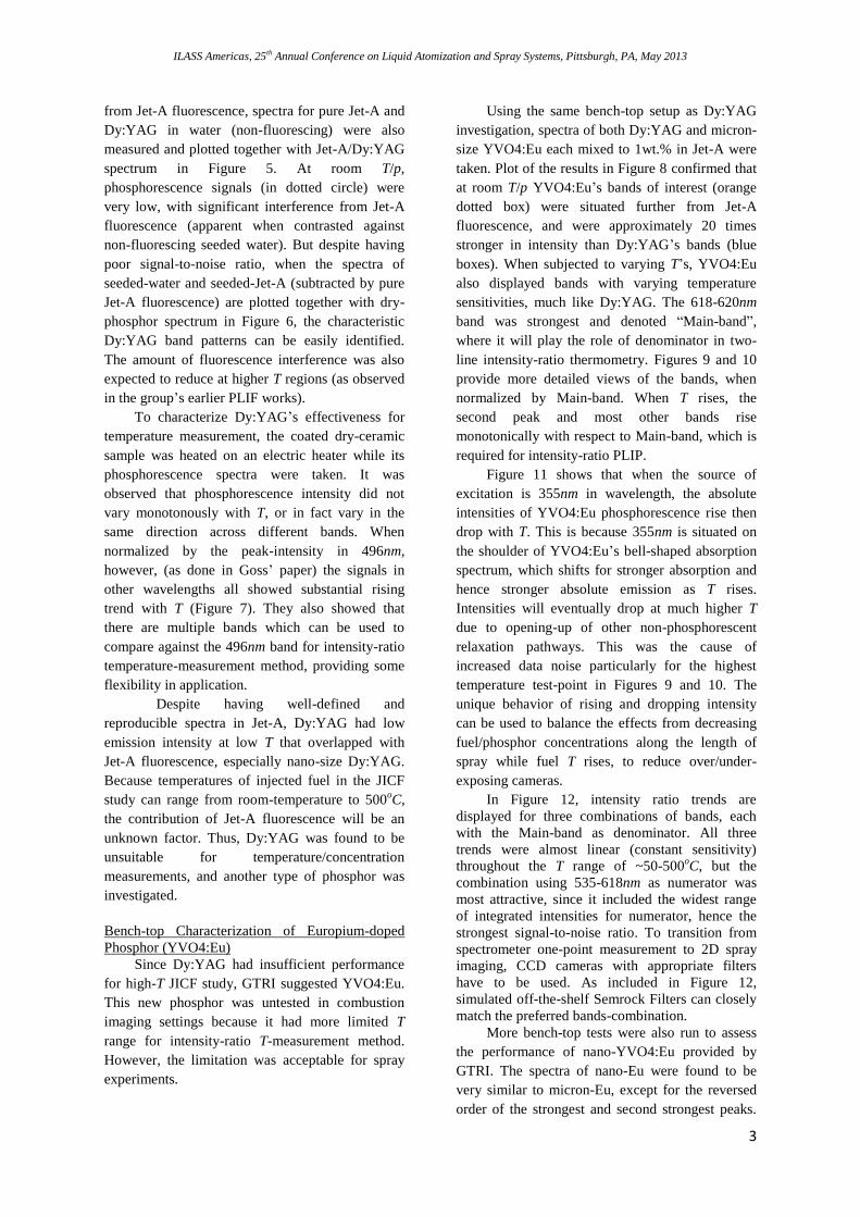
from Jet-A fluorescence, spectra for pure Jet-A and
Dy:YAG in water (non-fluorescing) were also
measured and plotted together with Jet-A/Dy:YAG
spectrum in Figure 5. At room T/p,
phosphorescence signals (in dotted circle) were
very low, with significant interference from Jet-A
fluorescence (apparent when contrasted against
non-fluorescing seeded water). But despite having
poor signal-to-noise ratio, when the spectra of
seeded-water and seeded-Jet-A (subtracted by pure
Jet-A fluorescence) are plotted together with dry-
phosphor spectrum in Figure 6, the characteristic
Dy:YAG band patterns can be easily identified.
The amount of fluorescence interference was also
expected to reduce at higher T regions (as observed
in the group’s earlier PLIF works).
To characterize Dy:YAG’s effectiveness for
temperature measurement, the coated dry-ceramic
sample was heated on an electric heater while its
phosphorescence spectra were taken. It was
observed that phosphorescence intensity did not
vary monotonously with T, or in fact vary in the
same direction across different bands. When
normalized by the peak-intensity in 496nm,
however, (as done in Goss’ paper) the signals in
other wavelengths all showed substantial rising
trend with T (Figure 7). They also showed that
there are multiple bands which can be used to
compare against the 496nm band for intensity-ratio
temperature-measurement method, providing some
flexibility in application.
Despite having well-defined and
reproducible spectra in Jet-A, Dy:YAG had low
emission intensity at low T that overlapped with
Jet-A fluorescence, especially nano-size Dy:YAG.
Because temperatures of injected fuel in the JICF
study can range from room-temperature to 500oC,
the contribution of Jet-A fluorescence will be an
unknown factor. Thus, Dy:YAG was found to be
unsuitable for temperature/concentration
measurements, and another type of phosphor was
investigated.
Bench-top Characterization of Europium-doped
Phosphor (YVO4:Eu)
Since Dy:YAG had insufficient performance
for high-T JICF study, GTRI suggested YVO4:Eu.
This new phosphor was untested in combustion
imaging settings because it had more limited T
range for intensity-ratio T-measurement method.
However, the limitation was acceptable for spray
experiments.
Using the same bench-top setup as Dy:YAG
investigation, spectra of both Dy:YAG and micron-
size YVO4:Eu each mixed to 1wt.% in Jet-A were
taken. Plot of the results in Figure 8 confirmed that
at room T/p YVO4:Eu’s bands of interest (orange
dotted box) were situated further from Jet-A
fluorescence, and were approximately 20 times
stronger in intensity than Dy:YAG’s bands (blue
boxes). When subjected to varying T’s, YVO4:Eu
also displayed bands with varying temperature
sensitivities, much like Dy:YAG. The 618-620nm
band was strongest and denoted “Main-band”,
where it will play the role of denominator in two-
line intensity-ratio thermometry. Figures 9 and 10
provide more detailed views of the bands, when
normalized by Main-band. When T rises, the
second peak and most other bands rise
monotonically with respect to Main-band, which is
required for intensity-ratio PLIP.
Figure 11 shows that when the source of
excitation is 355nm in wavelength, the absolute
intensities of YVO4:Eu phosphorescence rise then
drop with T. This is because 355nm is situated on
the shoulder of YVO4:Eu’s bell-shaped absorption
spectrum, which shifts for stronger absorption and
hence stronger absolute emission as T rises.
Intensities will eventually drop at much higher T
due to opening-up of other non-phosphorescent
relaxation pathways. This was the cause of
increased data noise particularly for the highest
temperature test-point in Figures 9 and 10. The
unique behavior of rising and dropping intensity
can be used to balance the effects from decreasing
fuel/phosphor concentrations along the length of
spray while fuel T rises, to reduce over/under-
exposing cameras.
In Figure 12, intensity ratio trends are
displayed for three combinations of bands, each
with the Main-band as denominator. All three
trends were almost linear (constant sensitivity)
throughout the T range of ~50-500oC, but the
combination using 535-618nm as numerator was
most attractive, since it included the widest range
of integrated intensities for numerator, hence the
strongest signal-to-noise ratio. To transition from
spectrometer one-point measurement to 2D spray
imaging, CCD cameras with appropriate filters
have to be used. As included in Figure 12,
simulated off-the-shelf Semrock Filters can closely
match the preferred bands-combination.
More bench-top tests were also run to assess
the performance of nano-YVO4:Eu provided by
GTRI. The spectra of nano-Eu were found to be
very similar to micron-Eu, except for the reversed
order of the strongest and second strongest peaks.

ILASS Americas, 25th Annual Conference on Liquid Atomization and Spray Systems, Pittsburgh, PA, May 2013
4
Using the same bands as micron-Eu, intensity ratios
of nano-Eu provided similar T-sensitivity to
micron-Eu. But, nano-YVO4:Eu had very weak
signals and was particularly difficult to capture
within the spectrometer’s limited dynamic range.
As a matter of fact, under the same conditions,
micron-size YVO4:Eu was up to 30 times brighter
than nano-Eu. Thus, if injector material is resistant
to abrasion, micron-size YVO4:Eu is preferred.
The degree to which micron-particles influence
spray characteristics is a subject for future
investigations.
Concept-verification Experiment at High Crossing
Flow Temperature
Following bench-top characterization of
YVO4:Eu, a high-temperature JICF test was
conducted to get visual evidence of Eu-
phosphorescence for concept-verification. While
the final optical system for phosphorescence
imaging will include three cameras (one for Mie-
scattering, and one each for the two
phosphorescence bands), only a simplified one-
camera system was installed for the first test. The
camera can capture fluorescence, Mie-scattering
and Eu-phosphorescence simultaneously in color at
1280x1024px resolution, and was synchronized to
355nm Nd:YAG laser pulse rate of 10Hz. Test
conditions for the first test were set to high-T and
relatively low-p, which promoted vaporization of
fuel so multi-phase flow-detection can be tested.
Found in Figure 13 are 150-frames-averaged
images representing one set of test conditions with
and without YVO4:Eu seeding. First, with only
pure Jet-A at high-T, low-p, the fuel fluoresced
strongly (blue) near the injector when it was still
cold. Then as it was heated by hot ambient air,
fluorescence disappeared, and only Mie-scattering
signal (green) remained. Further down, as droplets
vaporized into gaseous Jet-A, Mie-scattering
disappeared too, and the spray was no longer
visible. With seeded Jet-A, the same trends with
fluorescence and Mie-scattering occurred, but the
gaseous portion of spray glowed orange with
phosphorescence. Regions with fluorescence and
Mie-scattering were also brighter, due to
underlying phosphorescence signal. In general, the
in-rig phosphorescence test showed promising
results for YVO4:Eu. It produced easily visible
signals without requiring any special camera
intensifier, and had no issue providing detection
capability for gaseous fuel.
It is also worth remarking that Dy:YAG and
YVO4:Eu powders were both not naturally soluble
in liquid Jet-A. Undisturbed, micron-size phosphor
particles will quickly agglomerate and sediment at
the fuel tank’s bottom. During tests with seeded
fuel, sedimentation was a noticeable concern.
Figure 14 shows concentrations of phosphor in
seeded Jet-A at different tank depths after 2hr in
the tank. During the entire period, three 40W
ultrasonic transducers mounted on the tank’s
bottom provided ultrasonic agitation to the mixture.
The figure suggests that majority of YVO4:Eu
powder sank to the bottom, leaving a highly
concentrated lower zone, and a nearly uniform-
concentration upper zone. At the point of writing,
other techniques are being investigated to more
effectively disperse phosphor, including: fuel
recirculation/sediment re-deposition, more
powerful ultrasound, and surface-coating treatment.
Concluding Remarks and Future Plans
Despite efforts at optimization, Dy:YAG
phosphor, which was used by Hasegawa and
many other researchers (mostly in the form of
surface coating T measurement), was found to
be unsuitable for cross-flow spray studies
because of its weak phosphorescence signals.
This is especially the case when the spray
consist of Jet-A, which had natural
fluorescence wavelengths that overlap with
Dy:YAG phosphorescence.
Extensive bench-top tests and subsequent
high-T jet in cross-flow test showed that
YVO4:Eu had brighter phosphorescence
emission than Dy:YAG, and also emission
bands of interest in longer wavelengths
(further away from Jet-A fluorescence). It had
more limited applicable T-range compared to
Dy:YAG, but was adequate for spray
experiments. As such, YVO4:Eu is the
preferable phosphor for detecting temperature,
concentration distribution, and gas/liquid fuel
interface in JICF studies.
For future work, a 3-camera optical diagnostic
system for the high-temperature rig will be
completed, and the PLIP technique will be
fully tested for concentration, temperature and
phase-detection capabilities. At the same time,
investigation into nano-size phosphors and
more phosphor dispersion techniques will
continue.

ILASS Americas, 25th Annual Conference on Liquid Atomization and Spray Systems, Pittsburgh, PA, May 2013
5
References
1. Gopala, Y. et al., “Liquid Fuel Jet in
Crossflow- Trajectory Correlations based on
the Column Breakup Point,” 48th AIAA
Aerospace Sciences Meeting Including the
New Horizons Forum and Aerospace
Exposition, Orlando, Florida, USA, January
2010.
2. Omrane, A. et al., “Development of
Temperature Measurements Using
Thermographic Phosphors: Applications for
Combustion Diagnostics.”
3. Omrane, A. et al., “2D-Temperature Imaging
of Single Droplets and Sprays Using
Thermographic Phosphors,” Appl. Phys. B, 79:
431-434 (2004).
4. Someya, S. et al., “Lifetime-based Phosphor
Thermometry of an Optical Engine Using a
High-speed CMOS Camera,” International
Journal of Heat and Mass Transfer, 54: 3927-
3932 (2011).
5. J. Brubach et al., “Spray Thermometry Using
Thermographic Phosphors,” Appl. Phys. B, 83:
499-502 (2006).
6. Hasegawa, R. et al., “Two-dimensional Gas-
phase Temperature Measurements Using
Phosphor Thermometry,” Appl. Phys. B, 88:
291-296 (2007).
7. Alden, M. et al., “Thermographic Phosphors
for Thermometry: A Survey of Combustion
Applications,” Progress in Energy and
Combustion Science, 37: 422-461 (2011).
8. Heyes, A. L., “On the Design of Phosphors for
High-Temperature Thermometry,” Journal of
Luminescence, 129: 2004-2009 (2009).
9. Jain, N. et al., “Characterization of
Thermophosphor Particles for Simultaneous
Imaging of Velocity and Temperature,” 49th
AIAA Aerospace Sciences Meeting Including
the New Horizons Forum and Aerospace
Exposition, Orlando, Florida, USA, January
2011.
10. Goss, L. P. et al., “Surface Thermometry by
Laser-Induced Fluorescence,” Rev. Sci. Intrum.,
60: 3702 (1989).
Figure 1. Pictures and schematic of samples and bench-top phosphor-characterization setup.

ILASS Americas, 25th Annual Conference on Liquid Atomization and Spray Systems, Pittsburgh, PA, May 2013
6
Figure 2. Schematic of jet in cross-flow test section.
Figure 3. Schematic of cross-flow rig’s fuel system.

ILASS Americas, 25th Annual Conference on Liquid Atomization and Spray Systems, Pittsburgh, PA, May 2013
7
Figure 4. Spectra of GTRI and UK Dy:YAG coated on ceramic with and without glue binder, at room T/p.
Background: spectra of Dy:YAG as measured by Goss et al. [10].
Figure 5. Spectra of pure Jet-A, and GTRI Dy:YAG mixed in Jet-A and water at 1wt.% concentration. Peak for
water mixture at ~532nm was due to misaligned 532nm laser that lit the sample during measurement.
0
500
1000
1500
2000
2500
427.8 447.8 467.8 487.8 507.8
Inte
nsi
ty L
eve
l
Wavelength (nm)
Dy:YAG Coated Ceramic Phosphorescence (355nm Laser)
GTRI w/ Binder
GTRI w/o Binder
UK w/ Binder
UK w/o Binder
0
500
1000
1500
2000
2500
3000
3500
4000
300 350 400 450 500 550 600 650 700
Inte
nsi
ty L
eve
l
Wavelength (nm)
Dy:YAG Phosphorescence (355nm Laser, GTRI second batch)
DyYAG in Jet-A
DyYAG in Water
Jet-A Only (Normalized)

ILASS Americas, 25th Annual Conference on Liquid Atomization and Spray Systems, Pittsburgh, PA, May 2013
8
Figure 6. Phosphorescence spectra from samples on dry-ceramic (arbitrarily scaled to fit), in water, and in Jet-A
(minus Jet-A fluorescence).
Figure 7. Dy:YAG spectra at different temperatures, normalized by the 496nm peak.
0
50
100
150
200
250
300
450 460 470 480 490 500 510 520 530
Inte
nsi
ty L
eve
l
Wavelength (nm)
Dy:YAG Phosphorescence (355nm Laser, GTRI second batch)
DyYAG in Jet-A w/ Fluor Subtracted
DyYAG in Water
GTRI 1st Batch Ceramic (arbitrary absolute intensity)
0
0.2
0.4
0.6
0.8
1
1.2
440 460 480 500 520 540 560 580 600
49
6.9
5nm
-No
rmal
ize
d I
nte
nsi
ty
Wavelength (nm)
496nm-Normalized Spectra of GTRI Dy:YAG at Different T's
50oC
150oC
250oC
350oC
450oC
552oC
650oC

ILASS Americas, 25th Annual Conference on Liquid Atomization and Spray Systems, Pittsburgh, PA, May 2013
9
Figure 8. Comparison of Dy:YAG and YVO4:Eu spectra. Dotted boxes contain the bands of interest for
intensity-ratio PLIP: Dy:YAG (blue) and YVO4:Eu (orange).
Figure 9. Main-band-normalized spectra of YVO4:Eu (zoomed in at 600-640nm region).
0
0.2
0.4
0.6
0.8
1
1.2
600 605 610 615 620 625 630 635 640
Pak
-No
rmal
ize
d I
nte
nsi
ty
Wavelength (nm)
Peak-Normalized Spectra of YVO4:Eu at Different T's
59oC
150oC
249oC
350oC
450oC

ILASS Americas, 25th Annual Conference on Liquid Atomization and Spray Systems, Pittsburgh, PA, May 2013
10
Figure 10. Main-band-normalized spectra of YVO4:Eu (zoomed in at 530-600nm region).
Figure 11. Absolute intensity variations of three bands at different T’s, with 355nm laser excitation.
Peak-Normalized Spectra of YVO4:Eu at Different T's
0
0.05
0.1
0.15
0.2
0.25
0.3
530 540 550 560 570 580 590 600Wavelength (nm )
Pak
-No
rmal
ize
d In
ten
sity
59oC
150oC
249oC
350oC
450oC
0
5000
10000
15000
20000
25000
30000
35000
40000
0 100 200 300 400 500 600
Inte
nsi
ty
Temperature (oC)
YVO4:Eu Integral Intensity vs. T @ 355nm Excitation
Main-band (618-620nm)
Band 1 (600-618nm)
Band 2 (535-592nm)

ILASS Americas, 25th Annual Conference on Liquid Atomization and Spray Systems, Pittsburgh, PA, May 2013
11
Figure 12. Intensity ratios using different combinations of bands. Intensity ratio trend using off-the-shelf
Semrock filters (simulated with data from Semrock) is also included.
Figure 13. Averaged spray images from cross-flow test with/without YVO4:Eu-seeding.
0
0.5
1
1.5
2
2.5
3
3.5
0 100 200 300 400 500
Inte
nsi
ty R
atio
Temperature (oC)
YVO4:Eu Intensity Ratios vs. T
(535-592nm)/(618-620nm)
(600-618nm)/(618-620nm)
(535-618nm)/(618-620nm)
Simulated Semrock Filters set

ILASS Americas, 25th Annual Conference on Liquid Atomization and Spray Systems, Pittsburgh, PA, May 2013
12
Figure 14. Concentration results from tank-sampling test. Left-side of abscissa represents top of tank, and vice-
versa. Horizontal iso-concentration lines are established from known-concentration samples. Initial tank mixture
was 1wt.% phosphor.