Embed Size (px)
Citation preview
A FRACTION OF TUBERCLE BACILLI POSSESSING PRIMARY T O X I C I T Y
BY JOHN K. SPITZNAGEL,* M.D., AND REN~ J. DUBOS, P~.D.
(From the Laboratories of The Rockefeller Institule for Medic~ Research)
(Received for publication, November 17, 1954)
Much progress has been made during recent years towards the identification of the components of tubercle bacilli which are responsible for tubercle formation and for the development of tuberculin allergy. I t has never been proved how- ever that these two phenomena can account--singly or jointly--for all the systemic manifestations of tuberculosis. Indeed, there are a few facts which suggest that tubercle bacilli contain or produce substances capable of exerting deleterious effects on the host without participation of allergic processes and without productive tissue reaction.
The toxicity of cultures of tubercle bacilli has been observed under a wide range of conditions. In rabbits and guinea pigs, the injection of bacillary bodies killed by heat or disinfectants always causes loss of weight and commonly elicits the production of tubercles. The cachexia is slowly progressive and is eventually fatal if the amount of bacilli injected is large enough (1-5). While these effects can be obtained by injecting bacilli resuspended in aqueous medium (1-3), the results are more dramatic when an oil (mineral or vegetable) is used as a vehicle (4, 5).
I t has been found during recent years that acute toxic effects can be produced also in mice by injecting whole bacilli living or dead by either the intravenous or the intraperitoneal route (6-8). In this ease again the toxic effects are much increased by using an oil vehicle to inject the bacillary suspension.
Particulate fragments and soluble extracts obtained from cultures by various techniques have been shown to possess toxicity. Thus, non-acid-fast, minute (sub- microscopic) granules separated from autolyzed bacilli by high speed centrifugatlon exhibit a high degree of biological activity (9). There have been many claims that the toxic material is soluble in organic solvents (10-16). The toxic properties of some of the tuberculolipids will be considered later in the discussion of this report in relation to our own findings. The toxic effects which have been obtained with aqueous extracts of cultures have been rarely emphasized. Although they may not be directly relevant to the specific object of the present paper, they seem worth some mention here. Dramatic lesions have been produced in guinea pigs by the intravenous injection of large amounts of water-soluble tuberculoproteins (17). Indeed, large doses of tuber-
* Major, Medical Corps. Present address: United States Army Hospital, Fort Bragg, North Carolina.
291
292 TOXIC FRACTION FROM TUBERCLE BACILLI
culin can be acutely lethal to these animals (18). On the other hand, injection by the subcutaneous route of 5 ml. of filtrates of old cultures--free of bacilli--brings about a set of symptoms (filtrate disease) characterized by loss of muscular tone, cachexia, often resulting in death, but without production of tubercles (19).
While there is convincing evidence that the injection of whole tubercle bacilli, fragments of them, or soluble extracts of cultures, can cause a great variety of toxic reactions, it is not possible at the present time to evaluate or even relate the various findings. It is very likely that most of the preparations obtained by extraction with either aqueous media or organic solvents contained minute non-acid-fast granules which were overlooked; these have been shown to possess high biological activity (9). It is certain on the other hand that the injection of killed bacilli, or extracts of them, can bring about sensitization to some of the bacillary products (20, 21); some of the delayed toxic effects may have been therefore the manifestation of allergic processes. Indeed, several authors have expressed the view that the toxic manifestations of experimental as well as natural tuberculosis depend to a large extent, if not ex- cluslvely, upon reactions elicited by tuberculin allergy (22-24).
Ever since Auclair's pioneer studies, there have been many attempts to separate chemically the cellular components--particularly lipidic in nature--- responsible for the biological properties of tubercle bacilli (11, 25). The experi- ments to be reported here were focussed upon the possibility of producing with bacillary substances acute toxic reactions, unrelated to the development of al- lergy. Efforts were first directed toward the discovery of a solvent more effective than the ones commonly in use for the primary extraction of the bacillary lipids. I t was found that treatment with monochlorobenzene removed rapidly from living and dead bacilli a large amount of a fraction capable of exerting powerful systemic and local toxic effects in experimental animals. The toxic material which could be concentrated by fractional precipitation from cold petroleum ether, appeared unable to cause tubercle formation, or to elicit in guinea pigs a state of tuberculin allergy. I t is the purpose of the present paper to describe the mode of preparation, and some biological properties, of this toxic, non-tubercu- linogenic fraction, obtained from four different strains of mammalian bacilli.
EXPERIMENTAL
Cu/~rez.--The following strains of tubercle bacilli were used : - - Vallee M.V. (bovine, type, virulent).---Originally obtained from the Pasteur Institute,
Paris; this strain has been maintained for the past 5 years in a state of high virulence by re- peated isolation from the spleen of mice 2 weeks after infection.
R1Rv (human type, attenuated).--Obtained through the courtesy of Mr. W. Steenken, Jr., from the Standard Culture Depot maintained by the National Tuberculosis Association at the Trudeau Laboratory, Saranac, New York. The history of this strain is described in reference 26.
BCG-P (bovine type, attenuated).--A substrain of BCG obtained through the courtesy of Doctor J. Aronson, Henry Phipps Institute, Philadelphia.
BCG-T (bovine type, attenuated).--A substrain of BCG, obtained through the courtesy of Doctor S. Rosenthal, "rice Laboratory. Chicago.

JOHN K. SPITZNAGEL AND REN~ J. DUBOS 293
H37Ra (human type, avirulent).--Obtained through the courtesy of Mr. W. Steenken, Jr., from the Standard Culture Depot maintained by the National Tuberculosis Association at the Trudeau Laboratory. The h/story of this strain is presented in reference 26.
The comparative morphology of these strains and their ability to multiply in experimental animals has been discussed in several recent papers (27).
The bacteria used for extraction were obtained by inoculating 20 ml. of culture grown for 10 to 14 days in liquid tween-albumin medium into 1000 mL of liqu/d medium without tween as described in reference 28. The inoculated medium in 5 liter Blake bottles was incubated at 37.4°C., the bottles lying on their deep sides. After 3 days a light growth became visible; the bottles were quickly tipped upright and then returned to the original position. This ma- neuver assured the prompt appearance of a surface pellicle. Total time of incubation varied from 2 to 6 weeks. Heavy growth was obtained usually within 2 weeks except in the case of H37Ra which grew slowly and formed pellicles only after prolonged incubation. The yields varied from 1.2 to 3.8 gin. (dry weight) of bacteria per liter depending on the strain of or- ganism and the length of incubation.
The cultures of BCG-P were harvested in the viable state by collecting the pellicles over cheese-cloth with aseptic precautions for operators and cultures. Two batches were prepared, one from a culture grown for 2 weeks and the other for 4 weeks.
For reasons of safety the virulent cultures were killed by treatment with approximately 3.5 per cent phenol at room temperature for 24 hours prior to harvesting (40 ml. 88 per cent phenol per I000 cc. of culture). As the cultures of the RIRv and H37Ra strains were to be used in comparison with the material prepared from the virulent M.V. they were killed by the same technique. The M.V. and RIRv cultures were both incubated 58 days. The H37Ra culture was incubated 69 days in order to obtain a larger yield, more comparable to that ob- tained from the M.V. and RIRv cultures. The final yield (dry weight per liter of medium) was respectively: H37Ra 1.8 gin., R1Rv 2.4 gin., M.V. 3.8 gin. The cultures were collected on filter paper at reduced pressure on Buchner funnels.
The bacterial mass of BCG-P was dried in va~uo over P~O~ at room temperature. The bacterial masses of H37Ra, R1Rv, and M.V. were washed three times with 300 ml. of cold acetone to remove phenol and water. The last wash was negative for phenol as shown by failure to react with 10 per cent ferric chloride reagent. The acetone was then allowed to evaporate by spreading the bacteria on a glass plate. Final drying was accomplished in va~uo over P~O~. The dried bacteria were then stored over a desiccant at room temperature either in va~uo or in a polyethylene plastic bag.
Agimals.--Albino mice (so called Rockefeller Swiss strain) were obtained from the Rocke- feller Institute colony.
Black mice of the C57 BL6 strain were obtained from the Jackson Memorial Laboratories, Bar Harbor, Maine.
All mice were received at about 4 weeks of age, newly weaned. They were housed in groups of four in metal cages, bedded in cedar shavings, and given standard pellets and water ad lib.
The guinea pigs were from the stock raised at The Rockefeller Institute for Medical Re- search by Mr. Paul Marangello under the supervision of Dr. Merrill Chase. They were housed in metal wire cages, bedded in cedar shavings, and fed oats, hay, and cabbage.
Toxicity Assay.--Systemic toxicity was determined by observing changes in appearance, behavior, and weight of albino or C57 BL6 mice following intmperitoneal injection of test materials. Mice were weighed in groups once before injection and then at 3 to 4 day intervals until death ensued or until the original weight was regained.
In order to achieve conditions as uniform as possible, the mice were transferred to indi- vidual cages at 4 p.m. on the day prior to weighing. No food, water, or shavings were pro- vided. 18 hours later they were weighed by groups and the total weight of the group recorded

294 TOXIC FRACTION ]~ROM TUBERCLE BACILLI
in grams. The mice which died during the toxicity tests were autopsied and observations were made on gross changes in the organs. No histological examinations were performed.
Local toxicity was estimated by the reaction caused in the skin of guinea pigs by intra- dermal injections of test materials. Normal guinea pigs were restrained on a guinea pig board with a metal head harness and their backs carefully shaved with small animal clippers. In- jections were made from chemically cleaned 0.25 cc. tuberculin syringes with No. 26 gauge needles. Blebs containing the test materials were made by intradermal injection of the desired amount in 0.05 ml. of an inert vehicle. The size, color, and consistency of the lesions were ob- served at intervals over a 72 hour period. The method did not lend itself to exact quantitation.
Induction of Delayed Type Hypersensitivity to Tuberculin.--The fractions of interest were tested for their ability to induce tuberculous allergy in guinea pigs. Two methods known to be useful in establishing this type of allergy were employed. In the first, 50/~g. of the test substance was injected intraperitoneally in 0.5 ml. mineral oil. Tuberculin testing was done with either 0.1 ml. of 1:100 old tuberculin, 0.1 ml. of 1:10 old tuberculin, or 50 #g. of PPD in 0.1 ml. of diluent. Testing was done 3 weeks after the sensitizing injection.
The second method involved several injections of 5 #g. of test substance in 0.05 ml. sesame oil intradermally into guinea pigs over a 20 day period. The last two injections were spaced 6 days apart. Tuberculin testing was done 3 weeks after the last sensitizing injection using the tuberculin preparations mentioned above. These methods are regarded as likely to reveal any significant sensitizing ability.
Extraction with Monoctdorobenzene of BCG-P, H37Ra, RIRv and Vagee.--(Table I). A weighed aliquot of dried bacterial mass was placed in a glass vessel which could be closed to prevent the escape of solvent vapor. Monochlorobensene was added in the ratio of 8 ml. per gin. of dry bacteria. A magnetic agitator was placed in the vessel which was then closed and placed on a magnetic stirrer. The stirrer speed was so adjusted that the bacteria were main- tained completely suspended and moving through the solvent. The extraction was allowed to proceed at room temperature (26-27°C.) for 6 days. The bacteria were quite grumose when first introduced into the solvent. However, they became evenly dispersed throughout the solvent after a few hours stirring.
The major portion of the insoluble matter was separated from the extracting solvent by centrifugation, leaving a clear amber supernatant fluid. I t should be noted that although this fluid appeared crystal-clear, it still contained bacterial debris as revealed by a positive Tyndal effect. No special effort was made to separate the debris at this point as this was achieved with greater ease in a subsequent step. The mass of insoluble residue was then subjected to further extractions under the same conditions. Extraction of the residue was continued to exhaustion of extractable substance. The extracts from each aliquot of solvent were worked up separately.
Recovery of the Crude ~xtrazt.--The extract was rapidly evaporated to dryness in a Craig evaporator (29), at a temperature never exceeding 50°C. Residual solvent was removed from the aromatic, brownish soft, waxy mass by further evaporation in vacuo in a stream of nitro- gen. Altogether approximately 10 per cent of the total dry cell mass weight could thus be extracted in solution from the BCG-P culture. This proportion was somewhat different from other cultures and dependent upon the length of incubation.
Exttoxtion with Alcohol-Ether and Clgoroform.--Dried culture masses of BCG-P and M.V. organisms were extracted at room temperature (26°-27°C.) with a mixture in equal proper- tions of alcohol and ether, 10 ml. of solvent being used per gin. of bacteria. The solvent was replaced from time to time and extraction continued until evaporation of samples no longer revealed the presence of extracted material. The bacterial residue was then dried and ex- tracted at the same temperature with chloroform, using 10 ml. of this solvent per gm. of original weight of bacteria. Extraction was continued with repeated changes of chloroform until nothing further could be removed from the residue. The extracts were freed of most of
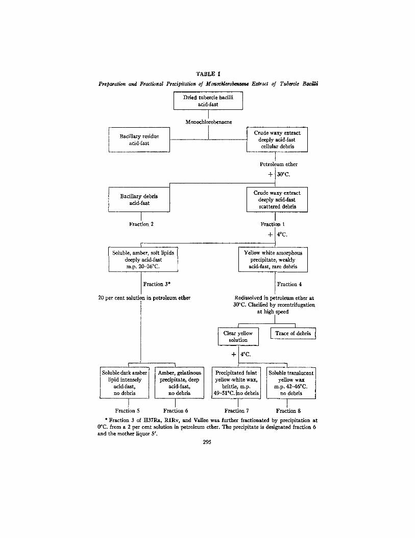
TABLE I
Preparation and Fractional Precipitation of Monocklorobenzens Extract of
Bacillary residue acid-fast
Bacillary debris acid-fast
I Fraction 2
Soluble, amber, soft lipids deeply acid-fast m.p. 20-26°C.
1 Dried tubercle bacilli |
/ acid-fast
I Monochlorobermene
I
Fraction 3*
20 per cent solution in petroleum ether
Soluble dark amber lipid intensely
acid-fast, no debris
Amber, gelatinous precipitate, deep
acid-fast, no debris
Tuberde Bacilli
Crude waxy extract deeply acid-fast ceUular debris
I Petroleum ether
+ 30°C.
Crude waxy extract deeply acid-fast scattered debris
I Fraction 1
+14°C.
Yellow white amorphous precipitate, weakly
acid-fast, rare debris
I Fraction 4
Redissolved in petroleum ether at 30°C. Clarified by recentrifugation
at high speed
I I I
Clear yellow I Trace of debris I solution
+ J 4°C"
Precipitated faint yellow-whlte wax,
brittle, m.p. 49-51°C. Ino debris
J J I Fraction 5 Fraction 6 Fraction 7
I Soluble translucent
yellow wax m.p. 42-46°C.
no debris
I Fraction 8
* Fraction 3 of H37Ra, R1Rv, and Vallee was further fractionated by precipitation at 0°C. from a 2 per cent solution in petroleum ether. The precipitate is designated fraction 6 and the mother liquor 5'.
295

296 TOXIC FRACTION FROM TUBERCLE BACILLI
the bacillary bodies by filtration through Schleicher and Schull No. 576 paper on a Buchner funnel. They were then recovered by evaporation in the Craig evaporator and were stored in vacuo after all solvent had been removed.
Fractionation of Extracts of BCG-P.--The crude monochlorobenzene extract was frac- tionated by taking advantage of the limited solubility of certain of its components in petroleum ether (BP 30-60°C.). Whenever petroleum ether is mentioned in this paper the low boiling point variety is meant. 4 gm. of crude extract was dissolved in 100 ml. of petroleum ether at room temperature. The insoluble material was centrifuged out of the cloudy amber solution. Microscopic examination revealed that the sediment thus obtained consisted, of acid-fast and non-acid-fast bacterial debris and of granules similar to those seen in intact cells of Myco- bacterium tuberculosis. Continued centrifugation for 1 hour at 2100 R.p.x~. in glass-stoppered centrifuge tubes sufficed to clarify the solution and to render it free of bacillary debris. The soluble material was designated fraction 1 and the bacillary debris fraction 2. (Table I.)
Fraction 1 was restored to its original volume with fresh petroleum ether. The solution had a dark amber color with a strong odor similar to that of phenyl ethyl alcohol. I t was placed at +4°C. for 24 hours during which time a heavy amorphous precipitate formed. This was removed by centrifugation in the cold, but the superuatant fluid could not be entirely clari- fied under the conditions employed. The sediment was washed twice with small portions of ether. I t was designated fraction 4, and the mother liquor fraction 3 (Table I).
Fraction 4 was redissolved with difficulty in 10 ml. of petroleum ether at room temper- ature and the solution centrifuged. This procedure removed a very small amount of insoluble bacterial debris leaving a clear, slightly aromatic, yellowish solution. This solution was placed at +4°C. and after 24 hours a heavy white precipitate formed which was removed by centrif- ugation in the cold. This was termed fraction 7 and the material remaining in the mother liquor fraction 8. Microscopic examination revealed no bacterial debris in either fraction 7 or fraction 8 (Table I).
The volume of the solution of fraction 3 (mother liquor of fraction 4) was reduced 80 per cent; chilling to +4°C. for 24 hours resulted in formation of a gelatinous insoluble phase in the tube. This was separated from a very dark amber highly aromatic supernatant liquid. The supernatant fluid was designated fraction 5 and the gelatinous mass fraction 6 (Table I).
Fractionation of Extracts of M.V., R1Rv, H37Ra.--The differences in petroleum ether solubilities of certain components present in extracts of these three strains necessitated minor modifications in procedure and terminology. Fraction 1 of M.V. gave no precipitate at +4°C. However, precipitation occurred when the temperature was reduced to 0°C. for 24 hours. This precipitate, designated fraction 6', was found to be the most toxic fraction of the M.V. extract. The mother liquor separated from this material and freed of solvent, was designated fraction 5'.
In the case of the R1Rv extract, precipitation occurred at +4°C. and the precipitate was designated fraction 4. Further precipitation from the mother liquor occurred at 0°C. and the precipitate was designated fraction 6'; the material in the mother liquor, fraction 5'. The tI37Ra extract gave a precipitate at +4"C. but no further precipitate at 0°C. The precipitate was designated fraction 4 and the material from the mother liquor fraction 5. In the case of fraction 4 from R1Rv and H37Ra, subsequent treatment was similar to that used in fractionat- ing the BCG-P extract. Fraction 7 was prepared from both of these extracts.
Physical and Chemical Properties of Fraction 7 of B C C r - - F r a c t i o n 7 a c c o u n t e d
for a p p r o x i m a t e l y 1 p e r c en t of t h e or ig ina l d r y we igh t of t he whole bac t e r i a l
mass . A l t h o u g h th i s f r ac t i on does n o t c o n s t i t u t e a well def ined chemica l en-
t i t y some of i t s phys i ca l a n d chemica l cha rac t e r i s t i c s will b e desc r ibed in m o r e
de ta i l because i t p r o v e d to possess h i g h biological a c t i v i t y .

JOttN K. SPITZNAGEL AND REN]~ ~'. DUBOS 297
As stated in Table I the fraction 7 freed of solvents appeared as a faintly yellowish white, brittle, waxy amorphous material. I t did not give the neutral red reaction (fraction 3 did) (30). When spread on a glass slide and stained by the Ziehl-Neelsen technique, it revealed no bacterial bodies or debris, but left a faintly pink film even after vigorous decolorization. Fraction 7 was soluble at room temperature in monochlorobenzene, nitrobenzene, benzene, chloroform, petroleum ether, and diethyl ether, the solubility decreasing in the order indi- cated. I t could be dissolved to a very limited extent in n-butanol, n-propanol, ethanol, methanol, and acetone with warming, and was completely insoluble in water regardless of pH.
15
~" 0.5 o
Conc. 15m9./~.4cc. pet.ethe~ Cell length I can.
0210 250 300 ~0 Wave leagm (m/~)
FIo. 1. Infrared absorption curve of fraction 7 of monochlorobenzene extract of BCG-P.
I t had a sharp melting point at 49-51°C. Its phosphorous content was 0.4 per cent suggesting that the material might be a phospholipid. Its nitrogen content was 0.14 per cent, indicating that little or no protein was present.
Fig. 1 represents absorption curves of fraction 7 in the infrared range. ~ The absorption in the ultraviolet was probably non-specific. The infrared absorption curve suggests that the bulk of the fraction was an aliphatic substance with carbonyl and alcohol groups. Since the material was not acidic there is a possi- bility that the carbonyl groups were ketonic or a part of esterified carboxylic groups.
Solubility Characteristics.--The bacillary fractions obtained with various primary lipid solvents behaved with some individuality when subjected to fractional precipitation from cold petroleum ether. This technique was used to
1 Kindly carried out by Dr. Herbert Jaffe of The Rockefeller Institute for Medical Re- search.
298 TOXIC :FRACTION FROM TUBERCLE BACILLI
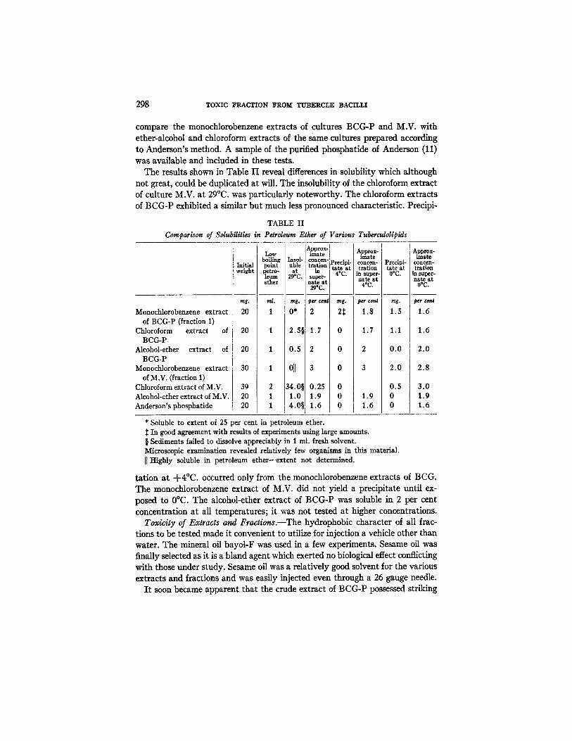
compare the monochlorobenzene extracts of cultures BCG-P and M.V. with ether-alcohol and chloroform extracts of the same cultures prepared according to Anderson's method. A sample of the purified phosphatide of Anderson (11) was available and included in these tests.
The results shown in Table II reveal differences in solubility which although not great, could be duplicated at will. The insolubility of the chloroform extract of culture M.V. at 29°C. was particularly noteworthy. The chloroform extracts of BCG-P exhibited a similar but much less pronounced characteristic. Precipi-
TABLE II
Comparison o/Solubili t ies in Petroleum Ether of Various Tuberculoliplds
Monochlorobenzene extract of BCG-P (fraction 1)
Chloroform extract of BCG-P
Alcohol-ether extract of BCG-P
iV[onochlorobenzene extract
of M.V. (fraction 1) Chloroform extract of M.V. Alcohol-ether extract of M.V. Anderson's phosphatide
Approx- I Approx- Low [ ira ate I I boiling Insol- [ concen-P~ • • F ,mate rreclpl- Initial point uble traUon [ --re t I coneen-
weight petro- at [ in I taAl*oa tration leum 29°C. [ super- [ = ~" [ in super-
h a t e at e ther a~ca.t l o • 4oc,
[ mg. ml. mg. ~er cenl mg. per cent
O* 2 2~
rag.
20 1 1.8 1.5
20 1 2.5 1.7 0 1.7 1.1
20 1 0.5 2 0 2 0 .0
30 1 0]l 3 0 3 2.0 I
39 2 [34.0§ 0.25 0 0.5 20 1 ,I 1.0 1.9 0 1.9 0 20 1 ' 4 . 0 § 1.6 0 1.6 0 I
Precipi- tate at OoC.
Approx- tmat~
coneen- trstlon
in super- nate a t
O°C.
per cen#
1.6
1.6
2 .0
2.8
3 .0 1.9 1.6
* Soluble to extent of 25 per cent in petroleum ether. :~ In good agreement with results of experiments using large amounts. § Sediments failed to dissolve appreciably in 1 ml. fresh solvent. Microscopic examination revealed relatively few organisms in this material. [[ Highly soluble in petroleum ether---extent not determined.
tation at +4°C. occurred only from the monochlorobenzene extracts of BCG. The monochlorobenzene extract of M.V. did not yield a precipitate until ex- posed to 0°C. The alcohol-ether extract of BCG-P was soluble in 2 per cent concentration at all temperatures; it was not tested at higher concentrations.
Toxicity of Extracts and Fractions.--The hydrophobic character of all frac- tions to be tested made it convenient to utilize for injection a vehicle other than water. The mineral oil bayol-F was used in a few experiments. Sesame oil was finally selected as it is a bland agent which exerted no biological effect conflicting with those under study. Sesame oil was a relatively good solvent for the various extracts and fractions and was easily injected even through a 26 gauge needle.
I t soon became apparent that the crude extract of BCC~P possessed striking
JOHN K. SPITZNAGEL AND R E I ~ J. DUBOS 299
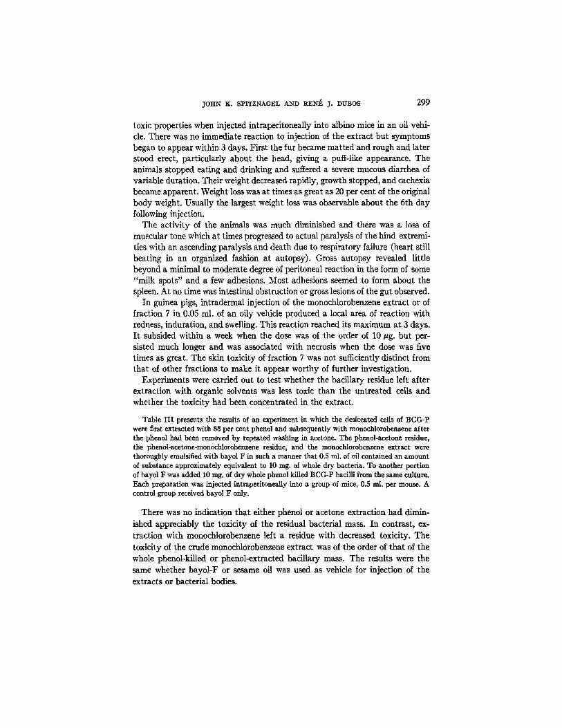
toxic properties when injected intraperitoneally into albino mice in an oil vehi- cle. There was no immediate reaction to injection of the extract but symptoms began to appear within 3 days. First the fur became matted and rough and later stood erect, particularly about the head, giving a puff-like appearance. The animals stopped eating and drinking and suffered a severe mucous diarrhea of variable duration. Their weight decreased rapidly, growth stopped, and cachexia became apparent. Weight loss was at times as great as 20 per cent of the original body weight. Usually the largest weight loss was observable about the 6th day following injection.
The activity of the animals was much diminished and there was a loss of muscular tone which at times progressed to actual paralysis of the hind extremi- ties with an ascending paralysis and death due to respiratory failure (heart still beating in an organized fashion at autopsy). Gross autopsy revealed little beyond a minimal to moderate degree of peritoneal reaction in the form of some "milk spots" and a few adhesions. Most adhesions seemed to form about the spleen. At no time was intestinal obstruction or gross lesions of the gut observed.
In guinea pigs, intradermal injection of the monochlorobenzene extract or of fraction 7 in 0.05 ml. of an oily vehicle produced a local area of reaction with redness, induration, and swelling. This reaction reached its maximum at 3 days. I t subsided within a week when the dose was of the order of 10/zg. but per- sisted much longer and was associated with necrosis when the dose was five times as great. The skin toxicity of fraction 7 was not sufficiently distinct from that of other fractions to make it appear worthy of further investigation.
Experiments were carried out to test whether the bacillary residue left after extraction with organic solvents was less toxic than the untreated cells and whether the toxicity had been concentrated in the extract.
Table I I I presents the results of an experiment in which the desiccated cells of BCG-P were first extracted with 88 per cent phenol and subsequently with monochlorobenzene after the phenol had been removed by repeated washing in acetone. The phenol-acetone residue, the phenol-acetone-monochlorobenzene residue, and the monochlorobenzene extract were thoroughly emulsified with bayol F in such a manner that 0.5 ml. of oil contained an amount of substance approximately equivalent to 10 rag. of whole dry bacteria. To another portion of bayol F was added l0 rag. of dry whole phenol killed BCG-P bacilli from the same culture. Each preparation was injected intraperitoneaUy into a group of mice, 0.5 rol. per mouse. A control group received bayol F only.
There was no indication that either phenol or acetone extraction had dimin- ished appreciably the toxicity of the residual bacterial mass. In contrast, ex- traction with monochlorobenzene left a residue with decreased toxicity. The toxicity of the crude monochlorobenzene extract was of the order of that of the whole phenol-killed or phenol-extracted bacillary mass. The results were the same whether bayol-F or sesame oil was used as vehicle for injection of the extracts or bacterial bodies.

300 TOXIC FRACTION :FROM TUBERCLE BACILLI
Concentration of Toxic Material in tke Various Fractions of the Crude Ex- tract.-
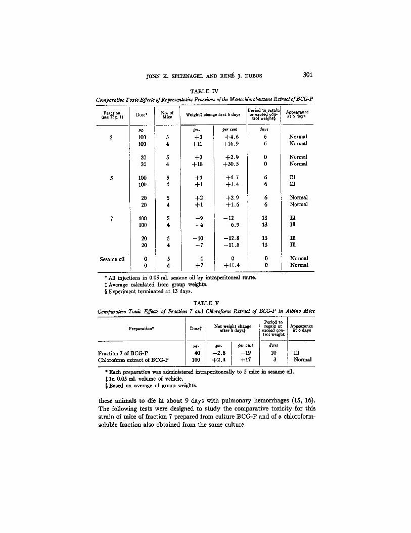
Fractions 2, 5, 6, 7, and 8 were emulsified in sesame oil to give doses of either 100 or 20 /Jg. per 0.05 ml. Mice were given one injection intraperitoneally of each dose of each prepara- tion, the controls receiving 0.05 ml. of sesame oil alone. The weight changes and other repre- sentative data for groups of mice receiving fractions 2, 5, 7 and for the controls are recorded in Table IV.
The results presented in Table IV show that fraction 7 was the most toxic of the bacillary lipids; it caused marked weight loss at both dose levels. Frac- tion 5 was next most effective and fraction 2 practically inactive. Fractions 6
TABLE I I I ToxiviSy of Various Fractions of BCG-P
Prelmmtions* Doses Average weight change in 6 days
Whole phenol-killed bacilli Bacilli extracted with phenol-acetone Bacilli extracted with phenol-acetone-monochloro-
benzene Monochlorobenzene extract Bayol-F control
mg.
10 7.3 5.5
0.59 0
g~,n.
- -1 .8 --3.1 +1.5
--2.6 +2.8
#er 6anJ
-17.4 -24.4 +12.8
--20.4 +28.0
* All preparations injected in 0.5 ml. bayol-F v/a the intraperitoneal route. Each prepa- ration was given to a group of 5 mice.
Selected as approximately equivalent to 10 mg. of whole phenol-killed tubercle bacilli (dry weight).
and 8 gave results related to their position in the extraction procedure, frac- tion 8 being indistinguishable from fraction 7, and fraction 6 intermediate between fractions 5 and 7.
Comparison in Swiss Albino Mice of Fraction 7 and the Chloroform-Soluble Fraction of BCG-P.--It is apparent from published descriptions that fraction 7 presents similarities with Anderson's chloroform soluble "wax" (11). In order to determine the comparative toxicity of these two materials, prepara- tions obtained from a culture of BCG-P were emulsified in sesame oil and in- jected intraperitoneally into albino mice.
As shown in Table V fraction 7 was toxic in a dose of 40/~g. whereas injec- tion of 100 #g. of the chloroform-soluble fraction produced no untoward ef- fects.
Toxicity of Fraction 7for C57 BL6 Mice.--During the course of these investi- gations there appeared reports showing that chloroform extracts prepared by Anderson's technique contain a material toxic for C57 black mice under special conditions. Multiple successive injections of small doses were found to cause
JONN K. SPITZNAGEL AND RENE J. DUBOS 301
TABLE IV Compar~iveToxicEff~ts~Re~es~t~eFr~ticns~theM~oc~orob~z~eE~r~t~BCG-P
Fraction (see Fig. 1)
Sesame oil
Dose*
#g-
100 100
20 20
100 100
20 20
100 100
20 20
No. of Mice Weight:[: change first 6 days
gftZ.
+3 +11
+2 +18
+1 +1
+2 +1
- 9 --4
-10 - 7
0 +7
Period to regain o r exceed con-
per cen|
+4.6 +16.9
+2.9 +30.5
+1.7 +1.4
+ 2 . 9 +1.6
--12 --6.9
--12.8 --11.8
0 +11.4
trol weight§
days
6 6
13 13
13 13
Appearance at 6 days
Normal Normal
Normal Normal
III Ill
Normal Normal
Ill
In 131
Normal Normal
* All injections in 0.05 ral. sesame oil by intraperitonesl route. ~: Average calculated from group weights. § Experiment terminated at 13 days.
TABLE V Comparative Toxic l~.ffexts of FracHon 7 and Chloroform Extract of BCG-P in Albino Mice
Preparation*
Fraction 7 of BCG-P Chloroform extract of BCG-P
Net weight change Dose: after 6 days§
4O 100
gin. per cenl
--2.8 --19 +2 .4 +17
Period to regain o r
exceed con- trol weight
d~ys
10 Ill 3 Normal
* Each preparation was administered intraperitoneally to 5 mice m sesame oil. :~ In 0.05 mi. volume of vehicle. § Based on average of group weights.
Appearance at 6 days
these animals to die in about 9 days with pulmonary hemorrhages (15, 16). The following tests were designed to s tudy the comparat ive toxici ty for this s train of mice of fraction 7 prepared from culture BCG-P and of a chloroform- soluble fraction also obtained from the same culture.
302 TOXIC ]~RACTION FROM TUBERCLE BACILLI
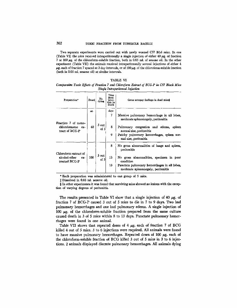
Two separate experiments were carried out with newly weaned C57 BL6 mice. In one (Table VI) the mice received intraperitoneally a single injection of either 40 ~tg. of fraction 7 or 100/~g. of the chloroform-soluble fraction, both in 0.05 ml. of sesame oil. In the other experiment (Table VII) the animals received intraperitoneaUy several injections of either 4 /tg. each of fraction 7 spaced at 3 day intervals, or of 100#g. of the chloroform-soluble fraction (both in 0.05 ml. sesame oil) at similar intervals.
TABLE VI Comparalit~ Toxic Effects of Fraction 7 and Ctdotoform Extract of BCG-P in C57 Black Mice
Single In~raperiloneal Injection
Preparation*
Fraction 7 of mono- chlorobenzene ex- tract of BCG-P
Chloroform extract of alcohol-ether ex- tracted BCG-P
)ose
ttg.
40
100
No. dying
3 ou|
of,
3 out of
Time from injec- :ion to death
days
7
8
9
8
13
13
Gross autopsy findings in dead mice§
Massive pulmonary hemorrhage in all lobes, moderate splenomegaly, peritonitis
Pulmonary congestion and edema, spleen normal size, peritonitis
Patchy pulmonary hemorrhages, spleen nor- mal size, peritonitis
No gross abnormalities of lungs and spleen, peritonitis
No gross abnormalities, specimen in poor condition
Punctate pulmonary hemorrhages in all lobes, moderate splenomegaly, peritonitis
* Each preparation was administered to one group of 5 mice. Dissolved in 0.05 ml. sesame oil.
§ In other experiments it was found that surviving mice showed no lesions with the excep- tion of varying degrees of peritonitis.
The results presented in Table VI show that a single injection of 40 gg. of fraction 7 of BCG-P caused 3 out of 5 mice to die in 7 to 9 days. Two had pulmonary hemorrhages and one had pulmonary edema. A single injection of 100 ttg. of the chloroform-soluble fraction prepared from the same culture caused death in 3 of 5 mice within 8 to 13 days. Punctate pulmonary hemor- rhages were found in one animal.
Table VII shows that repeated doses of 4 ttg. each of fraction 7 of BCG killed 4 out of 5 mice. 3 to 6 injections were required. All animals were found to have massive pulmonary hemorrhages. Repeated doses of 100 #g. each of the chloroform-soluble fraction of BCG killed 3 out of 5 mice in 3 to 6 injec- tions. 2 animals displayed discrete pulmonary hemorrhages. All arfimMs dying

JOHN K. SPITZNAGEL AND RENE J. DUBOS 303
in these experiments had some degree of peritonitis. No striking change in weights was noted even in animals receiving the most toxic preparations.
These and other experiments of similar design not presented here make it clear that fraction 7 contained a factor capable of causing pulmonary hemor- rhages and death in C57 BL6 mice. A larger dose of the chloroform-soluble
TABLE VII Comparison Toxic Effects of Multiple Intraperltoneal Injections of Fraction 7 and Chloroform
Extract of BCG-P in C57 Black Mice
Preparation*
Fraction 7 of mono- chlorebenzene ex- tract of BCG
Total Time injections
Dose~ No. from 1st in each dying injection mouse
to death prior to death
8 3
9 4 4 out
4 of 5 14 5
17 6
Gross autopsy findings in dead mice§
Massive pulmonary hemorrhage: spleen normal size, peritonitis
Massive pulmonary hemorrhage, spleen normal size, peritonitis
Massive pulmonary hemorrhage, splenomegaly, no gross peritonitis
Massive pulmonary hemorrhage, spleen not enlarged, no peritonitis
Chloroform extract 3 out of alcohol-ether 100 of 5 extracted BCG-P
8 3 No gross pulmonary pathology, spleen normal size, severe peri- tonitis
13 4 Many discrete pulmonary hemor. rhages, spleen normal size, no peri. tonitis
15 6 Patchy pulmonary hemorrhages, splenomegaly and peritonitis
* Ea£h preparation was administered to one group of 5 mice. :~ Given in 0.05 ml. sesame oil. § In other experiments it was found that surviving mice showed no lesions other than
varying degrees of peritonitis.
fraction also produced death but with less striking pulmonary lesions. As little as 12/~g. of fraction 7 in 3 divided doses sufficed to produce death with hemor- rhages in mice of this strain. Whether the toxic material responsible for these effects was the one that produced death without pulmonary hemorrhages in albino mice could not be determined from these experiments.
Toxicity of Fractions Prepared from Other Strains of Tubercle Bacillus.-- Table V I I I shows the results of toxicity tests carried out with fractions of the monochlorobenzene extracts prepared from cultures H37Ra, R1Rv and M.V.
In general the results indicate that it is possible to extract from the cells of

304 TOXIC FRACTION FROM TUBERCLE BACILLI
these three strains a toxic mater ial similar in propert ies to tha t obtained from BCG-P. As was to be expected, the weight loss was slightly less in groups of mice receiving 20 gg. than in groups receiving 100 ~tg. of any given prepara- tion,
TABLE VIII Comparatlv¢ Toxic EffecJs of Fractions of MonochIorobensene Ex~rads Obtained from 3 Different
Strains of Tubercle Ba¢~ll
Fraction and strain
(see Fig. 1)
7 + 8 of H37Ra
Dose* Period to
Weight change after I control weight 6 days~ regain or exceed
i pg. gm.
100 - 2 . 6 20 +3.0
5' of H37Ra 100 +7.6 20 +3.0
7 + 8 of R1Rv 100 --5.0 20 --3.6
5' of R1Rv 100 --5.3 20 - 4.8
5' of R1Rv 100 - 7 . 0 20 0
5' of Vallee 100 -- 10.3 20 --8.6
5' of Vallee 100 - 7.0 20 --4.6
Sesame oil 0 + 7 . 6
No injection 0 +6.3
Appearance at 6 days
- 4 . 4 +5.1
+13.4 +5.3
- 8 . 5 - 6 . 3
- 9 . 5 - 8 . 5
-12 .3 0
-18 .4 --15.1
--12.5 --8.1
+13.3
+10.7
days
10 Normal 7 Normal
3 Normal 7 Normal
10 Ill 7 Ill
14 Ill 10 Ill
14 Normal 7 Normal
Ill Ill
Ill Ill
3 Normal
3 Normal
No. dying
0 0
1 0
0 0
0 1
1 0
5 4
6 2
0
0
* Each dose level of each preparation was administered intraperitoueally to three groups of 4 mice each. Vehicle was sesame oil. Volume 0.05 ml..
2~ Average calculated from group weights.
Under the conditions of these experiments deaths were most frequent and weight loss most severe among mice receiving fractions prepared from the M.V. culture. The H37Ra fractions were the least toxic. The differences in temperature conditions required to precipi ta te the insoluble fractions obtained from H37Ra, R1Rv, and M.V. extracts did not seem to bear any relationship to the biological effects. There were indications tha t in any given extract the fraction which precipi ta ted first was the most toxic regardless of the source of the extract and regardless of whether precipi tat ion occurred at + 4 ° or 0°C.
JOHN K. SPITZNAGEL AND R E I ~ ~. DUBOS 305
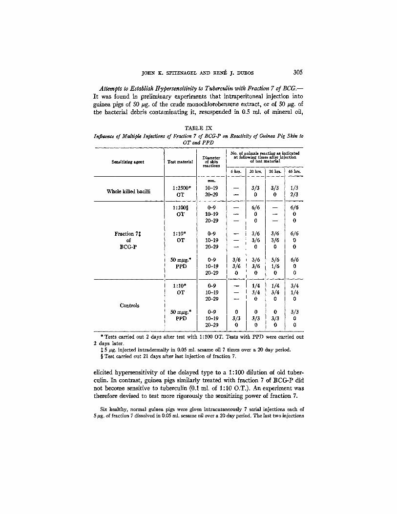
Attempts to Establisk ttypersensiti~ity to Tuberculin with Fraction 7 of BCG.-- It was found in preliminary experiments that intraperitoneal injection into guinea pigs of 50 gg. of the crude monochlorobenzene extract, or of 50 tzg. of the bacterial debris contaminating it, resuspended in 0.5 ml. of mineral oil,
TABLE IX
Influence of Mrdtiple Injections of Fraction 7 of BCG-P on Reactivity of Guinea Pig Skin to OT and PPD
Sensitizing agent
Whole killed bacilli
Fraction 7:~ of
BCG-P
Controls
Test material Diameter
of skin reactions
No. of animals reacting as indicated at following times after injection
of test material
6 hrs. 20 hrs. 26 hrs. 46 hrs,
1:2500" 10-19 - - 3/3 3/3 1/3 OT 20-29 - - 0 0 2/3
1 : 1 0 0 § 0-9 - - 6/6 - - 6/6 OT 10-19 ~ 0 - - 0
20-29 ~ 0 ~ 0
1:10" 0-9 - - 3/6 3/6 6/6 OT 10-19 - - 3/6 3/6 0
20-29 ~ 0 0 0
50 mgg.* 0-9 3/6 PPD 10-19 3/6
20-29 0
1 : 10" 0-9 OT 10-19
20-29 k
3/6 5/6 6/6 3/6 1/6 o
0 0 0
1/4 I 1/4 3/4 3/4 3/4 1/4 0 0 0
50 m~g.* 0-9 0 0 0 3/3 PPD 10-19 3/3 3/3 3/3 0
20-29 0 0 0 0
* Tests carried out 2 days after test with 1: I00 OT. Tests with PPD were carried out 2 days later.
~: 5/zg. injected intradermally in 0.05 ml. sesame oil 7 times over a 20 day period. § Test carried out 21 days after last injection of fraction 7.
elicited hypersensitivity of the delayed type to a 1:100 dilution of old tuber- culin. In contrast, guinea pigs similarly treated with fraction 7 of BCG-P did not become sensitive to tuberculin (0.1 ml. of 1:10 O.T.). An experiment was therefore devised to test more rigorously the sensitizing power of fraction 7.
Six healthy, normal guinea pigs were given intracutaneously 7 serial injections each of 5/~g. of fraction 7 dissolved in 0.05 ml. sesame oil over a 20 day period. The last two injections

306 TOXIC I~RACTION ]~ROM TUBERCLE BACILLI
were spaced 6 days apart. 2 Tuberculin testing was begun 21 days after the last injection of fraction 7. The results are tabulated in Table IX.
The results presented in Table IX show that animals having received re- peated intmdermal injections of fraction 7 and tested with either 50 /~g. of PPD, or 0.1 ml. of 1:100 O.T., or 0.1 ml. of 1:10 O.T. gave reactions which did not differ from those seen in the controls challenged with the same doses. The reactions to 0.1 ml. of a 1:2500 dilution of O.T. in animals previously sensitized with whole dead tubercle bacilli were far more severe than reactions to larger doses of O.T. in the test and control animals. It is dear therefore that treat- ment with fraction 7 did not elicit any significant degree of tuberculin sensi- tivity in guinea pigs.
DISCUSSION
Most species of Gram-negative microorganisms produce highly active, heat- stable endotoxins. In contrast, the heat-killed cells of Gram-positive species are on the whole remarkably inocuous for experimental animals. Mycobacteria appear to occupy an intermediate position between these two microbial groups with regard to toxicity. It has been shown repeatedly that injection of killed tubercle bacilli or of certain fragments of them can cause in rabbits and guinea pigs a slowly progressing ultimately fatal cachcxia (1-5). The subcutaneous injection into guinea pigs of 5 ml. of filtrates of old cultures, freed of bacterial bodies causes what has been called the "filtrate disease" characterized by loss of muscular tone, cachexia, and eventually death without any evidence of tubercle formation (19). But the amounts of bacillary material or of filtrates required to produce these effects are rather large, of the order of several miUi- grams or milliliters. Observations in our laboratory have revealed that the in- traperitoneal injection into albino mice of 10 rag. of bacillary bodies killed by heat, phenol, formaldehyde, or iodine and resuspended in physiological saline will cause these animals to die within a very few days (7, 8). In rabbits, guinea pigs, and mice the toxic effects can be greatly increased by injecting the bacilli in suspension in oil (mineral or vegetable) instead of in saline; the minimum toxic dose being thus reduced 5- to 10-fold.
There is no doubt therefore that dead tubercle bacilli contain some highly toxic components. It is probable that many of the slowly progressive deleterious effects that they cause are the expression of allergic phenomena--resulting from the reaction of sensitized tissue with bacillary material persisting for many months in the body. But the fact that mice commonly die within 1 to 3 days following intraperitoneal injection of a few milligrams of bacilli (living or killed) makes plain that some at least of the toxic effects that have been observed are independent of allergic processes. Indeed, the present study has shown that it
At no time during this procedure did the appearance of the injection sites suggest that the animals had become sensitive to the material itself.

~0HN K. SPITZNAGEL AND RENE ~. DUBOS 307
is possible to separate from various cultures of mammalian bacilli a lipid frac- tion which appears to account for much of the primary toxicity of the bacillary bodies.
I t is surprising that the existence of this particular toxic material had not been brought to light by earlier studies of the lipid constituents of tubercle bacilli. The extensive and thorough purification procedures developed by Ander- son and his school (11), and lately extended through the use of chromatographic techniques (31, 32) have made available in a purified form many bacillary lipids. Some of these have been shown to call forth a great variety of tissue reactions, but none have been found to display a marked degree of toxicity.
It is true that there have appeared recently claims of separation from tubercle bacilli of powerful toxins bearing a direct relation to virulence, but their sig- nificance is at best uncertain. According to one group of reports, extraction of bacillary bodies with mineral oil releases a lipid fraction more abundant in virulent than in avirulent cultures and possessing extraordinary lethal proper- ties for guinea pigs (14). Confirmation of these findings has not been forthcom- ing. (For a more extensive discussion of this topic see reference 33.) According to other claims, extraction of young bacillary bodies with petrolic ether re- leases from certain strains of bacilli a toxic lipid fraction, designated "cord factor," which would be an essential determinant of virulence (15, 16). This material (which is also present in the chloroform-soluble fraction) has been shown to be toxic by the artifice of multiple injections into mice at several days' interval. This technique of assay leaves open the possibility that the ef- fects observed are not due to the primary toxicity of the substance but rather to artefacts such as the evocation of latent pulmonary viruses, or sensitiza- tion phenomena. Moreover several of the statements made concerning this material appear invalid in the light of recent publications as well as of critical scrutiny of the early reports (see reference 33 for a more extensive discussion of this topic).
The toxic material described in the present study has been obtained from young cultures of four strains of mammalian bacilli: one highly virulent (bo- vine), two attenuated (human and bovine), and one avirulent (derived from a human strain). It appears that the success in obtaining it depended upon the use of monochlorobenzene as primary solvent for the bacilli. Even at fairly low temperature, this solvent extracted some 10 per cent of the total bacillary weight. By fractional precipitation with low boiling petrolic ether (at 0 ° or 4°C. depending upon the culture used), it was possible to separate from the monochlorobenzene extract a toxic material which was designated fraction 7. Although fraction 7 represented only 1 per cent of the original weight of the bacillary mass, it accounted for much of its primary toxicity.
Some attempts have been made to characterize fraction 7 chemically but their results have little significance since nothing is known of the degree of

308 TOXIC PRACTION FROM "-'u~ERCLE BACILLI
purity of the material available. Suffice it to state that the sample studied contained 0.14 per cent nitrogen and 0.4 per cent phosphorus, and that it had a fairly sharp melting point (49-51°C.). Needless to say, this is no assurance of purity as mixtures of different lipidic substances often exhibit this behavior. I t is of interest in this respect that some of the characteristics of the fraction most extensively studied (fraction 7 from BCG-P) were not unlike those of Anderson's "purified wax," a component of the chloroform-soluble fraction, and the starting material for subsequent chromatographic analysis. However while fraction 7 precipitated out of the monochlorobenzene extract upon addi- tion of petroleum ether at low temperatures, no similar precipitate could be obtained from the chloroform extract. Furthermore, there were great differences in toxicity between the "purified wax" and fraction 7.
Fraction 7 produced in mice symptoms that could not be differentiated from those caused by whole bacilli, but no disseminated or even pulmonary tubercles were ever observed to result from its injection (by the intraperitoneal route). In albino mice, acute death followed intraperitoneal injection of amounts of the order of 100 #g.; one single injection of 20 #g. was sufficient to produce cachexia; death commonly ensued in about 9 days without gross pathological lesions other than minimal to moderate peritonitis. In C57 BL mice, one single intraperitoneal dose also caused death in about 9 days, accompanied by massive pulmonary hemorrhages. The same type of pulmonary lesions could be ob- tained with somewhat smaller doses when these were introduced by multiple injections at several days intervals. As already pointed out, however, the re- suits obtained by this technique are of questionable significance.
Severe local reactions could be produced following injection of a few micro- grams of fraction 7 into the skin of guinea pigs. In contrast, it was found im- possible to elicit with it any evidence of tuberculin allergy even by using tech- niques most conducive to the development of allergic reactions. Nor was there any indication that the susceptibility of the animals to fraction 7 itself in- creased in the course of serial intracutaneous injection. Thus, it seems certain that allergic phenomena played no significant part in the toxic reactions elicited by the material.
The four strains of tubercle bacilli tested yielded preparations exhibiting slight differences in solubility and in toxicity. The material obtained from the virulent culture M.V. was the most toxic on a weight basis, and that from the avirulent H37Ra the least toxic. The fractions obtained from the two at- tenuated strains BCG-P and RIRv were intermediate in toxicity. These find- ings make it tempting to assume that fraction 7 plays an important role as a determinant of virulence in tubercle bacilli. Other facts, however, cast doubt on the validity of this assumption. First it must be emphasized that the differ- ences in solubility and toxicity observed among the various preparations were not large and might well have been due to differences in the degree of purity of the material.
~OHN K. SPITZNAGEL AND KEN~ ~. DUBOS 309
Probably more important is the fact that the toxicity of the total bacillary substance of cultures killed by either heat or phenol did not bear any relation to the virulence of the strain. Weight for weight, for example, suspensions of the avirulent culture H37Ra were fully as toxic as suspensions of the highly virulent culture M.V. In fact the most attenuated of the BCG substrains in use in our laboratory (BCG-T), was found to yield the most toxic suspensions of bacterial cells.
I t is probable finally, that the state of the cultures affected both the solu- bility in organic solvents and the biological activity of the various preparations obtained from them. On the one hand there is evidence that the conditions of growth determine the over-all toxicity of the bacillary bodies (34). On the other hand, it is certain that aging of the culture brings about profound alterations in its various chemical constituents. Even relatively young cultures of tubercle bacilli contain non-acid-fast elements---evidence of autolytic changes. As a rule, the less virulent the culture, the more rapidly the cells lose viability and are disrupted by autolysis; it has been repeatedly observed in our laboratory for example that certain BCG cultures are particularly delicate in this respect, and H37Ra even more so. The fact recently published that the percentage amount of wax D (chloroform-soluble) yielded by the culture H37Ra is smaller in cultures 6 weeks old than in younger cultures (32), constitutes evidence that some of the tuberculolipids, or at least their state in the cell, are altered by autolytic processes. I t is clear therefore, that any attempt to study the com- parative toxicity of extracts of various cultures will require very exacting con- trol over many steps of the culture methods and preparative procedures.
While there is no evidence so far that the toxic material present in fraction 7 accounts for the different degrees of virulence of the various strains of tubercle bacilli, there seems to be good reason to believe that it plays some part in the toxemia of tuberculosis. Two independent observations almost rule out the pos- sibility that the toxic material is an artefact created by preparative procedures. On the one hand, the procedure used for its preparation involved only extraction and precipitation by organic solvents at low temperature, techniques not likely to create new products out of the bacterial components. We have already men- tioned on the other hand that death always rapidly followed the injection of adequate amounts of whole bacilli into experimental animals. In mice, this acute toxic effect followed intraperitoneal injection of a few milligrams of killed bacillary bodies in suspension in an oil vehicle. I t is of particular interest that the same result could be obtained by injecting intravenously a similar amount of living organisms. Fraction 7, which makes up approximately 1 per cent of the total bacillary weight, could cause acute death of mice in amounts (approximately 20/~g.) one-hundredth that required of killed or living cells. I t appears in other words that its toxicity accounted for a large part of the primary toxicity of the whole organisms.

310 TOXIC FRACTION FROM T U B E R C L E BACILLI
SUMMARY
Tubercle bacilli separated from young cultures were thoroughly extracted with monochlorobenzene at temperatures never exceeding 50°C. From the soluble material, a fraction corresponding to approximately 1 per cent of the total bacillary weight was separated by fractional precipitation with petrolic ether at temperatures of 0 ° or 4°C.---depending upon the strain of bacilli.
The monochlorobenzene-soluble--ether-insoluble material (fraction 7) pre- pared from BCG-P was found to contain 0.14 per cent nitrogen and 0.4 per cent phosphorus. Some of its other chemical characteristics are described.
Fraction 7 proved unable to elicit tuberculin allergy in guinea pigs, but in- jection of 5 #g. of it into the skin produced severe local reactions.
In albino mice, a single intraperitoneal injection of 20 /zg. caused loss of muscular tone and of weight followed by death within 9 days. The only tissue reaction observed was a slight degree of peritonitis. In mice of the C57 BL strain, a single injection of 40 #g. also caused death in the same time, but with pulmonary hemorrhages--usually massive.
Material similar to fraction 7 was obtained from one virulent, two attenuated, and one avirulent strain of mammalian tubercle bacilli (bovine and human). The fractions obtained from the various strains differed somewhat in solubility and toxicity. The more virulent the culture, the more toxic was the fraction obtained from it; but it is possible that this relation was the result of differences in the effectiveness of the extraction procedures, rather than of characteristics inherent to the cultures.
Evidence is presented that the toxicity of fraction 7 accounts for much of the primary toxicity of tubercle bacilli.
BIBLIOGRAPHY
1. Prudden, T. M., and tIodenpyl, E. New York Med. J., 1891, 16. Columbia College Studies from Pathological Laboratory, 1890-92.
2. Strauss, I., and Gamaleia, N., Arch. m~d. exp. et argO. path., 1891, 3, 705. 3. Mafucci, A., Centr. allg. Path. u. path. Anat., 1890, 1, 825. 4. Pdst, N., Ann. Inst. Pasteur, 1938, 61, 121, 5. Saenz, A., and Canetfi, G., Ann. Inst. Pasteur, 1940, 65, 13. 6. Youmans, G. P., and Youmans, A. S., Am. Reg. Tuberc., 1951, 64, 541. 7. Dubos, R. J., Schaefer, W. B., and Pierce, C. H., J. Exp. Med., 1953, 9/, 221. 8. Weiss, D., and Dubos, R. J., J. Exp. Med., 1955, 101,313. 9. Laporte, R., Ann. Inst. Pasteur, 1945, 71, 51.
10. Auclair, J., Arch. ra2d. Exp. et anat. path., 1898, 11, 363; 1900, 12, 189. 11. Anderson, R. J., Physiol. Rew., 1932, 12, 166; Harvey Lectures, 1939--40, 271. 12. Dessy, G., and Francioli, M., Boll. Ist. sieroter, milan., 1938, 1'/, 134. 13. Solomides, J., Compt. rend. Soc. biol., 1939, 131, 706. 14. Choucroun, N., Science, 1943, 98, 327; Am. Reg. Tuberc., 1947, 56, 203; Carapt.
rend. Acad. sc., 1949, 210, 511.
JOHN K. SPITZNAGEL AND REN~ J. DUBOS 311
15. Bloch, H., J. Exp. Med., 1950, 91, 197. 16. Asselineau, J., Bloch, H., and Lederer, E., Am. Rev. Tuberc., 1953, 67, 853. 17. Sabin, F. R., and Doan, C. A., J. Exp. Med., 1927, 46, 645. 18. Seibert, F. B., J. Biol. Chem., 1928, 78, 345. 19. Pinner, M., and Voldrich, M., Am. Re~. Tuberc., 1931, 24, 73. 20. Choucroun, N., Compt. rend. Acad. sd., 1948, 226, 1477. 21. Raffel, S., J. Infect. Dis., 1948, 82, 267; Experientia, 1950, 6, 410. Raffel, S.,
Lederer, E., and Asselineau, J., Fed. Proc., 1954, 13, 509. 22. van Denies, F., and Solomides, J., Ann. Inst. Pasteur, 1940, 64, 73. 23. Pirmer, M., Pulmonary Tuberculosis in the Adult, Springfield, Illinois, Charles
C Thomas, 1945. 24. Rich, A. R., The Pathogenesis of Tuberculosis, Springtidd, Illinois, Charles C
Thomas, 1944. 25. Sabin, F. R., Am. R~. Tuberc., 1941, 44, 415; Physiol. R,v., 1932, 12, 141; Am.
R~. Tub~c., 1932, 25, 153. 26. Steenken, W., Jr., and Gardner, L. U., Am. Rev. Tuberc., 1946, 64, 51, 62. 27. Pierce, C. H., Dubos, R. J., and Schaefer, W. B., J. Exp. Med., 1953, 97, 189. 28. Dubos, R. J., and Middlebrook, G., Am. Rev. Tuberc., 1947, 56, 334. 29. Craig, L. C., Sregony, J. D., and I-Iausman, W., Anal Chem., 1950, 22, 1462. 30. Dubos, R. J., and Middlebrook, G., Am. Rev. Tuberc., 1948, 58, 698. Dubos,
R. J., and Surer, E., Am. Re~. Tuberc., 1949, 60, 384. 31. Lederer, E., in Colloquium of chemotherapy of tuberculosis, Dublin, July,
1951, 1; in Symposium sur le m6tabolisme microbien, H, Congr. Internat. Biochim., Paris, July 21-27, 1952, 1.
32. Asseline~u, J., Prog. Explor. Tuberc., 1952, 5, 1. Ann. Inst. Pasteur, 1951, 81, 306.
33. Dubos, R. J., Components and structure of tubercle bacilli involved in their pathogenicity in Symposium on Pathogenicity, Society of General Micro- biology, Cambridge University Press, 1955, 103.
34. Weiss, D., unpublished data.