Embed Size (px)
Citation preview

From the desk of:Dr. Asha Jain
Senior GynecologistNHMC & H
It is a process of spontaneous per vaginum expulsion of mature fetus with vertex presentation followed by after births and which ends without any artificial aids, complications and which is not delayed.
Abnormal labor is complicated labor
There are 3 stages
Stage 1: from onset of labor to full dilatation of cervix. Duration is 5-10 hours.
Stage 2: from full dilatation to expulsion of fetus. Duration ½ to 1 hour.
Stage 3: Stage of expulsion of after births. Duration is 5 min -1/2 hour.
Total duration of normal laborPrimi: 12-16 hoursMulti: 6-8 hours
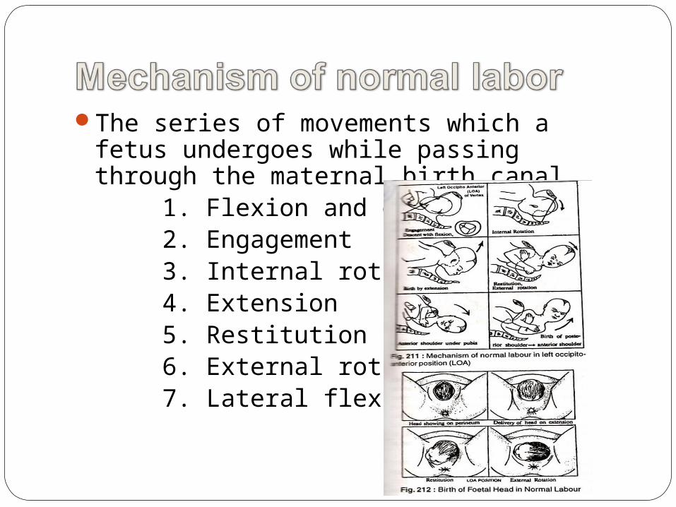
The series of movements which a fetus undergoes while passing through the maternal birth canal.
1. Flexion and descent2. Engagement3. Internal rotation4. Extension5. Restitution6. External rotation7. Lateral flexion
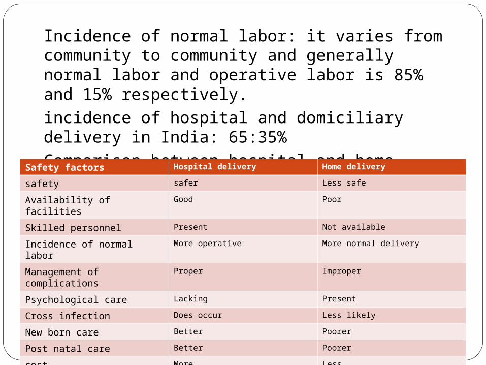
Incidence of normal labor: it varies from community to community and generally normal labor and operative labor is 85% and 15% respectively. incidence of hospital and domiciliary delivery in India: 65:35%Comparison between hospital and home deliverySafety factors Hospital delivery Home delivery
safety safer Less safe
Availability of facilities Good Poor
Skilled personnel Present Not available
Incidence of normal labor More operative More normal delivery
Management of complications
Proper Improper
Psychological care Lacking Present
Cross infection Does occur Less likely
New born care Better Poorer
Post natal care Better Poorer
cost More Less
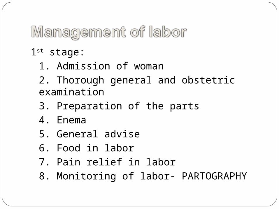
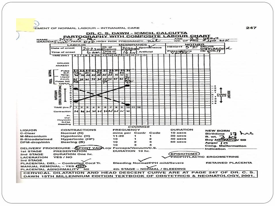
1st stage: 1. Admission of woman2. Thorough general and obstetric examination3. Preparation of the parts4. Enema5. General advise6. Food in labor7. Pain relief in labor8. Monitoring of labor- PARTOGRAPHY

2nd stage:
1. Observe signs of 2nd stage of labor
2. Woman taken to the delivery table
3. Continuous monitoring of the vitals and fetal heart rate every 15 minutes
4. Position of the woman and vaginal examination after cleaning and draping
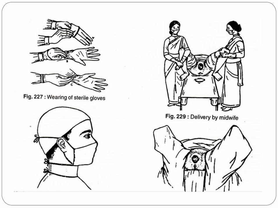
5. Preparation by the birth attendant
6. Evacuation of bladder
7. Patient is encouraged to bear down with further progress of labor
8. Perineal infilteration with episiotomy before crowning of the head
9. Observe crowning
10. Head is delivered by supporting perineum and flexing the coming head
11. Once head is delivered eyes are cleaned, looked for cord around the neck
12. Delivery of the shoulders and trunk
13. Care of new born – APGAR SCORE
3rd stage: Active management1. Watch for the signs of separation of placenta2. Delivery of placenta by control cord traction3. Receive and inspect placenta4. Watch for any signs of PPH and obtain proper hemostasis5. Suturing of episiotomy
4th stage:1. Observation of the female after delivery for at least ½ an hour
Post partum hemorrhage: (Atonic 80% and traumatic 20%) excessive hemorrhage from genital tract after the birth of baby amounting to a degree affecting general condition of the patient. Statistically if blood loss is more than 500 ml. Incidence 0.5 % of hospital delivery. Maternal morbidity 10%
Types: primary and secondary
Primary
Diagnosis and management: a) Atonic: Prolonged labor, over distended uterus, abnormal presentations, abnormal uterine contractions
b) Traumatic: cervical and perineal tears, ppt labor
Secondary
Retained placental pieces
Subinvolution of uterus
Deep lacerations of cervix
Infections
Choriocarcinoma
Management:
General : IV fluid, blood transfusion and uterotonic drugs
Surgical: Exploration of the genital tract, uterine compression
Late stage: ligation of uterine artery, ligation of anterior division of internal iliac artery, hysterectomy
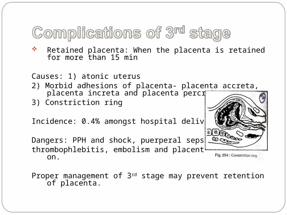
Retained placenta: When the placenta is retained for more than 15 min
Causes: 1) atonic uterus2) Morbid adhesions of placenta- placenta accreta,
placenta increta and placenta percreta3) Constriction ring
Incidence: 0.4% amongst hospital deliveries
Dangers: PPH and shock, puerperal sepsis, thrombophlebitis, embolism and placental polyp later on.
Proper management of 3rd stage may prevent retention of placenta.
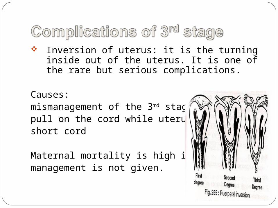
Inversion of uterus: it is the turning inside out of the uterus. It is one of the rare but serious complications.
Causes: mismanagement of the 3rd stage, pull on the cord while uterus is atonic, short cord
Maternal mortality is high if immediate management is not given.
Amniotic fluid embolism is spontaneous embolism of amniotic fluid debris in small pulmonary artery leading to varying degree of respiratory distress and circulatory collapse. It is rare but dramatic and deadliest complication.
Risk factors: 1)vigorous labor contractions2) Rent through fetal membranes as in marginal
separation of placenta
Prognosis varies on the severity and fatal for mother in severe cases.
Shock is a state of collapse of circulation so that there is a critical deduction of perfusion of tissues by blood with oxygen
Causes: Hemorrhagic- all 3rd stage complications Non hemorrhagic- trauma of obstetric operations
like breech extraction, MRP, internal podalic version etc., obstetric accidents like rupture of uterus, abruptio placenta, acute inversion etc., due to anesthesia and drugs, pulmonary embolism
Sepsis- delayed complication
Puerperal sepsis and septic shock
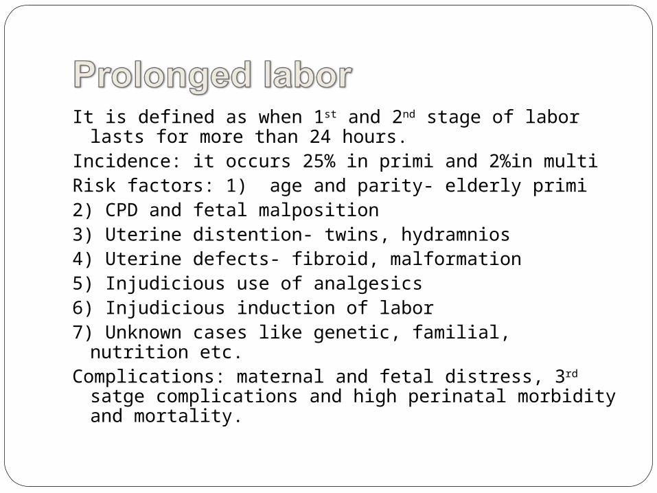
It is defined as when 1st and 2nd stage of labor lasts for more than 24 hours.
Incidence: it occurs 25% in primi and 2%in multiRisk factors: 1) age and parity- elderly primi2) CPD and fetal malposition3) Uterine distention- twins, hydramnios4) Uterine defects- fibroid, malformation5) Injudicious use of analgesics6) Injudicious induction of labor7) Unknown cases like genetic, familial, nutrition etc.Complications: maternal and fetal distress, 3rd satge
complications and high perinatal morbidity and mortality.
It is extremely fast labor due to over efficient uterine action and labor may end within 3 hours.
Causes: 1) common in multigravida rarely in primi
2) Injudicious use of oxytocin 3) Unusual lack of resistance in cervix and
lower uterine segmentDangers: maternal and fetal injuries
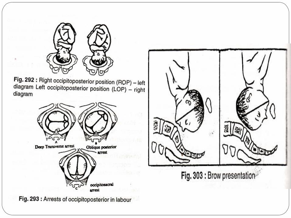
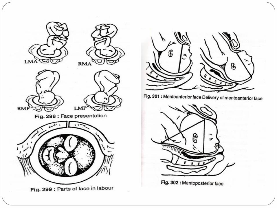
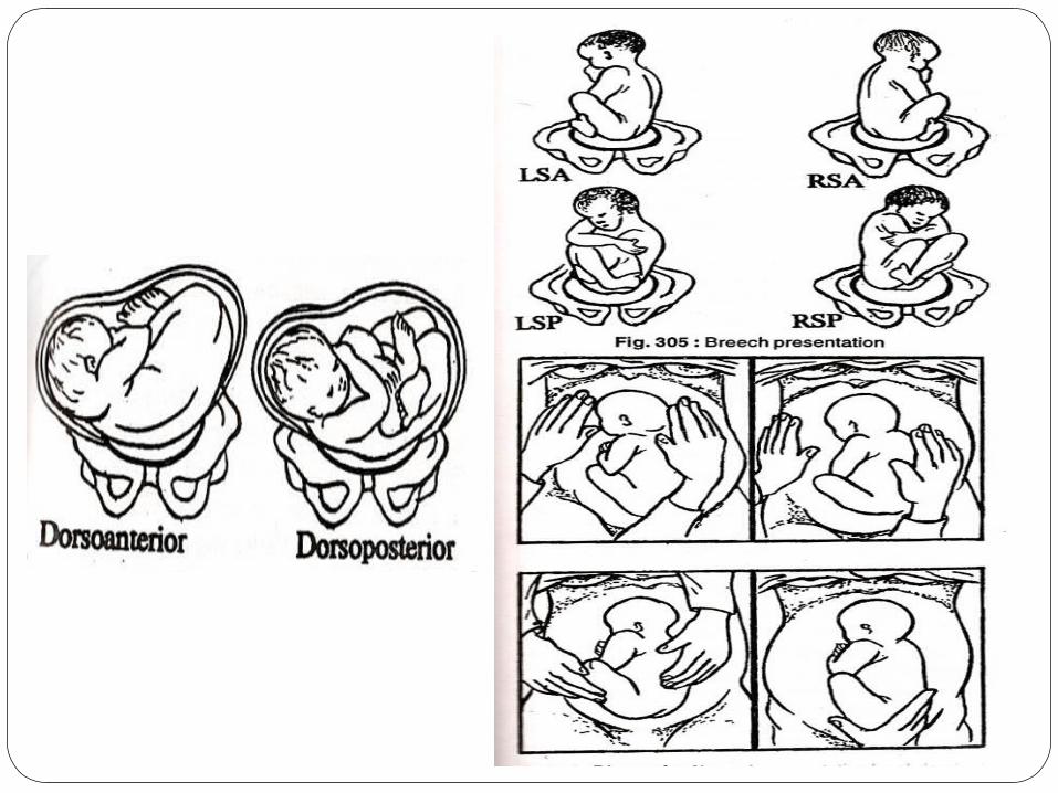
Total 14% of pregnancies have abnormal presentations
1. Occipito posterior 2. Face 0.2%3. Brow 0.1%4. Breech 3.2%5. Shoulder 0.5%
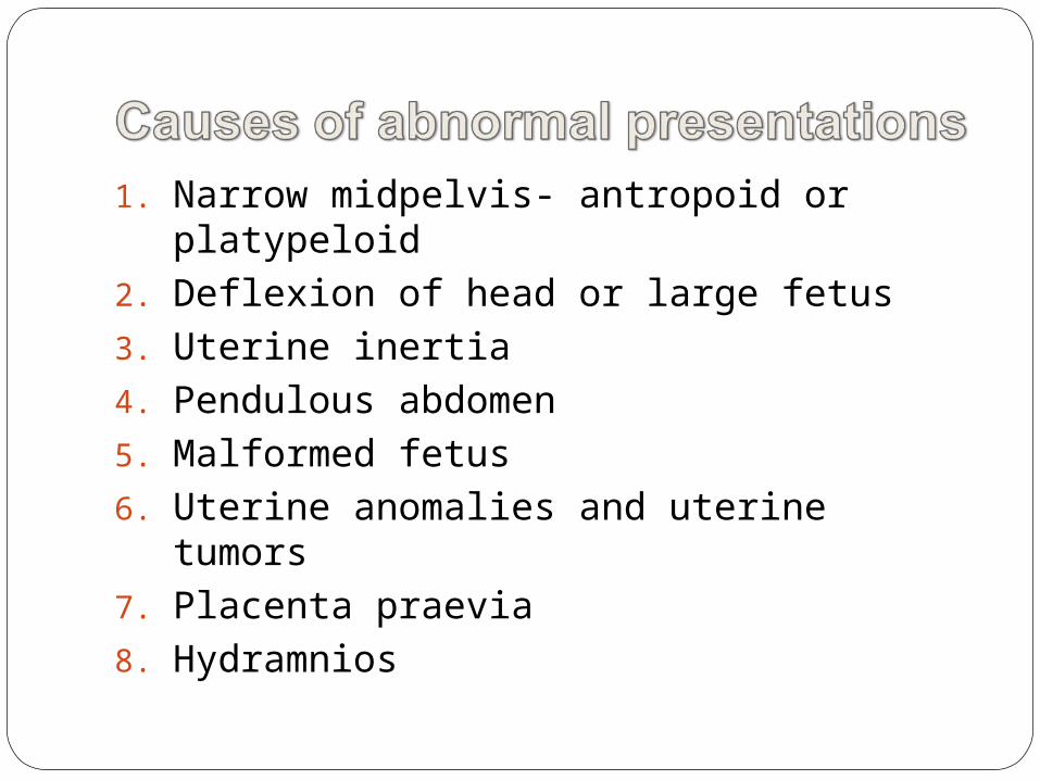
1. Narrow midpelvis- antropoid or platypeloid
2. Deflexion of head or large fetus3. Uterine inertia4. Pendulous abdomen5. Malformed fetus6. Uterine anomalies and uterine tumors7. Placenta praevia8. Hydramnios
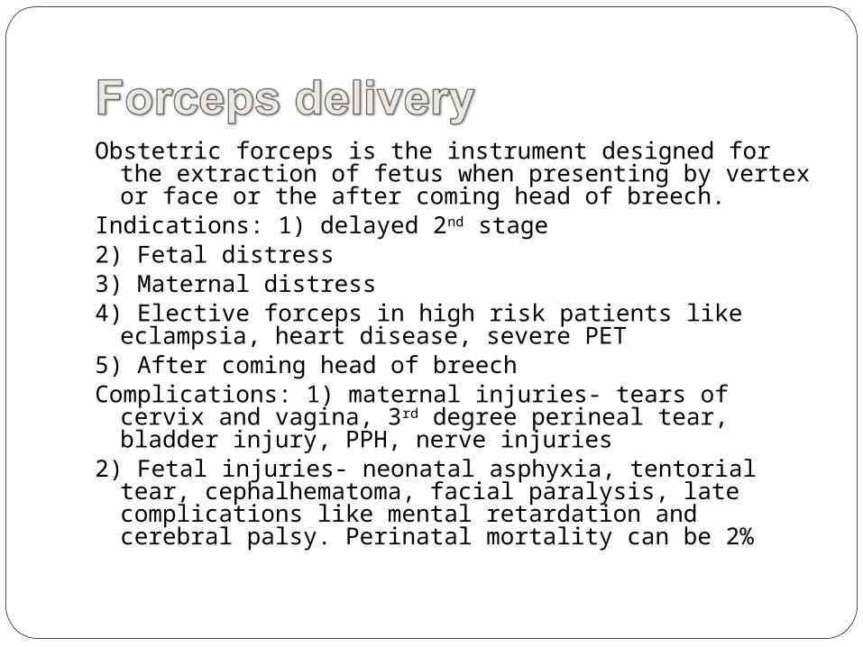
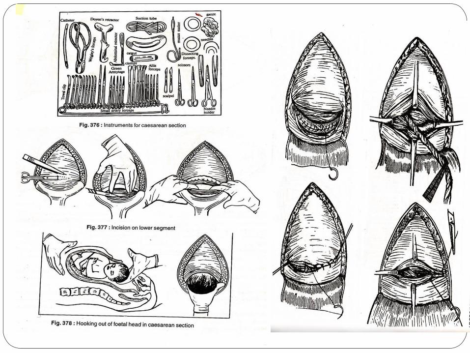
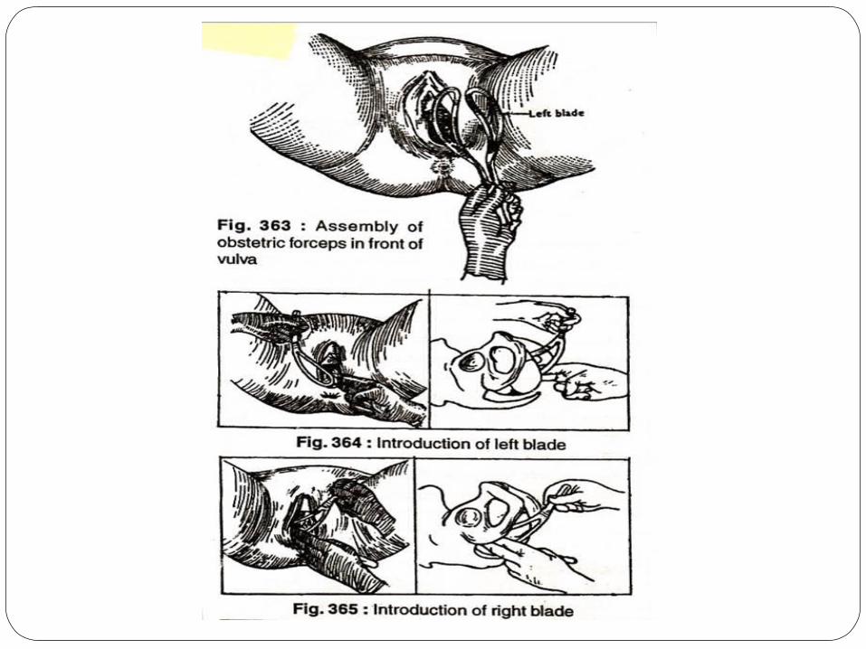
Obstetric forceps is the instrument designed for the extraction of fetus when presenting by vertex or face or the after coming head of breech.
Indications: 1) delayed 2nd stage2) Fetal distress3) Maternal distress4) Elective forceps in high risk patients like eclampsia,
heart disease, severe PET5) After coming head of breechComplications: 1) maternal injuries- tears of cervix and
vagina, 3rd degree perineal tear, bladder injury, PPH, nerve injuries
2) Fetal injuries- neonatal asphyxia, tentorial tear, cephalhematoma, facial paralysis, late complications like mental retardation and cerebral palsy. Perinatal mortality can be 2%
This is the abdominal delivery of the baby by laprotomy and section of the uterus after 28 weeks of pregnancy.
Indications:
1) Absolute: when vaginal delivery is impossible
a. severe degree of CPD
b. central placenta praevia
c. pelvic mass obstructing the passage
d. advanced CA cervix
e. vaginal obstruction like atresia, stenosis
2) Relative: here vaginal delivery may be possible with or without aids but maternal and fetal risk is very high.
a. CPD
b. previous caesarean 2 or more
c. ante partum hemorrhage
d. mal presentations
e. failed induction of labor
f. bad obstetric history
g. hypertensive disorders
h. other medical disorders
i. fetal distress
Time of operation: elective and emergency
Types:
1. lower segment section 2. classical or upper segment section
Caesarean hysterectomy: this is an abdominal delivery of the baby by CS followed by hysterecomy. Indiacations are uncontrollable PPDH placenta accreta
Risks and complications:
1. Hemorrahge 20%,
2. Anesthetic complication: aspiration pneumonitis, cardiac arrest
3. Infections: puerperal sepsis, wound infection, paralytic ilieus, burst abdomen, septic shock
4. Leg vein thrombosis
5. Remote gynecological: menstrual disorders, chronic pelvic pain, infertility, scar endometriosis, VVF, incisional hernia, scar rupture in future pregnancy
6. Maternal mortality is 0.2-0.5%
Main causes of death: infection, hemorrhage, thromboembolism and anesthetic accidents
Mortality is more in cases of late referral and high risk labors
Perinatal mortality: 5-10% due to high incidence of emergency CS and high risk pregnancies
Causes of perinatal death: asphyxia or hypoxia, RDS, prematurity, intracranial hemorrhage, infections