Embed Size (px)
Citation preview
From Exit Wave to Structure: Is the Phase Object Approximation Useless?
° University of Antwerp, Department of Physics, B-2020 Antwerp, Belgium°°NCEM, Lawrence Berkeley Laboratory, U.S.A.
D. Van Dyck°, P. Geuens°, C. Kisielowski°°, J.R. Jinschek°°
Cairns, Australia
July 2, 2003
Evolution in Science
describe understand design
macro micro nano

Evolution in theory
• Prediction of properties (materials, molecules from “first principles”
• Ingredients: atom positions with high precision (0.01 Å)
experiment theory
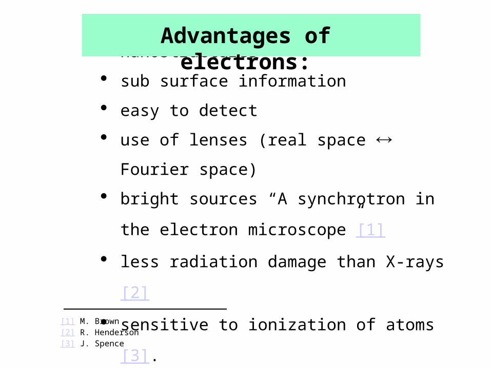
strong interaction nanostructures
sub surface information
easy to detect
use of lenses (real space Fourier space)
bright sources “A synchrotron in the electron
microscope”[1]
less radiation damage than X-rays[2]
sensitive to ionization of atoms[3].
[1] M. Brown[2] R. Henderson[3] J. Spence
Advantages of electrons:
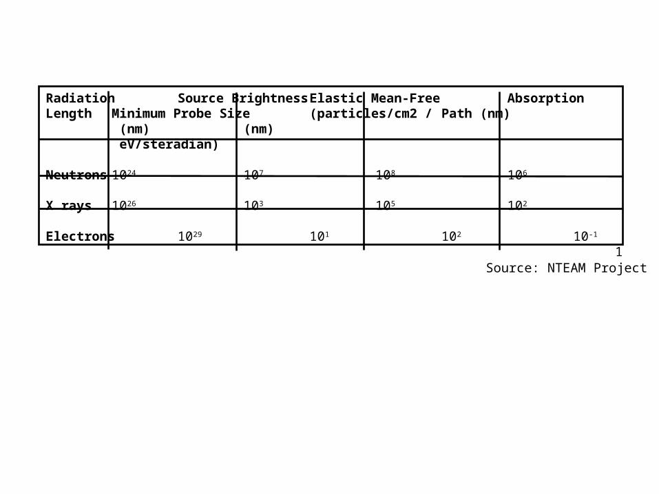
Radiation Source Brightness Elastic Mean-Free Absorption Length Minimum Probe Size (particles/cm2 / Path (nm) (nm) (nm)
eV/steradian)
Neutrons 1024 107 108 106
X rays 1026 103 105 102
Electrons 1029 101 102 10-1
1
Source: NTEAM Project
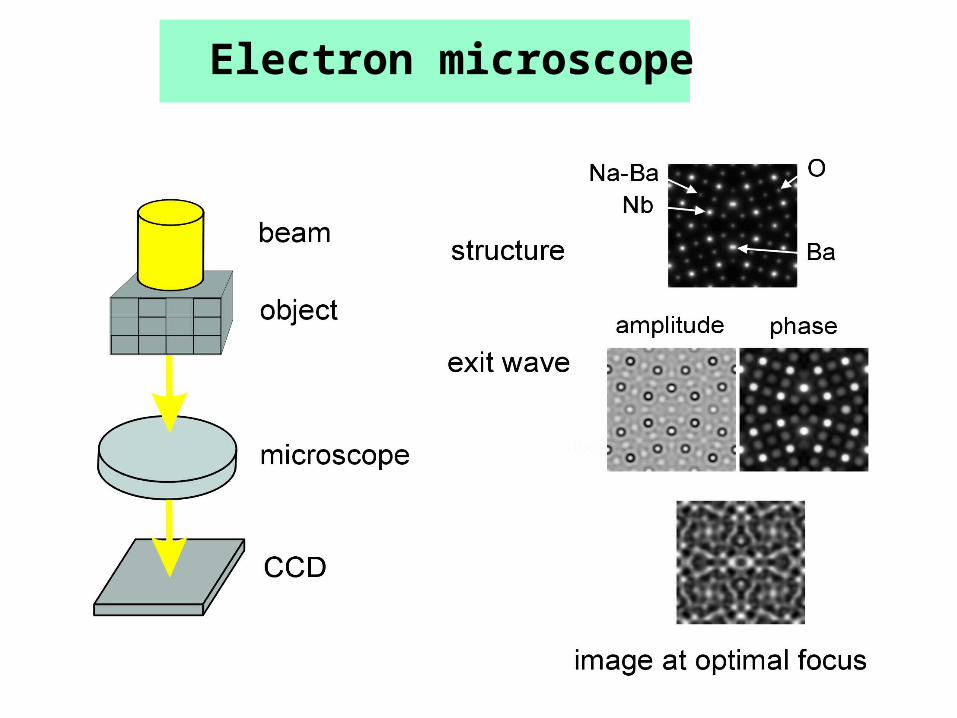
Electron microscope
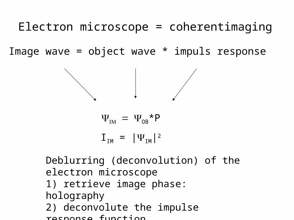
Electron microscope = coherentimaging
Image wave = object wave * impuls response
Deblurring (deconvolution) of the electron microscope1) retrieve image phase: holography2) deconvolute the impulse response function3) reconstruct exit (object) wave
OB*P
IIM = |IM|2

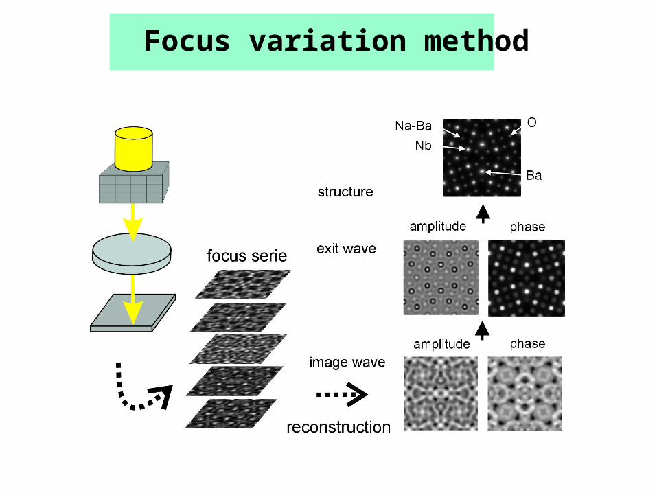
Focus variation method

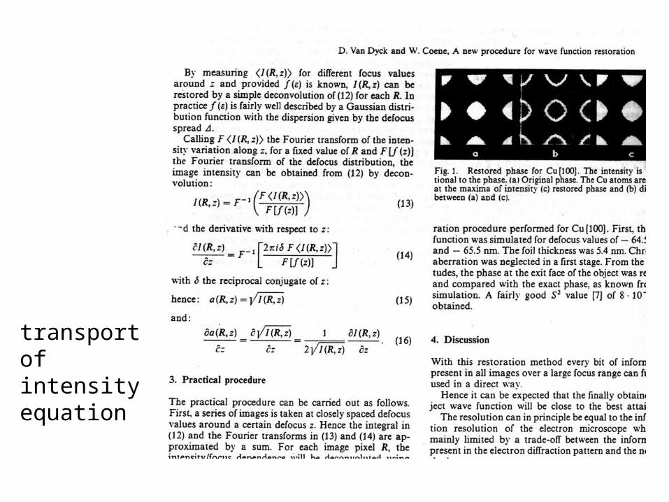
transport of intensity equation

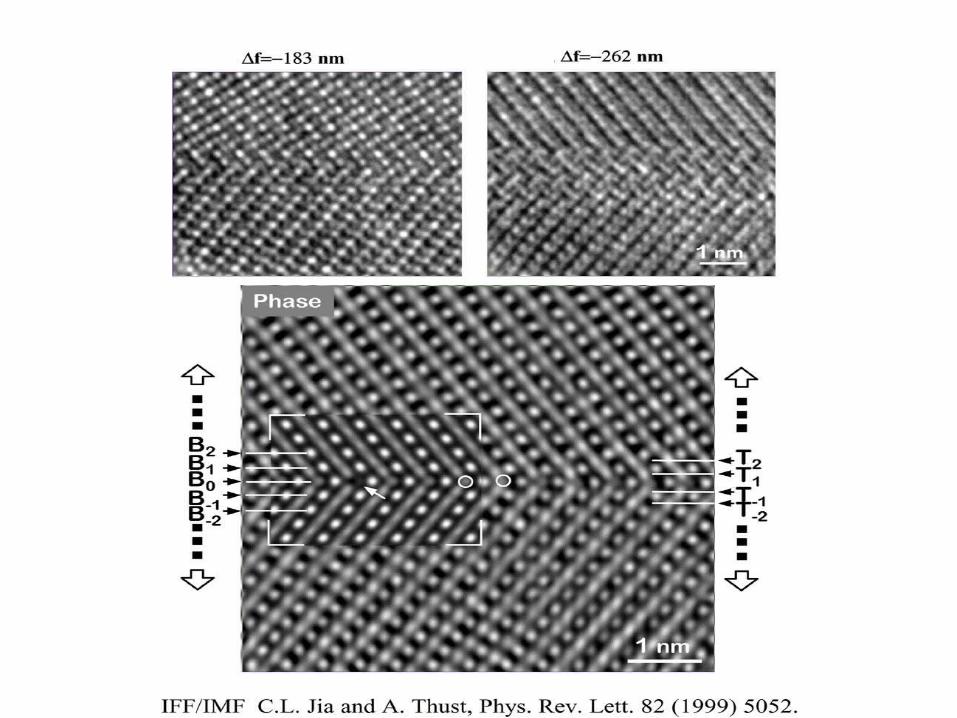
Phase of total exit wave5 Al: Cu
Courtesy C. Kisielowski (NCEM,Berkeley)
Phase of total exit waveAu [110] wedge
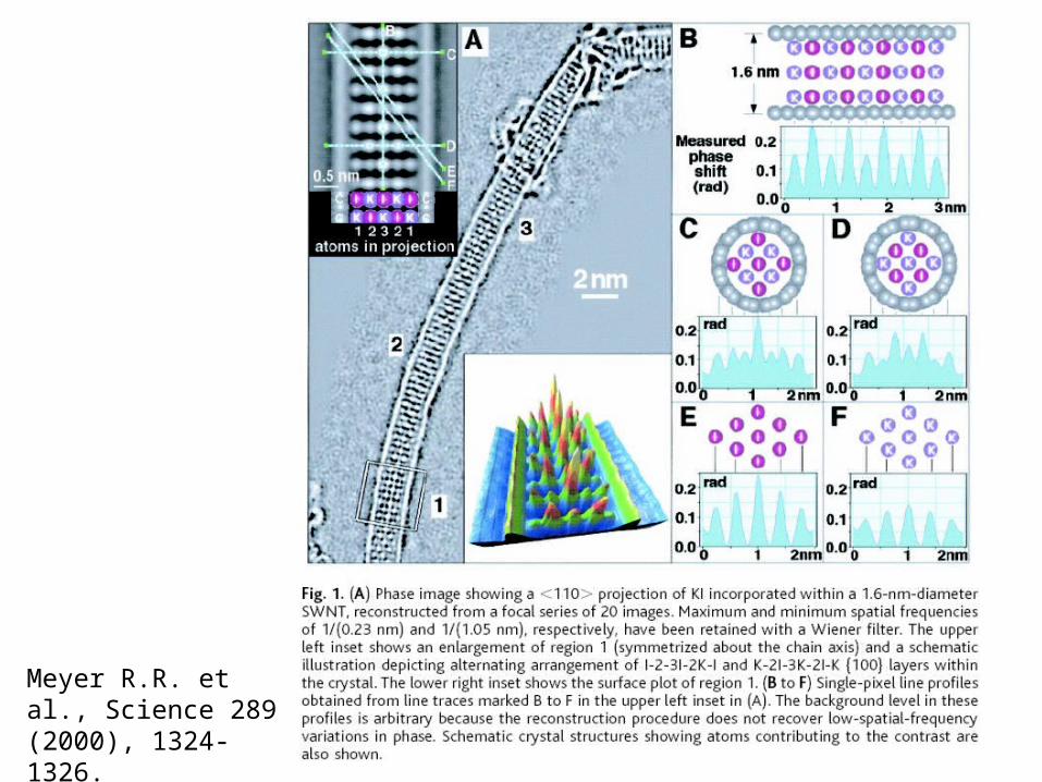
Meyer R.R. et al., Science 289 (2000), 1324-1326.
meE
h
2
),,(2),,('
zyxVEme
hzyx
d(x, y,z)2dz
' 2
dz
2
dz
EV (x,y,z)
E 1
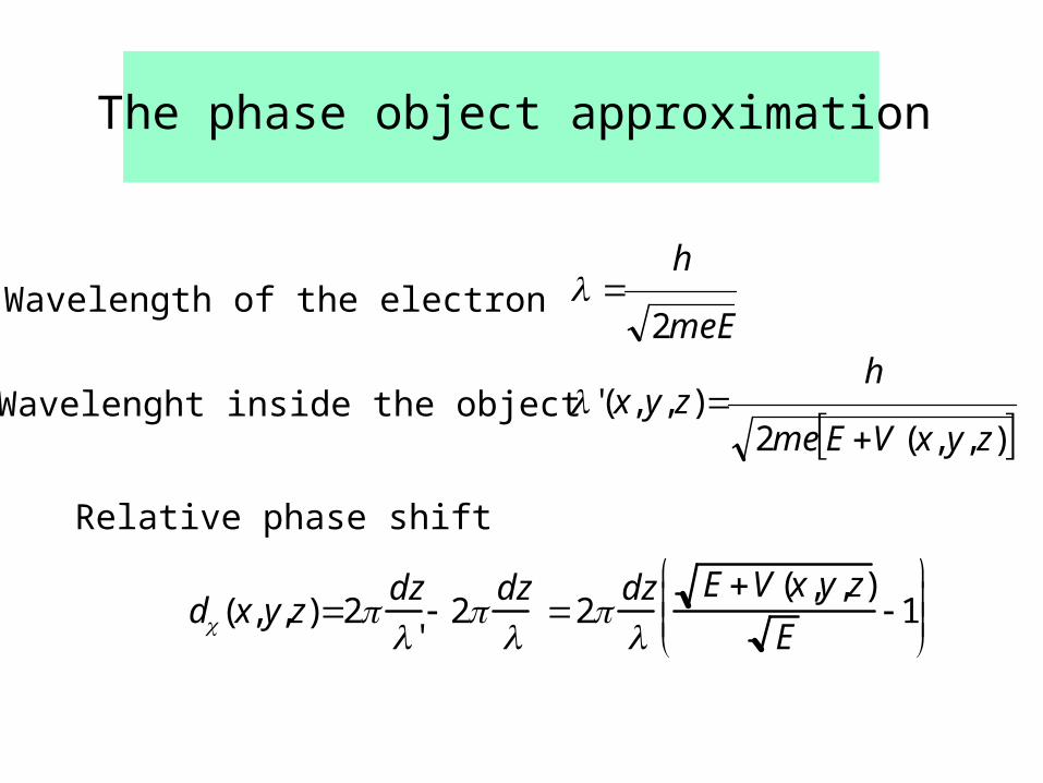
The phase object approximation
Wavelength of the electron
Wavelenght inside the object
Relative phase shift
Total phase shift
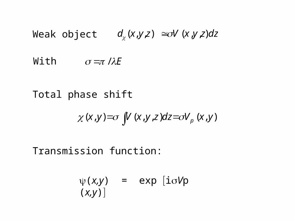
),(),,(),( yxVdzzyxVyx p
Transmission function:
(x,y) = exp iVp (x,y)
d(x, y,z)
V (x,y,z)dz
E /
Weak object
With
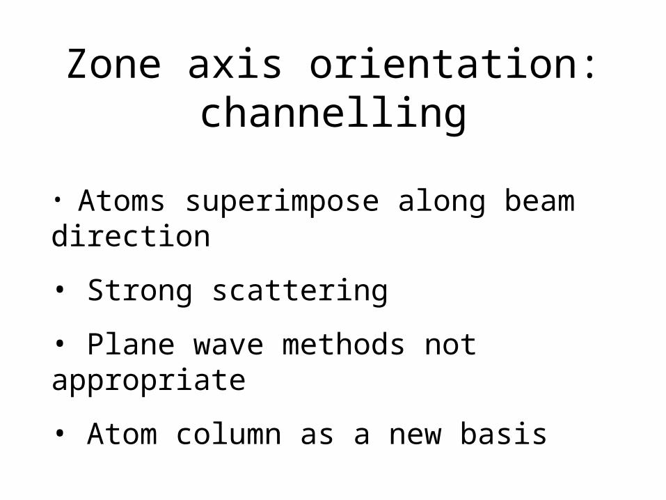
Zone axis orientation: channelling
• Atoms superimpose along beam direction
• Strong scattering
• Plane wave methods not appropriate
• Atom column as a new basis
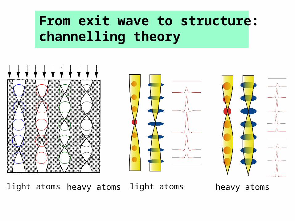
From exit wave to structure: channelling theory
light atoms heavy atoms light atoms heavy atoms
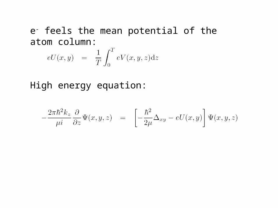
High energy equation:
e- feels the mean potential of the atom column:
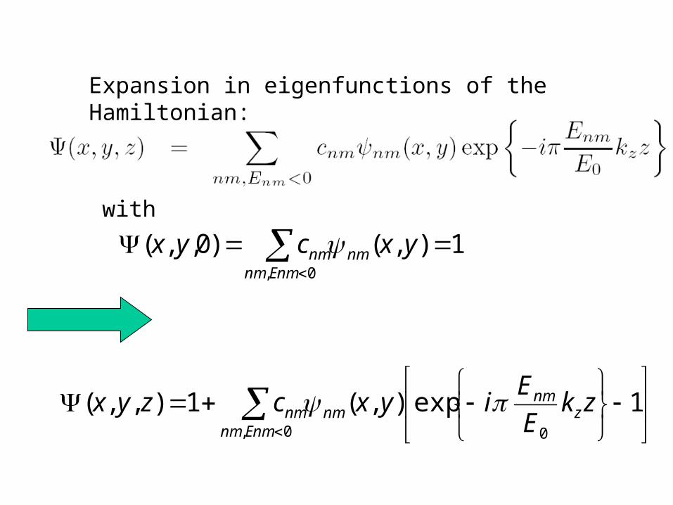
Expansion in eigenfunctions of the Hamiltonian:
1),()0,,(0,
yxcyxEnmnm
nmnm
1exp),(1),,(00,
zkE
Eiyxczyx z
nm
Enmnmnmnm
with
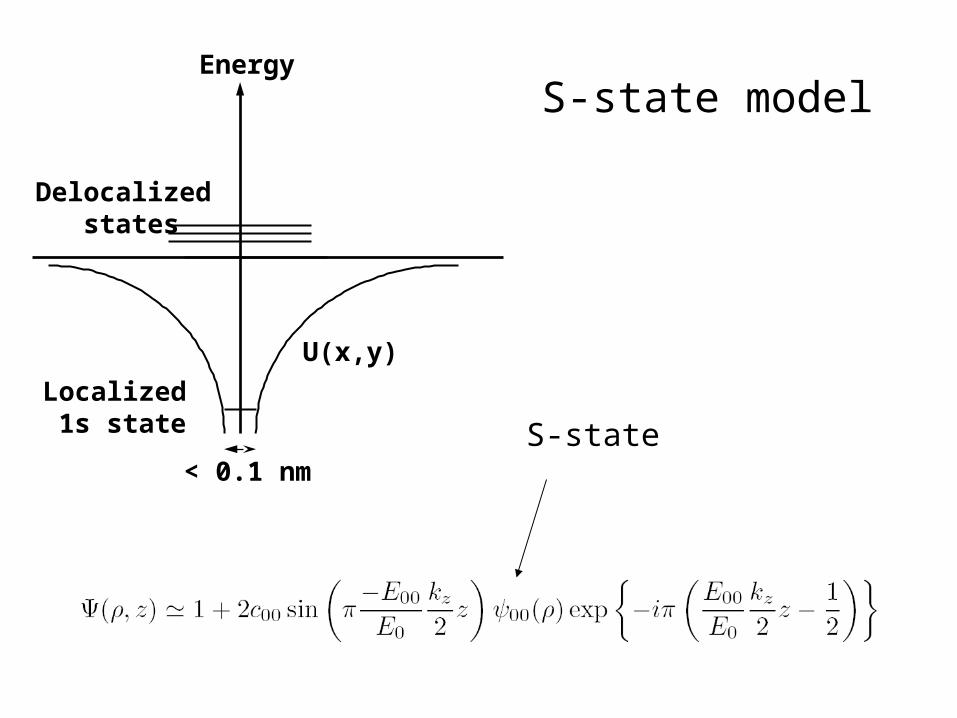
Energy
Delocalized states
Localized 1s state
U(x,y)
< 0.1 nm
S-state model
S-state
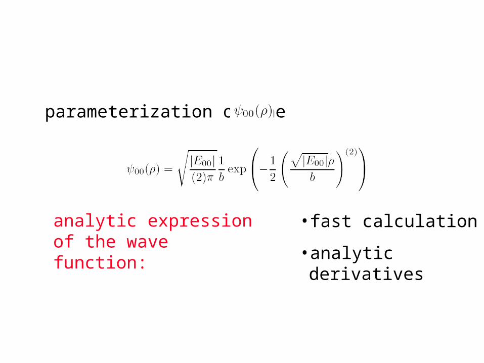
parameterization of the
analytic expression of the wave function:
• fast calculation
• analytic derivatives
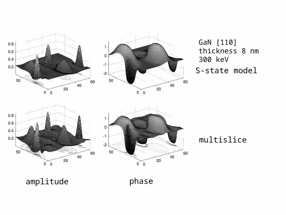
S-state model
multislice
phaseamplitude
GaN [110] thickness 8 nm 300 keV
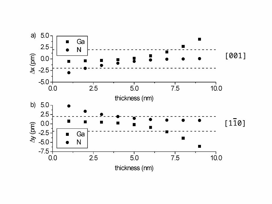
[001]
[110]
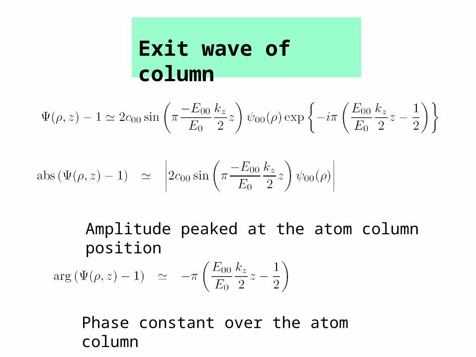
Exit wave of column
Amplitude peaked at the atom column position
Phase constant over the atom column
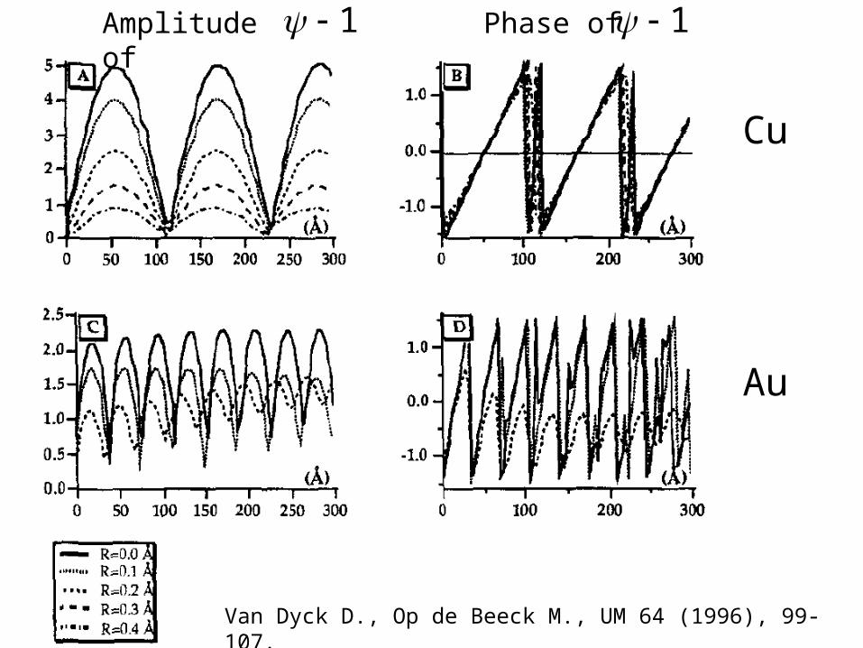
Van Dyck D., Op de Beeck M., UM 64 (1996), 99-107.
Amplitude of 1 Phase of 1
Cu
Au

Phase of total exit wave 5 Al: Cu
Amplitude of
Phase of
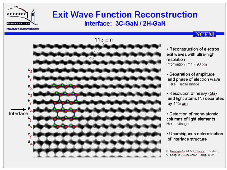
Courtesy C. Kisielowski, J.R. Jinschek (NCEM, Berkeley)
5 Al + Cu
Phase of
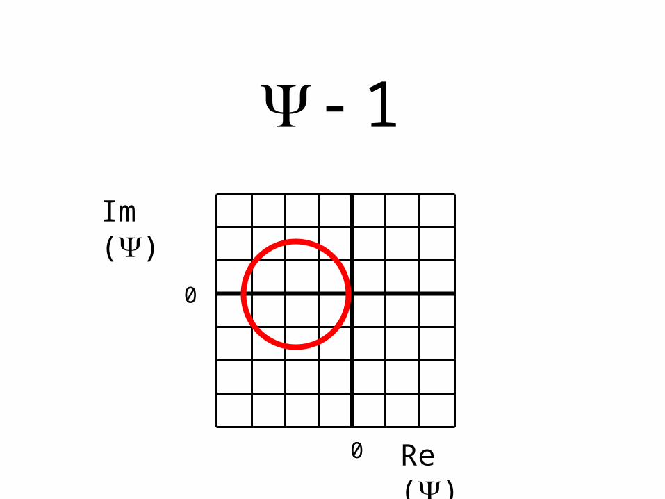
Im ()
1
Re ()
0
0

Au [110] – Vacuum wave
Courtesy C. Kisielowski, J.R. Jinschek (NCEM, Berkeley)

Re ()
= exit
wave
Im (
)
exit wave - vacuum
vacuum
=
Re ()
Im (
)
layer 1
layer 2
layer 10layer 9
Courtesy C. Kisielowski, J.R. Jinschek (NCEM, Berkeley)

Au [110] – Vacuum wave
Courtesy C. Kisielowski, J.R. Jinschek (NCEM, Berkeley)
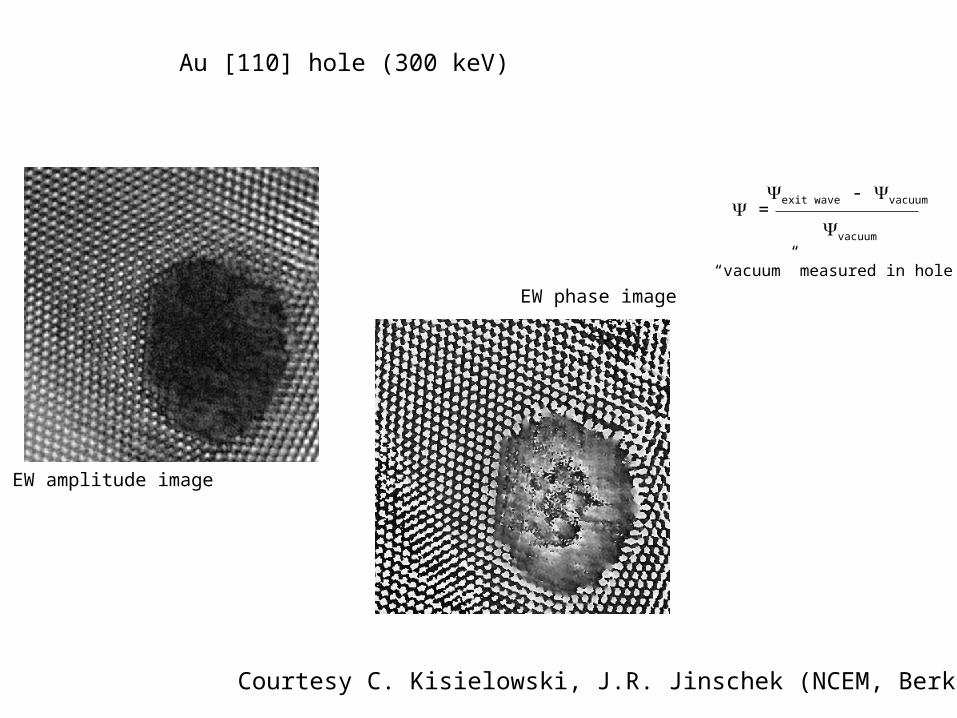
EW phase image
EW amplitude image
exit wave - vacuum
vacuum
=
“vacuum” measured in hole
Courtesy C. Kisielowski, J.R. Jinschek (NCEM, Berkeley)
Au [110] hole (300 keV)
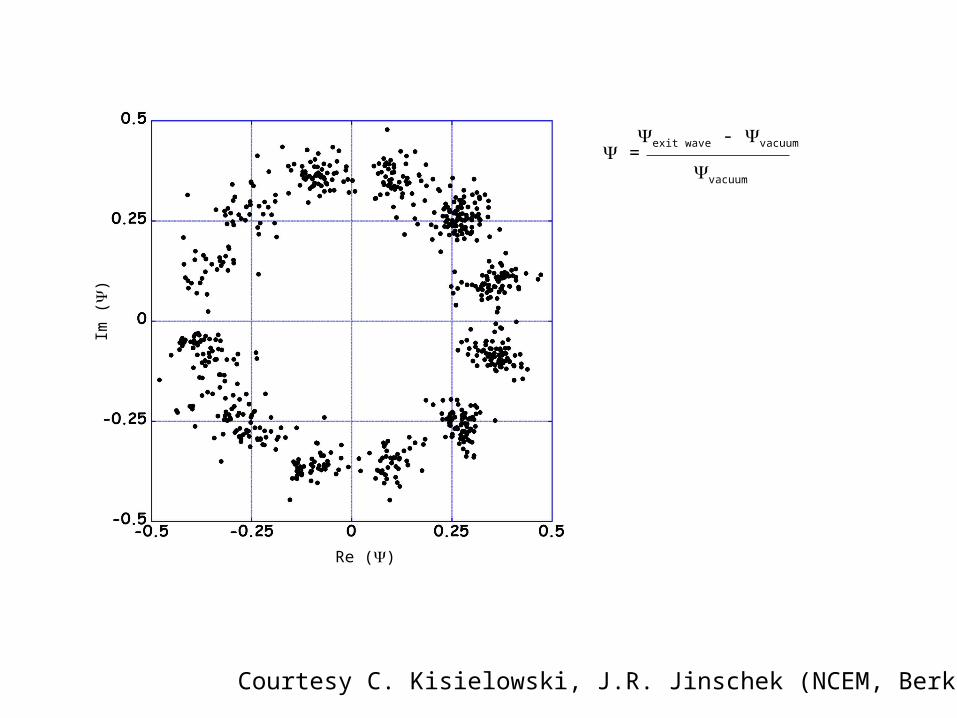
exit wave - vacuum
vacuum
=
Im (
)
Re ()
Courtesy C. Kisielowski, J.R. Jinschek (NCEM, Berkeley)
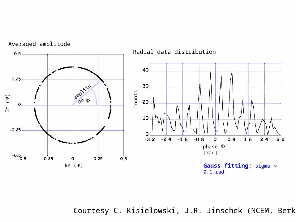
counts
phase [rad]
Im (
)
Re ()
amplit
ud
e
Gauss fitting: sigma 0.1 rad
Radial data distributionAveraged amplitude
Courtesy C. Kisielowski, J.R. Jinschek (NCEM, Berkeley)
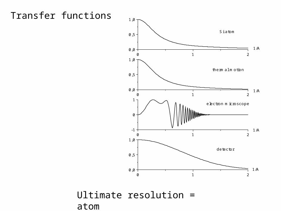
Ultimate resolution = atom
Transfer functions
0 1 20,0
0,5
1,0
1/A
1/A
1/A
1/A
detector
0 1 2-1
0
1electron microscope
0 1 20,0
0,5
1,0
thermal motion
0 1 20,0
0,5
1,0
Si atom
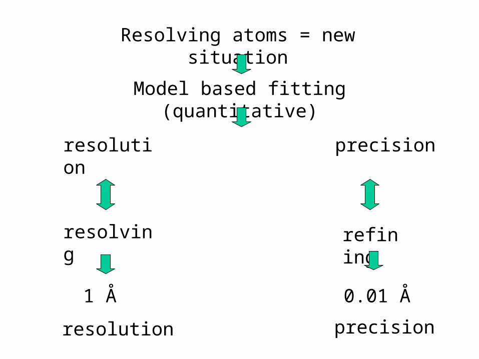
Resolving atoms = new situation
Model based fitting (quantitative)
resolution precision
resolving refining
resolution precision
1 Å 0.01 Å
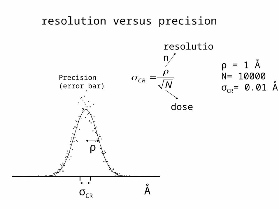
CRN
resolution
dose
ρ = 1 ÅN= 10000σCR= 0.01 Å
Å
ρ
σCR
resolution versus precision
Precision (error bar)
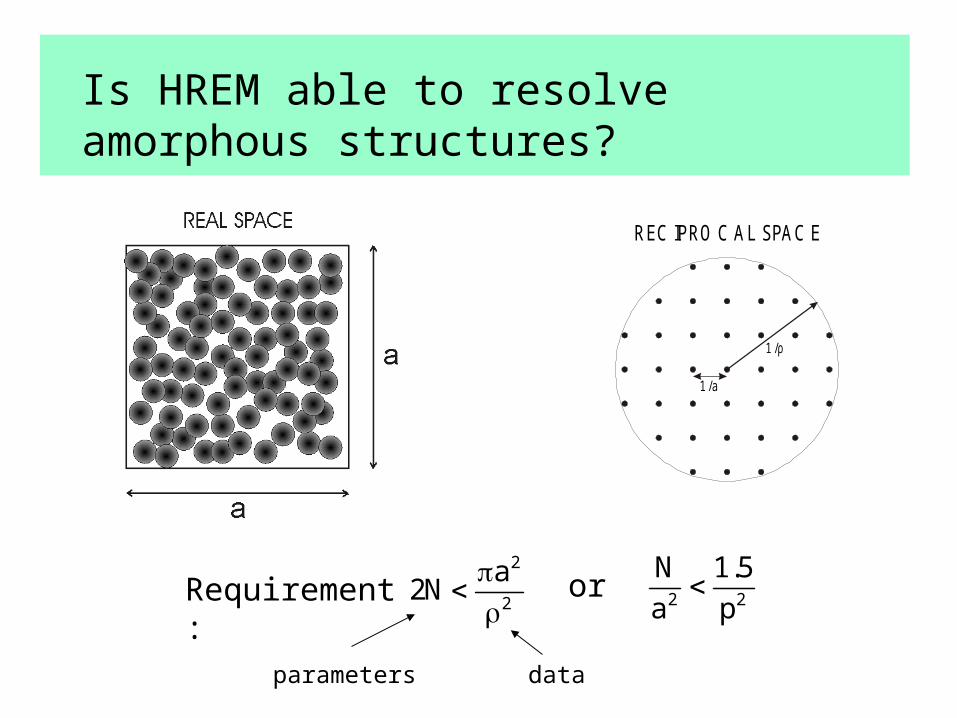
Is HREM able to resolve amorphous structures?
1/a
1/ρ
REC IPRO C AL SPAC E
2
2
a2N
2 2
N 1.5
a pRequirement: or
parameters data

3D HR Electron Tomography (HRET)
3
3
14
3N13a
parameters data
Amorphous structures never resolvable in 2D
N/a3 < 1.5/
2 Ångstrom resolution sufficient in 3D
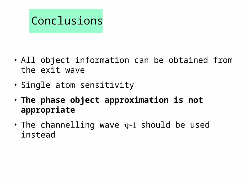
Conclusions
• All object information can be obtained from the exit wave
• Single atom sensitivity
• The phase object approximation is not appropriate
• The channelling wave should be used instead

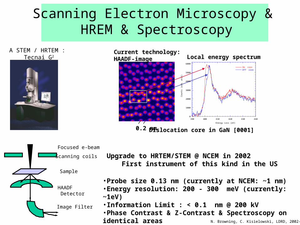
Scanning Electron Microscopy & HREM & Spectroscopy
A STEM / HRTEM : Tecnai G2
Scanning coils
Sample
Focused e-beam
HAADF Detector
Image Filter
Upgrade to HRTEM/STEM @ NCEM in 2002 First instrument of this kind in the US
•Probe size 0.13 nm (currently at NCEM: ~1 nm)•Energy resolution: 200 - 300 meV (currently: ~1eV)•Information Limit : < 0.1 nm @ 200 kV•Phase Contrast & Z-Contrast & Spectroscopy on identical areas
0
1000
2000
3000
4000
5000
6000
390 400 410 420 430 440
On core
Off core
Co
un
ts (
arb
. un
it)
Energy Loss (eV)
Current technology: HAADF-image Local energy spectrum
Dislocation core in GaN [0001] 0.2 nm
N. Browning, C. Kisielowski, LDRD, 2002-2003
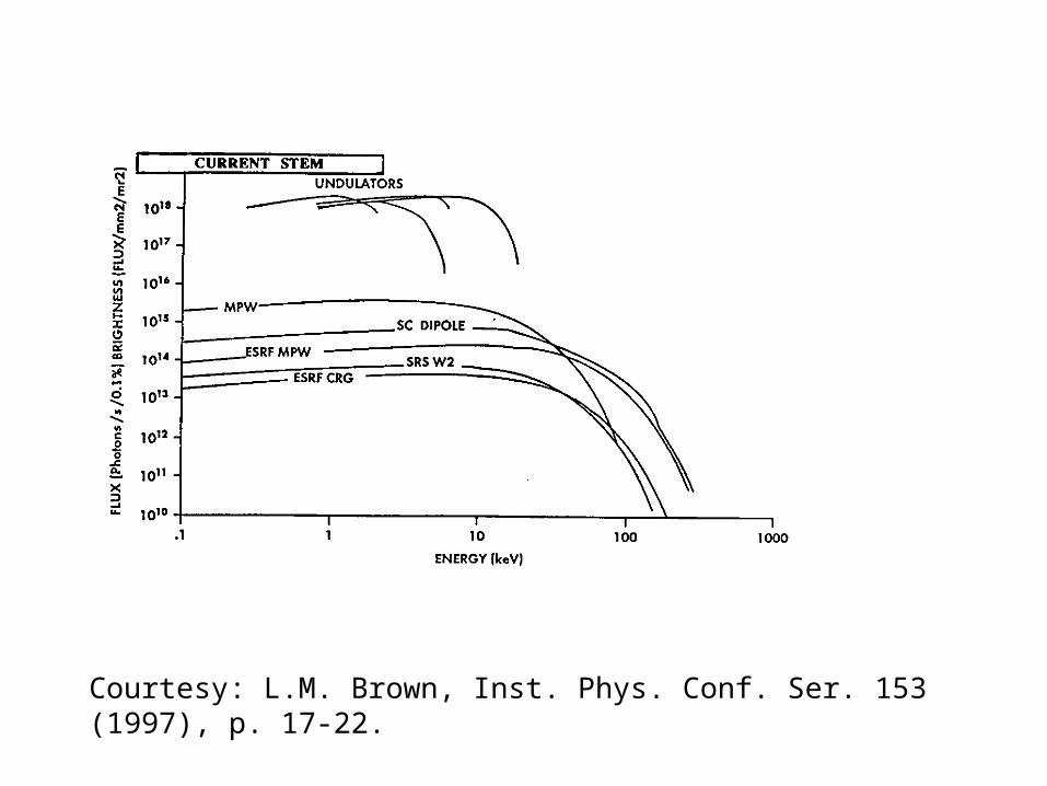
Courtesy: L.M. Brown, Inst. Phys. Conf. Ser. 153 (1997), p. 17-22.
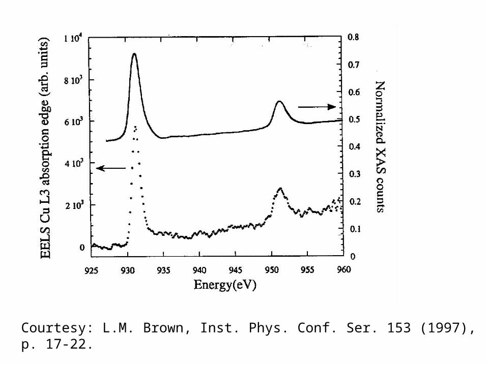
Courtesy: L.M. Brown, Inst. Phys. Conf. Ser. 153 (1997), p. 17-22.

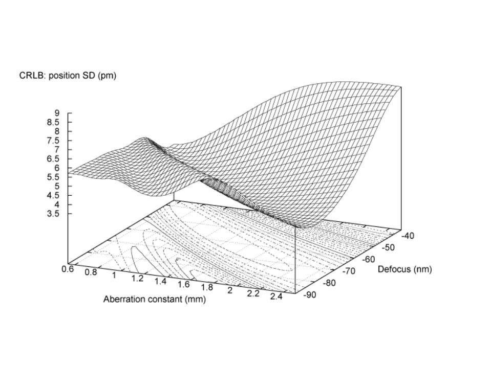
Experiment design
Intuition is misleading
“Ideal” HREM: Cs = 0f = 0
“Ideal object”:phase object
we need a strategy
no image contrast

• Spherical aberration corrector?improves the point resolution
• Chromatic aberration corrector?improves the information limit
• Monochromator?improves the information limit reduction of electrons
Ultramicroscopy 89(2001), 275-290
Do these correctors improve the precision as well?
The electron microscope of the future
• Quantitative 3D structure determination on atomic scale
• Spectroscopy on atomic scale
• Flexibility, experiment design
• Nanolab
The ideal instrument for the characterisation of nanostructures