Embed Size (px)
Citation preview

RESEARCH ARTICLE
From early stress to 12-month development
in very preterm infants: Preliminary findings
on epigenetic mechanisms and brain growth
Monica Fumagalli1☯, Livio Provenzi2☯, Pietro De Carli2, Francesca Dessimone1,
Ida Sirgiovanni1, Roberto Giorda3, Claudia Cinnante4, Letizia Squarcina5, Uberto Pozzoli6,
Fabio Triulzi4, Paolo Brambilla5,7, Renato Borgatti8, Fabio Mosca1, Rosario Montirosso2*
1 NICU, Department of Clinical Sciences and Community Health, Università degli Studi di Milano,
Fondazione IRCCS Ca’ Granda Ospedale Maggiore Policlinico, Milano, Italy, 2 0–3 Centre for the at-Risk
Infant, Scientific Institute, IRCCS Eugenio Medea, Bosisio Parini, LC, Italy, 3 Molecular Biology Lab,
Scientific Institute, IRCCS Eugenio Medea, Bosisio Parini, LC, Italy, 4 Neuroradiology Unit, Fondazione
IRCCS Ca’ Granda Ospedale Maggiore Policlinico, Milano, Italy, 5 Department of Neurosciences and Mental
Health, Fondazione IRCCS Ca’ Granda Ospedale Maggiore Policlinico, University of Milan, Milano, Italy,
6 Bioinformatics Lab, Scientific Institute, IRCCS Eugenio Medea, Bosisio Parini, LC, Italy, 7 Department of
Psychiatry and Behavioral Neurosciences, University of Texas at Houston, Houston, TX, United States of
America, 8 Neuropsychiatry and Neurorehabilitation Unit, Scientific Institute, IRCCS Eugenio Medea, Bosisio
Parini, LC, Italy
☯ These authors contributed equally to this work.
Abstract
Very preterm (VPT) infants admitted to Neonatal Intensive Care Unit (NICU) are at risk for
altered brain growth and less-than-optimal socio-emotional development. Recent research
suggests that early NICU-related stress contributes to socio-emotional impairments in VPT
infants at 3 months through epigenetic regulation (i.e., DNA methylation) of the serotonin
transporter gene (SLC6A4). In the present longitudinal study we assessed: (a) the effects of
NICU-related stress and SLC6A4 methylation variations from birth to discharge on brain
development at term equivalent age (TEA); (b) the association between brain volume at
TEA and socio-emotional development (i.e., Personal-Social scale of Griffith Mental Devel-
opment Scales, GMDS) at 12 months corrected age (CA). Twenty-four infants had complete
data at 12-month-age. SLC6A4 methylation was measured at a specific CpG previously
associated with NICU-related stress and socio-emotional stress. Findings confirmed that
higher NICU-related stress associated with greater increase of SLC6A4 methylation at
NICU discharge. Moreover, higher SLC6A4 discharge methylation was associated with
reduced anterior temporal lobe (ATL) volume at TEA, which in turn was significantly associ-
ated with less-than-optimal GMDS Personal-Social scale score at 12 months CA. The
reduced ATL volume at TEA mediated the pathway linking stress-related increase in
SLC6A4 methylation at NICU discharge and socio-emotional development at 12 months
CA. These findings suggest that early adversity-related epigenetic changes might contribute
to the long-lasting programming of socio-emotional development in VPT infants through epi-
genetic regulation and structural modifications of the developing brain.
PLOS ONE | https://doi.org/10.1371/journal.pone.0190602 January 5, 2018 1 / 15
a1111111111
a1111111111
a1111111111
a1111111111
a1111111111
OPENACCESS
Citation: Fumagalli M, Provenzi L, De Carli P,
Dessimone F, Sirgiovanni I, Giorda R, et al. (2018)
From early stress to 12-month development in very
preterm infants: Preliminary findings on epigenetic
mechanisms and brain growth. PLoS ONE 13(1):
e0190602. https://doi.org/10.1371/journal.
pone.0190602
Editor: Olivier Baud, Hopital Robert Debre, FRANCE
Received: June 28, 2017
Accepted: December 18, 2017
Published: January 5, 2018
Copyright: © 2018 Fumagalli et al. This is an open
access article distributed under the terms of the
Creative Commons Attribution License, which
permits unrestricted use, distribution, and
reproduction in any medium, provided the original
author and source are credited.
Data Availability Statement: All relevant data are
within the paper and its Supporting Information
files.
Funding: This study was supported by the Italian
Ministry of Health under Grant RC01-05, 2015-
2017 to RB for a longitudinal research project (i.e.,
Preterm Behavioral Epigenetics) on the genetic and
epigenetic effects of early adverse exposures on
socio-emotional development in very preterm
infants.

Introduction
Even in the absence of severe comorbidities, very preterm (VPT) infants (gestational age at
birth< 32 weeks) need long-lasting hospitalization in the Neonatal Intensive Care Units
(NICU) [1] and are at risk for altered socio-emotional development [2]. During NICU stay,
VPT infants are exposed to life-saving yet invasive interventions including mechanical ventila-
tion and painful skin-breaking procedures. These sources of NICU-related stress have been
found to contribute to VPTs’ socio-emotional development during infancy [3, 4] and child-
hood [5, 6]. Both functional (e.g., epigenetic mechanisms [7]) and structural (e.g., brain vol-
ume alterations [8]) factors have been suggested to be involved in setting the risk of less-than-
optimal socio-emotional development in VPT infants and children. Nonetheless, the specific
mechanisms implicated in the effects of NICU-related stress on socio-emotional developmen-
tal outcomes are unknown. Here, using data from a longitudinal research study, we examined
potential links between early NICU stress exposure and socio-emotional development at 12
months corrected age (CA) in VPT children, assessing both epigenetic variations (i.e., seroto-
nin transporter gene (SLC6A4) methylation [9]) and brain growth (i.e., anterior temporal lobe
volume [10, 11]).
Preterm infants’ developing brain is vulnerable to adverse environmental stimulations and
advanced Magnetic Resonance Imaging (MRI) techniques have been used to investigate the
pathophysiological basis of neurodevelopmental disorders that VPT infants may manifest later
in childhood [12]. NICU-related stress might affect VPT infants’ cerebral growth [8, 13]. Previ-
ous studies suggested that the anterior temporal lobe (ATL) plays a critical role in socio-emo-
tional functioning and emotion regulation [14, 15] and preterm infants present reduced ATL
volume at term age compared to their full-term counterparts [16]. Moreover, from the ana-
tomical point of view, the ATL is of particular interest since it contains the amygdala, extended
amygdala and anterior hippocampus, anatomic brain structures that are well-known for their
involvement in socio-emotional development and functioning [17, 18]. In the light of this evi-
dence, the ATL is a candidate region of interest (ROI) to examine preterm infants brain
growth, consistent with the aims of the present study.
Recent research suggests that early life adversities may contribute to the long-lasting pro-
gramming of socio-emotional development through functional modifications (e.g., DNA
methylation) of stress-related genes, without structural modifications of the chromatin struc-
ture [19]. DNA methylation consists in the addition of a methyl group to cytosine/guanine
DNA dinucleotides (i.e., CpG sites) within the promoter region of a specific gene usually
resulting in reduced transcriptional activity (i.e., gene silencing) [20]. The SLC6A4 gene codes
for the serotonin transporter, it is susceptible to epigenetic regulation by DNA methylation
[21] and it acts as the key regulator of the serotonergic system under stress [22]. Greater expo-
sure to NICU-related stress has been recently associated with increased CpG-specific methyla-
tion of the SLC6A4 gene in VPT infants [9], which in turn was predictive of socio-emotional
development at 3 months CA [23].
Our group has previously documented the epigenetic effects of early pain-related stress
exposure on the socio-emotional developmental outcomes of VPT infants during the first
months of life [9, 23]. In the present study, we extended previous research assessing the poten-
tial links between SLC6A4 CpG-specific methylation and brain volumes at term equivalent age
(TEA). Furthermore, we assessed whether functional (i.e., DNA methylation) and structural
brain variations (i.e., ATL volume) might be associated with socio-emotional development at
12 months CA (see Fig 1). The main aims were: (1) to assess the association between increased
SLC6A4 methylation at NICU discharge and ATL volume at TEA; (2) to investigate the effects
of altered SLC6A4 methylation at discharge and ATL volume at TEA on VPT infants
Behavioral epigenetics and preterm infants’ development
PLOS ONE | https://doi.org/10.1371/journal.pone.0190602 January 5, 2018 2 / 15
Competing interests: The authors have declared
that no competing interests exist.

performance at the Griffith Mental Development Scales (GMDS [24]) Personal-Social scale at
12 months CA. Finally, through a path analysis, we speculatively tested the hypothesis that an
altered ATL volume might mediate the association between early NICU-related SLC6A4 epige-
netic alterations (i.e., increased CpG methylation) and socio-emotional development at 12
months CA.
Materials and methods
Participants
Fifty-six VPT infants (<32 weeks of gestation and/or <1500 g at birth) were recruited between
October 2011 and April 2014, at the NICU of the Department of Clinical Sciences and Com-
munity Health, Fondazione IRCCS Ca’ Granda Ospedale Maggiore Policlinico of Milan.
Exclusion criteria included: mothers with documented cognitive impairment and under psy-
chotropic treatment during and after pregnancy; any kind of hemodynamic disturbances expe-
rienced during NICU stay (defined as need of inotropic drugs to maintain normal values of
arterial blood pressure); need of surgery; major brain lesions as documented by cerebral ultra-
sound (intraventricular hemorrhage > grade 2 according to Papile [25], cystic periventricular
leukomalacia); neuro-sensorial deficits (retinopathy of prematurity� stage 2 [26]); genetic
syndromes and/or major malformations. All mothers were Italian, 18-year-old or more,
cohabitant with the father of the infant.
Procedures
The research was conducted in accordance with the Declaration of the World Medical Associ-
ation and with the 7th revision of the Declaration of Helsinki for ethical principles regarding
human experimentation. The study protocol was approved by the ethical committees of the
Scientific Institute IRCCS E. Medea in Bosisio Parini and of the Fondazione IRCCS Ca’
Granda Ospedale Maggiore Policlinico of Milan. All parents signed a written informed con-
sent form. For the purposes of epigenetic analyses, cord blood at birth and peripheral blood at
NICU discharge were collected from the included VPT infants. Blood samples were obtained
by trained nurses to avoid hemolysis and immediately stored at -20˚C. Infants’ clinical vari-
ables were obtained from medical records at the end of the NICU hospitalization. Mothers
completed a socio-demographic form during the first days after birth. Among the initial sam-
ple of 56 VPT infants, 12 babies did not undergo brain magnetic resonance imaging (MRI), 11
Fig 1. Schematic time-line of the longitudinal project, limitedly to the variables of interest. Note. NICU,
Neonatal Intensive Care Unit; SLC6A4, serotonin transporter gene; Δmet, mean change in SLC6A4 methylation
from birth to NICU discharge at CpG chr17: 28562786–28562787; ATL, anterior temporal lobe; MRI, Magnetic
Resonance Imaging; GMDS, Griffith Mental Development Scales; PCA, Post-Conceptional Age; TEA, term-
equivalent age; CA, Corrected Age for prematurity.
https://doi.org/10.1371/journal.pone.0190602.g001
Behavioral epigenetics and preterm infants’ development
PLOS ONE | https://doi.org/10.1371/journal.pone.0190602 January 5, 2018 3 / 15
were scanned after 43 weeks post-menstrual age and 6 MRI scans were not suitable for volu-
metric analysis due to movement artifacts. As such, 27 subjects (48%) had available MRI at
TEA (39+0–42+6weeks). At 12 months (corrected age for prematurity), developmental func-
tioning was assessed with the GMDS. Three infants (11%) had no GMDS assessment at 12
months.
Measures
Neonatal and clinical variables. Gestational age (weeks) and weight (grams) were
recorded at birth. Other neonatal variables included: gender, Apgar at minute 1, twin preg-
nancy, mode of delivery, being small for gestational age. NICU stress-related variables included:
total length of hospitalization (days); number of skin-breaking procedures (e.g., heel lance, arte-
rial and venous punctures, peripheral venous line insertion); total days of invasive ventilation.
Other clinical variables included: sepsis, bronchopulmonary dysplasia, necrotizing enterocolitis,
retinopathy of prematurity. The Clinical Risk Index for Babies (CRIB II [27]) score was com-
puted to obtain an overall assessment of the clinical risk associated with preterm birth.
Socio-demographic variables. Socio-demographic data (i.e., maternal age, years of study
and occupation) were obtained from all the mothers. Hollingshead’s classification [28] was
used to assess maternal socio-economic status (SES). SES ranged from 0 (occupations that do
not require a high school degree) to 90 (occupations that require highly specialized education
and training).
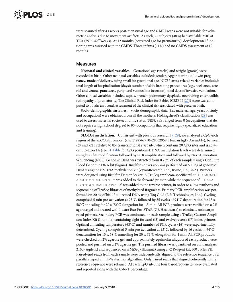
SLC6A4 methylation. Consistent with previous research [9, 29], we analyzed a CpG-rich
region of the SLC6A4 promoter (chr17:28562750–28562958, Human hg19 Assembly), between
-69 and -213 relative to the transcriptional start site, which contains 20 CpG sites and is adja-
cent to exon 1A (see S1 Table for CpG positions). DNA methylation levels were determined
using bisulfite modification followed by PCR amplification and followed by Next-Generation
Sequencing (NGS). Genomic DNA was extracted from 0.2 ml of each sample using a GenElute
Blood Genomic DNA kit (Sigma). Bisulfite conversion was performed on 500 ng of genomic
DNA using the EZ DNA methylation kit (ZymoResearch, Inc., Irvine, CA, USA). Primers
were designed using Bisulfite Primer Seeker. A TruSeq amplicon-specific tail 50 CCTACACGACGCTCTTCCGATCT 30 was added to the forward primer, while the sequence 50 TCAGACGTGTGCTCAACCGATCT 30 was added to the reverse primer, in order to allow synthesis and
sequencing of TruSeq libraries of methylated fragments. Primary PCR-amplification was per-
formed on 20 ng of bisulfite- treated DNA using Taq Gold (Life Technologies, Inc.). Cycling
comprised 5 min pre-activation at 95˚C, followed by 35 cycles of 94˚C denaturation for 15 s,
58˚C annealing for 20 s, 72˚C elongation for 1.5 min. All PCR products were verified on a 2%
agarose gel and treated with Ilustra Exo Pro-STAR (GE Healthcare) to eliminate unincorpo-
rated primers. Secondary PCR was conducted on each sample using a TruSeq Custom Ampli-
con Index Kit (Illumina) containing eight forward (i5) and twelve reverse (i7) index primers.
Optimal annealing temperature (68˚C) and number of PCR cycles (16) were experimentally
determined. Cycling comprised 5 min pre-activation at 95˚C, followed by 16 cycles of 94˚C
denaturation for 15 s, 68˚C annealing for 20 s, 72˚C elongation for 1 min. All PCR products
were checked on 2% agarose gel, and approximately equimolar aliquots of each product were
pooled and purified on a 2% agarose gel. The purified library was quantified on a Bioanalyzer
2100 (Agilent) and sequenced on a MiSeq (Illumina) using a v2 Reagent kit, 300 cycles PE.
Paired-end reads from each sample were independently aligned to the reference sequence by a
parallel striped Smith-Waterman algorithm. Only paired reads that aligned coherently to the
reference sequence were retained. At each CpG site, the four base-frequencies were evaluated
and reported along with the C-to-T percentage.
Behavioral epigenetics and preterm infants’ development
PLOS ONE | https://doi.org/10.1371/journal.pone.0190602 January 5, 2018 4 / 15

Brain volumes. Brain MRI was performed at TEA on a 3T Philips Achieva scanner (Phil-
ips Medical Systems, Best, The Netherlands) according to the internal scanning protocol,
reported in Table 1. Patients were fed and wrapped, noise attenuators (MiniMuffs1 Natus
Medical Inc, San Carlos, California) were applied for hearing protection; heart rate and arterial
oxygen saturation were continuously monitored and a neonatologist was present throughout
the entire examination. All babies were scanned during spontaneous sleep.
Socio-emotional development. The GMDS [24] assesses mental development of infants
from birth to 8 years. It is based on five subscales tapping the following domains: locomotor,
personal-social development, hearing and speech, hand and eye coordination, and perfor-
mance. For the aims of the current study we used the Personal-Social dimension of the
GMDS. This scale assesses the children proficiency in daily living activities, levels of indepen-
dence, as well as ability to interact with other children. The evaluation of this dimension at 12
months includes items assessing specific competences of personal-social functioning such as
visual recognizing of maternal face, following moving persons with eyes, social smiling and
vocalizations, self-soothing capacities, smiling and playing in response to mirror image, play-
ing actively in interactive games with others, being interested in activities of others. A final
standardized score (ranging from 50 to 150) is obtained, with mean equal to 100 and standard
deviation equal to 15. Notably, previous research has documented that this scale is particularly
sensitive in capturing early developmental difficulties in VPT infants [30].
Data reduction
NICU-related stress. In order to provide a global measure of NICU-related stress, a Prin-
cipal Component Analyses was performed on the selected indexes reported above (i.e., numberof skin-breaking procedures including heel lance, arterial and venous punctures, peripheralvenous line insertion; total days of invasive ventilation), leading to a one factor-solution that
explained 74% of the variance with factor loadings ranging from .66 to .94. After extracting the
principal component (i.e., NICU-related stress) it has been weighted on length of NICU stay
(days), in order to obtain a global index of stress exposure in NICU and to avoid confounding
effects of multicollinearity in further analyses.
SLC6A4 methylation. Previous research documented that CpG-specific (i.e., chr17:
28562786–28562787) SLC6A4 methylation occurs in VPT infants in response to NICU-related
stress [9] and is linked with further socio-emotional development at 3 months CA [23]. This
CpG site showed the greatest association with early exposure to NICU-related stress and pain
in VPT infants. As such, it could be a possible CpG site candidate for potential epigenetic
effects of the adverse NICU environment on VPT infants’ methylation status of the SLC6A4
gene. Consistently, in the present study, we adopted a CpG-specific approach, focusing on the
delta-score obtained subtracting SLC6A4 methylation at birth from SLC6A4 methylation at
NICU discharge for the CpG chr17: 28562786–28562787 (i.e., Δmet).
Table 1. Scanning protocol for included infants.
Sequences Slice orientation / thickness FOV TR / TE Matrix
T1 3D FFE Coronal/ voxel size: 0.9x0.9x0.9 mm 130x140 8.5 / 4.6 144 x 132
T2 TSE Axial / 2 mm 130x130 7000 / 140 324 x 260
T2 TSE Coronal / 2 mm 130x130 9000 / 150 324 x 260
T2 FFE Axial / 3 mm 160x160 652/16 159 x 200
DWI Axial / 3 mm 180x180 4388 / 55 148 x 127
Note. FOV, Field of View; TR/TE, repetition time / echo time; FEE; Fast Field Echo, TSE, Turbo Spin Echo; DWI, Diffusion Weight Imaging.
https://doi.org/10.1371/journal.pone.0190602.t001
Behavioral epigenetics and preterm infants’ development
PLOS ONE | https://doi.org/10.1371/journal.pone.0190602 January 5, 2018 5 / 15

Brain segmentation and volumetric analysis. Automated segmentation was conducted
on each neonatal Axial T2 2 mm scan, in conjunction with the T1 scan. The two images were
registered, in order to segment brain tissue and extract volume measures using a neonatal spe-
cific segmentation approach [31] based on the Expectation–Maximisation (EM) technique
[32]. The target areas were visually checked and manual editing was performed with ITK-S-
NAP [33]. Volumetric measures of the structures of each neonate were extracted from each
segmentation. All measures are expressed in mm3. For the purposes of the present study, the
following ROIs were selected: anterior temporal lobe lateral part left (ATL-LPL) and right
(ATL-LPR) and anterior temporal lobe medial part left (ATL-MPL) and right (ATL-MPR)
(Fig 2).
Plan of analysis
Descriptive statistics for neonatal, clinical and socio-demographic characteristics as well as for
variables of interest (i.e., NICU-related stress, Δmet, ATL volumes, socio-emotional develop-
ment) have been computed. The regression of NICU-related stress on Δmet has been carried
to confirm previous findings on a larger sample from the same longitudinal study.
Effects of NICU-related stress and SLC6A4 methylation on ATL brain volumes. In the
first step, we performed different multiple regressions to test the effects of NICU-related stress
and SLC6A4methylation (i.e., Δmet) on each ROI, controlling for the following covariates: ges-
tational age at birth, maternal SES and gestational age at MRI.
Fig 2. Brain MRI segmentation: A. axial and B. coronal view T1 images. Note. Colors highlight anterior temporal lobe (ATL) lateral part left (ATL-LPL,
yellow) and right (ATL-LPR, dark blue) as well as ATL medial part left (ATL-M;PL, light blue) and right (ATL-MPR, pink).
https://doi.org/10.1371/journal.pone.0190602.g002
Behavioral epigenetics and preterm infants’ development
PLOS ONE | https://doi.org/10.1371/journal.pone.0190602 January 5, 2018 6 / 15

Effects of brain volumes on developmental outcome. In the second step, we performed
a series of multiple regressions to predict the performance on the Griffith subscale Personal-
Social. In order to reduce the number of predictors in the face of limited sample size, we
weighted brain volumes for age at the MRI, extracting the residuals of the regression of gesta-
tional age at MRI on brain volumes. We added the extracted weighted measures (i.e.,
ATL-LPLw; ATL-LPRw; ATL-MPLw; ATL-MPRw) in separate regression models together with
NICU-related stress, gestational age at birth and maternal SES.
Exploratory assessment of the brain volume mediation hypothesis. Finally, we per-
formed four different path analyses, one for each ROI. Specifically, we tested the indirect effect
of methylation on the Griffiths’ Personal-Social scale, mediated by brain volumes. The good-
ness of the model was assessed separately for each ROI according to: non-significant chi-
square statistic; comparative fit index (CFI) and Tucker-Lewis index (TLI) close to .95 [34];
Root Mean Squared Error of Approximation (RMSEA) smaller than .05; Square Residual Root
Mean (SRMR) smaller than .08 [35]; non-significant p value associated with the CI of RMSEA
[36].
Results
Descriptive statistics for socio-demographic characteristics and variables of interest are
reported in Table 2. The association between NICU-related stress and Δmet from birth to dis-
charge was positive and significant (β = .43, p = .02, 95% C. I. [.06,.80]).
Effects of NICU-related stress and SLC6A4 methylation on brain
volumes
Results of the first step of analysis are presented in Table 3. A significant effect of Δmet on ROI
volumes emerged for all the ATL areas investigated (ATL-MPR, F(5,21) = 2.52, p = .06, R2 =
0.37; ATL-MPL, F(5,21) = 2.84, p = .04, R2 = 0.40; ATL-LPR, F(5,21) = 2.52, p = .07, R2 = 0.36;
ATL-LPL, F(5,21) = 2.60, p = .05, R2 = 0.38). Greater increase in methylation was associated
with smaller brain volume in the ROI (see Table 3).
Effects of brain volumes on socio-emotional development
Significant effects emerged for ATL-MPLw, R2 = .58, F(5,18) = 5.00, p = .005, ATL-LPRw, R2 =
.61, F(5,18) = 5.62, p = .003, and ATL-LPLw, R2 = .68, F(5,18) = 7.61, p = .0005,. A significant
effect of ATL volumes on the GMDS Personal-Social scale emerged for ATL-MPLw, ATL-
LPRw and ATL-LPLw (Table 4). The greater these areas were, the higher was the score of the
GMDS Personal-Social scale. In addition, higher scores in the GMDS Personal-Social scale
were significantly predicted by lower NICU-related stress and gestational age at birth.
Exploratory assessment of the brain volume mediation hypothesis
The path analysis is reported in S1 Fig. All the indexes indicated an acceptable fit of the data:
all chi-squares < 0.7, p> .6;; CFIs = 1.00; TLIs > 1.00; RMSEAs = 0.00, ps> .69. The mediated
path was significant for three out of four ROI analyzed (i.e., ATL-MPL, ATL-LPR, ATL-LPL).
More specifically, the analysis suggested that (1) higher NICU-related stress was significantly
associated with greater Δmet; (2) greater Δmet was significantly associated with reduced ATL
volumes at TEA; (3) reduced ATL volumes were significantly associated with lower scores at
the GMDS Personal-Social scale at 12 months CA. Finally, NICU-related remained a signifi-
cant predictor of GMDS Personal-Social score at 12 months CA.
Behavioral epigenetics and preterm infants’ development
PLOS ONE | https://doi.org/10.1371/journal.pone.0190602 January 5, 2018 7 / 15

Discussion
In this prospective longitudinal study, we tested the effects of NICU-related stress, CpG-spe-
cific SLC6A4 methylation and ATL volumes on socio-emotional development of VPT infants
Table 2. Descriptive statistics for the included subjects.
Infants’ characteristics Mean SD
Gestational age at birth (weeks) 30.10 2.00
Birth weight (grams) 1285.00 277.00
CRIB II score * 5.80 2.70
Days of ventilation 19.20 17.00
Number of skin-breaking procedures 30.80 19.00
Length of NICU stay (days) 52.80 22.90
Gestational age at MRI scan (weeks) 40.80 0.90
N %
Males 12.00 44.00
Singleton 12.00 44.00
Cesarean Section 27.00 100.00
Assisted ventilation 26.00 96.20
Small for gestational age † 3.00 11.00
Sepsis ‡ 5.00 18.50
Bronchopulmonary dysplasia [37] 1.00 3.70
Necrotizing enterocolitis [38] 2.00 7.40
Retinopathy of prematurity § 2.00 7.40
Maternal characteristics Mean SD
Age (years) 35.17 4.62
Education (years of study) 16.59 2.15
Socio-economic status (SES) ¶ 51.48 19.16
Variables of interest Mean SD
NICU-related stress 0.00 1.00
Δmet # 0.07 1.23
ATL-MPR 1674.31 309.91
ATL-MPL 1758.76 366.96
ATL-LPR 1957.52 306.05
ATL-LPL 1937.60 321.38
GMDS personal-social scale score 94.88 9.60
Note.
* Clinical Risk Index for Babies–II [27]† Small for gestational age is defined as birth weight inferior to the 10th centile‡ Sepsis is defined as clinical signs of infection (including tachycardia/bradycardia, hypotension, poor
perfusion, apnoea, cyanosis, tachypnea, need for ventilator, increased oxygen requirement, abnormal
temperature, lethargy, hypotonia and feeding problems) associated with increased plasmatic levels of C
reactive protein and a positive blood colture§ Stage I retinopathy of prematurity, according to the International Classification [26]¶ According to Hollingshead classification [28]# Δmet SLC6A4 methylation at NICU discharge minus SLC6A4 methylation at birth at CpG chr17: 28562786–
28562787; ATL-MPR, anterior temporal lobe–medial part right; ATL-MPL, anterior temporal lobe–medial
part left; ATL-LPR, anterior temporal lobe–lateral part right; ATL-LPL, anterior temporal lobe–lateral part left;
GMDS, Griffiths Mental Development Scales [24].
https://doi.org/10.1371/journal.pone.0190602.t002
Behavioral epigenetics and preterm infants’ development
PLOS ONE | https://doi.org/10.1371/journal.pone.0190602 January 5, 2018 8 / 15

at 12 months CA. First, we tested NICU-related stress and SLC6A4 CpG-specific methylation
in association with brain growth at TEA in VPT infants. After controlling for potential perina-
tal confounders, CpG-specific SLC6A4 methylation emerged as a significant predictor of bilat-
eral ATL volume, including both medial and lateral areas. In previous studies, we had shown
that the birth-to-discharge methylation increase of this specific CpG site within the promoter
region of the serotonin transporter gene was affected by early exposure to skin-breaking
Table 3. Effects of NICU-related stress and SLC6A4 methylation on brain volumes.
Predictors Outcome Variables
Anterior Temporal Lobe—Medial Part
ATL-MPR ATL-MPL
B SE t p B SE t p
Gestational age at birth .00 .15 -.03 .98 -.10 .15 -.66 .51
NICU-related stress -.06 .31 -.20 .84 -.08 .30 -.26 .80
Δmet -.48 .16 -3.06 .01 -.54 .15 -3.50 .00
SES .01 .01 .63 .54 .01 .01 .92 .37
Gestational age at MRI .11 .21 .54 .59 .24 .20 1.16 .26
ATL-LPR ATL-LPL
B SE t p B SE t p
Gestational age at birth -.17 .15 -1.15 .26 -.28 .15 -1.85 .08
NICU-related stress -.20 .31 -.64 .53 -.33 .31 -1.07 .30
Δmet -.44 .16 -2.76 .01 -.37 .16 -2.32 .03
SES .01 .01 .73 .47 .01 .01 1.00 .33
Gestational age at MRI .43 .21 2.02 .06 .53 .21 2.55 .02
Note. ATL-MPR, anterior temporal lobe–medial part right; ATL-MPL, anterior temporal lobe–medial part left; ATL-LPR, anterior temporal lobe–lateral part
right; ATL-LPL, anterior temporal lobe–lateral part left; NICU, Neonatal Intensive Care Unit; Δmet, mean change in SLC6A4 methylation from birth to NICU
discharge at CpG chr17: 28562786–28562787; SES, socio-economic status; MRI, magnetic resonance imaging.
https://doi.org/10.1371/journal.pone.0190602.t003
Table 4. Effects of brain volumes on GMDS Personal-Social scale.
Personal-Social scale Personal-Social scale
Predictors B SE t p B SE t p
Gestational age at birth -0.67 0.19 -3.62 0.00 Gestational age at birth -0.63 0.16 -3.83 0.00
NICU-related stress -1.40 0.36 -3.89 0.00 NICU-related stress -1.37 0.32 -4.27 0.00
Δmet 0.04 0.18 0.24 0.81 Δmet 0.17 0.17 1.01 0.33
ATL-MPRw 0.00 0.00 1.31 0.21 ATL-MPLw 0.01 0.00 2.57 0.02
SES 0.00 0.01 0.45 0.66 SES 0.00 0.01 0.25 0.81
Predictors B SE t p B SE t p
Gestational age at birth -0.59 0.16 -3.70 0.00 Gestational age at birth -0.47 0.15 -3.12 0.01
NICU-related stress -1.31 0.31 -4.23 0.00 NICU-related stress -1.13 0.29 -3.95 0.00
Δmet 0.14 0.15 0.92 0.37 Δmet 0.15 0.13 1.11 0.28
ATL-LPRw 0.01 0.00 2.89 0.01 ATL-LPLw 0.01 0.00 3.75 0.00
SES 0.00 0.01 0.29 0.78 SES 0.00 0.01 -0.10 0.93
Note. ATL-MPRw, anterior temporal lobe–medial part right weighted on gestational age at MRI; ATL-MPLw, anterior temporal lobe–medial part left weighted
on gestational age at MRI; ATL-LPRw, anterior temporal lobe–lateral part right weighted on gestational age at MRI; ATL-LPLw, anterior temporal lobe–
lateral part left weighted on gestational age at MRI; NICU, Neonatal Intensive Care Unit; Δmet, mean change in SLC6A4 methylation from birth to NICU
discharge at CpG chr17: 28562786–28562787; SES, socio-economic status; MRI, magnetic resonance imaging.
https://doi.org/10.1371/journal.pone.0190602.t004
Behavioral epigenetics and preterm infants’ development
PLOS ONE | https://doi.org/10.1371/journal.pone.0190602 January 5, 2018 9 / 15

procedures during the NICU hospitalization in VPT infants [9]. Moreover, higher methylation
of this CpG site was predictive of poorer socio-emotional regulation at 3 months CA, when
VPT infants were compared with full-term peers [23]. As such, the present findings extend
previous evidence suggesting that early NICU-related stress might also be associated with
reduced bilateral ATL volume at TEA in VPT infants, via epigenetic regulation (i.e., increased
promoter region methylation) of the serotonin transporter gene.
Second, we documented that reduced ATL volumes were significantly associated with
lower scores on the Personal-Social scale of the GMDS at 12 months CA. This finding corrobo-
rates previous research [39, 40] that already suggested that the ATL is involved in socio-emo-
tional functioning [14, 15]. Notably, the GMDS Personal-Social scale includes the assessment
of infants’ face recognition, adequate response to social stimuli and self-regulation, which are
specific socio-emotional processes associated with ATL activation. It should be noted that the
scores in the Personal-Social scale observed at 12 months CA were within normal range (min
81; max 103) and they did not exceed one standard deviation from the mean expected value.
Also, VPT infants enrolled in this study did not develop severe comorbidities related to pre-
term birth and had no major brain lesions at conventional MRI. Thus, it seems that early
NICU-related stress could, at least partially, explain the emergence of individual variability in
socio-emotional development at 12 months CA in VPT infants.
Notably, at an exploratory level, ATL volumes emerged as significant mediators of the asso-
ciation between SLC6A4 CpG-specific methylation and Personal-Social score at 12 months
CA. Given the well-recognized role played by the serotonergic system in socio-emotional regu-
lation and development [22], this finding is intriguing. A speculative interpretation might be
that the early epigenetic variations (i.e., SLC6A4 methylation) observed in association with
exposure to life adversities (i.e., NICU-related stress) might be embedded in the developing
biology of young at-risk individuals (i.e., VPT infants) through cerebral developmental
changes (i.e., ATL volume reduction), finally leading to long-term (i.e., 12 months CA)
reduced socio-emotional capacities. Future research is warranted to corroborate the potential
pathway through which early NICU-related epigenetic variations might contribute to the
long-term socio-emotional development of VPT infants by modifications of specific brain
structures.
This study has limitations. First, the sample size was relatively small. As such, limited con-
trol for confounders was possible and generalizability is not warranted. Second, the analyses
were performed on different tissues at birth (i.e., cord blood) and discharge (i.e., peripheral
blood). Tissue specificity and the relevance of non-central tissues in human behavioral epige-
netics research is discussed in literature [41]. Nonetheless, it should be highlighted that cord
blood and peripheral blood DNA are both commonplace in epidemiological studies [42].
Moreover, cord blood and peripheral blood samples have been suggested to reveal similar lev-
els of DNA methylation in humans [43]. Additionally, we did not perform any immunologic
analysis to ascertain the white blood cell distribution in our peripheral and cord blood samples;
therefore we are unable to correct our results for cell content. Third, the number of skin-break-
ing procedures has been collected retrospectively and only effective blood sampling and
venous line insertions have been recorded. Attempts were not recorded thus resulting in possi-
ble underestimation of the number of skin-breaking procedures the infants underwent during
NICU stay. We cannot rule out that other early NICU stressors (i.e., maternal separation)
might have contributed to our findings. Fourth, genetic variants of the SLC6A4 gene (e.g.,
5-HTTLPR) are known to be involved in the availability of serotonin transporter [44], in
socio-emotional development [45, 46] and they might interact with epigenetic mechanisms in
human beings [14]. We suggest that future research is needed to assess the effects of early epi-
genetic variations of SLC6A4 on VPT infants’ brain development in the context of different
Behavioral epigenetics and preterm infants’ development
PLOS ONE | https://doi.org/10.1371/journal.pone.0190602 January 5, 2018 10 / 15

5-HTTLPR genotypes. Fifth, a single-CpG approach has been adopted in the present study.
This approach was based on the fact the sample size was relatively small and multiple compari-
sons using all the 20 CpG sites would have resulted in critically underpowered statistics. More-
over, the choice of the specific CpG site of the SLC6A4 studied here was based on previous
research suggesting that this site shows the highest sensitivity to environmental stress in pre-
term infants among the pool of 20 CpG sites investigated by our group [47]. Future studies on
bigger samples are warranted to look at the association between methylation assessed at differ-
ent CpG sites and brain volume measures in this population. Finally, in the light of the limited
sample size, the mediation-testing path analysis must be intended as an exploratory apprecia-
tion of the complex relationships occurring between environmental, epigenetic, cerebral and
behavioral variables.
In conclusion, the present study suggests the intriguing hypothesis that the effects of early
NICU-related stress on VPT infants’ socio-emotional development at 12 months CA might be
affected and at least partially mediated by both functional mechanisms (i.e., increased SLC6A4methylation at discharge) and structural changes in the developing brain (i.e., reduced ATL
volumes at TEA). This is consistent with emerging hypotheses about the brain effects of early
adversity-related epigenetic alterations in humans [48] and the present findings have implica-
tions for both research and clinical practice.
First, these findings further expand our knowledge of the role that early NICU-related stress
might play in affecting brain growth and development in VPT infants. Indeed, previous work
have documented that formerly preterm infants and children might have altered brain devel-
opment in specific areas [49]. Moreover, exposure to pain and invasive procedures during the
NICU stay might have a long-standing impact on brain architecture during infancy and child-
hood [50]. The present work preliminarily suggests that epigenetics variations (i.e., methyla-
tion) might be a potential mechanism through which the early exposure to NICU-related
adversity might contribute to altered brain development in specific areas, which in turn could
be associated with long-term programming of VPT infants’ developmental outcomes even sev-
eral months after discharge [51]. Future prospective and longitudinal research is warranted to
help us understand the complex interplay among early NICU-related adversities, epigenetic
variations and brain development in contributing to setting the risk for less-than-optimal
socio-emotional development in VPT infants.
As for clinical implications, the present results are particularly intriguing in the context of
previous works suggesting that early interventions during the NICU stay might have an impact
on the neurodevelopment of preterm infants [52,53]. For example, Als and colleagues [54]
have demonstrated that a specific and comprehensive program of developmental care in the
NICU (i.e., the Newborn Individualized Developmental Care and Assessment Program, NID-
CAP) is associated with increased coherence between frontal and occipital brain regions,
higher anisotropy in left internal capsule with a trend for frontal white matter. More recently,
a randomized controlled trial on the effects of an early training of parental sensitivity during
the NICU stay demonstrated that both maturation and connectivity of white matter of VPT
infants were significantly improved in the intervention group compared to controls who did
not receive the same parental intervention [55]. In the light of this evidence, one might wonder
whether early NICU interventions involving parents of VPT infants might have long-lasting
impact on infants’ brain development through epigenetic mechanisms. Consistently, the study
of the epigenetic correlates of developmental care effects on VPT infants’ neurobehavioral,
cognitive and emotional development appear to be a promising direction of future research in
the context of Preterm Behavioral Epigenetics [47, 56].
Behavioral epigenetics and preterm infants’ development
PLOS ONE | https://doi.org/10.1371/journal.pone.0190602 January 5, 2018 11 / 15

Supporting information
S1 Fig. Exploratory path analysis for the mediation effect of ATL-MPL (A), ATL-LPR (B),
and ATL-LPL (C) on the relationship between birth-to-discharge SLC6A4 methylation
increase and GMDS Personal-Social score at 12 months CA. Note. NICU, Neonatal Inten-
sive Care Unit; Δmet, mean change in SLC6A4 methylation from birth to NICU discharge at
CpG chr17: 28562786–28562787; ATL-MPL, anterior temporal lobe–medial part left;
ATL-LPR, anterior temporal lobe–lateral part right; ATL-LPL, anterior temporal lobe–lateral
part left. Dotted lines represent non-significant associations. Mediated paths: a�b, β = -.31, p =
.02, 95% C.I. [-4.51, -.38]; c�d, β = -.28, p = .02, 95% C.I. [-4.00, -.28]; e�f, β = -.29, p = .02, 95%
C.I. [-4.18, -.32].
(TIF)
S1 Table. CpG sites position on the chromosome 17 and distance from the transcription
start site of the SLC6A4 gene. Note. Chr17 = Chromosome 17; TSS = Transcriptional Start
Site.
(DOCX)
S1 Dataset. This data set reports all relevant data for the present paper.
(XLSX)
Acknowledgments
Authors wish to thank the nurses of the NICU, Fondazione IRCCS Ca’ Granda Ospedale Mag-
giore Policlinico, Milan and to Prof. Silvano Milani for statistical support. We are grateful to
Federica Bernasconi, Niccolò Butti, Paola Desimone, Gaia Donadoni and Hilarj Tasca who
were under-graduate students in Psychology at the time of the research project and helped us
in data collection. Finally, special thanks go to the mothers and infants who engaged in the
present research.
Author Contributions
Conceptualization: Monica Fumagalli, Livio Provenzi, Rosario Montirosso.
Data curation: Monica Fumagalli, Livio Provenzi, Francesca Dessimone, Ida Sirgiovanni,
Roberto Giorda, Claudia Cinnante, Letizia Squarcina, Uberto Pozzoli, Fabio Triulzi, Paolo
Brambilla.
Formal analysis: Pietro De Carli, Francesca Dessimone, Fabio Triulzi, Paolo Brambilla.
Funding acquisition: Renato Borgatti.
Investigation: Monica Fumagalli, Livio Provenzi.
Methodology: Monica Fumagalli, Livio Provenzi, Roberto Giorda, Claudia Cinnante, Letizia
Squarcina.
Project administration: Livio Provenzi, Renato Borgatti, Rosario Montirosso.
Resources: Uberto Pozzoli.
Software: Pietro De Carli.
Supervision: Renato Borgatti, Fabio Mosca, Rosario Montirosso.
Validation: Uberto Pozzoli, Paolo Brambilla.
Visualization: Ida Sirgiovanni.
Behavioral epigenetics and preterm infants’ development
PLOS ONE | https://doi.org/10.1371/journal.pone.0190602 January 5, 2018 12 / 15

Writing – original draft: Monica Fumagalli, Livio Provenzi.
Writing – review & editing: Monica Fumagalli, Livio Provenzi, Fabio Mosca, Rosario
Montirosso.
References1. Lester BM, Miller RJ, Hawes K, Salisbury A, Bigsby R, Sullivan MC, et al. Infant neurobehavioral devel-
opment. Semin Perinatol. 2011; 35:8–19. https://doi.org/10.1053/j.semperi.2010.10.003 PMID:
21255702
2. Montagna A, Nosarti C. Socio-Emotional Development Following Very Preterm Birth: Pathways to Psy-
chopathology. Front Psychol. 2016; 7:80. https://doi.org/10.3389/fpsyg.2016.00080 PMID: 26903895
3. Gorzilio DM, Garrido E, Gaspardo CM, Martinez FE, Linhares MB. Neurobehavioral development prior
to term-age of preterm infants and acute stressful events during neonatal hospitalization. Early Hum
Dev. 2015; 91:769–775. https://doi.org/10.1016/j.earlhumdev.2015.09.003 PMID: 26422801
4. Provenzi L, Giusti L, Fumagalli M, Tasca H, Ciceri F, Menozzi G et al. Pain-related stress in the Neona-
tal Intensive Care Unit and salivary cortisol reactivity to socio-emotional stress in 3-month-old very pre-
term infants. Psychoneuroendocrinology. 2016; 72:161–165. https://doi.org/10.1016/j.psyneuen.2016.
07.010 PMID: 27428089
5. Brummelte S, Grunau RE, Zaidman-Zait A, Weinberg J, Nordstokke D, Cepeda IL. Cortisol levels in
relation to maternal interaction and child internalizing behavior in preterm and full-term children at 18
months corrected age. Dev Psychobiol. 2011; 53:184–195. https://doi.org/10.1002/dev.20511 PMID:
21298633
6. Valeri BO, Holsti L, Linhares MBM. Neonatal pain and developmental outcomes in children born pre-
term. Clin J Pain. 2014; 31(4):355–362. https://doi.org/10.1097/AJP.0000000000000114 PMID:
24866853
7. Montirosso R, Provenzi L. Implications of Epigenetics and Stress Regulation on Research and Develop-
mental Care of Preterm Infants. J Obstet Gynecol Neonatal Nurs. 2015; 44:174–182. https://doi.org/10.
1111/1552-6909.12559 PMID: 25712710
8. Vinall J, Miller SP, Bjornson BH, Fitzpatrick KP, Poskitt KJ, Brant R et al. Invasive procedures in preterm
children: brain and cognitive development at school age. Pediatrics. 2014; 133:412–421. https://doi.org/
10.1542/peds.2013-1863 PMID: 24534406
9. Provenzi L, Fumagalli M, Sirgiovanni I, Giorda R, Pozzoli U, Morandi F et al. Pain-related stress during
the Neonatal Intensive Care Unit stay and SLC6A4 methylation in very preterm infants. Front Behav
Neurosci. 2015; 9:1–9.
10. Nosarti C, Giouroukou E, Healy E, Rifkin L, Walshe M, Reichenberg A et al. Grey and white matter dis-
tribution in very preterm adolescents mediates neurodevelopmental outcome. Brain. 2008; 131:205–
217. https://doi.org/10.1093/brain/awm282 PMID: 18056158
11. Nosarti C, Nam KW, Walshe M, Murray RM, Cuddy M, Rifkin L et al. Preterm birth and structural brain
alterations in early adulthood. NeuroImage Clin. 2014; 6:180–191. https://doi.org/10.1016/j.nicl.2014.
08.005 PMID: 25379430
12. Chau V, Synnes A, Grunau RE, Poskitt KJ, Brant R, Miller SP. Abnormal brain maturation in preterm
neonates associated with adverse developmental outcomes. Neurology. 2013; 81(24):2082–2089.
https://doi.org/10.1212/01.wnl.0000437298.43688.b9 PMID: 24212394
13. Smith GC, Gutovich J, Smyser C, Pineda R, Newnham C, Tjoeng TH et al. Neonatal intensive care unit
stress is associated with brain development in preterm infants. Ann Neurol. 2011; 70:541–549. https://
doi.org/10.1002/ana.22545 PMID: 21976396
14. Olson IR, Plotzker A, Ezzyat Y. The Enigmatic temporal pole: a review of findings on social and emo-
tional processing. Brain. 2007; 130:1718–1731. https://doi.org/10.1093/brain/awm052 PMID:
17392317
15. Ross LA, Olson IR. Social cognition and the anterior temporal lobes. Neuroimage. 2010; 49:3452–
3462. https://doi.org/10.1016/j.neuroimage.2009.11.012 PMID: 19931397
16. Soria-Pastor S, Padilla N, Zubiaurre-Elorza L, Ibarretxe-Bilbao N, Botet F, Costas-Moragas C et al.
Decreased regional brain volume and cognitive impairment in preterm children at low risk. Pediatrics.
2009; 124:e1161–e1170. https://doi.org/10.1542/peds.2009-0244 PMID: 19948618
17. LeDoux JE. Emotion circuits in the brain. Annu Rev Neurosci. 2000; 23:155–184. https://doi.org/10.
1146/annurev.neuro.23.1.155 PMID: 10845062
Behavioral epigenetics and preterm infants’ development
PLOS ONE | https://doi.org/10.1371/journal.pone.0190602 January 5, 2018 13 / 15
18. Oler JA, Fox AS, Shelton SE, Rogers J, Dyer TD, Davidson JR et al. Amygdalar and hippocampal sub-
strates of anxious temperament differ in their heritability. Nature. 2010; 466:864–868. https://doi.org/10.
1038/nature09282 PMID: 20703306
19. Hyman SE. How adversity gets under the skin. Nat Neurosci. 2009; 12:241–243. https://doi.org/10.
1038/nn0309-241 PMID: 19238182
20. Meaney MJ, Szyf M. Maternal care as a model for experience-dependent chromatin plasticity? Trends
Neurosci. 2005; 28:456–463. S0166-2236(05)00189-X https://doi.org/10.1016/j.tins.2005.07.006
PMID: 16054244
21. Provenzi L, Giorda R, Beri S, Montirosso R. SLC6A4 methylation as an epigenetic marker of life adver-
sity exposures in humans: A systematic review of literature. Neurosci Biobehav Rev. 2016; 71:7–20.
https://doi.org/10.1016/j.neubiorev.2016.08.021 PMID: 27565518
22. Lesch KP. When the serotonin transporter gene meets adversity: the contribution of animal models to
understanding epigenetic mechanisms in affective disorders and resilience. Curr Top Behav Neurosci.
2011; 7:251–280. https://doi.org/10.1007/7854_2010_109 PMID: 21225411
23. Montirosso R, Provenzi L, Giorda R, Fumagalli M, Morandi F, Sirgiovanni I, et al. SLC6A4 promoter
region methylation and socio-emotional stress response in very preterm and full-term infants. Epige-
nomics. 2016; 8:895–907. https://doi.org/10.2217/epi-2016-0010 PMID: 27381173
24. Griffiths R. The abilities of Young Children. London (UK): Child Develiopment Research Center; 1979.
25. Papile LA, Burstein J, Burstein R, Koffler H. Incidence and evolution of subependymal and intraventricu-
lar hemorrhage: a study of infants with birth weights less than 1500 gm. J Pediatr. 1978; 92:529–534.
PMID: 305471
26. International Committee for the Classification of Retinopathy of Prematurity. The International Classifi-
cation of Retinopathy of Prematurity revisited. Arch Ophthalmol. 2015; 123(7):991–999. https://doi.org/
10.1001/archopht.123.7.991 PMID: 16009843
27. Parry G, Tucker J, Tarnow-Mordi W, UK Neonatal Staffing Study Collaborative Group. CRIB II: an
update of the clinical risk index for babies score. Lancet. 2003; 361:1789–1791. https://doi.org/10.1016/
S0140-6736(03)13397-1 PMID: 12781540
28. Hollingshead AB. Four factor index of socio-economic status. Yale University Press, unpublished man-
uscript; 1978.
29. Nikolova YS, Koenen KC, Galea S, Wang CM, Seney ML, Sibille E, et al. Beyond genotype: serotonin
transporter epigenetic modification predicts human brain function. Nat Neurosci. 2014; 17:1153–1155.
https://doi.org/10.1038/nn.3778 PMID: 25086606
30. Squarza C, Picciolini O, Gardon L, Gianni ML, Murru A, Gangi S, et al. Learning disabilities in extremely
low birth weight children and neurodevelopmental profiles at preschool age. Front Psychol. 2016;
7:998. https://doi.org/10.3389/fpsyg.2016.00998 PMID: 27445952
31. Makropoulos A, Gousias IS, Ledig C, Aljabar P, Serag A, Haynal JV, et al. Automatic whole brain MRI
segmentation of the developing neonatal brain. IEEE Trans Med Imaging. 2014; 33:1818–1831. https://
doi.org/10.1109/TMI.2014.2322280 PMID: 24816548
32. Van Leemput K, Maes F, Vandermeulen D, Suetens P. Automated model-based bias field correction of
MR images of the brain. IEEE Trans Med Imaging. 1999; 18:885–896. https://doi.org/10.1109/42.
811268 PMID: 10628948
33. Yushkevich PA, Piven J, Hazlett HC, Smith RG, Ho S, Gee JC, et al. User-guided 3D active contour
segmentation of anatomical structures: Significantly improved efficiency and reliability. Neuroimage.
2006; 31:1116–1128. https://doi.org/10.1016/j.neuroimage.2006.01.015 PMID: 16545965
34. Jackson DL, Gillaspy JA, Purc-Stephenson R. Reporting practices in confirmatory factor analysis: an
overview and some recommendations. Psychol Methods. 2009; 14:6–23. https://doi.org/10.1037/
a0014694 PMID: 19271845
35. Hu L, Bentler PM. Cutoff criteria for fit indexes in covariance structure analysis: Conventional criteria
versus new alternatives. Struct Equ Model A Multidiscip J. 1999; 6:1–55. https://doi.org/10.1080/
10705519909540118
36. Sivo SA, Fan X, Witta EL, Willse JT. The search for optimal cutoff properties: Fit index criteria in struc-
tural equation modeling. J Exp Educ. 2006; 74:267–288. https://doi.org/10.3200/JEXE.74.3.267–288
37. Jobe AH, Bancalari E. Bronchopulmonary dysplasia. Am J Respir Crit Care Med. 2001; 163(7):1723–
1729. https://doi.org/10.1164/ajrccm.163.7.2011060 PMID: 11401896
38. Bell M, Bell MJ, Ternberg JL, Feigin RD, Keating JP, Marshall R, et al. Neonatal necrotizing enterocoli-
tis. Therapeutic decisions absed upon clinical staging. Ann Surg. 1978; 18(1):1–7.
39. Wong CL, Harris JA, Gallate JE. Evidence for a social function of the anterior temporal lobes: low-fre-
quency rTMS reduces implicit gender stereotypes. Soc Neurosci. 2012; 7:90–104. https://doi.org/10.
1080/17470919.2011.582145 PMID: 21954929
Behavioral epigenetics and preterm infants’ development
PLOS ONE | https://doi.org/10.1371/journal.pone.0190602 January 5, 2018 14 / 15

40. Wong C, Gallate J. The function of the anterior temporal lobe: a review of the empirical evidence. Brain
Res. 2012; 1449:1494–116. https://doi.org/10.1016/j.brainres.2012.02.017 PMID: 22421014
41. Wan J, Oliver VF, Wang G, Zhu H, Zack DJ, Merbs SL, et al. Characterization of tissue-specific differen-
tial DNA methylation suggests distinct modes of positive and negative gene expression regulation.
BMC Genomics. 2015; 16:49. https://doi.org/10.1186/s12864-015-1271-4 PMID: 25652663
42. Relton CL, Groom A, St Pourcain B, Sayers AE, Swan DC, Embleton ND, et al. DNA methylation pat-
terns in cord blood DNA and body size in childhood. PLoS One. 2012; 7:e31821. https://doi.org/10.
1371/journal.pone.0031821 PMID: 22431966
43. Tabano S, Colapietro P, Cetin I, Grato FR. Zamittp S. MamdòC, et al. Epigenetic modulation of the
IGF2/H19 imprinted domain in human embryonic and extra-embryonic compartments and its possible
role in fetal growth restriction. Epigenetics. 2010; 5:313–324. https://doi.org/10.4161/epi.5.4.11637
PMID: 20418667
44. Canli T, Lesch KP. Long story short: The serotonin transporter in emotion regulation and social cogni-
tion. Nat Neurosci. 2007; 10:1103–1109. https://doi.org/10.1038/nn1964 PMID: 17726476
45. Pauli-Pott U, Friedl S, Hinney A, Hebebrand J. Serotonin transporter gene polymorphism (5-HTTLPR),
environmental conditions, and developing negative emotionality and fear in early childhood. J Neural
Transm. 2009; 116:503–512. https://doi.org/10.1007/s00702-008-0171-z PMID: 19137235
46. Montirosso R, Provenzi L, Tavian D, Morandi F, Bonanomi A, Missaglia S, et al. Social stress regulation
in 4-month-old infants: Contribution of maternal social engagement and infants’ 5-HTTLPR genotype.
Early Hum Dev. 2015; 91:173–179. https://doi.org/10.1016/j.earlhumdev.2015.01.010 PMID:
25676184
47. Provenzi L, Guida E, Montirosso R. Preterm behavioral epigenetics: A systematic review. Neurosci Bio-
behav Rev. 2018; 84_262–271. https://doi.org/10.1016/j.neubiorev.2017.08.020 PMID: 28867654
48. Nikolova YS, Hariri AR. Can we observe epigenetic effects on human brain function? Trends Cogn Sci.
2015; 19:366–73. https://doi.org/10.1016/j.tics.2015.05.003 PMID: 26051383
49. Rogers CE, Sylvester CM, Mintz C, Kenley JK, Shimony JS, Barch DM, et al. Neonatal amygdala func-
tional connectivity at rest in healthy and preterm infants and early internalizing symptoms. J Am Acad
Child Adolesce Psychiatry. 2017; 56(2):157–166. https://doi.org/10.1016/j.jaac.2016.11.005 PMID:
28117062
50. Rogers CE, Anderson PJ, Thompson DK, Kidokoro H, Wallendorf M, Treyvaud K, et al. Regional cere-
bral development at term relates to school-age social-emotional development in very preterm children.
J Am Acad Child Adolesc Psychiatry. 2012; 51(2):181–191. https://doi.org/10.1016/j.jaac.2011.11.009
PMID: 22265364
51. Provenzi L, Borgatti R, Montirosso R. Why are prospective longitudinal studies needed in preterm
behavioral epigenetic research? JAMA Pediatr. 2017; 171(1):92. https://doi.org/10.1001/
jamapediatrics.2016.2464 PMID: 27893869
52. Samra HA, McGrath JM, Wehbe M, Clapper J. Epigeneitcs and family-centered developmental care for
the preterm infant. Adv Neonatal Care. 2012; 12(5):S2–S9. https://doi.org/10.1097/ANC.
0b013e318265b4bd PMID: 22968002
53. Montirosso R, Tronick E, Borgatti R. Promoting neuroprotective care in Neonatal Intensive Care Units
and preterm infant development: Insights from the Neonatal Adequate Care for Quality of Life Study.
Child Dev Perspectives. 2017; 11(1):9–15. https://doi.org/10.1111/cdep.12208
54. Als H, Duffy FH, McAnulty GB, Rivkin MJ, Vajapeyam S, Mulkern RV, et al. Early experience alters
brain function and structure. Pediatrics. 2004; 113(4):846–857. https://doi.org/10.1542/peds.113.4.846
PMID: 15060237
55. Milgrom J, Newnham C, Anderson PJ, Doyle LW, Gemmill AW, Lee K, et al. Early sensitivity training for
parents of preterm infants: Impact on the developing brain. Ped Res. 2010; 67:330–335. https://doi.org/
10.1203/PDR.0b013e3181cb8e2f PMID: 19952869
56. Provenzi L, Barello S. Behavioral epigenetics of family-centered care in the Neonatal Intensive Care
Unit. JAMA Pediatr. 2015; 169(7):697–698. https://doi.org/10.1001/jamapediatrics.2015.0367 PMID:
26147053
Behavioral epigenetics and preterm infants’ development
PLOS ONE | https://doi.org/10.1371/journal.pone.0190602 January 5, 2018 15 / 15