Embed Size (px)
Citation preview
From design of bio-based biocomposite electrospun scaffoldsto osteogenic differentiation of human mesenchymal stromal cells
Julien Ramier • Daniel Grande • Thibault Bouderlique • Olya Stoilova •
Nevena Manolova • Iliya Rashkov • Valerie Langlois • Patricia Albanese •
Estelle Renard
Received: 31 October 2013 / Accepted: 7 February 2014
� Springer Science+Business Media New York 2014
Abstract Electrospinning coupled with electrospraying
provides a straightforward and robust route toward prom-
ising electrospun biocomposite scaffolds for bone tissue
engineering. In this comparative investigation, four types
of poly(3-hydroxybutyrate) (PHB)-based nanofibrous
scaffolds were produced by electrospinning a PHB solu-
tion, a PHB/gelatin (GEL) mixture or a PHB/GEL/nHAs
(hydroxyapatite nanoparticles) mixed solution, and by
electrospinning a PHB/GEL solution and electrospraying a
nHA dispersion simultaneously. SEM and TEM analyses
demonstrated that the electrospun nHA-blended framework
contained a majority of nHAs trapped within the consti-
tutive fibers, whereas the electrospinning-electrospraying
combination afforded fibers with a rough surface largely
covered by the bioceramic. Structural and morphological
characterizations were completed by FTIR, mercury
intrusion porosimetry, and contact angle measurements.
Furthermore, an in vitro investigation of human mesen-
chymal stromal cell (hMSC) adhesion and proliferation
properties showed a faster cell development on gelatin-
containing scaffolds. More interestingly, a long-term
investigation of hMSC osteoblastic differentiation over
21 days indicate that hMSCs seeded onto the nHA-sprayed
scaffold developed a significantly higher level of alkaline
phosphatase activity, as well as a higher matrix biominer-
alization rate through the staining of the generated calcium
deposits: the fiber surface deposition of nHAs by elec-
trospraying enabled their direct exposure to hMSCs for an
efficient transmission of the bioceramic osteoinductive and
osteoconductive properties, producing a suitable biocom-
posite scaffold for bone tissue regeneration.
1 Introduction
The clinical need for adaptative materials in bone tissue
engineering increases continuously since essential diffi-
culties in bone grafting efficiency and quality without side
effects still remain [1]. Autologous and allogeneic grafting
techniques are mainly employed; nevertheless, they present
significant limitations, including limited number of donor,
the difficulty to match properly the defect area, tissue and
cells morbidity at the transplanted sites, and/or disease
transmission [2–4]. Multidisciplinary strategies in tissue
engineering combine the latest developments in materials
science and engineering, cellular and molecular biology, as
well as (bio) chemistry and medicine, thus offering new
paradigms for the restoration of tissue functions [5]. Hence,
bone tissue regeneration approaches coupling bone scaf-
folds and stem/progenitor cells can be considered as
effective alternative routes that permit to engineer viable
bone substitutes [6].
Bone scaffolds play a pivotal role by providing an
appropriate substrate for cell growth and differentiation
within the bone defect as well as a structural and functional
J. Ramier � D. Grande � V. Langlois � E. Renard (&)
Systemes Polymeres Complexes, Institut de Chimie et des
Materiaux Paris-Est, Equipe UMR 7182 CNRS, Universite Paris
Est Creteil, 2, rue Henri Dunant, Thiais 94320, France
e-mail: [email protected]
T. Bouderlique � P. Albanese (&)
Laboratoire ‘‘Croissance, Reparation et Regeneration
Tissulaires’’, EAC 7149 CNRS, Universite Paris Est Creteil, 61,
avenue du General de Gaulle, Creteil 94010, France
e-mail: [email protected]
O. Stoilova � N. Manolova � I. Rashkov
Laboratory of Bioactive Polymers, Institute of Polymers,
Bulgarian Academy of Sciences, Acad. G. Bonchev St., bl.
103A, 1113 Sofia, Bulgaria
123
J Mater Sci: Mater Med
DOI 10.1007/s10856-014-5174-8
support for new tissue formation. Ideal three-dimensional
tissue-engineered scaffolds should be highly porous, bio-
compatible and biodegradable frameworks leading to the
production of non-toxic products, thus acting as suitable
substitutes for the natural extracellular matrix (ECM) by
providing cells the appropriate microenvironment of
chemical and physical cues, including cell–cell and cell–
matrix interactions [7]. To engineer nanofibrous polymeric
biomaterials that mimic the ECM, the electrospinning pro-
cess has emerged as a powerful technique for the generation
of tailored scaffolds [8]. Electrospun polymeric scaffolds
have received widespread interest, as their peculiar charac-
teristics, including a large surface area to volume ratio, and a
high porosity with interconnected voids, may enhance cell
attachment and growth, nutrient diffusion, as well as vas-
cularization of the scaffold to support efficient cell survival
all along tissue regeneration process [9, 10].
To design useful tailored scaffolds, it is of paramount
significance to tune the biomaterial functionalization and the
controlled release of bioactive agents like growth or differ-
entiation factors [11–13]. In this context, miscellaneous
biocomposite scaffolds formed by synthetic polyesters (i.e.,
polylactide) and/or natural polymers (i.e., gelatin), as well as
inorganic compounds, have been engineered to be used as
biomaterials supporting cell development [14–17]. Gelatin
(GEL), which is derived from collagen, is generally chosen
as a biofunctionalization component, thanks to its bioaffinity
due to integrin binding sites for cell adhesion and prolifer-
ation and its lack of antigenicity [17–19]. It possesses also
carboxylate groups that binds calcium ions present in HA,
with ionic interactions. On the other hand, hydroxyapatite
(HA) has emerged as the most suitable bioceramic for bone
replacement therapies according to its ability to adsorb
protein from serum, which should promote better binding
with integrins for osteoconductivity. Despite its excellent
osteoinductive and osteoconductive properties [20–23]. HA
possesses poor mechanical stability. Blending of HA within
natural and synthetic polymers has facilitated the formation
of HA nanofibers with better mechanical strength but con-
versely it may lead to masking of its osteoinductive prop-
erties as the HA particles are completely embedded inside
the fibers. Alternatively, an original strategy consisting in
depositing HA by electrospraying, while engineering the
fibers via electrospinning, enables the generation of a uni-
form layer of nanoparticles on the fiber surface, with con-
trolled surface topography and parameters. Promising
results were described on rare studies on osteoblasts [15, 16,
24]. Finally, poly(3-hydroxybutyrate) (PHB) represents the
simplest member of the poly(3-hydroxyalkanoate) family,
and these aliphatic biopolyesters have attracting interest for
biomedical applications, due to their renewability, bio-
compatibility and biodegradability, as well as the non-
cytotoxicity of their metabolic products [25, 26]. Only a few
scaffolds based on PHB or its statistical copolymers (i.e.,
poly(3-hydroxybutyrate-co-3-hydroxyvalerate) and poly(3-
hydroxybutyrate-co-3-hydroxyhexanoate)) have been
reported, using fibroblasts for skin regeneration [27, 28],
osteoblasts [29] and very recently mesenchymal stromal
cells (MSCs) for bone tissue engineering [30].
Human mesenchymal stromal cells (hMSCs) are multi-
potent progenitor cells that can be used in various clinical
applications. In this purpose, scaffolds have to be biocom-
patible and able to commit hMSCs towards specific lineages.
These hMSCs are primary cultured cells currently tested as
promising cell therapeutic product in numerous pre-clinical
and clinical approaches for the treatment of bone and cartilage
lesion. Such transplantation approaches are based on MSC
properties, that are not shared with fibroblasts and osteoblasts:
(1) differentiating towards osteoblastic, chondrocytic and
adipocytic lineages, thereby replacing deficient endogenous
cells during degenerative or regenerative processes, (2) self-
renewing (replicating and proliferating without differentia-
tion) thereby obtaining large cell numbers for efficient ther-
apeutic doses; (3) providing support and maintenance for the
other major stem cell population in the bone marrow, the
hematopoietic stem cells; (4) modulating immunological
responses of the body after cellular transplantation in vivo,
and so optimizing engraftment efficiency.
In the present paper, we report on and compare the one-
step fabrication and characterization of new nanofibrous
biocomposite materials engineered by two different
approaches, namely electrospinning a PHB/GEL/HA mix-
ture and combining electrospinning of PHB/GEL solutions
and electrospraying of HA nanoparticles dispersion. More-
over, we examined in vitro hMSC development on the
generated scaffolds during the first steps of adhesion and cell
proliferation. Further, hMSCs osteogenic differentiation was
evaluated with quantitative assessment of alkaline phos-
phatase (ALP) activity and matrix biomineralization through
the staining of calcium deposits.
2 Materials and methods
2.1 Materials
Poly(3-hydroxybutyrate) (PHB) with a number-average
molar mass (Mn) of 330,000 g mol-1 and trifluoroethanol
(TFE) were respectively purchased from Biomer (Germany)
and Roth (France). Synthetic hydroxyapatite nanoparticles
(nHAs, particle size \ 200 nm), type B gelatin (GEL) from
bovine skin, glutaraldehyde solution (25 wt %), and hex-
amethyldisilazane (HMDS) were purchased from Sigma-
Aldrich. All other reagents were of analytical grade and
were used without further purification. Human Mesenchy-
mal Stem Cells (hMSCs) were purchased from ABCell-Bio
J Mater Sci: Mater Med
123
(France). Alpha Minimum Essential Medium (aMEM),
Fetal Bovine Serum (FBS), penicillin–streptomycin (PS),
and Phosphate Buffered Saline (PBS) solutions were pur-
chased from Gibco (Invitrogen, USA) for hMSC culture.
2.2 Methods
2.2.1 Preparation of PHB, PHB/GEL, and PHB/GEL/nHA
scaffolds
Four types of PHB-based fibrous scaffolds were fabricated:
(i) PHB mats were prepared by electrospinning a 10 % (w/v)
PHB solution in TFE. (ii) mats of PHB/GEL blends fibrous
material were prepared by electrospinning of a polymer
mixture in a ratio of 75/25 (w/w) previously dissolved in
TFE overnight to form a 10 % (w/v) solution. (iii) mats of
PHB/GEL/nHA (blend) were prepared as follows: a 10 %
(w/v) PHB/GEL (75/25 w/w) solution in TFE was mixed
with a previously sonicated dispersion of nHAs (20 wt %
with respect to PHB/GEL). The electrospinning setup was
described elsewheren [31]. Briefly, the polymer solution was
placed in a syringe and electrospun directly onto an alumi-
num rotating grounded drum collector at a flow rate of
1 mL h-1 (syringe pump NE-1000, New Era Pump Systems,
Inc, Farmingdale, USA). The rotating grounded drum was
placed at 25 cm from the needle tip, and the rotating speed
was maintained at 2,500 rpm; a voltage of 25 kV generated
by a high-voltage power supply (Linari, Italy) was applied.
20 mL of each solution were electrospun to achieve pure
PHB, mixed PHB/GEL, and composite PHB/GEL/nHA
(blend) electrospun mats. The electrospun mats were placed
under reduced pressure at 30 �C to remove any solvent
residues. (iv) mats of PHB/GEL/nHA (spray) scaffolds were
prepared by simultaneous electrospinning and electrosp-
raying using a 10 % (w/v) PHB/GEL (75/25 w/w) solution
and a nHA dispersion simultaneously prepared as described
above. The PHB/GEL solution and the nHA dispersion were
placed in separate syringes using two pumps with a common
angle of 120�. A flow rate of 1 mL h-1 and a tip-to-collector
distance of 25 cm were used for the electrospinning process,
while a flow rate of 2 mL h-1 and a 5 cm tip-to-collector
distance was applied for the electrospraying process. A
voltage of 25 kV was also applied. The selected angle
between the pumps permitted an efficient deposition of HA
nanoparticles during the PHB fiber formation on the rotating
collector. The collected mats were placed under vacuum at
30 �C to remove any solvent residues.
2.2.2 Characterization of electrospun scaffolds
The scaffold morphology was studied using scanning
electron microscopy (SEM) with an accelerating voltage of
1 kV. The samples (1 cm2) were first vacuum-covered with
a 4-nm layer of palladium/platinum alloy in a Cressington
208 HR sputter-coater for a better conductivity during the
imaging, and then examined by a LEO 1,530 SEM
microscope equipped with an In Lens detector. The SEM
images were analyzed with the Image J software (U.S.
National Institutes of Health, Bethesda, MD) to determine
the average fiber diameter by measuring a total of 20
randomly selected fibers from each image.
Transmission electron microscopy (TEM) observations
were carried out with a FEI Tecnai F20 ST microscope
operating at a voltage of 200 kV. The field-emission gun
was operated at a 3.8 kV extraction voltage. The samples
were previously prepared by depositing nanofibers on a
copper grid.
Fourier-transform infrared (FTIR) spectroscopy analy-
ses were performed using a Bruker Tensor 27 spectrometer
equipped with an attenuated total reflection (ATR) acces-
sory (diamond crystal). The spectra were recorded from
3,500 to 500 cm-1 with a spectral resolution of 4 cm-1 and
an accumulation of 32 scans. For the Fourier transforma-
tion of the interferogram, a Blackman-Harris-3-term apo-
dization function was selected as well as a zero-filling
factor of 2 and a standard Mertz procedure for phase
correction.
Static contact angles of deionized water (MilliQ water)
on the surface of the electrospun mats were measured with
a Kruss G10 goniometer at 25 �C using the Drop Shape
Analysis software. The electrospun materials were tightly
attached to a glass side, thus allowing for the measurement
of the contact angles for 10 droplets of 20 lL to obtain the
corresponding average value.
Pore size distribution and porosity percentage of the
electrospun mats were measured using a mercury intrusion
porosimeter (Autopore IV, Micromeritics Instruments,
Georgia, USA). The determination of porosity features was
based on the relationship between the applied pressure
(from 1.03 to 206.8 MPa) and the pore diameter into which
mercury intrudes (Washburn’s equation).
2.2.3 In vitro study
2.2.3.1 Human mesenchymal stromal cell cultures Human
mesenchymal stromal cells (ABCell-Bio SAS, Paris,
France) were cultured in amplification medium based on
aMEM medium supplemented with 10 % (v/v) FBS and
1 % (v/v) PS solution in a 175 cm2 cell culture flask. Cells
were incubated at 37 �C in a humidified atmosphere con-
taining 5 % CO2 for 2 weeks, and the culture medium was
changed twice per week. Each fibrous scaffold (1 cm2) was
sterilized under UV light, carefully loaded on a cell-crown
mold (Scaffdex, Finland), and placed in a 24-well plate. The
scaffolds were washed with PBS, and subsequently
immersed in the culture medium overnight before cell
J Mater Sci: Mater Med
123
seeding. Third passage hMSCs were grown to confluence,
detached by trypsin, counted with trypan blue exclusion, and
seeded on the scaffolds, or on tissue-cultured plates (TCPs)
as positive controls, at a density of 20,000 cells/cm2 for
further adhesion, proliferation and differentiation studies.
2.2.3.2 Cell adhesion and proliferation studies in control
and differentiation media Cell adhesion was evaluated
after 3 h of incubation whereas short term proliferation was
evaluated after 1, 3, and 6 days of culture, using the MTS
assay (3-(4,5-dimethylthiazol-2-yl)-5-(3-carboxymethoxy-
phenyl)-2-(4-sulfophenyl)-2H-tetrazolium, inner salt) (Pro-
mega). Cell medium and unattached cells were removed,
then the samples were incubated with 20 % (v/v) MTS
reagent in RPMI 1,640 medium (Roswell Park Memorial
Institute) for 2 h at 37 �C. The reduction of yellow tetrazo-
lium salt in MTS, by the dehydrogenase enzymes secreted by
mitochondria of metabolically active cells, formed purple
formazan crystals. The absorbance of the formazan dye
crystals formed was measured at 492 nm, using a spectro-
photometric plate reader (Infinite 1,000, Tecan, North Car-
olina, USA), and was directly proportional to the number of
cells.
To assess osteogenic differentiation and matrix miner-
alization on the biocomposite scaffold tested, hMSC were
cultivated in osteogenic differentiation medium based on
aMEM with 10 % FBS, 1 % PS, 10-8 M dexamethasone,
10 mM b-glycerophosphate, 50 lg mL-1 ascorbic acid,
over 21 days and compared to control amplification media.
The long-term hMSC development on the nanofibrous
scaffolds was investigated after 7, 14, and 21 days in a
culture medium changed twice per week, according to
MTS assays as described above.
2.2.3.3 Study of alkaline phosphatase (ALP) activity of
hMSCs The osteoblastic differentiation potential of see-
ded hMSCs was analyzed after 7, 14, and 21 days of
osteogenic culture, first by measurement of ALP activity
using ALP liquid substrate for ELISA (Sigma-Aldrich,
USA). In this reaction, ALP catalyzes the hydrolysis of
colorless organic phosphate ester substrate (p-nitrophe-
nylphosphate, p-NPP), to a yellow product, p-nitrophenol
(p-NP). Briefly, scaffolds and TCPs were washed twice
with PBS and immersed in 300 lL of an alkaline lysis
buffer (Tris–HCl 10 mM, EDTA 1 mM and 0.1 % Triton
X-100). The samples were scrapped and the cell lysates
underwent 3 freeze–thaw cycles. Fixed volume of samples
(between 50 and 100 lL) of each supernatant was incu-
bated with 100 lL of the p-NPP solution at 37 �C for
30 min in a 96-well plate, and thereafter the reaction was
stopped by adding 100 lL of a 0.4 mol.L-1 NaOH solu-
tion. Absorbance was measured at 405 nm with the spec-
trophotometric plate reader, and the amount of the formed
p-NP was determined according to a standard curve of
concentrations. Analysis of each sample was performed in
triplicate and ALP activity was normalized per cell with
DNA quantification according to PicoGreen assay as
described by manufacturer: Briefly, using the same lysate
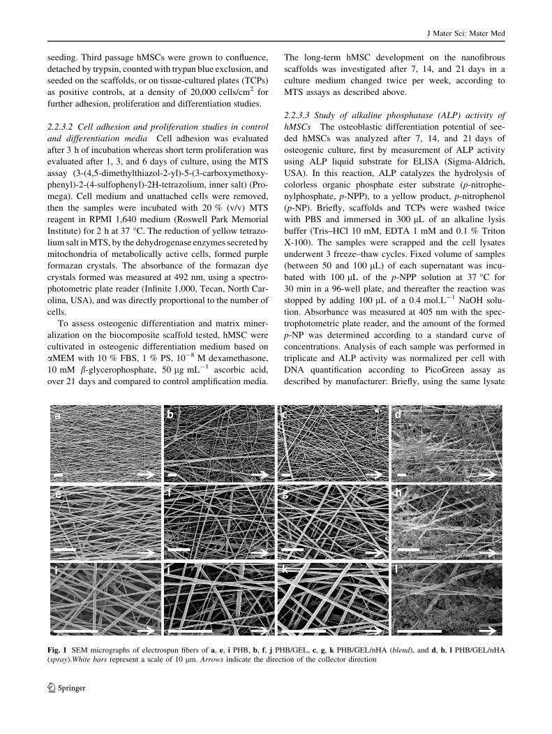
Fig. 1 SEM micrographs of electrospun fibers of a, e, i PHB, b, f, j PHB/GEL, c, g, k PHB/GEL/nHA (blend), and d, h, l PHB/GEL/nHA
(spray).White bars represent a scale of 10 lm. Arrows indicate the direction of the collector direction
J Mater Sci: Mater Med
123
solution, proteinase K was added to samples at a final
concentration of 0.2 mg mL-1, and incubated at 56 �C
overnight. Then PicoGreen assay was performed in the
dark at room temperature in a 96-well plate, on samples of
lysates diluted in Tris EDTA-buffer before adding 50 lL of
the PicoGreen solution. After 10 min incubation, fluores-
cence was measured with the Infinite 1,000 plate reader
using an excitation wavelength of 480 nm and an emission
wavelength of 520 nm.
2.2.3.4 Mineralization of hMSCs Calcium minerals pro-
duction and deposit by hMSC were both qualitatively and
quantitatively measured by Alizarin Red Staining (ARS)
which consists in selectively staining selectively calcium
salt. The scaffolds were rinsed twice with PBS and fixed in
70 % ethanol during 1 h at 4 �C. Then, the constructs were
washed twice with deionized water and immersed in ARS
(2 % (w/w) in deionized water at pH 4.2) during 15 min.
After five washes with deionized water, scaffolds were
dried overnight and observed with an optical microscope
(Aristoplan, Leitz) by taking images using the CoolSnap
software. Staining was then extracted by incubating the
mats in a 10 % cetylpyridinium chloride solution (Sigma-
Aldrich, USA) for 1 h at room temperature. Finally, 50 lL
of each sample was diluted by the addition of 50 lL of
deionized water in a 96-well plate, to fit with the detection
range of the spectrophotometric plate reader and the
absorbance was measured at 540 nm.
2.2.3.5 hMSC morphology studies SEM investigation of
in vitro cultured hMSCs morphology on the scaffolds was
performed after 3 h, 3, 6, and 21 days of cell culture. Prior
scaffolds were rinsed with PBS, and fixed in a 3 % (w/w)
glutaraldehyde solution for 30 min. After washing steps in
deionized water cells were dehydrated with increased
concentrations of ethanol (50, 70, 90, 100 vol. %) for
10 min each. Specimens were then dried in 100 % HMDS
and air-dried by keeping them in a fume hood. Finally,
scaffolds were coated with a 4-nm layer of palladium/
platinum and observed using an accelerating voltage of
1 kV to analyze hMSC morphology. The surface elemental
composition of the samples was also determined by
energy-dispersive X-ray (EDX) spectroscopy with a
10 mm2 germanium diode as an X-ray detector (lumix,
Princeton Gamma-Tech) attached to the SEM equipment.
EDX analyses were thus performed to detect mineraliza-
tion (calcium and phosphorus) on the samples. Sample
surfaces were analyzed for 5 min at 10 kV and a magni-
fication of 1,000. Spatial element mapping was performed
by grouping pixels with similar atomic spectra.
2.2.3.6 Statistical analyses All the data are expressed as
mean ?/6 standard deviation (SD) of n = 9 values
obtained from triplicate values per conditions from 3
independent experiments Statistical analyses were per-
formed using Student’s t test for the calculation of the
significance level of biological data. Differences were
considered statistically significant at p \ 0.05.
3 Results
3.1 Morphological and structural characterizations
of nanofibrous scaffolds
SEM images of the PHB-based biocomposite scaffolds
(Fig. 1) indicate that the PHB, PHB/GEL, and PHB/GEL/
nHA (blend) materials exhibited a porous morphology
constituted of uniform and beadless fibers which were
rather oriented in the collector rotation direction (Fig. 1a–
c, e–g, i–k). Interestingly, the PHB/GEL/nHA (blend)
composite fibers displayed a smooth surface with very few
nanoparticles (Fig. 1k), whereas a rough surface topogra-
phy was generated by the nHA covering for the PHB/GEL/
nHA (spray) analogues (Fig. 1d, h, l). It seemed that the
HA nanoparticles were trapped within the PHB/GEL/nHA
(blend) fibers, while the electrospinning-electrospraying
combination provided fibers with a surface largely covered
by the bioceramic.
The major difference between both one-step strategies
implemented, i.e. electrospinning of a polymer/HA mixture
and electrospinning-electrospraying coupled techniques,
was corroborated by TEM analyses of single fibers from
biocomposite scaffolds (not shown). The PHB/GEL/nHA
(blend) framework (Fig. 2a, b) contained a vast majority of
HA nanoparticles within the constitutive fibers, while a
very small amount lined the fiber surface, indicating that
HA minerals were trapped within the fibers. By means of
the electrospinning-electrospraying process, the nanopar-
ticles were mainly deposited on the surface of the as-
formed fibers. We hypothesis that this tandem technique,
that do not trap nanoparticles on polymer film, will be most
suitable to offer close interactions between cells and nHAs.
ATR-FTIR spectra of the fibrous materials exhibited the
occurrence of PHB characteristic signals with the carbonyl
stretching band at 1,722 cm-1, the C–O stretching band at
1,055 cm-1, and the –CH– bands at 2,970 cm-1. The FTIR
analysis reveals the appearance of the characteristic bands
of functional groups from gelatin on spectra b, c, d with a
signal at 3,306 cm-1 typical of N–H stretching, suggesting
the presence of amine functions. The amide bands of the
gelatin were also observed for the spectra of all gelatin-
containing materials: the amide I band corresponding to
C=O stretching and the amide II band corresponding to N–
H bending. The phosphate ion (PO43-), which is a major
component of HA, with characteristic vibrational bands at
J Mater Sci: Mater Med
123
1,018 cm-1 (stretching) and 569 cm-1 (deformation), were
observed in PHB/GEL/nHA (blend) and PHB/GEL/nHA
(spray).
The use of trifluoroethanol as the solvent for electros-
pinning permitted to produce frameworks with fiber
diameters lower than 500 nm, due to its relative high
dielectric constant (e = 26.14). This order of magnitude
for the average fiber diameter may be particularly suitable
for cell development by making nanofibrous structures
similar to the morphology of the ECM, which is constituted
of natural collagen fibers with diameters typically ranging
from 50 to 500 nm [32]. High porosity in fibrous materials
constitutes an important feature in order to enable an effi-
cient circulation of nutrients and wastes, but also to provide
enough void for the ECM regeneration [14]. PHB frame-
works were produced with fiber diameters lower than
500 nm (Table 1). Electrospinning of gelatin clearly
increases the diameter as compared to PHB, whereas nHA
do not. Porosity ratios and pore size ranges, as determined
by mercury intrusion porosimetry varied from 62 to 83 %
and from 0.4 to 8 lm respectively, thus demonstrating
highly porous frameworks. Obtaining a lower pore size
range for the PHB material could be correlated to the
smaller average fiber diameter, as a decrease in the fiber
diameter is generally associated with a reduction of the
inter-fiber space [33]. Interestingly, the incorporation of
HA nanoparticles by electrospraying led to the generation
of a fibrous scaffold with higher porosity and pore size.
Moreover whereas the neat PHB nanofibers displayed a
highly hydrophobic surface (h = 119�), the gelatin-con-
taining scaffolds exhibited a drastic reduction of the con-
tact angle with a equal to 0�. This indicates that the
hydroxyl groups of gelatin were located on the external
surfaces, thus dramatically increasing the hydrophilic nat-
ure of the surfaces.
3.2 Adherence, short-term proliferation
and morphological evolution of hMSCs
on nanofibrous scaffolds
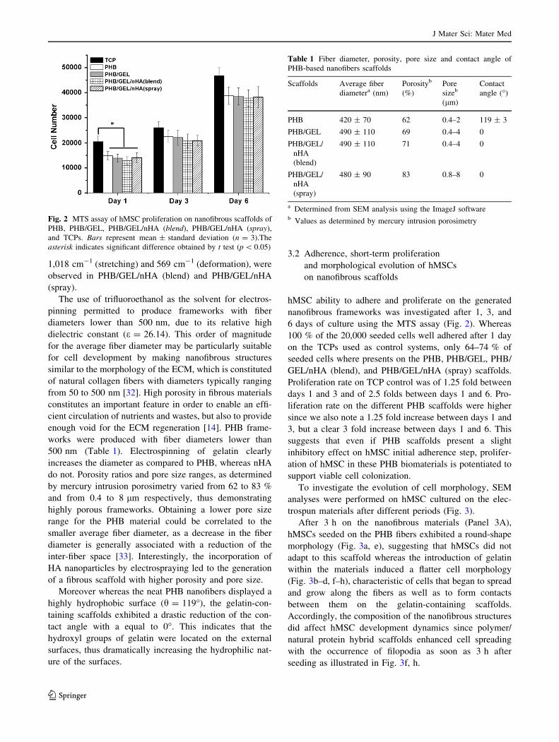
hMSC ability to adhere and proliferate on the generated
nanofibrous frameworks was investigated after 1, 3, and
6 days of culture using the MTS assay (Fig. 2). Whereas
100 % of the 20,000 seeded cells well adhered after 1 day
on the TCPs used as control systems, only 64–74 % of
seeded cells where presents on the PHB, PHB/GEL, PHB/
GEL/nHA (blend), and PHB/GEL/nHA (spray) scaffolds.
Proliferation rate on TCP control was of 1.25 fold between
days 1 and 3 and of 2.5 folds between days 1 and 6. Pro-
liferation rate on the different PHB scaffolds were higher
since we also note a 1.25 fold increase between days 1 and
3, but a clear 3 fold increase between days 1 and 6. This
suggests that even if PHB scaffolds present a slight
inhibitory effect on hMSC initial adherence step, prolifer-
ation of hMSC in these PHB biomaterials is potentiated to
support viable cell colonization.
To investigate the evolution of cell morphology, SEM
analyses were performed on hMSC cultured on the elec-
trospun materials after different periods (Fig. 3).
After 3 h on the nanofibrous materials (Panel 3A),
hMSCs seeded on the PHB fibers exhibited a round-shape
morphology (Fig. 3a, e), suggesting that hMSCs did not
adapt to this scaffold whereas the introduction of gelatin
within the materials induced a flatter cell morphology
(Fig. 3b–d, f–h), characteristic of cells that began to spread
and grow along the fibers as well as to form contacts
between them on the gelatin-containing scaffolds.
Accordingly, the composition of the nanofibrous structures
did affect hMSC development dynamics since polymer/
natural protein hybrid scaffolds enhanced cell spreading
with the occurrence of filopodia as soon as 3 h after
seeding as illustrated in Fig. 3f, h.
Fig. 2 MTS assay of hMSC proliferation on nanofibrous scaffolds of
PHB, PHB/GEL, PHB/GEL/nHA (blend), PHB/GEL/nHA (spray),
and TCPs. Bars represent mean ± standard deviation (n = 3).The
asterisk indicates significant difference obtained by t test (p \ 0.05)
Table 1 Fiber diameter, porosity, pore size and contact angle of
PHB-based nanofibers scaffolds
Scaffolds Average fiber
diametera (nm)
Porosityb
(%)
Pore
sizeb
(lm)
Contact
angle (�)
PHB 420 ± 70 62 0.4–2 119 ± 3
PHB/GEL 490 ± 110 69 0.4–4 0
PHB/GEL/
nHA
(blend)
490 ± 110 71 0.4–4 0
PHB/GEL/
nHA
(spray)
480 ± 90 83 0.8–8 0
a Determined from SEM analysis using the ImageJ softwareb Values as determined by mercury intrusion porosimetry
J Mater Sci: Mater Med
123
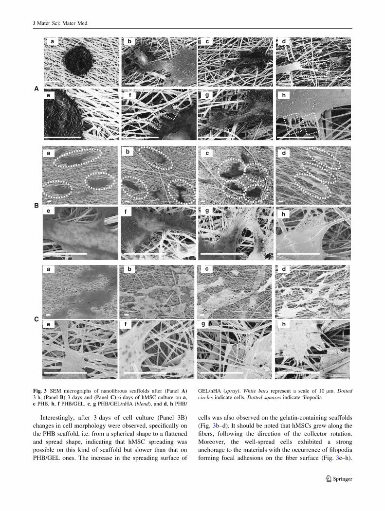
Interestingly, after 3 days of cell culture (Panel 3B)
changes in cell morphology were observed, specifically on
the PHB scaffold, i.e. from a spherical shape to a flattened
and spread shape, indicating that hMSC spreading was
possible on this kind of scaffold but slower than that on
PHB/GEL ones. The increase in the spreading surface of
cells was also observed on the gelatin-containing scaffolds
(Fig. 3b–d). It should be noted that hMSCs grew along the
fibers, following the direction of the collector rotation.
Moreover, the well-spread cells exhibited a strong
anchorage to the materials with the occurrence of filopodia
forming focal adhesions on the fiber surface (Fig. 3e–h).
a b c d
e f gh
a b c d
e f g h
a b c d
e f g h
A
B
C
Fig. 3 SEM micrographs of nanofibrous scaffolds after (Panel A)3 h, (Panel B) 3 days and (Panel C) 6 days of hMSC culture on a,
e PHB, b, f PHB/GEL, c, g PHB/GEL/nHA (blend), and d, h PHB/
GEL/nHA (spray). White bars represent a scale of 10 lm. Dotted
circles indicate cells. Dotted squares indicate filopodia
J Mater Sci: Mater Med
123
The generation of these inter-cellular communication
channels was noticeable with the formation of natural ECM
(Fig. 3g, h) with spherical pores mostly generated between
two or more close cells through the production of slender
cytoplasmic projection,
After 6 days of cell culture, hMSCs were no longer inde-
pendent, but formed a cell network connected by the ECM
(Panel 3C) on all scaffolds analyzed. The spreading surface of
growing cells was increased as compared to day 3 of cell
culture, suggesting that they tended toward colonization of the
entire scaffold surface up to reaching confluence.
3.3 Long-term proliferation and differentiation
of hMSCs on nanofibrous scaffolds
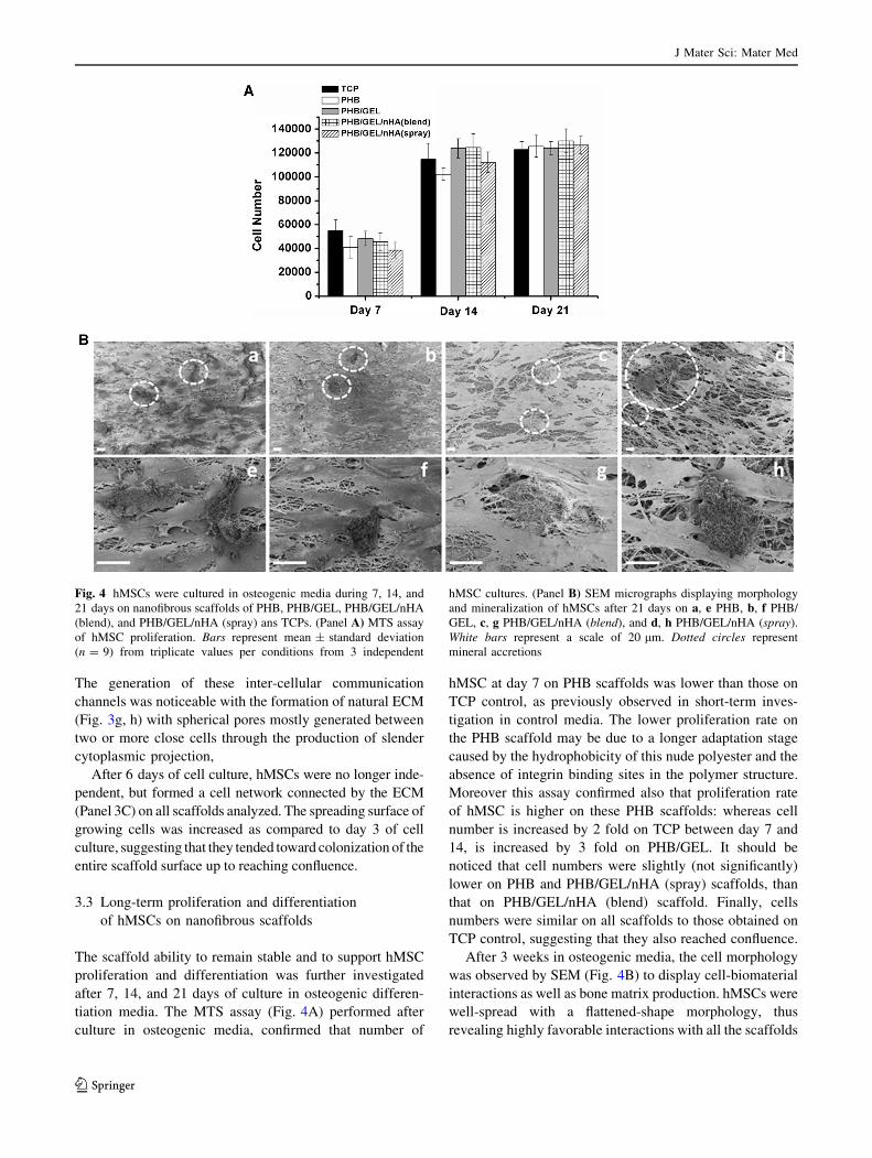
The scaffold ability to remain stable and to support hMSC
proliferation and differentiation was further investigated
after 7, 14, and 21 days of culture in osteogenic differen-
tiation media. The MTS assay (Fig. 4A) performed after
culture in osteogenic media, confirmed that number of
hMSC at day 7 on PHB scaffolds was lower than those on
TCP control, as previously observed in short-term inves-
tigation in control media. The lower proliferation rate on
the PHB scaffold may be due to a longer adaptation stage
caused by the hydrophobicity of this nude polyester and the
absence of integrin binding sites in the polymer structure.
Moreover this assay confirmed also that proliferation rate
of hMSC is higher on these PHB scaffolds: whereas cell
number is increased by 2 fold on TCP between day 7 and
14, is increased by 3 fold on PHB/GEL. It should be
noticed that cell numbers were slightly (not significantly)
lower on PHB and PHB/GEL/nHA (spray) scaffolds, than
that on PHB/GEL/nHA (blend) scaffold. Finally, cells
numbers were similar on all scaffolds to those obtained on
TCP control, suggesting that they also reached confluence.
After 3 weeks in osteogenic media, the cell morphology
was observed by SEM (Fig. 4B) to display cell-biomaterial
interactions as well as bone matrix production. hMSCs were
well-spread with a flattened-shape morphology, thus
revealing highly favorable interactions with all the scaffolds
Fig. 4 hMSCs were cultured in osteogenic media during 7, 14, and
21 days on nanofibrous scaffolds of PHB, PHB/GEL, PHB/GEL/nHA
(blend), and PHB/GEL/nHA (spray) ans TCPs. (Panel A) MTS assay
of hMSC proliferation. Bars represent mean ± standard deviation
(n = 9) from triplicate values per conditions from 3 independent
hMSC cultures. (Panel B) SEM micrographs displaying morphology
and mineralization of hMSCs after 21 days on a, e PHB, b, f PHB/
GEL, c, g PHB/GEL/nHA (blend), and d, h PHB/GEL/nHA (spray).
White bars represent a scale of 20 lm. Dotted circles represent
mineral accretions
J Mater Sci: Mater Med
123
and a high cytocompatibility of the letters. Furthermore, the
hMSC layers on the nanofibrous scaffolds exhibited the
occurrence of filopodia that linked cells with the nanofibers,
and above all generate their own direct communication
channels in this osteogenic induction media, as observed
previously in control amplification media. Cell development
tended to a complete covering of the scaffold surface, con-
firming that they have reached confluence, and the secretion
of ECM was clearly observed (Fig. 4a–d). The newly
formed ECM is characterized by several aggregations of
globular accretions, which were in intimate contact with
cells (Fig. 4e–h). Noticeably, the PHB/GEL/nHA (spray)
scaffold enabled the highest extent of deposits with the
formation of miscellaneous larger aggregates (Fig. 4d, h).
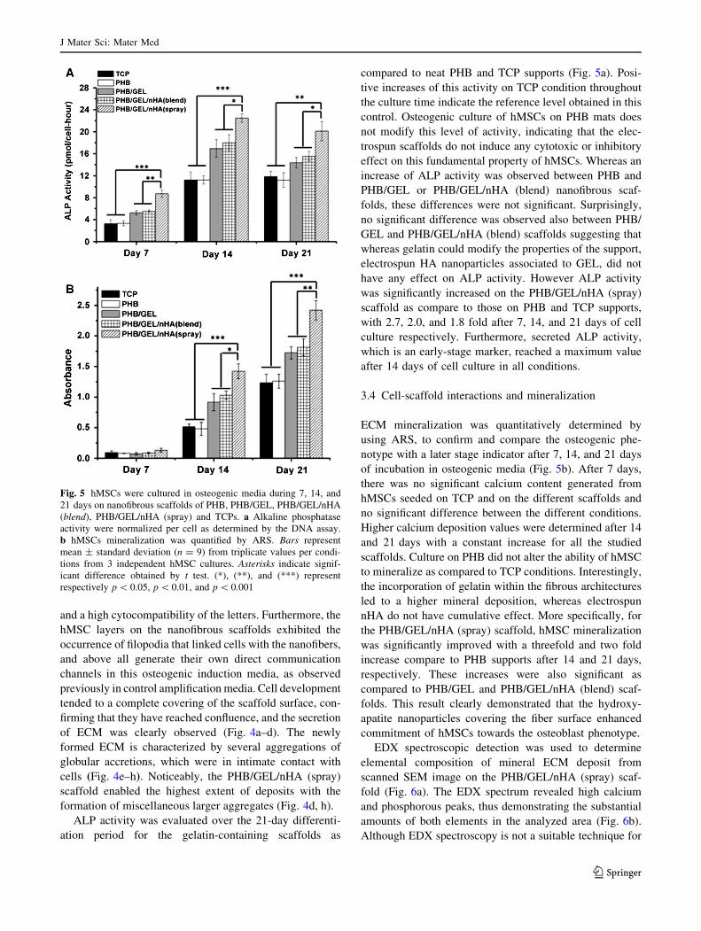
ALP activity was evaluated over the 21-day differenti-
ation period for the gelatin-containing scaffolds as
compared to neat PHB and TCP supports (Fig. 5a). Posi-
tive increases of this activity on TCP condition throughout
the culture time indicate the reference level obtained in this
control. Osteogenic culture of hMSCs on PHB mats does
not modify this level of activity, indicating that the elec-
trospun scaffolds do not induce any cytotoxic or inhibitory
effect on this fundamental property of hMSCs. Whereas an
increase of ALP activity was observed between PHB and
PHB/GEL or PHB/GEL/nHA (blend) nanofibrous scaf-
folds, these differences were not significant. Surprisingly,
no significant difference was observed also between PHB/
GEL and PHB/GEL/nHA (blend) scaffolds suggesting that
whereas gelatin could modify the properties of the support,
electrospun HA nanoparticles associated to GEL, did not
have any effect on ALP activity. However ALP activity
was significantly increased on the PHB/GEL/nHA (spray)
scaffold as compare to those on PHB and TCP supports,
with 2.7, 2.0, and 1.8 fold after 7, 14, and 21 days of cell
culture respectively. Furthermore, secreted ALP activity,
which is an early-stage marker, reached a maximum value
after 14 days of cell culture in all conditions.
3.4 Cell-scaffold interactions and mineralization
ECM mineralization was quantitatively determined by
using ARS, to confirm and compare the osteogenic phe-
notype with a later stage indicator after 7, 14, and 21 days
of incubation in osteogenic media (Fig. 5b). After 7 days,
there was no significant calcium content generated from
hMSCs seeded on TCP and on the different scaffolds and
no significant difference between the different conditions.
Higher calcium deposition values were determined after 14
and 21 days with a constant increase for all the studied
scaffolds. Culture on PHB did not alter the ability of hMSC
to mineralize as compared to TCP conditions. Interestingly,
the incorporation of gelatin within the fibrous architectures
led to a higher mineral deposition, whereas electrospun
nHA do not have cumulative effect. More specifically, for
the PHB/GEL/nHA (spray) scaffold, hMSC mineralization
was significantly improved with a threefold and two fold
increase compare to PHB supports after 14 and 21 days,
respectively. These increases were also significant as
compared to PHB/GEL and PHB/GEL/nHA (blend) scaf-
folds. This result clearly demonstrated that the hydroxy-
apatite nanoparticles covering the fiber surface enhanced
commitment of hMSCs towards the osteoblast phenotype.
EDX spectroscopic detection was used to determine
elemental composition of mineral ECM deposit from
scanned SEM image on the PHB/GEL/nHA (spray) scaf-
fold (Fig. 6a). The EDX spectrum revealed high calcium
and phosphorous peaks, thus demonstrating the substantial
amounts of both elements in the analyzed area (Fig. 6b).
Although EDX spectroscopy is not a suitable technique for
Fig. 5 hMSCs were cultured in osteogenic media during 7, 14, and
21 days on nanofibrous scaffolds of PHB, PHB/GEL, PHB/GEL/nHA
(blend), PHB/GEL/nHA (spray) and TCPs. a Alkaline phosphatase
activity were normalized per cell as determined by the DNA assay.
b hMSCs mineralization was quantified by ARS. Bars represent
mean ± standard deviation (n = 9) from triplicate values per condi-
tions from 3 independent hMSC cultures. Asterisks indicate signif-
icant difference obtained by t test. (*), (**), and (***) represent
respectively p \ 0.05, p \ 0.01, and p \ 0.001
J Mater Sci: Mater Med
123
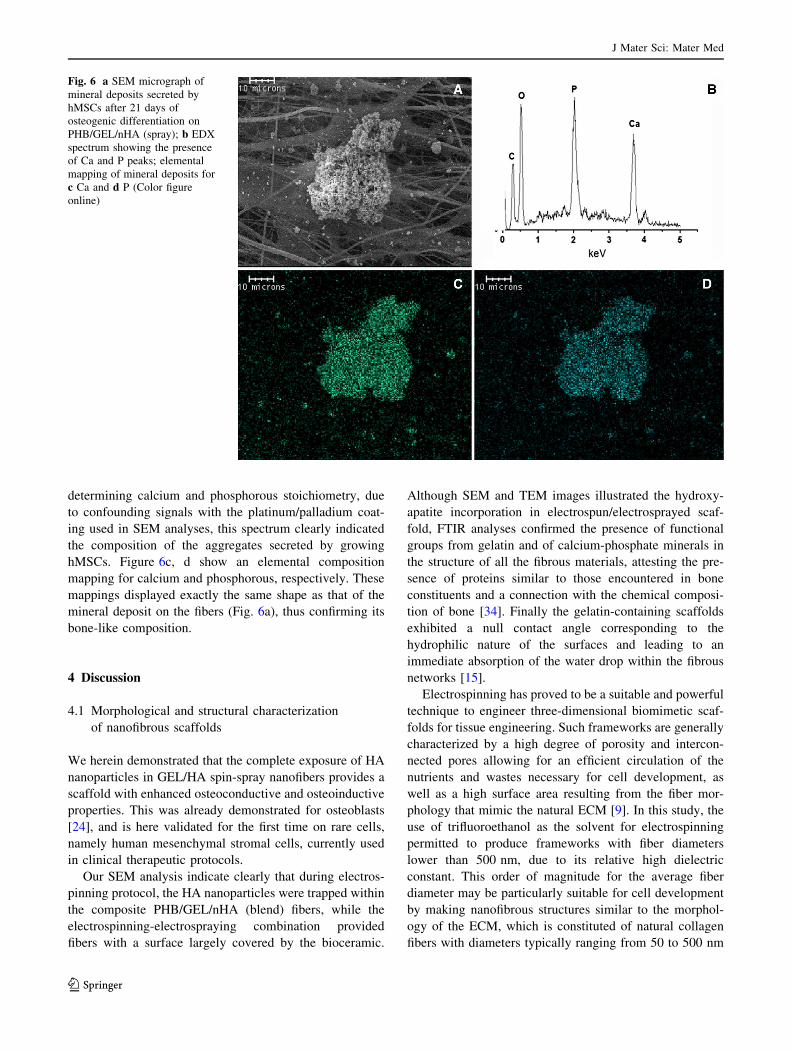
determining calcium and phosphorous stoichiometry, due
to confounding signals with the platinum/palladium coat-
ing used in SEM analyses, this spectrum clearly indicated
the composition of the aggregates secreted by growing
hMSCs. Figure 6c, d show an elemental composition
mapping for calcium and phosphorous, respectively. These
mappings displayed exactly the same shape as that of the
mineral deposit on the fibers (Fig. 6a), thus confirming its
bone-like composition.
4 Discussion
4.1 Morphological and structural characterization
of nanofibrous scaffolds
We herein demonstrated that the complete exposure of HA
nanoparticles in GEL/HA spin-spray nanofibers provides a
scaffold with enhanced osteoconductive and osteoinductive
properties. This was already demonstrated for osteoblasts
[24], and is here validated for the first time on rare cells,
namely human mesenchymal stromal cells, currently used
in clinical therapeutic protocols.
Our SEM analysis indicate clearly that during electros-
pinning protocol, the HA nanoparticles were trapped within
the composite PHB/GEL/nHA (blend) fibers, while the
electrospinning-electrospraying combination provided
fibers with a surface largely covered by the bioceramic.
Although SEM and TEM images illustrated the hydroxy-
apatite incorporation in electrospun/electrosprayed scaf-
fold, FTIR analyses confirmed the presence of functional
groups from gelatin and of calcium-phosphate minerals in
the structure of all the fibrous materials, attesting the pre-
sence of proteins similar to those encountered in bone
constituents and a connection with the chemical composi-
tion of bone [34]. Finally the gelatin-containing scaffolds
exhibited a null contact angle corresponding to the
hydrophilic nature of the surfaces and leading to an
immediate absorption of the water drop within the fibrous
networks [15].
Electrospinning has proved to be a suitable and powerful
technique to engineer three-dimensional biomimetic scaf-
folds for tissue engineering. Such frameworks are generally
characterized by a high degree of porosity and intercon-
nected pores allowing for an efficient circulation of the
nutrients and wastes necessary for cell development, as
well as a high surface area resulting from the fiber mor-
phology that mimic the natural ECM [9]. In this study, the
use of trifluoroethanol as the solvent for electrospinning
permitted to produce frameworks with fiber diameters
lower than 500 nm, due to its relative high dielectric
constant. This order of magnitude for the average fiber
diameter may be particularly suitable for cell development
by making nanofibrous structures similar to the morphol-
ogy of the ECM, which is constituted of natural collagen
fibers with diameters typically ranging from 50 to 500 nm
Fig. 6 a SEM micrograph of
mineral deposits secreted by
hMSCs after 21 days of
osteogenic differentiation on
PHB/GEL/nHA (spray); b EDX
spectrum showing the presence
of Ca and P peaks; elemental
mapping of mineral deposits for
c Ca and d P (Color figure
online)
J Mater Sci: Mater Med
123
[32]. Interestingly, whereas electrospinning of nHA do not
modify PHB/Gel parameters, the incorporation of HA
nanoparticles by electrospraying led to the generation of a
fibrous scaffold with higher porosity and pore size. The
presence of nanoparticles that covered the fiber surface
may prevent a tight layering of PHB/GEL nanofibers
formed during the electrospinning process, thus generating
a looser fiber organization of the scaffold with higher pore
content. Such high porosity in fibrous materials is of
interest, since it constitutes an important feature in order to
not only enable an efficient circulation of elements and
cells, but also to provide enough void for the ECM
regeneration [14].
Our hypothesis was that the major difference observed
on the scaffold, corresponding to the morphological
observation of the HA mineral location, inside or outside
the fibers, could affect cell/bioceramic interactions. Dis-
tinct effects could be attempted: such electrospraying of
nHA particles on electrospun nanofibers that permit to
obtain rough surface morphology, should be ideal to cell
attachment and proliferation, and also achieve improved
mechanical properties compared to blended nanofibers.
Conversely on electrospun nHA, such a thin polymer film
covering the HA nanoparticles could hide and inhibit there
osteogenic properties. We decided to test these hypotheses
on hMSCs.
hMSCs represent a prospective therapeutic cell source
for many tissue engineering applications, since they can be
obtained from various origins, and have a multilineage
differentiation potential leading to several specific cell
types, such as osteoblasts, and chondrocytes, necessary for
bone repair. Cell adhesion and proliferation represent the
first step of communication between hMSCs and a bio-
material, which is critical for their subsequent differentia-
tion [35]. Moreover, the scaffold properties have a
fundamental impact on the cell growth and morphology
[36]. Our data indicate that PHB scaffolds present a slight
inhibitory effect on hMSC initial adherence step as com-
pared to TCP. Similar results have previously been repor-
ted, revealing that electrospun scaffolds may hinder the
first steps of cell adhesion and proliferation compared to
TCPs [17, 37]. This phenomenon seems to be mediated by
fiber topography, which leads to a decrease in the cyto-
skeleton spreading and results in a slower initial develop-
ment [8, 38]. However in our conditions, proliferation of
hMSC in the 4 different PHB biomaterials is potentiated to
the same extend to support viable cell colonization, in
control media as well as in osteogenic media. Even if any
quantitative differences were notable between the 4 scaf-
folds, morphological differences were observed as soon as
3 h after seeding. Whereas hMSCs seeded on the PHB
fibers exhibited a round-shape morphology suggesting that
these cells did not adapt to the scaffold, the introduction of
gelatin induced flatter cell morphology, cells began to
spread and grow along the fibers, as well as to form con-
tacts between them and to exhibit strong anchorage to the
materials with the occurrence of filopodia. Such early
increase in the spreading surface of cells, observed on the
gelatin-containing scaffolds, could be attributed to a sig-
nificant affinity and favorable interactions with scaffolds.
In fact, gelatin possesses several integrin binding sites, thus
leading to a rearrangement of the cytoskeleton structure
composed of actin filaments that promote the filopodia
generation. These filopodia are known to be responsible for
the proper hMSC adhesion and spreading [16, 39, 40].
During cell spreading and proliferation process, the inter-
cellular communication is essential in order to exchange
miscellaneous signals concerning the differentiation for
example. The generation of these communication channels
was noticeable and was also associated with the formation
of natural ECM with spherical pores mostly generated
between two or more close cells through the production of
slender cytoplasmic projection.
One of the critical organic components produced during
osteoblastic differentiation is the ALP enzyme, because of
its activity in the formation of mineralized matrix [41].
ALP is considered as an early-stage biochemical marker of
immature osteoblast activity, and its transient activity was
reported in the hMSC-induced process toward osteolineage
[42]. Even if ALP can be generated in miscellaneous tis-
sues including liver, kidney or placenta, elevated levels are
typically observed within cell metabolisms for bone
regeneration during the initial phase of mineralization [43,
44]. The stabilization of secreted ALP levels, which is an
early-stage marker, is currently related to the cellular
process switching into further steps, such as mineralization
[45], and then decreases when the cell phenotypic shift
occurs. hMSC ability to mineralize their surrounding
matrix is considered as an evidence of fully differentiated
stem cells toward mature osteoblasts [42, 46].
Our data indicate that whereas gelatin induce an
increase of secreted ALP and mineralization, electrospun
nHA associated to GEL did not have any effect on these 2
processes. This could be explained by the inclusion of
nHAs within the latter fibers, thus hindering close nano-
particle/cell interactions, and potentially inhibiting the bi-
oceramic osteoinductive properties. This was confirmed by
the upregulation of ALP activity and mineralization when
nHAs were sprayed on the fiber surface: we hypothesize an
enhanced effect of nHA that are in direct contact with
hMSCs and are known to induce in vitro osteoblastic dif-
ferentiation [20]. In addition, Jung et al. have demonstrated
that Ca2? released from HA activates the expression of
bone-associated proteins in MC3T3-E1 cells [47]. More-
over, such higher osteogenic activities observed for hMSCs
seeded on the PHB/GEL/nHA (spray) scaffold could
J Mater Sci: Mater Med
123
explain the particular slowing down of the proliferation
rate in this condition. Indeed, Owen et al. [48] have evi-
denced the competition between hMSC proliferation and
differentiation behaviors, revealing that the increased
expression of osteoblastic markers leads to a decrease in
the proliferation rate
It is noteworthy that Gupta et al., which compared PLACL/
GEL/HA(blend) and PLACL/GEL/HA(spray) effect on fetal
osteoblast cells have observed a slight effect of HA (blend or
sprayed) on proliferation as compared to PLACL/GEL levels,
whereas differentiation effect characterized by ALP activity
and ARS quantification was only significant with sprayed HA
[15]. These different effects of HA on cell proliferation as
compared to our data could be explained by different signal-
ing pathway activated on transformed cell lines, that are silent
on hMSCs, thus confirming the interest to work with primary
cultures. Finally, newly formed ECM, are characterized by
several secreted aggregations of globular mineral accretions
that were in intimate contact with cells. The interfacial
properties between ceramic, cells and secreted matrix are
crucial, since the sizes of calcium phosphate particles and
their respective distribution have been recognized as impor-
tant parameters on the mechanical behavior of bioceramic
system. Whereas these accretions were observed on all scaf-
folds, the PHB/GEL/nHA (spray) scaffold enabled the highest
extent of mineral deposits with the formation of miscella-
neous larger aggregates. This is of interest since, as previously
reported, this mineral deposition occurred as the first step of
osteogenesis, and therefore is very important for bone
regeneration processes [22, 49].
5 Conclusion
The present investigation describes straightforward and
robust routes toward promising electrospun/electrosprayed
biocomposite scaffolds with a potentially mineralized surface
for bone tissue engineering. The short-term biological
investigation has shown that the scaffolds exhibit a continuous
cell proliferation with a faster evolution of cell morphology
for the gelatin-containing materials. In addition, the long-term
biological investigation has clearly demonstrated that hMSCs
seeded onto the nHA-sprayed scaffold develop a significantly
higher level of ALP activity, as well as a higher biomineral-
ization rate compared to the other scaffolds, and especially to
PHB/GEL/nHA-blend supports. The fiber surface deposition
of nHAs by the electrospraying process enables their direct
exposure to hMSCs for an efficient transmission of the bi-
oceramic osteoinductive and osteoconductive properties to
cells, thus inducing a synergistic enhancement of the stimu-
latory effect of the osteogenic medium toward cell differen-
tiation. Consequently, the nHA-sprayed framework has
shown its biocompatibility for hMSC properties and more
specifically its strong capacity for enhancing osteoblastic
differentiation of hMSC. The exact mechanism leading to the
beneficial effects brought by nHAs on the differentiation is
still unclear, while the bioceramic may influence the cell
surface receptors that induce the activation of signaling
pathways related to osteogenesis [22].
Accordingly, we believe that electrospinning/electro-
spraying-derivatized biocomposite scaffolds may facilitate
hMSC colonization and improve bone formation, when
implanted in vivo in tissue engineering strategies, to induce
bone formation in vivo through ectopic model or to induce
bone regeneration after injury on bone orthotropic models.
Acknowledgments The authors thank the CNRS (France) and the
Bulgarian Academy of Sciences (Bulgaria) for financial support
through the bilateral project No. 135751. Financial support from the
National Fund of Scientific Research (Grant DO 02-237/2008) is also
gratefully acknowledged (J.R., O.S., N.M., and I.R.).
References
1. Frohlich M, Grayson W, Wan L, Marolt D, Drobnic M, Vunjak-
Novakovic G. Tissue engineered bone grafts: biological
requirements, tissue culture and clinical relevance. Curr Stem
Cell Res Ther. 2008;3:254–64.
2. Conway JD. Autograft and nonunions: morbidity with intra-
medullary bone graft versus iliac crest bone graft. Orthop Clin
North Am. 2010;41:75–84.
3. Grayson WL, Frohlich M, Yeager K, Bhumiratana S, Chan ME,
Cannizzaro C, et al. Engineering anatomically shaped human
bone grafts. Proc Natl Acad Sci USA. 2010;107:3299–304.
4. Kohn D, Sander-Beuermann A. Donor-site morbidity after har-
vest of a bone-tendon-bone patellar tendon autograft. Knee Surg
Sports Traumatol Arthrosc. 1994;2:219–23.
5. Langer R, Vacanti J. Tissue engineering. Science. 1993;260:920–6.
6. Petite H, Viateau V, Bensaid W, Meunier A, de Pollak C,
Bourguignon M, et al. Tissue-engineered bone regeneration. Nat
Biotechnol. 2000;18:959–63.
7. Peter SJ, Miller MJ, Yasko AW, Yaszemski MJ, Mikos AG.
Polymer concepts in tissue engineering. J Biomed Mater Res.
1998;43:422–7.
8. Pham QP, Sharma U, Mikos AG. Electrospinning of polymeric
nanofibers for tissue engineering applications: a review. Tissue
Eng. 2006;12:1197–211.
9. Sill TJ, von Recum HA. Electrospinning: applications in drug
delivery and tissue engineering. Biomaterials. 2008;29:1989–2006.
10. Yoo HS, Kim TG, Park TG. Surface-functionalized electrospun
nanofibers for tissue engineering and drug delivery. Adv Drug
Deliv Rev. 2009;61:1033–42.
11. Martins A, Duarte ARC, Faria S, Marques AP, Reis RL, Neves NM.
Osteogenic induction of hBMSCs by electrospun scaffolds with
dexamethasone release functionality. Biomaterials. 2010;31:5875–85.
12. Ravichandran R, Venugopal JR, Sundarrajan S, Mukherjee S,
Ramakrishna S. Precipitation of nanohydroxyapatite on PLLA/
PBLG/Collagen nanofibrous structures for the differentiation of
adipose derived stem cells to osteogenic lineage. Biomaterials.
2012;33:846–55.
13. Spadaccio C, Rainer A, Centola M, Trombetta M, Chello M,
Lusini M, et al. Heparin-releasing scaffold for stem cells: a dif-
ferentiating device for vascular aims. Regen Med. 2010;5:
645–57.
J Mater Sci: Mater Med
123
14. Agrawal CM, Ray RB. Biodegradable polymeric scaffolds for mus-
culoskeletal tissue engineering. J Biomed Mater Res. 2001;55:141–50.
15. Gupta D, Venugopal J, Mitra S, Giri Dev VR, Ramakrishna S.
Nanostructured biocomposite substrates by electrospinning and
electrospraying for the mineralization of osteoblasts. Biomateri-
als. 2009;30:2085–94.
16. Prabhakaran MP, Venugopal J, Ramakrishna S. Electrospun
nanostructured scaffolds for bone tissue engineering. Acta Bio-
mater. 2009;5:2884–93.
17. Han I, Shim KJ, Kim JY, Im SU, Sung YK, Kim M, et al. Effect of
poly(3-hydroxybutyrate-co-3-hydroxyvalerate) nanofiber matrices
cocultured with hair follicular epithelial and dermal cells for bio-
logical wound dressing. Artif Organs. 2007;31:801–8.
18. Kim HW, Song JH, Kim HE. Nanofiber generation of gelatin-
hydroxyapatite biomimetics for guided tissue regeneration. Adv
Funct Mater. 2005;15:1988–94.
19. Meng W, Xing ZC, Jung KH, Kim SY, Yuan J, Kang IK, et al.
Synthesis of gelatin-containing PHBV nanofiber mats for bio-
medical application. J Mater Sci-Mater Med. 2008;19:2799–807.
20. Jang JH, Castano O, Kim HW. Electrospun materials as potential
platforms for bone tissue engineering. Adv Drug Deliv Rev.
2009;61:1065–83.
21. Jose MV, Thomas V, Johnson KT, Dean DR, Nyairo E. Aligned
PLGA/HA nanofibrous nanocomposite scaffolds for bone tissue
engineering. Acta Biomater. 2009;5:305–15.
22. Seyedjafari E, Soleimani M, Ghaemi N, Shabani I. Nanohy-
droxyapatite-coated electrospun poly(l-lactide) nanofibers
enhance osteogenic differentiation of stem cells and induce
ectopic bone formation. Biomacromolecules. 2010;11:3118–25.
23. Whited BM, Whitney JR, Hofmann MC, Xu Y, Rylander MN.
Pre-osteoblast infiltration and differentiation in highly porous
apatite-coated PLLA electrospun scaffolds. Biomaterials.
2011;32:2294–304.
24. Francis L, Venugopal J, Prabhakaran MP, Thavasi V, Marsano E,
Ramakrishna S. Simultaneous electrospin–electrosprayed bio-
composite nanofibrous scaffolds for bone tissue regeneration.
Acta Biomater. 2010;6:4100–9.
25. Chen GQ, Wu Q. The application of polyhydroxyalkanoates as
tissue engineering materials. Biomaterials. 2005;26:6565–78.
26. Misra SK, Valappil SP, Roy I, Boccaccini AR. Polyhydrox-
yalkanoate (PHA)/inorganic phase composites for tissue engi-
neering applications. Biomacromolecules. 2006;7:2249–58.
27. Asran AS, Razghandi K, Aggarwal N, Michler GH, Groth T.
Nanofibers from blends of polyvinyl alcohol and polyhydroxy
butyrate as potential scaffold material for tissue engineering of
skin. Biomacromolecules. 2010;11:3413–21.
28. Kuppan P, Vasanthan KS, Sundaramurthi D, Krishnan UM, Se-
thuraman S. Development of poly(3-hydroxybutyrate-co-3-hy-
droxyvalerate) fibers for skin tissue engineering: effects of
topography, mechanical, and chemical stimuli. Biomacromole-
cules. 2011;12:3156–65.
29. Sombatmankhong K, Sanchavanakit N, Pavasant P, Supaphol P.
Bone scaffolds from electrospun fiber mats of poly(3-hydroxy-
butyrate), poly(3-hydroxybutyrate-co-3-hydroxyvalerate) and
their blend. Polymer. 2007;48:1419–27.
30. Wang Y, Gao R, Wang P–P, Jian J, Jiang X-L, Yan C, et al. The
differential effects of aligned electrospun PHBHHx fibers on
adipogenic and osteogenic potential of MSCs through the regu-
lation of PPARc signaling. Biomaterials. 2012;33:485–93.
31. Mincheva R, Manolova N, Paneva D, Rashkov I. Preparation of
polyelectrolyte-containing nanofibers by electrospinning in the
presence of a non-ionogenic water-soluble polymer. J Bioact
Compat Polym. 2005;20:419–35.
32. Jeong SI, Ko EK, Yum J, Jung CH, Lee YM, Shin H. Nanofibrous
poly(lactic acid)/hydroxyapatite composite scaffolds for guided
tissue regeneration. Macromol Biosci. 2008;8:328–38.
33. Li D, Frey MW, Joo YL. Characterization of nanofibrous membranes
with capillary flow porometry. J Membr Sci. 2006;286:104–14.
34. Boskey A, Pleshko Camacho N. FT-IR imaging of native and
tissue-engineered bone and cartilage. Biomaterials. 2007;28:
2465–78.
35. Venugopal JR, Low S, Choon AT, Kumar AB, Ramakrishna S.
Nanobioengineered electrospun composite nanofibers and osteo-
blasts for bone regeneration. Artif Organs. 2008;32:388–97.
36. Beachley V, Wen X. Polymer nanofibrous structures: fabrication,
biofunctionalization, and cell interactions. Prog Polym Sci. 2010;
35:868–92.
37. Prabhakaran MP, Venugopal JR, Ramakrishna S. Mesenchymal
stem cell differentiation to neuronal cells on electrospun nanofi-
brous substrates for nerve tissue engineering. Biomaterials.
2009;30:4996–5003.
38. Badami AS, Kreke MR, Thompson MS, Riffle JS, Goldstein AS.
Effect of fiber diameter on spreading, proliferation, and differ-
entiation of osteoblastic cells on electrospun poly(lactic acid)
substrates. Biomaterials. 2006;27:596–606.
39. Li WJ, Danielson KG, Alexander PG, Tuan RS. Biological
response of chondrocytes cultured in three-dimensional nanofi-
brous poly(e-caprolactone) scaffolds. J Biomed Mater Res.
2003;67A:1105–14.
40. Anselme K. Osteoblast adhesion on biomaterials. Biomaterials.
2000;21:667–81.
41. Anderson HC, Sipe JB, Hessle L, Dhamyamraju R, Atti E,
Camacho NP, et al. Impaired calcification around matrix vesicles
of growth plate and bone in alkaline phosphatase-deficient mice.
Am J Pathol. 2004;164:841–7.
42. Jaiswal N, Haynesworth SE, Caplan AI, Bruder SP. Osteogenic
differentiation of purified, culture-expanded human mesenchymal
stem cells in vitro. J Cell Biochem. 1997;64:295–312.
43. Heinemann C, Heinemann S, Bernhardt A, Worch H, Hanke T. Novel
textile chitosan scaffolds promote spreading, proliferation, and dif-
ferentiation of osteoblasts. Biomacromolecules. 2008;9:2913–20.
44. Liu F, Malaval L, Aubin JE. Global amplification polymerase
chain reaction reveals novel transitional stages during osteopro-
genitor differentiation. J Cell Sci. 2003;116:1787–96.
45. Yu HS, Hong SJ, Kim HW. Surface-mineralized polymeric
nanofiber for the population and osteogenic stimulation of ratbone-marrow stromal cells. Mater Chem Phys. 2009;113:873–7.
46. Sottile V, Thomson A, McWhir J. In vitro osteogenic differen-
tiation of human es cells. Cloning Stem Cells. 2003;5:149–55.
47. Jung GY, Park YJ, Han JS. Effects of HA released calcium ion on
osteoblast differentiation. J Mater Sci Mater Med. 2010;21:
1649–54.
48. Owen TA, Aronow M, Shalhoub V, Barone LM, Wilming L,
Tassinari MS, et al. Progressive development of the rat osteoblast
phenotype in vitro: Reciprocal relationships in expression of
genes associated with osteoblast proliferation and differentiation
during formation of the bone extracellular matrix. J Cell Physiol.
1990;143:420–30.
49. Li C, Vepari C, Jin HJ, Kim HJ, Kaplan DL. Electrospun silk-
BMP-2 scaffolds for bone tissue engineering. Biomaterials.
2006;27:3115–24.
J Mater Sci: Mater Med
123