Embed Size (px)
Citation preview

From Body Surface Potential to Activation Maps on the
Atria: a Machine Learning Technique
Nejib Zemzemi, Simon Labarthe, Remi Dubois, Yves Coudiere
To cite this version:
Nejib Zemzemi, Simon Labarthe, Remi Dubois, Yves Coudiere. From Body Surface Potentialto Activation Maps on the Atria: a Machine Learning Technique. CINC - Computing inCardiology 2012, Sep 2012, Krakow, Poland. 39, pp.125-128, 2012, Computing in Cardiology2012,. <www.cinc.org/archives/2012/pdf/0125.pdf>. <hal-00759210>
HAL Id: hal-00759210
https://hal.inria.fr/hal-00759210
Submitted on 30 Nov 2012
HAL is a multi-disciplinary open accessarchive for the deposit and dissemination of sci-entific research documents, whether they are pub-lished or not. The documents may come fromteaching and research institutions in France orabroad, or from public or private research centers.
L’archive ouverte pluridisciplinaire HAL, estdestinee au depot et a la diffusion de documentsscientifiques de niveau recherche, publies ou non,emanant des etablissements d’enseignement et derecherche francais ou etrangers, des laboratoirespublics ou prives.

From Body Surface Potential to Activation Maps on the Atria:a Machine Learning Technique
Nejib Zemzemi 1, Simon Labarthe 1, Remi Dubois 2, Yves Coudiere 1
1 INRIA Bordeaux Sud-Ouest, Talence, France2 LIRYC Universite de Bordeaux, Pessac, France
Abstract
The treatment of atrial fibrillation has greatly changedin the past decade. Ablation therapy, in particular pul-monary vein ablation, has quickly evolved. However, thesites of the trigger remain very difficult to localize. Inthis study we propose a machine-learning method able tonon-invasively estimate a single site trigger. The machinelearning technique is based on a kernel ridge regression al-gorithm. In this study the method is tested on a simulateddata. We use the monodomain model in order to simulatethe electrical activation in the atria. The ECGs are com-puted on the body surface by solving the Laplace equationin the torso.
1. Introduction
The inverse problem in cardiac electrophysiology alsoknown as electrocardiography imaging (ECGI) is a newand a powerful diagnosis technique. It allows the recon-struction of the electrical potential on the heart surfacefrom electrical potentials measured on the body surface.This non-invasive technology and other similar techniqueslike the electroencephalography imaging interest more andmore medical industries. The success of these technol-ogy would be considered as a breakthrough in the cardiacand brain diagnosis. However, in many cases the qual-ity of reconstructed electrical potential is not sufficientlyaccurate. The difficulty comes from the fact that the in-verse problem in cardiac electrophysiology is well knownas a mathematically ill-posed problem. Different methodsbased on Thikhnov regularization [1] have been used inorder to regularize the problem, but still the reconstructedelectrical potential is not sufficiently satisfactory. In thisstudy we present a machine learning method based on Re-producing Kernel Hilbert Space (RKHS) [2]. The idea isto learn on a data base of body surface potentials (BSPs)and their correspondent electrograms (EMGs) in order toconstruct a metamodel. This is called the ”learning phase”.Afterwards the evaluation of the metamodel on a new BSP
provides a statical guess of the correspendant heart poten-tial (EGM). This second phase is called the ”reconstructionphase”.
2. Methods
In the present work the data base used for the learningphase is made of a set of couples (BSPs,EGMs) providedby our ECG simulator. In paragraph 2.1, we present themathematical models and methods used in order to simu-late the electrical potential on the atria and its correspon-dent BSP. In paragraph 2.2, we present the machine learn-ing method solving the inverse problem.
2.1. Forward problem
The bidomain model is considered as the state-of-theart model describing the electrical wave propagation in-side the heart [3]. Less complex and easier to solve is themonodomain model. It has been shown that the modomainmodel is a good approximation of the bidomain model[3, 4]. The monodomain model is a reaction-diffusionequation coupled to an ordinary differential equations sys-tem,
Am
(Cm∂tvm + Iion(vm, w)
)− div(σm∇vm) = Istim,
in ΩH,
σm∇vm.nH = 0, on Σ.
∂tw + g(vm,w) = 0, in ΩH.
vm(0, .) = v0, and w(0, .) = w0 in ΩH.(1)
where vm is the transmembrane potential.Constants Am
and Cm represent the rate of membrane surface per unit ofvolume and the membrane capacitance, respectively. Themyocardium conductivity tensor is represented by σm.The function Istim and Iion are the stimulation and thetransmembrane ionic currents. The field of variables w isa vector containing different chemical concentrations andvarious gate variables. Its time derivative is given by thevector of functions g. Constants v0 and w0 are the initial

!T
!H
!ext
!
Figure 1. Two-dimensional geometrical description: heartdomain ΩH, torso domain ΩT (extramyocardial regions),heart-torso interface Σ and torso external boundary Γext.
conditions of the monodomain problem. The precise def-initions of g and Iion depend on the electrophysiologicaltransmembrane ionic model. In the present work we makeuse of one of the human myocyte model Beeler Reuter[5].ΩH stands for the heart domain which is in our casethe atria. In order to simulate the body surface potentialwe first need to compute the extracellular potential in theheart. Supposing the the extracellular conductivity tensorσe = λσi, whereσi is the intracellular conductivity tensorand λ ∈ R. The extracellular potential satisfies
(λ+ 1) div(σi∇ue) = −div(σi∇vm). (2)
One can easily check that
ue =1
(1 + λ)(< vm > −vm) (3)
is a solution of equation (2). Morover, it is the unique so-
lution satisfying(∫
ΩH
ue
)= 0.
Here, < vm >=1
|ΩH|
∫ΩH
vm is the mean value of vm in
space. In order to compute the body surface potential weneed to solve a Laplace equation on the torso with a Dirich-let boundary condition on the heart-torso interface [4].
div(σT∇uT) = 0, in ΩT,
σT∇uT.n = 0, on Γext,
uT = ue, on Σ.
(4)
The forward problem algorithm is: (a) compute the trans-membrane potential by solving equation (1), (b) computethe extracellular potential using the formula (3), (c) com-pute the torso potential by solving (4). As it was explainedin the introduction we build a synthetic data base of BSPsand their correspondant EGMs. Each sample of BSPs andEGMs corresponds to a stimulation location. We simulatedn heart beats, each one corresponds to a given Istim. Weuse finite element method in order to solve equations (1)and (4), a space discretisation of the heart and torso do-mains is then needed. Since we are interested in targetingectopic beats in the atria, we only consider the electrical
Figure 2. Finite element computational domains: Atriageometry with the different locations of stimulus used toconstruct the training data set (lfet). Torso geometry withdifferent BSP measurements locations (right).
activation in the atria. The finite element geometry of atriais given in Figure 2.1 (left). it was embedded in a torsogeometry given in Figure 2.1 (right) [6].
2.2. Inverse problem: a regression method
As explained in the previous paragraph, we haven samples of BSP and EGMs. The sequence(BSPi, EGMi)i=1...n ∈ Rp×m × Rq×m is the data setof our metamodel. Here p (respectively, q) is the num-ber of potential measurement locations on the body sur-face (respectively, on the heart surface), and m is thenumber of time steps. We denote by (xk)k=1...p (re-spectively, (yk)k=1...q) the positions of the BSP measure-ments (respectively, positions of EGMs measurements)and (tl)l=0...m−1 the times of the recordings. For thei th element of our data set we have, BSPi ∈ Rp×m,where BSPi(l × p + k) = uT(xk, tl), for k = 1, ..., pand l = 0, ...,m − 1, and EGMi ∈ Rq×m where,EGMi(l × q + k) = ue(yk, tl), for k = 1, ..., q andl = 0, ...,m− 1.
The main goal is to build a function f able to accuratelymap a BSP to an EGM. We use a kernel ridge regressionmethod based on the gaussian kernel
K(x,y) = e−|x− y|2
2σ2 , ∀x,y ∈ R m×p.
We look for f in a Reproducing Kernel Hilbert Space(RKHS) (H, 〈·, ·〉H) characterized by the following prop-erty,
∀f ∈ H, ∀x ∈ Rm×p, f(x) = 〈f(·),K(·,x)〉H.(5)
where 〈·, ·〉H is the inner product in H. The use of theGaussian kernel can be motivated by the following prop-erty (see [7]): given a compact subset K of Rm×p, the setof the restriction to K of functions from H is dense in theset of continuous functions from K to R. In other word,
H is rich enough to approximate any possible continuousmapping from the BSP to a given lead of the EGM.
Consider in H the following regularized least squareproblem: given a training set (xi,yi)ni=1 ∈ (Rm×p ×R)n, solve
minf∈H
1
n
n∑i=1
(f(xi)− yi)2 + λ‖f‖2H
, (6)
where λ > 0 is a given regularization coefficient and n isthe number of the data set element. The representer theo-rem ( [6]) states that the solution to (6) can be written as:
n∑i=1
αiK(·,xi),
where α = (αi)i=1...n ∈ Rn is the new unknown. Defin-ing the Gram matrix K = (Kij)i,j=1...n with Kij =K(xi,xj), the training problem (6) is equivalent to solv-ing:
minα∈Rn
1
n(Kα− y)T(Kα− y) + λαTKα
,
whose solution is given by
α = (K + λnI)−1y. (7)
Here y ∈ Rn, and the ith element of y represents an ele-ment of EGMi meaning the value of ue in a fixed positionat at a given time. In order to consider all the positions onthe atria surface and all the time sequence. The trainingphase consists in solving this system:
A = (K + λnI)−1Y, (8)
with A,Y ∈ Rn×(q×m), where Y = (EGMTi )i=1...n.
Once the training phase is completed, the reconstruction ofEGM from a given heart beat BSP ∈ Rp×m is obtained,according to (5), by
EGM = KA, (9)
with Ki = K(BSP,BSPi), i = 1 . . . n.
3. Results
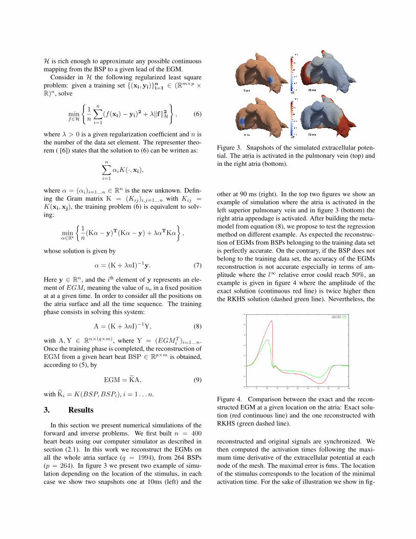
In this section we present numerical simulations of theforward and inverse problems. We first built n = 400heart beats using our computer simulator as described insection (2.1). In this work we reconstruct the EGMs onall the whole atria surface (q = 1994), from 264 BSPs(p = 264). In figure 3 we present two example of simu-lation depending on the location of the stimulus, in eachcase we show two snapshots one at 10ms (left) and the
Figure 3. Snapshots of the simulated extracellular poten-tial. The atria is activated in the pulmonary vein (top) andin the right atria (bottom).
other at 90 ms (right). In the top two figures we show anexample of simulation where the atria is activated in theleft superior pulmonary vein and in figure 3 (bottom) theright atria appendage is activated. After building the meta-model from equation (8), we propose to test the regressionmethod on different example. As expected the reconstruc-tion of EGMs from BSPs belonging to the training data setis perfectly accurate. On the contrary, if the BSP does notbelong to the training data set, the accuracy of the EGMsreconstruction is not accurate especially in terms of am-plitude where the l∞ relative error could reach 50%, anexample is given in figure 4 where the amplitude of theexact solution (continuous red line) is twice higher thenthe RKHS solution (dashed green line). Nevertheless, the
-20
-10
0
10
20
30
40
50
0 50 100 150 200 250 300 350 400 450 500
Exact solutionRKHS solution
Figure 4. Comparison between the exact and the recon-structed EGM at a given location on the atria: Exact solu-tion (red continuous line) and the one reconstructed withRKHS (green dashed line).
reconstructed and original signals are synchronized. Wethen computed the activation times following the maxi-mum time derivative of the extracellular potential at eachnode of the mesh. The maximal error is 6ms. The locationof the stimulus corresponds to the location of the minimalactivation time. For the sake of illustration we show in fig-

ure 4 a comparison of the activation times computed fromthe original signal (left) and the reconstructed (right). Theactivation site is localized with a 0.5 cm of accuracy, whichis expected since the average of the distance between thestimulus sites in the training set is 0.42 cm.
Figure 5. Comparison of the activation times: exact acti-vation times (left) and activation times reconstructed withRKHS metamodel (right).
4. Discussion
In this work we showed an original approach based ona machine learning technique in order to solve the inverseproblem in electrocardiography. This method allows solv-ing the inverse problem in real time: since the metamodelcould be build off-line, in practice we only need to eval-uate the metamodel according to equation (9). This doesnot require solving any linear system. The reconstructionof EGMs is accurate if the training data base is sufficientlyrich. The accuracy of the produced EGMs depends on howclose is the BSP from the elements of the training set. Thismeans that it is difficult to reproduce an information whichis far from the training data base. It is for instance difficultto reproduce accurate EGM for a multiple sites activationbased on a data set containing only simulation with singlesite stimulus. This limitation could be tackled by includ-ing multiple site stimulations in the training data set, at thesame time this will increase the complexity of the trainingphase. But since this phase is off line, the computationaltime of the reconstruction would be slightly affected.
5. Conclusion
In this work we have considered a machine learning ap-proach, based on the kernel ridge regression method, inorder to construct activation maps on the atria from a setof BSP measurements. The procedure has been trained us-ing synthetic data from numerical simulations, based ona 3D mathematical model of the ECG involving a math-ematical description of the electrical activity of the heartand the torso. Several examples showed the method is ableto reconstruct conveniently the EGM information included
in the training data set, and is robust when reconstruct-ing situations close to the training data set. In this studywe showed that a single site stimulus is localized with 0.5cm error. The accuracy could be improved if the trainingset is much rich. In the proposed approach, the solutiondepends on all training examples, which may be too ex-pensive. This point could be improved in future works. Infuture works this method would be trained and tested onclinical data. The ECG simulator would be used in orderto enrich the clinical data.
Acknowledgements
This work was partially supported by an ANR grant partof Investissements dAvenir program reference ANR-10-IAHU-04
References
[1] Ghosh S, Rudy Y. Application of l1-norm regularizationto epicardial potential solution of the inverse electrocardio-graphy problem. Annals of Biomedical Engineering 2009;37(5):902912.
[2] Scholkopf B, Smola AJ. Learning with Kernels: Sup-port Vector Machines, Regularization, Optimization, and Be-yond. Adaptive Computation and Machine Learning. Cam-bridge, MA, USA: MIT Press, 2001.
[3] Sundnes J, Lines G, Cai X, Nielsen B, Mardal KA, TveitoA. Computing the electrical activity in the heart. Springer-Verlag, 2006.
[4] Boulakia M, Cazeau S, Fernandez M, Gerbeau J, ZemzemiN. Mathematical modeling of electrocardiograms: a nu-merical study. Annals of biomedical engineering 2010;38(3):1071–1097. ISSN 0090-6964.
[5] Beeler G, Reuter H. Reconstruction of the action potential ofventricular myocardial fibres. J Physiol Lond 1977;268:177–210.
[6] Klepfer R, Johnson C, MacLeod R. The effects of inhomo-geneities and anisotropies on electrocardiographic fields:athree-dimensional finite element study. IEEE Eh4BC andCMBEC 1995;.
[7] Saunders C, Gammerman A, Vovk V. Ridge regression learn-ing algorithm in dual variables. In ICML. 1998; 515–521.
Address for correspondence:
Nejib ZemzemiINRIA Bordeaux Sud-Ouest, 200 rue de la vieille tour, 33405Talence [email protected]


![Object Extent Pooling for Weakly Supervised Single …...the concept of Class Activation Maps (CAM) [28] into the very first weakly-supervised ‘single-shot’ detector that does](https://img.pdfslide.us/doc/110x75/5f36b4183626d940fd2971f5/object-extent-pooling-for-weakly-supervised-single-the-concept-of-class-activation.jpg)


![Actor-centric Relation Networkopenaccess.thecvf.com/content_ECCV_2018/papers/Chen_Sun...Actor-Centric Relation Network 3 maps with classification activation mapping [63]. Figure 1](https://img.pdfslide.us/doc/110x75/5e38f85be503391a984beb8d/actor-centric-relation-actor-centric-relation-network-3-maps-with-classiication.jpg)


![arXiv:2004.10586v2 [stat.ML] 23 Apr 2020 · 2020-04-24 · Gaussian Process Manifold Interpolation for Probabilistic Atrial Activation Maps and Uncertain Conduction Velocity Sam Coveney](https://img.pdfslide.us/doc/110x75/5f34b988ff052d76eb3b8e63/arxiv200410586v2-statml-23-apr-2020-2020-04-24-gaussian-process-manifold.jpg)