Embed Size (px)
Citation preview
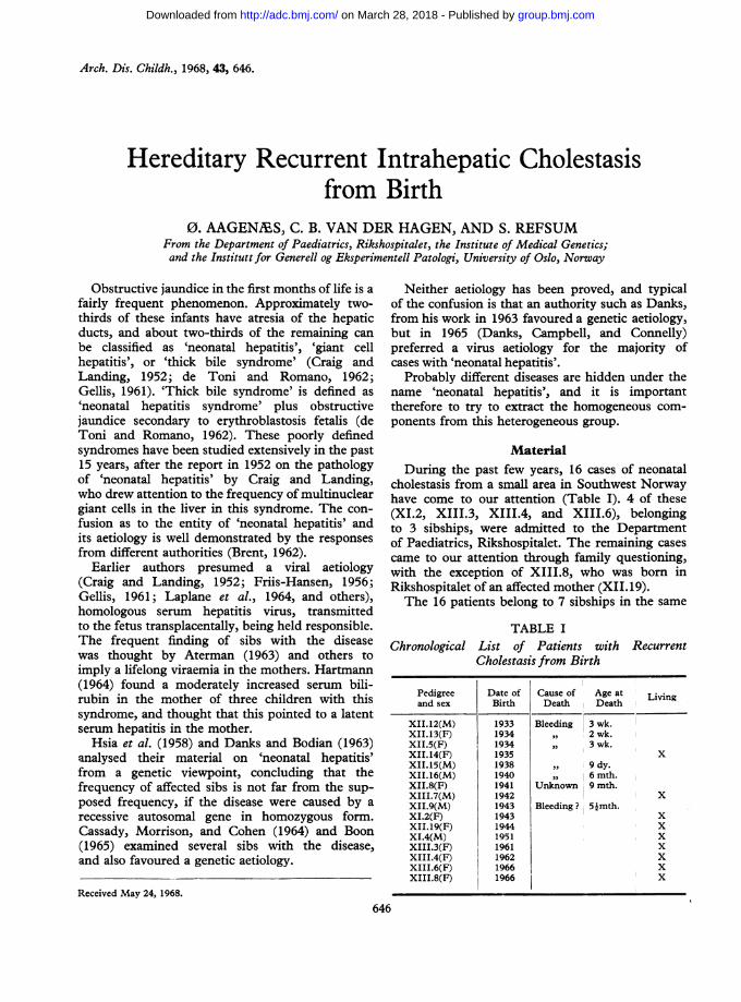
Arch. Dis. Childh., 1968, 43, 646.
Hereditary Recurrent Intrahepatic Cholestasisfrom Birth
0. AAGEN&S, C. B. VAN DER HAGEN, AND S. REFSUMFrom the Department of Paediatrics, Rikshospitalet, the Institute of Medical Genetics;and the Institutt for Generell og Eksperimentell Patologi, University of Oslo, Norway
Obstructive jaundice in the first months of life is afairly frequent phenomenon. Approximately two-thirds of these infants have atresia of the hepaticducts, and about two-thirds of the remaining canbe classified as 'neonatal hepatitis', 'giant cellhepatitis', or 'thick bile syndrome' (Craig andLanding, 1952; de Toni and Romano, 1962;Gellis, 1961). 'Thick bile syndrome' is defined as'neonatal hepatitis syndrome' plus obstructivejaundice secondary to erythroblastosis fetalis (deToni and Romano, 1962). These poorly definedsyndromes have been studied extensively in the past15 years, after the report in 1952 on the pathologyof 'neonatal hepatitis' by Craig and Landing,who drew attention to the frequency of multinucleargiant cells in the liver in this syndrome. The con-fusion as to the entity of 'neonatal hepatitis' andits aetiology is well demonstrated by the responsesfrom different authorities (Brent, 1962).
Earlier authors presumed a viral aetiology(Craig and Landing, 1952; Friis-Hansen, 1956;Gellis, 1961; Laplane et al., 1964, and others),homologous serum hepatitis virus, transmittedto the fetus transplacentally, being held responsible.The frequent finding of sibs with the diseasewas thought by Aterman (1963) and others toimply a lifelong viraemia in the mothers. Hartmann(1964) found a moderately increased serum bili-rubin in the mother of three children with thissyndrome, and thought that this pointed to a latentserum hepatitis in the mother.
Hsia et al. (1958) and Danks and Bodian (1963)analysed their material on 'neonatal hepatitis'from a genetic viewpoint, concluding that thefrequency of affected sibs is not far from the sup-posed frequency, if the disease were caused by arecessive autosomal gene in homozygous form.Cassady, Morrison, and Cohen (1964) and Boon(1965) examined several sibs with the disease,and also favoured a genetic aetiology.
Received May 24, 1968.
Neither aetiology has been proved, and typicalof the confusion is that an authority such as Danks,from his work in 1963 favoured a genetic aetiology,but in 1965 (Danks, Campbell, and Connelly)preferred a virus aetiology for the majority ofcases with 'neonatal hepatitis'.
Probably different diseases are hidden under thename 'neonatal hepatitis', and it is importanttherefore to try to extract the homogeneous com-ponents from this heterogeneous group.
MaterialDuring the past few years, 16 cases of neonatal
cholestasis from a small area in Southwest Norwayhave come to our attention (Table I). 4 of these(XI.2, XIII.3, XIII.4, and XIII.6), belongingto 3 sibships, were admitted to the Departmentof Paediatrics, Rikshospitalet. The remaining casescame to our attention through family questioning,with the exception of XIII.8, who was born inRikshospitalet of an affected mother (XII.19).The 16 patients belong to 7 sibships in the same
ChronologicalTABLE I
List of Patients with RecurrentCholestasis from Birth
Pedigree Date of Cause of Age at Livingand sex Birth Death Death
XII.12(M) 1933 Bleeding 3 wk.XII.13(F) 1934 2 wk.XII.5(F) 1934 ,, 3 wk.XII.14(F) 1935 XXII.15(M) 1938 ,, 9 dy.XII.16(M) 1940 ,, 6 mth.XII.8(F) 1941 Unknown 9 mth.XIII.7(M) 1942 XXII.9(M) 1943 Bleeding ? 5imth.XI.2(F) 1943 XXII.19(F) 1944 XXI.4(M) 1951 XXIII.3(F) 1961 XXIII.4(F) 1962 XXIII.6(F) 1966 XXIII.8(F) 1966 X
646
group.bmj.com on March 28, 2018 - Published by http://adc.bmj.com/Downloaded from
Hereditary Recurrent Intrahepatic Cholestasis from Birth
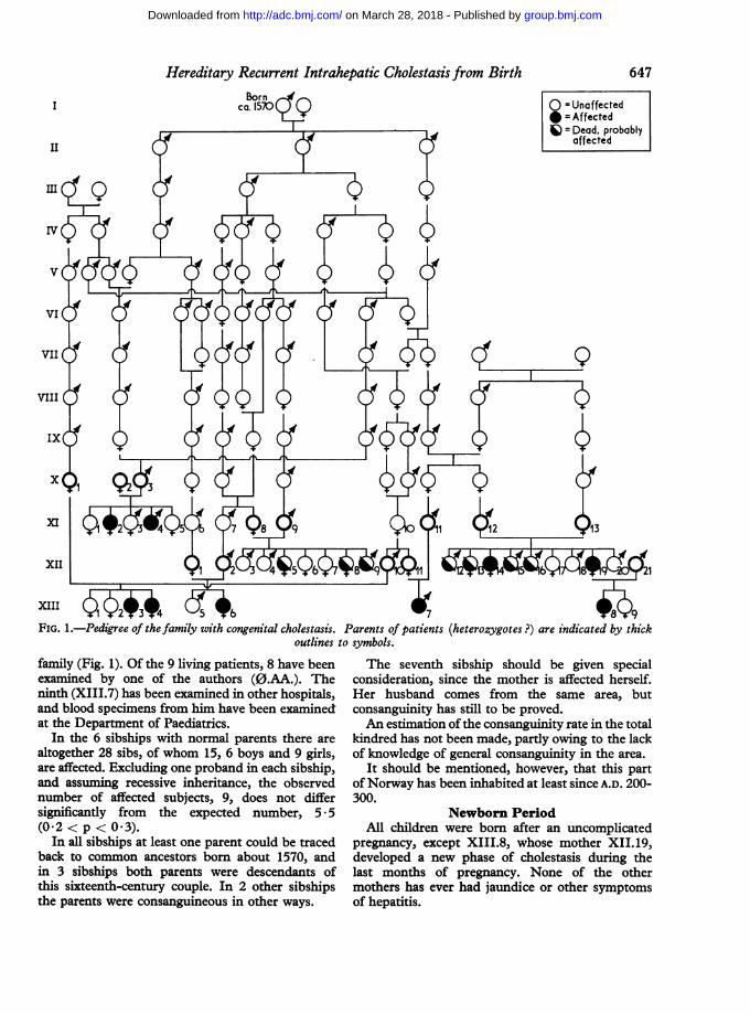
Y)1 Y( Y2'3Y4 (9J5 b *7 Y8Y9FIG. 1.-Pedigree of the family with congenital cholestasis. Parents of patients (heterozygotes ?) are indicated by thick
outlines to symbols.
family (Fig. 1). Of the 9 living patients, 8 have beenexamined by one of the authors (0.AA.). Theninth (XIII.7) has been examined in other hospitals,and blood specimens from him have been examinedat the Department of Paediatrics.
In the 6 sibships with normal parents there arealtogether 28 sibs, of whom 15, 6 boys and 9 girls,are affected. Excluding one proband in each sibship,and assuming recessive inheritance, the observednumber of affected subjects, 9, does not differsignificantly from the expected number, 5 * 5(0t2 < p < 0.3).
In all sibships at least one parent could be tracedback to common ancestors born about 1570, andin 3 sibships both parents were descendants ofthis sixteenth-century couple. In 2 other sibshipsthe parents were consanguineous in other ways.
The seventh sibship should be given specialconsideration, since the mother is affected herself.Her husband comes from the same area, butconsanguinity has still to be proved.An estimation of the consanguinity rate in the total
kindred has not been made, partly owing to the lackof knowledge of general consanguinity in the area.
It should be mentioned, however, that this partof Norway has been inhabited at least since A.D. 200-300.
Newborn PeriodAll children were born after an uncomplicated
pregnancy, except XIII.8, whose mother XII.19,developed a new phase of cholestasis during thelast months of pregnancy. None of the othermothers has ever had jaundice or other symptomsof hepatitis.
647
group.bmj.com on March 28, 2018 - Published by http://adc.bmj.com/Downloaded from
Aagenes, Van der Hagen, and RefsumDelivery was uncomplicated in all cases, and
where we know the birthweight, this was normal.All 16 children were jaundiced at birth or before1 week of age. The stools were pale shortly after thetermination of meconium, and urines were dark, butthe children remained otherwise in good condition.
XIII.8 was observed by one of the authors(0.AA.) from birth. Cord blood was examined,with the following findings: bilirubin 6 * 8 mg./100 ml. (unconj. 3-1 mg./100 ml., conj. 3-7 mg./100 ml.). Lipids: cholesterol 134 mg./100 ml.(norm. < 100 rig./100 ml.), phospholipids 172(norm. < 150 mg./100 ml.), triglycerides 146mg./100 ml. (norm. < 60 mg./100 ml.). Theapproximate normal values are taken from aSwedish source (Persson and Gentz, 1966) and anAmerican source (Kaplan and Lee, 1965). Lipo-protein electrophoresis showed a considerableincrease in f-lipoproteins, when compared with thepattern in cord blood from normal infants, where5-lipoprotein concentration is small. SGOT 91,SGPT 51 Karmen units. Protein electrophoresiswas normal. The hyperbilirubinaemia, hyper-lipaemia, and high transaminases all point to aprenatal liver disease.
Faecal fat at the age of 14 days was excreted at therate of 6 g./day (on an intake of about 10 g./day).
In the 7 children who died in infancy (Table I)the diagnosis is based on the history of jaundice,dark urine, and pale stools from shortly after birthuntil death. Three of these (XII.16, XII.8, andXII.9) were examined in local hospitals whereno important examinations were performed. Theother 4 children were seen only by the parentsand the local practitioners, but the facts were welldocumented.Of 5 children born before 1939, when vitamin K
was introduced, 4 died of haemorrhages in thefirst weeks of life and 1 is still alive.Of the next 4 children who received one dose of
vitamin K shortly after birth, 3 died between theages of 5 and 9 months, at least 1 and probably 2ofhaemorrhages.The last 7 patients with the disease, most of
whom have received vitamin K more consistently,are living; but without regular vitamin K treatmentthey have low prothrombin values and a bleedingtendency.
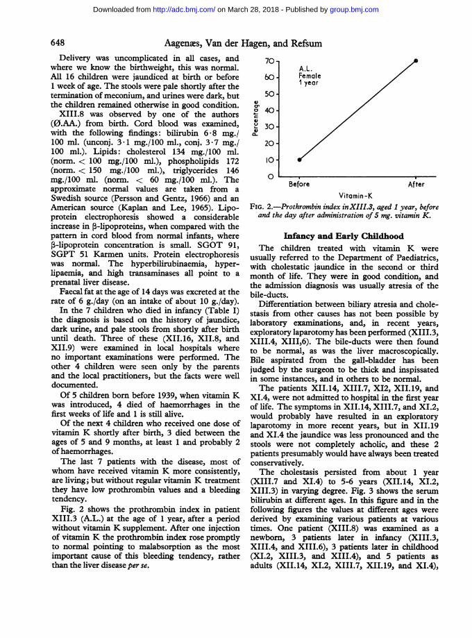
Fig. 2 shows the prothrombin index in patientXIII.3 (A.L.) at the age of 1 year, after a periodwithout vitamin K supplement. After one injectionof vitamin K the prothrombin index rose promptlyto normal pointing to malabsorption as the mostimportant cause of this bleeding tendency, ratherthan the liver disease per se.
4)cr0
a,
L-a0
70-
60.
50-
40-
30-
20 -
10-
0
A.L.Female1 year
Before AfterVitamin -K
FIG. 2.-Prothrombin index inXIII.3, aged 1 year, beforeand the day after administration of 5 mg. vitamin K.
Infancy and Early ChildhoodThe children treated with vitamin K were
usually referred to the Department of Paediatrics,with cholestatic jaundice in the second or thirdmonth of life. They were in good condition, andthe admission diagnosis was usually atresia of thebile-ducts.
Differentiation between biliary atresia and chole-stasis from other causes has not been possible bylaboratory examinations, and, in recent years,exploratory laparotomy has been performed (XIII.3,XIII.4, XIII,6). The bile-ducts were then foundto be normal, as was the liver macroscopically.Bile aspirated from the gall-bladder has beenjudged by the surgeon to be thick and inspissatedin some instances, and in others to be normal.The patients XII.14, XIII.7, XI2, XII.19, and
XI.4, were not admitted to hospital in the first yearof life. The symptoms in XII.14, XIII.7, and XI.2,would probably have resulted in an exploratorylaparotomy in more recent years, but in XII.19and XI4 the jaundice was less pronounced and thestools were not completely acholic, and these 2patients presumably would have always been treatedconservatively.The cholestasis persisted from about 1 year
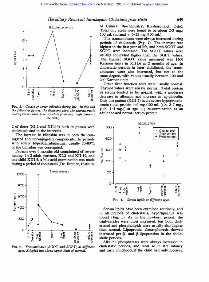
(XIII.7 and XI.4) to 5-6 years (XII.14, XI.2,XIII.3) in varying degree. Fig. 3 shows the serumbilirubin at different ages. In this figure and in thefollowing figures the values at different ages werederived by examining various patients at varioustimes. One patient (XIII.8) was examined as anewborn, 3 patients later in infancy (XIII.3,XIII.4, and XIII.6), 3 patients later in childhood(XI.2, XIII.3, and XIII.4), and 5 patients asadults (XII.14, XI.2, XIII.7, XII.19, and XI.4),
648
group.bmj.com on March 28, 2018 - Published by http://adc.bmj.com/Downloaded from
Hereditary Recurrent Intrahepatic Cholestasis from Birth
12
10-
8-
6-IE8E
cio
Bilirubin in serum
0
E
4) ao1<,4 04x)
FIG. 3.-Course of serum bilirubin during 1
the following figures, the diagrams show thcourse, rather than precise values from anv
see text.)
2 of these (XI.2 and XII.19) both in phases withcholestasis and in the intervals.The increase in bilirubin was in both the con-
jugated and unconjugated components. In periodswith severe hyperbilirubinaemia, usually 70-80%of the bilirubin was conjugated.
Patients over 6 months old complained of severe
itching. In 2 adult patients, XI.2 and XII.19, andone child XIII.8, a bile acid examination was madeduring a period of cholestasis (Dr. Bremer, Institute
1000"
800-
vs
c
400-
2001
C-'
TTransaminases
of Clinical Biochemistry, Rikshospitalet, Oslo).Total bile acids were found to be about 2-3 mg./100 ml. (normal < 0-23 mg./100 ml.).The transaminases were always increased during
periods of cholestasis (Fig. 4). The increase washighest in the first year of life, and both SGOT andSGPT were increased. The SGOT values wereusually somewhat higher than the SGPT values.The highest SGOT value measured was 1400
/.l Karmen units in XIII.6 at 2 months of age. Incholestatic periods in later childhood, the trans-aminases were also increased, but not to thesame degree, with values usually between 100 and200 Karmen units.
Other liver function tests were usually normal.Thymol values were always normal. Total protein
c ,75 in serum tended to be normal, with a moderate, decrease in albumin and increase in M2-globulin.
Only one patient (XIII.7) had a severe hypoprotein-Yfe. (In this and aemia (total protein 4 0 mg./100 ml. (alb. 2 7 mg.,te characteristic glob. 1 3 mg.)) at age 11; re-examination as ansinle Patient: adult showed normal serum protein.
500
400
E 300
8" 200-
00-
0
-o.
~-0ca _;r 1E.
0
0
x
Serum lipids*
x Cholesterolo O Triglyceridesx Phospholipids
x o
x
0
x 0
0
x
x
0
0
_~- 7-oI I
-o V Da
4-
00cX._1
FIG. 5.-Serum lipids at different ages.
4-i
0 C~~~
FIG. 4.-Transaminases (SGOT and SGPT) at differentages. Stippled line shows upper limit of normal.
Serum lipids have been examined routinely, andin all periods of cholestasis, hyperlipaemia wasfound (Fig. 5). As in the newbom period, thetriglycerides were most increased, but both chol-esterol and phospholipids were usually also higherthan normal. Lipoprotein electrophoresis showedincreased pre-,- and 5-lipoproteins in the chole-static periods.
Alkaline phosphatases were always increased incholestatic periods, and most so in late infancyand early childhood, if the child had only received
649
. . I
~ ~
group.bmj.com on March 28, 2018 - Published by http://adc.bmj.com/Downloaded from
650 ~~~~Aagenms, Van der Hagen, and Refsum
100-90-
80-
70*60.-50.
40-
30-
20-
Alkaline phosphatase
T
(6q.) (lOg.) (20g.) (3g.) (40g.)
T
1 4~~~~~1
,,'sL I~~~~
-C
On0t)
c.w
14
-.E-4j C,,. 0
E
0) ,0LC
0 04) 40: )
C.-u u-75a
7 C:C)
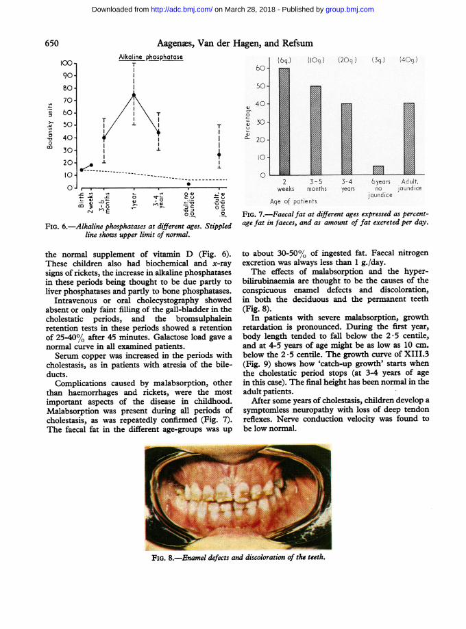
FIG. 6.-Alkaline phosphatases at different ages. Stippled
line shows upper limit of normal.
the normal supplement of vitamin D (Fig. 6).
These children also had biochemical and x-ray
signs of rickets, the increase in alkaline phosphatases
in these periods being thought to be due partly to
liver phosphatases and partly to bone phosphatases.
Intravenous or oral cholecystography showed
absent or only faint filling of the gall-bladder in the
cholestatic periods, and the bromsulphalein
retention tests in these periods showed a retention
of 25-40% after 45 minutes. Galactose load gave a
normal curve in all examined patients.
Serum copper was increased in the periods with
cholestasis, as in patients with atresia of the bile-
ducts.
Complications caused by malabsorption, other
than haemorrhages and rickets, were the most
important aspects of the disease in childhood.
Malabsorption was present during all periods of
cholestasis, as was repeatedly confirmed (Fig. 7).
The faecal fat in the different age-groups was up
:u 30.
oqL )n
10.
02 3-5 3-4 byeors .Adult,
weeks months years no jaundice
Age of patients.junk
FIG. 7.-Faecal fat at different ages expressed as percent-
age fat in faeces, and as amount of fat excreted per day.
to about 30-50% of ingested fat. Faecal nitrogen
excretion was always less than 1 g./day.
The effects of malabsorption and the hyper-
bilirubinaemia are thought to be the causes of the
conspicuous enamel defects and discoloration,
in both the deciduous and the permanent teeth
(Fig. 8).
In patients with severe malabsorption, growth
retardation is pronounced. During the first year,
body length tended to fall below the 2 -5 centile,
and at 4-5 years of age might be as low as 10 cm.
below the 2-5 centile. The growth curve of XIII.3
(Fig. 9) shows how 'catch-up growth' starts when
the cholestatic period stops (at 3-4 years of age
in this case). The final height has been normal in the
adult patients.After some years of cholestasis, children develop a
symptomless neuropathy with loss of deep tendon
reflexes. Nerve conduction velocity was found to
be low normal.
FIG. 8.-Enamel defects and discoloration of the teeth.
0On
650
tu -
group.bmj.com on March 28, 2018 - Published by http://adc.bmj.com/Downloaded from
Hereditary Recurrent Intrahepatic Cholestasis from Birth130-
- 100--Y-C 90*I,mxqr
0 2 3 4 5 6 7Age (years)
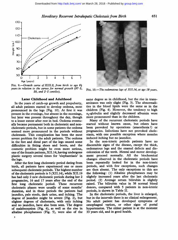
FIG. 9.-Growth curve ofXIII.3, from birth to age 64years in relation to the curves for normal growth (97.5,
50, and 2 5 centiles).
Later Childhood and AdulthoodIn the years of catch-up growth and prepuberty,
all adult patients started to develop oedema, mostpronounced in the legs (Fig. 10). At first it waspresent in the evenings, but absent in the mornings,but later was present throughout the day, thoughto a lesser extent after rest in bed. Oedema eventu-ally became permanent both in cholestatic and non-cholestatic periods, but in some patients the oedemaseemed more pronounced in the periods withoutcholestasis. This complication has been the mostsevere problem for the adult patients. The oedemain the feet and distal part of the legs caused somedifficulties in fitting shoes and boots, and thecosmetic problem might be even more serious,one ofthe female patients, XII. 14, having undergoneplastic surgery several times for 'elephantiasis' inthe legs.
After the first long cholestatic period dating frombirth, all patients have experienced one or moresubsequent cholestatic periods. The highest numberof the cholestatic periods is 5 (XII.14), while XII.19has had only 2 new cholestatic periods during her 2pregnancies, 16 and 17 years after the end of thefirst long cholestatic period. These recurrentcholestatic phases were usually of some months'duration, and in these periods the patients hadjaundice, pale stools, dark urine, and itching. Thedegree of jaundice might be severe (Fig. 2), butslighter degrees of cholestasis, with only itchingand no jaundice, have also been seen. The degreeof malabsorption (Fig. 6), as well as the rise inalkaline phosphatases (Fig. 5), were also of the
FIG. 10.-The oedematous legs of XII.14, at age 19 years.
same degree as in childhood, but the rise in trans-aminases was only slight (Fig. 3). The abnormali-ties in the blood lipids were the same as in thechildren (Fig. 4). However, the tendency to highM2-globulin and slightly decreased albumin weremore pronounced than in the children.Many of the recurrent cholestatic periods have
started without known cause, but others havebeen provoked by operations (anaesthesia?) orpregnancies. Infections have not provoked chole-stasis, with one possible exception where measlesinduced itching but no jaundice.
In the non-icteric periods patients have nodiscernible signs of the disease, except the thick,oedematous legs and the enamel defects and dis-coloration of the teeth. Mental and motor develop-ment proceed normally. All the biochemicalchanges observed in the cholestatic periods havebeen repeatedly looked for in the non-ictericperiods, and with few exceptions, such changesare then absent. The only exceptions to this arethe following: (1) Alkaline phosphatases may beslightly increased years after the last cholestaticperiod. (2) Average serum bilirubin is slightlyraised. The bilirubin value in 38 normal blooddonors, compared with 5 patients in non-ictericperiods, is shown in Table II.
In the cholestatic periods, the liver is enlarged,but in the intervals there is no clinical enlargement.No adult patient has developed symptoms ofoesophageal varices, or other signs of portalhypertension. The oldest patient is at the moment33 years old, and in good health.
651
group.bmj.com on March 28, 2018 - Published by http://adc.bmj.com/Downloaded from
Aagenxs, Van der Hagen, and RefsumTABLE II
Average Total Serum Bilirubin in 5in Phases Without Clinical Cholestasi.
and in 38 Controls
Patients in phases without clinicalcholestasis (5) ..
Parents of patients with cholestasis (13)Control material (38)
PathologyA surgical liver biopsy was taker
4, and 6, and at the ages of 5, 3i, a
respectively. A needle biopsy was aXIII.4 at the age of about 5 yeaepisode of icterus, needle biopsies w2 of the adults (XII.19 and XI.2).
Microscopically the liver biopsies frcshowed a preserved architecture. Therpartial collapse of some of the reticulslight increase in the amount of conn
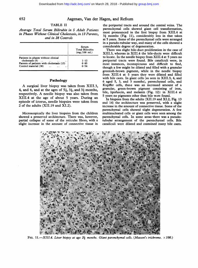
the periportal tracts and around the central veins. TheAdult Patients parenchymal cells showed giant cell transformation,in 13 Parents most pronounced in the first biopsy from XIII.4 at31 months (Fig. 11), considerably less in that taken
at 5 years. Some of the parenchymal cells were arrangedin a pseudo-tubular way, and many of the cells showed a
Serum considerable degree of degeneration.Total Bilirubin There was slight bile-duct proliferation in the case of(mg./100 ml.) XIII.3, whereas in XIII.4 the bile-ducts were difficult
to locate. In the needle biopsy from XIII.4 at 5 years no1-12 periportal tracts were found. Bile canaliculi were, in0-85 most instances, inconspicuous and difficult to find,0 54 though a few might be dilated and filled with a granular
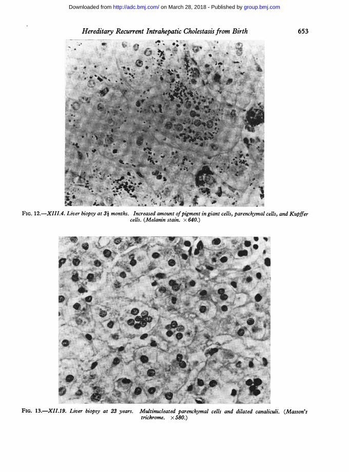
greenish-brown pigment, while in the needle biopsyfrom XIII.4 at 5 years they were dilated and filledwith bile casts. In giant cells (as seen in XIII.3, 4, and6 aged 5, 3, and 3 months), parenchymal cells, and
i from XIII 3, Kupffer cells, thete was an increased amount of a
nd 3- months' granular, green-brown pigment consisting of iron,lso taken from bile, lipofuscin, and melanin (Fig. 12): in XIII.4 at
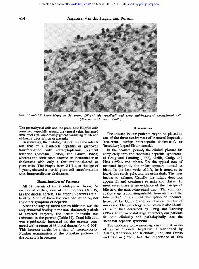
fsDroga 5 years no pigments other than bile were found.,rs. Durng an In biopsies from the adults (XII.19 and XI.2, Fig. 13tere taken from and 14) the architecture was preserved, with a slight
increase in the amount of connective tissue. Some of theparenchymal cells showed slight degeneration. A few
am the children multinucleated cells or giant cells were seen among thee was, however, parenchymal cells. In some areas there was a pseudo-at fibres, with a tubular arrangement of the parenchymal cells. BileLective tissue in canaliculi were dilated and contained many bile casts.
v~~~~~~~~~~kv4~ ~ ~ ~
Stb.* *s.4*
Jt
FIG. I11.-XIII.4. Liver biopsy at age 31 months. Giant parenchymal cells. (Masson's trichrome. x 100.)
652
group.bmj.com on March 28, 2018 - Published by http://adc.bmj.com/Downloaded from
Hereditary Recurrent Intrahepatic Cholestasis from Birth 653
..0 0
FIG. 12.-XIII.4. Liver biopsy at 3i months. Increased amount ofpigment in giant cells, parenchymal cells, and Kupifercells. (Melanin stain. x 640.)
FIG. 13.-XII.19. Liver biopsy at 23 years. Multinucleated parenchymal cells and dilated canaliculi. (Masson'strichrome. x 580.)
group.bmj.com on March 28, 2018 - Published by http://adc.bmj.com/Downloaded from
Aagenxs, Van der Hagen, and Refsum
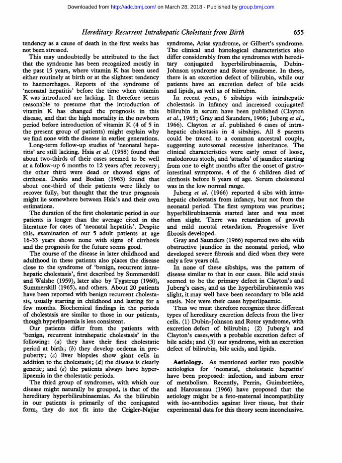
FIG. 14.-XI.2. Liver biopsy at 24 years. Dilated bile canaliculi and some multinucleated parenchymal cells.(Masson's trichrome. x 640.)
The parenchymal cells and the prominent Kupffer cellscontained, especially around the central veins, increasedamount of a yellow-brown pigment consisting of bile andwithout a trace of iron or melanin.
In summary, the histological picture in the infantswas that of a giant-cell hepatitis or giant-celltransformation with intracytoplasmic pigmentretention (Smetana, Edlow, and Glunz, 1965);whereas the adult cases showed an intracanalicularcholestasis with only a few multinucleated or
giant cells. The biopsy from XIII.4, at the age of5 years, showed a partial giant-cell transformationwith intracanalicular cholestasis.
Examination of ParentsAll 14 parents of the 7 sibships are living. As
mentioned earlier, one of the mothers (XII. 19)has the disease herself. The other 13 parents are allhealthy. None of them has ever had jaundice, nor
any other symptom of hepatitis.Since the slightly raised serum bilirubin was the
only abnormal finding in the non-cholestatic periodsof affected subjects, the serum bilirubin was
estimated in the parents (Table II). Total bilirubinwas significantly increased in the parents com-
pared with a group of 38 blood donors (p < 0 001).This increase might be a sign of heterozygosity.Further examination of the bilirubin patterns ofthe parents is in progress.
DiscussionThe disease in our patients might be placed in
one of the three syndromes: of 'neonatal hepatitis','recurrent, benign intrahepatic cholestasis', or'hereditary hyperbilirubinaemia'.
In the neonatal period, the clinical picture fitscompletely into the 'neonatal hepatitis syndrome'of Craig and Landing (1952), Gellis, Craig, andHsia (1954), and others. 'In the typical case ofneonatal hepatitis, the infant appears normal atbirth. In the first weeks of life, he is noted to beicteric, his stools pale, and his urine dark. The liverbegins to enlarge. Usually the infant does notappear ill and continues to gain and thrive. Inmost cases there is no evidence of the passage ofbile into the gastro-intestinal tract. The conditionat this stage is indistinguishable from atresia of thebile ducts.' This clinical description of 'neonatalhepatitis' by Gellis (1961) is identical to that ofour cases. The pathology in our cases is also identi-cal with that described by Craig and Landing(1952). In the neonatal stage, therefore, our patientsfit both clinically and pathologically into the'neonatal hepatitis syndrome'.The tendency to haemorrhages in the first weeks
of life in 'neonatal hepatitis' is mentioned byAdams, Anderson, and Richdorf (1952) and Danksand Bodian (1963), but the importance of this
654
group.bmj.com on March 28, 2018 - Published by http://adc.bmj.com/Downloaded from
Hereditary Recurrent Intrahepatic Cholestasis from Birthtendency as a cause of death in the first weeks hasnot been stressed.
This may undoubtedly be attributed to the factthat the syndrome has been recognized mostly inthe past 15 years, where vitamin K has been usedeither routinely at birth or at the slightest tendencyto haemorrhages. Reports of the syndrome of'neonatal hepatitis' before the time when vitaminK was introduced are lacking. It therefore seemsreasonable to presume that the introduction ofvitamin K has changed the prognosis in thisdisease, and that the high mortality in the newbornperiod before introduction of vitamin K (4 of 5 inthe present group of patients) might explain whywe find none with the disease in earlier generations.Long-term follow-up studies of 'neonatal hepa-
titis' are still lacking. Hsia et al. (1958) found thatabout two-thirds of their cases seemed to be wellat a follow-up 6 months to 12 years after recovery;the other third were dead or showed signs ofcirrhosis. Danks and Bodian (1963) found thatabout one-third of their patients were likely torecover fully, but thought that the true prognosismight lie somewhere between Hsia's and their ownestimations.The duration of the first cholestatic period in our
patients is longer than the average cited in theliterature for cases of 'neonatal hepatitis'. Despitethis, examination of our 5 adult patients at age16-33 years shows none with signs of cirrhosisand the prognosis for the future seems good.The course of the disease in later childhood and
adulthood in these patients also places the diseaseclose to the syndrome of 'benign, recurrent intra-hepatic cholestasis', first described by Summerskilland Walshe (1959), later also by Tygstrup (1960),Summerskill (1965), and others. About 20 patientshave been reported with benign recurrent cholesta-sis, usually starting in childhood and lasting for afew months. Biochemical findings in the periodsof cholestasis are similar to those in our patients,though hyperlipaemia is less consistent.Our patients differ from the patients with
'benign, recurrent intrahepatic cholestasis' in thefollowing: (a) they have their first cholestaticperiod at birth; (b) they develop oedema in pre-puberty; (c) liver biopsies show giant cells inaddition to the cholestasis; (d) the disease is clearlygenetic; and (e) the patients always have hyper-lipaemia in the cholestatic periods.The third group of syndromes, with which our
disease might naturally be grouped, is that of thehereditary hyperbilirubinaemias. As the bilirubinin our patients is primarily of the conjugatedform, they do not fit into the Crigler-Najjar
syndrome, Arias syndrome, or Gilbert's syndrome.The clinical and histological characteristics alsodiffer considerably from the syndromes with heredi-tary conjugated hyperbilirubinaemia, Dubin-Johnson syndrome and Rotor syndrome. In these,there is an excretion defect of bilirubin, while ourpatients have an excretion defect of bile acidsand lipids, as well as of bilirubin.
In recent years, 6 sibships with intrahepaticcholestasis in infancy and increased conjugatedbilirubin in serum have been published (Claytonet al., 1965; Gray and Saunders, 1966; Juberg et al.,1966). Clayton et al. published 6 cases of intra-hepatic cholestasis in 4 sibships. All 8 parentscould be traced to a common ancestral couple,suggesting autosomal recessive inheritance. Theclinical characteristics were early onset of loose,malodorous stools, and 'attacks' of jaundice startingfrom one to eight months after the onset of gastro-intestinal symptoms. 4 of the 6 children died ofcirrhosis before 8 years of age. Serum cholesterolwas in the low normal range.
Juberg et al. (1966) reported 4 sibs with intra-hepatic cholestatis from infancy, but not from theneonatal period. The first symptom was pruritus;hyperbilirubinaemia started later and was mostoften slight. There was retardation of growthand mild mental retardation. Progressive liverfibrosis developed.Gray and Saunders (1966) reported two sibs with
obstructive jaundice in the neonatal period, whodeveloped severe fibrosis and died when they wereonly a few years old.
In none of these sibships, was the pattern ofdisease similar to that in our cases. Bile acid stasisseemed to be the primary defect in Clayton's andJuberg's cases, and as the hyperbilirubinaemia wasslight, it may well have been secondary to bile acidstasis. Nor were their cases hyperlipaemic.Thus we must therefore recognize three different
types of hereditary excretion defects from the livercells. (1) Dubin-Johnson and Rotor syndrome, withexcretion defect of bilirubin; (2) Juberg's andClayton's cases,with a probable excretion defect ofbile acids; and (3) our syndrome, with an excretiondefect of bilirubin, bile acids, and lipids.
Aetiology. As mentioned earlier two possibleaetiologies for 'neonatal, cholestatic hepatitis'have been proposed: infection, and inborn errorof metabolism. Recently, Perrin, Guimbretiere,and Harousseau (1966) have proposed that theaetiology might be a feto-maternal incompatibilitywith iso-antibodies against liver tissue, but theirexperimental data for this theory seem inconclusive.
655
group.bmj.com on March 28, 2018 - Published by http://adc.bmj.com/Downloaded from
Aagenes, Van der Hagen, and RefsumThe occurrence of 16 well-defined cases of this
rare disease in 7 sibships of the same kindred isindicative of a genetic aetiology, and the segrega-tion is wholly consistent with an autosomal recessivemode of inheritance. All parents except one areunaffected by the disease, and it is reasonable toassume that they are heterozygotes for the gene.The one affected subject, who has conceived,
produced one affected and one unaffected child;this would be expected assuming she is homozygousand her husband heterozygous for the gene.
It might be argued that in an isolated communitysuch as this, the chances of finding consanguinityover 10 to 13 generations are bound to be high.On the other hand, our cases do not all live in thesame area today, nor were all disclosed throughfamily questioning. Moreover, the area has beeninhabited for almost 2000 years, so that it is hardlylikely that all or nearly all of the inhabitants aredescended from one ancient pair of settlers.
Pathogenesis and pathology. The recessiveautosomal inheritance is suggestive of a bio-chemical defect, but no specific defect has beendetected. A possible biochemical defect mightcause a general metabolic block, a localized ex-cretion defect in the liver, or a structural changeof the liver architecture.
Smetana et al. (1965) have suggested that as thegiant cells fill the entire space between the sinusoids,no normal bile canaliculi can be formed betweenthe parenchymal cells. Both the conjugated bileand other pigments normally excreted with thebile are therefore retained intracytoplasmicallyin the parenchymal cells. Due to the increasingamount of pigments within the parenchymal cells,these will eventually degenerate and die, withliberation of the pigments to macrophages andKupffer cells. If the giant cell transformation istotal, degenerated parenchymal cells will not bereplaced; the structure will collapse and be replacedby reticular fibres and fibroblasts. Such a situationwill usually lead to death within the first 6 to 8months. With a partial giant cell transformation,the degenerated parenchymal cells may be replacedby normal parenchymal cells which have the abilityto form normal bile canaliculi. Provided there areno other obstructions in the biliary tree (no biliaryatresia), both the bile and the other pigments maybe excreted. The liver may therefore return tonearly normal structure; there may be some residualcollapse of the structure with an increased amountof reticular and fibrocytic fibres, but no liver cir-rhosis.The replacement of giant cells by normal paren-
chymal cells seems, in our cases, to be a rather slowand incomplete process, inasmuch as we still findsome giant cells in the adult livers. Though bilecanaliculi have been formed, there still persists arecurrent defect in the bile excretion, with intra-canalicular and intracellular bile retention as theresult.The cause of this defect in the bile excretion,
which seems to affect all components of the bile, isunknown.
It is impossible to differentiate histologically thegiant cell transformation in the livers in our patientsfrom that found in other liver disorders in theneonatal period. The differentiation from otherintrahepatic cholestatic conditions in adult livers(Summerskill, 1965) is also difficult, though giantcells in the liver in the Summerskill type of chole-stasis (Williams et al., 1964) have not been reported.
SummarySixteen cases of neonatal cholestasis, all related,
with consanguinity in 6 of 7 parental couplesare reported. The children had an obstructive typeof jaundice from the first week of life.The jaundice lasted from about 1 to 6 years, and
in this period the patients complained of severeitching. Other symptoms, such as bleeding ten-dency, rickets, anaemia, and growth retardationwere ascribed to malabsorption.
Seven children died in infancy, at least 5 as aresult of haemorrhages, but no death from haemor-rhage has occurred since vitamin K has been regu-larly used.
Laboratory examinations showed hyperbili-rubinaemia (conjugated bilirubin), increasedtransaminases and alkaline phosphatases, hyper-lipaemia, and increase in pre-p and 5-lipoproteins,slight decrease in albumin, and increase in a2-globulin.
After the end of the cholestatic period, the child-ren started to regain normal stature, and adultheight was normal.From prepuberty patients tended to develop
oedema of the legs, for no known reason.One or more further periods of cholestasis
have recurred in all adult patients.Liver histology showed 'giant cell hepatitis',
but this did not progress to fibrosis.The parents showed a slight but significant
increase in serum bilirubin, possible evidence ofheterozygosity.The term 'hereditary, recurrent, intrahepatic
cholestasis from birth' is proposed for this newinborn error of metabolism.
656
group.bmj.com on March 28, 2018 - Published by http://adc.bmj.com/Downloaded from
Hereditary Recurrent Intrahepatic Cholestasis from Birth 657We are grateful to Mr. Ola Knutrud who performed
the laparotomies, to Drs. Helge Sigstad and Egil Gjonefor examination of the adult patients and for the needlebiopsies, to Dr. Georg Omland for the ambulatoryspecimen taken, to Drs. Jorgen Fog and Arne Bakkenfor the bilirubin examinations and for many fruitfuldiscussions, and to Dr. Per Seland for supporting uswith his knowledge of local genealogy.This study was supported by a grant from Nordisk
Insulinfond.
REFERENCES
Adams, F. H., Anderson, R. C., and Richdorf, L. F. (1952). Foursiblings with hepatic disease leading to cirrhosis. Amer. J. Dis.Child., 84, 168.
Aterman, K. (1963). Neonatale hepatitis and its relation to viralhepatitis of mother. ibid., 105, 395.
Boon, J. M. (1965). Neonatale hepatitis of familiale neonatalestuwingsicterus. Maandschr. Kindergeneesk., 33, 465.
Brent, R. L. (1962). Persistent jaundice in infancy. J. Pediat.,61, 111.
Cassady, G., Morrison, A. B., and Cohen, M. M. (1964). Familial'Giant-cell hepatitis' in infancy. Amer. J3. Dis. Child., 107, 456.
Clayton, R. J., Iber, F. L., Ruebner, B. H., and McKusick, V. A.(1965). Byler's disease: fatal familial intrahepatic cholestasisin an Amish kindred. J. Pediat., 67, 1025.
Craig, J. M., and Landing, B. H. (1952). Form of hepatitis inneonatal period simulating biliary atresia. Arch. Path., 54, 321.
Danks, D., and Bodian, M. (1963). A genetic study of neonatalobstructive jaundice. Arch. Dis. Childh., 38, 378.
-, Campbell, P. E., and Connelly, J. F. (1965). An aetiologicalstudy of neonatal jaundice in a children's hospital. Aust.paediat.J., 1, 193.
Friis-Hansen, B. (1956). Neonatal hepatitis with osteomalacia.Acta paediat. (Uppsala), 45, 376.
Gellis, S. S. (1961). Current problems in liver disease in infancyand childhood. Neonatal hepatitis. Progr. Liver Dis., 1, 61.
-, Craig, J. M., and Hsia, D. Y. -Y. (1954). Prolonged obstructivejaundice in infancy. IV. Neonatal hepatitis. Amer. Jt. Dis.Child., 88, 285.
Gray, O. P., and Saunders, R. A. (1966). Familial intrahepaticcholestatic jaundice in infancy. Arch. Dis. Childh., 41, 320.
Hartmann, W. (1964). Zur Kenntnis der Neugeborenenhepatitis(Riesenzellenhepatitis). Z. Kinderheilk., 89, 146.
Hsia, D. Y. -Y., Boggs, J. D., Driscoll, S. G., and Gellis, S. S.(1938). Prolonged obstructive jaundice in infancy. V. Thegenetic components in neonatal hepatitis. Amer. J. Dis. Child.,95, 485.
Juberg, R. C., Holland-Moritz, R. M., Henley, K. S., and Gonzalez,C. F. (1966). Familial intrahepatic cholestasis with mentaland growth retardation. Pediatrics, 38, 819.
Kaplan, A., and Lee, V. F. (1965). Serum lipid levels in infants andmothers at parturition. Clin. chim. Acta, 12, 258.
Laplane, R., Graveleau, D., Lods, C., and Noum, I. (1964). Leshepatites n6onatales familiales. Pediatrie, 19, 217.
Perrin, D., Guimbretiere, J., and Harousseau, H. (1966). Unenouvelle forme d'incompatibilite foeto-maternelle: l'hepatiteneo-natale familiale. Presse med., 74, 1307.
Persson, B., and Gentz, J. (1966). The pattern of blood lipids,glycerol and ketone bodies during the neonatal period, infancyand childhood. Acta paediat. (Uppsala), 55, 353.
Smetana, H. F., Edlow, J. B., and Glunz, P. R. (1965). Neonataljaundice. Arch. Path., 80, 553.
Summerskill, W. H. J. (1965). The syndrome of benign recurrentcholestasis. Amer. J3. Med., 38, 298.
- , and Walshe, J. M. (1959). Benign recurrent intrahepatic'obstructive' jaundice. Lancet, 2, 686.
de Toni G., and Romano, C. (1962). La sindroma da bile spessanel primo trimestre di vita. Minerva pediat., 14, 1429.
Tygstrup, N. (1960). Intermittent possibly familial intrahepaticcholestatic jaundice. Lancet, 1, 1171.
Williams, R., Cartter, M. A., Sherlock, S., Scheuer, P. J., and Hill,K. R. (1964). Idiopathic recurrent cholestasis: a study ofthe functional and pathological lesions in four cases. Quart. J3.Med., n.s 33,387.
group.bmj.com on March 28, 2018 - Published by http://adc.bmj.com/Downloaded from
cholestasis from birth.Hereditary recurrent intrahepatic
O. Aagenaes, C. B. van der Hagen and S. Refsum
doi: 10.1136/adc.43.232.6461968 43: 646-657 Arch Dis Child
http://adc.bmj.com/content/43/232/646.citationUpdated information and services can be found at:
These include:
serviceEmail alerting
online article. article. Sign up in the box at the top right corner of the Receive free email alerts when new articles cite this
Notes
http://group.bmj.com/group/rights-licensing/permissionsTo request permissions go to:
http://journals.bmj.com/cgi/reprintformTo order reprints go to:
http://group.bmj.com/subscribe/To subscribe to BMJ go to:
group.bmj.com on March 28, 2018 - Published by http://adc.bmj.com/Downloaded from


![Lecture - Birth · 2015. 10. 28. · [Expand] [Expand] Historic model of birth MRI of birth today Lecture - Birth From Embryology Introduction This lecture will finish prenatal human](https://img.pdfslide.us/doc/110x75/60a744b45ac90746d07b5138/lecture-birth-2015-10-28-expand-expand-historic-model-of-birth-mri-of.jpg)