Embed Size (px)
Citation preview
July 2008 11−−Frank William Ernest Gibson. 22 July 1923
A. J. Pittard and G. B. Cox
published online April 28, 2010Biogr. Mems Fell. R. Soc.
Supplementary data
/27/rsbm.2009.0020.DC1http://rsbm.royalsocietypublishing.org/content/suppl/2010/04"Data Supplement"
P<Pjournal. Published online 28 April 2010 in advance of the print
Errata
/content/57/463.1.full.pdf or: next pagesee
An erratum has been published regarding this article. Please
Email alerting service
hereor click sign up in the box at the top right-hand corner of the article Receive free email alerts when new articles cite this article -
and date of initial publication. Citations to Advance online articles must include the digital object identifier (DOIs)establish publication priority; they are indexed by PubMed from initial publication. when available prior to final publication). Advance online articles are citable andhave not yet appeared in the paper journal (edited, typeset versions may be posted Advance online articles have been peer reviewed and accepted for publication but
on July 8, 2018http://rsbm.royalsocietypublishing.org/Downloaded from on July 8, 2018http://rsbm.royalsocietypublishing.org/Downloaded from on July 8, 2018http://rsbm.royalsocietypublishing.org/Downloaded from
Frank William ErnEst Gibson22 July 1923 — 11 July 2008
Biogr. Mems Fell. R. Soc.
on July 8, 2018http://rsbm.royalsocietypublishing.org/Downloaded from
on July 8, 2018http://rsbm.royalsocietypublishing.org/Downloaded from
Frank William ErnEst Gibson
22 July 1923 — 11 July 2008
Elected Frs 1976
By A. J. PittArd1 AM FAA And G. B. Cox2 FAA
1Department of Microbiology and Immunology, University of Melbourne, Parkville, Victoria 3010, Australia
2The John Curtin School of Medical Research, GPO Box 334, Canberra City, ACT 2601, Australia
Frank Gibson rose from humble beginnings to become one of the most respected bacterial physiologists of his era. His identification of the elusive branch-point compound in the path-way of aromatic biosynthesis served as an initiation point for a sustained period of investiga-tion in which the genes, enzymes and intermediates in the various pathways to phenylalanine and tyrosine, the quinones, enterochelin and 4-aminobenzoate were identified and examined in detail. studies on the function of ubiquinone led to an examination of oxidative phospho-rylation and to the F1F0-atPase of the bacterium Escherichia coli. With Graeme Cox he established a group of researchers who in the 1980s applied the various techniques of micro-bial genetics to construct a molecular profile of the proteins, which constituted the F0 mem-brane-embedded part of the F1F0-atPase. this work resulted in the formulation in 1986 of a rotational model and the identification of several residues that could comprise a pore through which the protons, which drove the rotation, could pass. He trained many research students during his lifetime and was an exemplary role model. outside the laboratory he lived a full life, being an ardent skier, scuba diver and tennis player to name but a few of his pursuits. He is survived by his wife robin and their son, mark; by Frances, the daughter from his first marriage; and by grandchildren teresa, luke and simon. His first wife, margaret, and their second daughter, ruth (mother of teresa, luke and simon), are both deceased.
doi:10.1098/rsbm.2009.0020 3 this publication is © 2010 the royal society
a similar memoir appears in Historical Records of Australian Science, June 2010.
on July 8, 2018http://rsbm.royalsocietypublishing.org/Downloaded from
4 Biographical Memoirs
EArly dAys
Frank William Ernest Gibson was born in melbourne on 22 July 1923 to John William (bill) Gibson and alice ruby Gibson (née Hancock). He was the eldest of three children, having two younger sisters. He described himself as a third-generation australian of irish and scottish extraction. His father was employed by the adelaide steamship Company and eventually became a foreman stevedore working on coal ships. Frank had a happy childhood and remembered very supportive parents who shielded the children from the worst effects of the Great Depression. in a typical Frank understatement he once said that the only hardship that he remembered was eating bread and dripping and having cardboard in his shoes to cover up the holes.
He attended a public primary school and then went to Collingwood technical school, where it was planned that he should become a draftsman. after two years he decided that drafting was not his thing and at the age of 14 years he left school and looked for work. He was fortunate to obtain a position as junior technician in the bacteriology Department at the University of melbourne. While he learnt how to wash and plug test-tubes and make media, he attended evening classes in a chemistry diploma course that included scientific German and leaving Certificate English. after three years he had passed the early stages of his apprenticeship in the department and graduated to the research laboratory of Dr (later Professor) syd rubbo. the head of department, Harold Woodruff, and syd rubbo both encouraged Frank to consider tak-ing a university course but his qualifications at the time would not have allowed him to gain entrance to melbourne University. in 1940 the introduction of a new course in bacteriology at the University of Queensland provided Frank with the opportunity that he needed. Dr (later Professor) D. F. Gray, who was based in the Veterinary school, was to run the course and he needed a technician. Frank was appointed and, with David Gray in charge, worked hard on this new course. the University of Queensland had a more helpful attitude towards his qualifications and decided that if he completed chemistry and physics at matriculation level they would accept his other studies as having provided the necessary requirements for matriculation. Furthermore, the university was prepared to pay his fees. after one year of night school he was able to fulfil all the requirements and then began a science course, again at night school. by the end of 1946 he had completed two years of the three-year bsc degree. as it turned out he had also spent 12 months in the army before the university successfully recalled him to the medical school. at the beginning of 1947 he returned to the bacteriology Department at melbourne University and on the basis of his studies in Queensland was accepted into the third year of a bsc, which he completed at the end of 1948. For the next two years he was employed as a demonstrator (later temporary lecturer) and given the opportunity to start some research on the role of acridines as antibacterials. this led to his first publication, which may subsequently have been of significant importance in winning a scholarship to proceed to a DPhil (1)*.
in 1949 he married margaret burvill, who was working with syd rubbo and adrian albert on the mode of action of acridines and 8-hydroxyquinoline. the australian national University (anU) had just been established in Canberra and although they were not yet accepting PhD students they had a generous postgraduate scholarship scheme that enabled successful students to undertake postgraduate studies in britain. Frank applied for one of these but was initially unsuccessful. However, to his surprise and delight, not long after receiving a letter rejecting his application he received another reversing the first and offering him a scholarship to work for
* numbers in this form refer to the bibliography at the end of the text.
on July 8, 2018http://rsbm.royalsocietypublishing.org/Downloaded from
Frank William Ernest Gibson �
his DPhil at oxford with D. D. Woods (Frs 19�2), who was well known for his discoveries in elucidating the mode of action of the sulphonamides. in oxford, margaret obtained a grant to study with sir Cyril Hinshelwood Frs (Prs 19��–60). For his DPhil, Frank studied the biosyn-thesis of methionine by using washed cell suspensions of various mutants of E. coli. With this approach he was able to show that serine was the source of the methyl group of methionine (3). Unfortunately this work, apart from a brief abstract, was not published for about seven years, by which time it had been generally accepted that serine was the source of the methyl group of methionine. before leaving oxford, Frank received two offers of academic positions. one was a fellowship in the microbiology school at the anU and the other was a senior lectureship in the bacteriology Department at melbourne University, where s. D. rubbo was now head of depart-ment. Frank chose melbourne and took up his position in 19�3.
rEturn to AustrAliA
as a senior lecturer he taught a course on bacterial physiology to third-year students, and one on general microbiology to second-year students. He was an excellent teacher and was very popular with students. He also established a research laboratory applying the methodology that he had used at oxford, namely the use of washed cell suspensions of mutant strains of Aerobacter aerogenes and E. coli bacteria to further elucidate reactions and intermediates in the pathway of biosynthesis of the aromatic amino acids.
the laboratory equipment was fairly primitive in those days. the laboratory featured a Unicam manual spectrophotometer (to be replaced later by the Carey automatic machine), a sonicator and a Hughes press for breaking cells, two centrifuges (down the hall), a rotary evaporator that worked off the suction provided by the water taps and filled with water every time the pressure fluctuated, and several glass battery jars that Frank had scrounged from somewhere and were used for paper chromatography. Even though several highly toxic sol-vents such as benzene, pyridine and toluene were used in the chromatography, when the run was finished the paper chromatograms were removed from the jars and attached to a piece of string strung across the laboratory to dry. it is remarkable that the inhabitants of the laboratory seem to have survived these noxious fumes without any adverse effects.
Early studies with Colin Doy resulted in the identification of a new compound, namely 1-(o-carboxyphenylamino-1-deoxyribulose), accumulated by certain tryptophan auxotrophs. subsequent work showed this to be a dephosphorylated form of the true intermediate, now known as CDrP (2).
in 19�9 Frank received a travel grant from the Carnegie Foundation of new York that enabled him and margaret to travel to the Usa, where they worked with Charley Yanofsky (Formemrs 198�) at stanford University. at the same time Frank also attended C. b. van niel’s famous summer course in general microbiology held at the Johns Hopkins marine station each year. this experience greatly inspired his teaching of general microbiology on his return to melbourne. the work with Yanofsky involved a study of cell-free extracts of E. coli to establish whether one or more enzymes were involved in the conversion of CDrP to indoleglycerol phosphate. Frank achieved a partial purification of indoleglycerol phosphate synthetase and showed that a single protein was involved in the reaction (4).
on his return to australia after his study leave, Frank was promoted to reader in Chemical microbiology, and the laboratory switched from whole-cell suspensions to cell-free extracts
on July 8, 2018http://rsbm.royalsocietypublishing.org/Downloaded from
6 Biographical Memoirs
and also focused on the one remaining unsolved problem of the pathway, namely the position and the nature of the hypothetical branch-point compound. all the work leading to the discov-ery of this intermediate was performed in the old bacteriology school.
ChorisMiC ACid, thE BrAnCh-Point CoMPound
other workers had shown that the biosynthesis of the aromatic amino acids proceeded via a set of common reactions (the common pathway), with three terminal pathways leading to the amino acids tryptophan, phenylalanine and tyrosine. a compound referred to as ‘Z1 phosphate’ (3-enolpyruvylshikimate �-phosphate) and formed from shikimate �-phosphate was the last intermediate to be identified in the common pathway.
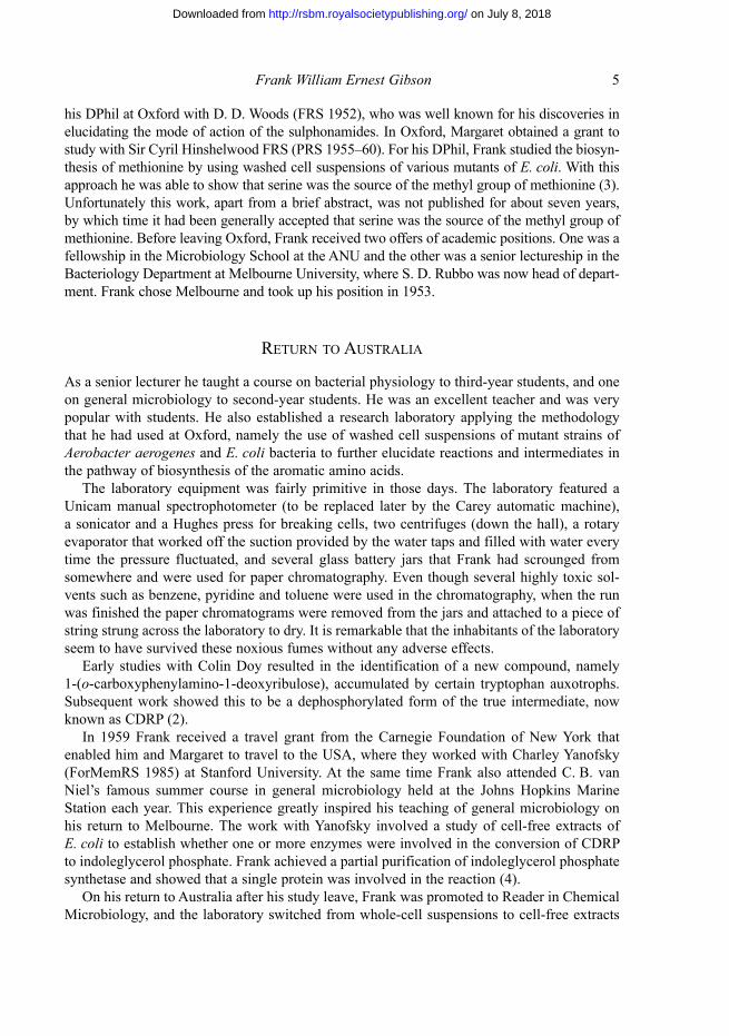
one proposal current at the time was that the tryptophan pathway diverged at shikimate �-phosphate and that the phenylalanine and tyrosine pathways diverged at Z1 phosphate (sprinson 1960). an alternative model suggested by Davis was that all three pathways diverged from the same compound, but whether it was Z1 phosphate or some unknown compound beyond this was not known (see figure 1) (7).
Doy and Gibson investigated these models in one final experiment with washed cell suspensions in which cells of a tryptophan auxotroph whose synthetic pathway was blocked after anthranilic acid were grown in a nitrogen-rich medium and then transferred to a nitro-gen-free medium. after incubation, the supernatants were examined for accumulated prod-ucts. it was postulated that in the nitrogen-free medium the intermediate directly preceding anthranilic acid might accumulate. the experiment revealed that under these conditions three major aromatic compounds accumulated: 4-hydroxybenzaldehyde, phenylpyruvic acid and 4-hydroxyphenylpyruvic acid. because the two phenylpyruvic acids were the first known intermediates in the terminal pathways to phenylalanine and tyrosine it was argued, correctly as it turned out, that when the tryptophan pathway was shut off because of the absence of a nitrogen source, the precursor of anthranilic acid was directed along the termi-nal pathways to phenylalanine and tyrosine. although the hypothetical compound was not
Z1 phosphate Prephenic acid
tyrosine
phenylalanine
Anthranilic acid
tryptophan
Shikimic acid-5-
phosphate
(Sprinson)
Shikimic acid-5-
phosphate
Z1 phosphate
tyrosine
phenylalanine
tryptophan(Davis)
Z phosphate Prephenic acid Prephenic acid
tyrosine
Prephenic acid
phenylalanine
Anthranilic acid
tryptophan
Z phosphate
tyrosine
phenylalanine phosphate phosphate phenylalanine phosphate
tryptophan
phosphate phenylalanine
Figure 1. the two schemes of sprinson and Davis for the branching of the aromatic pathway. in the Davis scheme the branch point could be at Z1 phosphate or an unknown compound beyond this point.
on July 8, 2018http://rsbm.royalsocietypublishing.org/Downloaded from
Frank William Ernest Gibson 7
identified, the results offered support for the Davis model and also provided some direction for the next critical experiment.
before taking steps to block the conversion of the hypothetical branch-point compound to the phenylpyruvic acids, experiments were undertaken to establish conditions under which cell-free extracts could convert shikimic acid (a readily available substrate) into anthranilic acid, phenylpyruvic acid and 4-hydroxyphenylpyruvic acid. During these experiments phosphoenol pyruvic acid, which is required for the formation of Z1 phosphate, was shown to stimulate the formation of anthranilic acid as well as the phenylpyruvic acids; this observa-tion added further support for the Davis model (9). in a further refinement of these studies, by using cell-free extracts from mutants blocked in successive reactions in the common pathway, it was possible to separate the overall conversion into two discrete steps. in the first, using extracts of A. aerogenes a170-44, which was blocked in the reaction immediately after Z1 phosphate, shikimic acid could be converted to a compound with all the characteristics of Z1 phosphate. in the second, using extracts from A. aerogenes poly 3, which was blocked between shikimate �-phosphate and Z1 phosphate, this compound could be converted to anthranilic acid and to a lesser extent to phenylpyruvic acid and 4-hydroxyphenylpyruvic acid. this second reaction required DPnH and was postulated to involve the production of the hypothetical branch-point compound (7). starting with a tryptophan auxotroph of A. aerogenes unable to convert anthranilic acid to tryptophan, a strategy was developed to block reactions converting the hypothetical branch-point compound to phenylpyruvic acid and 4-hydroxyphenylpyruvic acid. this involved irradiating the tryptophan auxotroph and isolating a mutant that now required both tryptophan and tyrosine for growth. this strain was further irradiated and a second strain was isolated that now required tryptophan, phenyl-alanine and tyrosine for growth. the observation that this mutant was still able to accumulate anthranilic acid confirmed that the common pathway was still intact and distinguished it from several other mutants that had acquired a requirement for phenylalanine because of a second block in the common pathway. the triple mutant was called 62-1. Cell-free extracts of this strain prepared from cells that had been grown in limiting tryptophan could readily convert shikimic acid to anthranilic acid. When Frank and his colleagues omitted glutamine from the reaction mixture, to their great excitement a new compound was formed. this compound, which seemed to be the hypothetical branch-point compound formerly called compound X, could be extracted by ethyl acetate or ether after acidification and used as a substrate with extracts of a170-44, which were able to convert it in the presence of glutamine to anthranilic acid, and in the presence of an oxidized form of diphosphopyridine nucleotide (DPn+; now known as nicotinamide adenine dinucleotide, naD+) it was converted into phenylpyruvic acid and 4-hydroxyphenylpyruvic acid. on more prolonged incubation, extracts of 62-1 were also able to convert shikimic acid to 4-hydroxybenzoic acid (6, 11, 13). after consultation with Frank’s father-in-law, archdeacon W. burvill, who was an accomplished Greek scholar, the name chorismic acid (meaning separation) was proposed for the elusive compound X (�1). attempts to isolate quantities of this compound in a pure state involved what Frank later described as several esoteric and potentially hazardous purification techniques such as chromatography in ether on columns of sucrose. as he stated, these were performed under conditions that would give a present-day safety officer nightmares. in the end, chorismic acid was shown to be less labile than had originally been feared; a relatively simple procedure was developed using ion-exchange columns in the cold and subsequent precipitation in methanol as a barium salt (11).
on July 8, 2018http://rsbm.royalsocietypublishing.org/Downloaded from
8 Biographical Memoirs
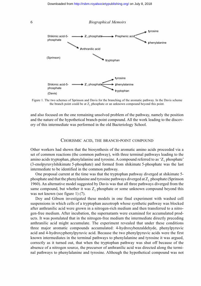
the hypothetical structure that had been proposed for chorismic acid was confirmed and the stereochemistry defined when lloyd Jackman, a new professor of chemistry at melbourne University, ran a nuclear magnetic resonance (nmr) spectrum on a pure sample of barium chorismate provided by Frank (8). as had been predicted, it was shown to be a hexadiene (3-enolpyruvic ether of trans-3,4-dihydroxycyclohexa-1,�-diene carboxylic acid; figure 2).
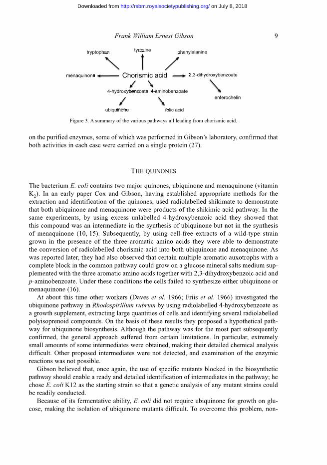
the availability of pure chorismic acid created a great opportunity to study the many diver-gent pathways that used this compound as a starting point (figure 3).
the next 10 years or so were extremely productive as Frank and his graduate students and later postdoctoral researchers pursued many of the pathways with vigour. His first wife, margaret, had made significant contributions to much of the work leading to the successful identification and isolation of chorismic acid. Unfortunately, illness prevented her from par-ticipating in the next phase of the work. this work was started in a laboratory in the Chemistry Department at the University of melbourne, where Frank’s group was temporarily housed while the new building for the Department of microbiology was being finished. in 196� the building was finished and Frank was promoted to Professor of Chemical microbiology. During the next two years, work began in earnest on the phenylalanine and tyrosine pathways and the pathways for ubiquinone, menaquinone, p-aminobenzoic acid and tryptophan. at this stage he also made the switch to E. coli k12, and genetic analysis became an important part of his future research. in 1967 Frank was offered and accepted the chair in biochemistry at the anU and moved there with many members of his group. the further exploration of these pathways and the extensive investigation of atP synthase and oxidative phosphorylation all took place at the anU.
PhEnylAlAninE And tyrosinE PAthwAys
it had been known that an early precursor in both these pathways was the compound pre-phenic acid, and its formation from chorismic acid had already been demonstrated. Cotton and Gibson investigated the enzymes involved in its formation. Chromatography of cell-free extracts on diethylaminoethyl (DEaE)-cellulose revealed that there were two discrete and separable proteins with enzymic activity (called chorismate mutase) converting chorismate to prephenate. of considerable interest was the finding that associated with one of these peaks of mutase activity was activity for the second reaction of the phenylalanine pathway (prephenate dehydratase), which converted prephenate into phenylpyruvate, and associated with the other peak of mutase activity was the second reaction of the tyrosine pathway (prephenate dehydro-genase), which converted prephenate into 4-hydroxyphenylpyruvate (14). subsequent work
C
CH2
COOH
OH
COOH
C
CH
OH
C
CH
C
CH
COOH
OO
Figure 2. Chorismic acid.
on July 8, 2018http://rsbm.royalsocietypublishing.org/Downloaded from
Frank William Ernest Gibson 9
on the purified enzymes, some of which was performed in Gibson’s laboratory, confirmed that both activities in each case were carried on a single protein (27).
thE QuinonEs
the bacterium E. coli contains two major quinones, ubiquinone and menaquinone (vitamin k2). in an early paper Cox and Gibson, having established appropriate methods for the extraction and identification of the quinones, used radiolabelled shikimate to demonstrate that both ubiquinone and menaquinone were products of the shikimic acid pathway. in the same experiments, by using excess unlabelled 4-hydroxybenzoic acid they showed that this compound was an intermediate in the synthesis of ubiquinone but not in the synthesis of menaquinone (10, 1�). subsequently, by using cell-free extracts of a wild-type strain grown in the presence of the three aromatic amino acids they were able to demonstrate the conversion of radiolabelled chorismic acid into both ubiquinone and menaquinone. as was reported later, they had also observed that certain multiple aromatic auxotrophs with a complete block in the common pathway could grow on a glucose mineral salts medium sup-plemented with the three aromatic amino acids together with 2,3-dihydroxybenzoic acid and p-aminobenzoate. Under these conditions the cells failed to synthesize either ubiquinone or menaquinone (16).
at about this time other workers (Daves et al. 1966; Friis et al. 1966) investigated the ubiquinone pathway in Rhodospirillum rubrum by using radiolabelled 4-hydroxybenzoate as a growth supplement, extracting large quantities of cells and identifying several radiolabelled polyisoprenoid compounds. on the basis of these results they proposed a hypothetical path-way for ubiquinone biosynthesis. although the pathway was for the most part subsequently confirmed, the general approach suffered from certain limitations. in particular, extremely small amounts of some intermediates were obtained, making their detailed chemical analysis difficult. other proposed intermediates were not detected, and examination of the enzymic reactions was not possible.
Gibson believed that, once again, the use of specific mutants blocked in the biosynthetic pathway should enable a ready and detailed identification of intermediates in the pathway; he chose E. coli k12 as the starting strain so that a genetic analysis of any mutant strains could be readily conducted.
because of its fermentative ability, E. coli did not require ubiquinone for growth on glu-cose, making the isolation of ubiquinone mutants difficult. to overcome this problem, non-
Chorismic acid
tryptophan tyrosine phenylalanine
menaquinone
ubiquinone folic acid
enterochelin
4-hydroxybenzoate 4-aminobenzoate
2,3-dihydroxybenzoate
tryptophan tyrosine phenylalanine
2,3-dihydroxybenzoate
enterochelin
4-hydroxybenzoate 4-aminobenzoate 4-hydroxybenzoate
folic acid ubiquinone
menaquinone
Figure 3. a summary of the various pathways all leading from chorismic acid.
on July 8, 2018http://rsbm.royalsocietypublishing.org/Downloaded from
10 Biographical Memoirs
fermentable substrates such as succinate or malate were substituted for glucose; under these conditions, cells unable to make ubiquinone failed to grow (16).
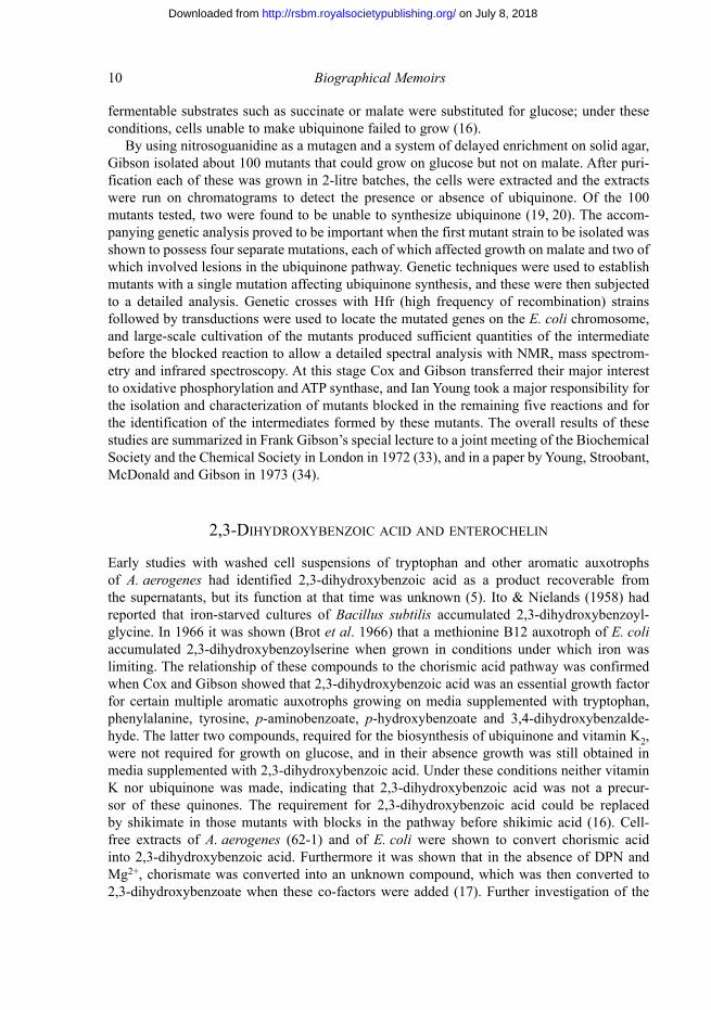
by using nitrosoguanidine as a mutagen and a system of delayed enrichment on solid agar, Gibson isolated about 100 mutants that could grow on glucose but not on malate. after puri-fication each of these was grown in 2-litre batches, the cells were extracted and the extracts were run on chromatograms to detect the presence or absence of ubiquinone. of the 100 mutants tested, two were found to be unable to synthesize ubiquinone (19, 20). the accom-panying genetic analysis proved to be important when the first mutant strain to be isolated was shown to possess four separate mutations, each of which affected growth on malate and two of which involved lesions in the ubiquinone pathway. Genetic techniques were used to establish mutants with a single mutation affecting ubiquinone synthesis, and these were then subjected to a detailed analysis. Genetic crosses with Hfr (high frequency of recombination) strains followed by transductions were used to locate the mutated genes on the E. coli chromosome, and large-scale cultivation of the mutants produced sufficient quantities of the intermediate before the blocked reaction to allow a detailed spectral analysis with nmr, mass spectrom-etry and infrared spectroscopy. at this stage Cox and Gibson transferred their major interest to oxidative phosphorylation and atP synthase, and ian Young took a major responsibility for the isolation and characterization of mutants blocked in the remaining five reactions and for the identification of the intermediates formed by these mutants. the overall results of these studies are summarized in Frank Gibson’s special lecture to a joint meeting of the biochemical society and the Chemical society in london in 1972 (33), and in a paper by Young, stroobant, mcDonald and Gibson in 1973 (34).
2,3-dihydroxyBEnzoiC ACid And EntEroChElin
Early studies with washed cell suspensions of tryptophan and other aromatic auxotrophs of A. aerogenes had identified 2,3-dihydroxybenzoic acid as a product recoverable from the supernatants, but its function at that time was unknown (�). ito & nielands (19�8) had reported that iron-starved cultures of Bacillus subtilis accumulated 2,3-dihydroxybenzoyl-glycine. in 1966 it was shown (brot et al. 1966) that a methionine b12 auxotroph of E. coli accumulated 2,3-dihydroxybenzoylserine when grown in conditions under which iron was limiting. the relationship of these compounds to the chorismic acid pathway was confirmed when Cox and Gibson showed that 2,3-dihydroxybenzoic acid was an essential growth factor for certain multiple aromatic auxotrophs growing on media supplemented with tryptophan, phenylalanine, tyrosine, p-aminobenzoate, p-hydroxybenzoate and 3,4-dihydroxybenzalde-hyde. the latter two compounds, required for the biosynthesis of ubiquinone and vitamin k2, were not required for growth on glucose, and in their absence growth was still obtained in media supplemented with 2,3-dihydroxybenzoic acid. Under these conditions neither vitamin k nor ubiquinone was made, indicating that 2,3-dihydroxybenzoic acid was not a precur-sor of these quinones. the requirement for 2,3-dihydroxybenzoic acid could be replaced by shikimate in those mutants with blocks in the pathway before shikimic acid (16). Cell-free extracts of A. aerogenes (62-1) and of E. coli were shown to convert chorismic acid into 2,3-dihydroxybenzoic acid. Furthermore it was shown that in the absence of DPn and mg2+, chorismate was converted into an unknown compound, which was then converted to 2,3-dihydroxybenzoate when these co-factors were added (17). Further investigation of the
on July 8, 2018http://rsbm.royalsocietypublishing.org/Downloaded from
Frank William Ernest Gibson 11
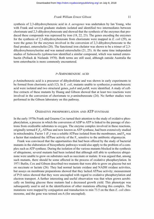
synthesis of 2,3-dihydroxybenzoic acid in A. aerogenes was undertaken by ian Young, who with Frank and several graduate students isolated and identified two intermediates between chorismate and 2,3-dihydroxybenzoate and showed that the synthesis of the enzymes that pro-duced these compounds was repressed by iron (18, 22, 23). the genes encoding the enzymes for the synthesis of 2,3-dihydroxybenzoate from chorismate were mapped in E. coli (29), as were the genes for the enzymes involved in the conversion of 2,3-dihydroxybenzoate to the final product, enterochelin (28). the functional iron chelator was shown to be a trimer of 2,3-dihydroxybenzoylserine and was named enterochelin (21, 2�). at the same time independent studies of Salmonella typhimurium identified a similar compound, which was named entero-bactin (Pollack & nielands 1970). both terms are still used, although outside australia the term enterobactin is more commonly encountered.
p-AMinoBEnzoiC ACid
p-aminobenzoic acid is a precursor of dihydrofolate and was shown in early experiments to be formed from chorismic acid (12). in E. coli, mutants unable to synthesize p-aminobenzoic acid were isolated and two structural genes, pabA and pabB, were identified. a study of cell-free extracts of these mutants by Huang and Gibson showed that at least two reactions were involved in the conversion of chorismate to p-aminobenzoate (24). no further studies were performed in the Gibson laboratory on this pathway.
oxidAtivE PhosPhorylAtion And AtP synthAsE
in the early 1970s Frank and Graeme Cox turned their attention to the study of oxidative phos-phorylation, a process in which the conversion of aDP to atP is linked to the passage of elec-trons from oxidizable substrates to oxygen. the enzyme complex involved in these reactions, originally termed F1F0-atPase and now known as atP synthase, had been extensively studied in mitochondria. Factor 1 (F1) was a soluble atPase isolated from the membranes, and F0 was a factor that rendered the atPase activity of the F1 sensitive to the antibiotic oligomycin.
Frank was convinced that the opportunities that had been offered by the study of bacterial mutants in the elaboration of biosynthetic pathways would also apply to the problem of a com-plex such as atP synthase. During the isolation of the various mutants blocked in the synthesis of ubiquinone, several mutants had been isolated that although still able to synthesize ubiqui-none were unable to grow on substrates such as succinate or malate. it was argued that, among such mutants, there should be some affected in the process of oxidative phosphorylation. in 1971 butlin, Cox and Gibson described two mutants that were able to grow on glucose but not on succinate or lactate (26). they had normal lactate oxidase and naDH oxidase activities, but assays on membrane preparations showed that they lacked atPase activity; measurement of P:o ratios showed that they were uncoupled with regard to oxidative phosphorylation and electron transport. a further interesting and useful observation was that when grown aerobi-cally on limiting glucose these mutants had a decreased growth yield. this phenotype was subsequently used to aid in the identification of other mutations affecting this complex. the mutations were mapped by conjugation and transduction to min 73.� on the then E. coli chro-mosome, and the gene was termed unca (for uncoupled).
on July 8, 2018http://rsbm.royalsocietypublishing.org/Downloaded from
12 Biographical Memoirs
on the basis of their phenotypes these mutants were assumed to be altered in the F1 component of atP synthase. this discovery, which was made at a time when bacteria such as E. coli were largely discounted as having any great relevance to the extensive studies of mitochondrial atP synthase, provided Frank with an opportunity to express his conviction of the potential for these bacterial studies. in this paper he stated:
the use of bacteria with their simpler cellular organization than eukaryotic cells, and of E. coli in particular, with its amenity to genetic manipulation, seems a promising experimental system for a combined genetic and biochemical approach to the problem of coupling of phosphorylation to electron transport.
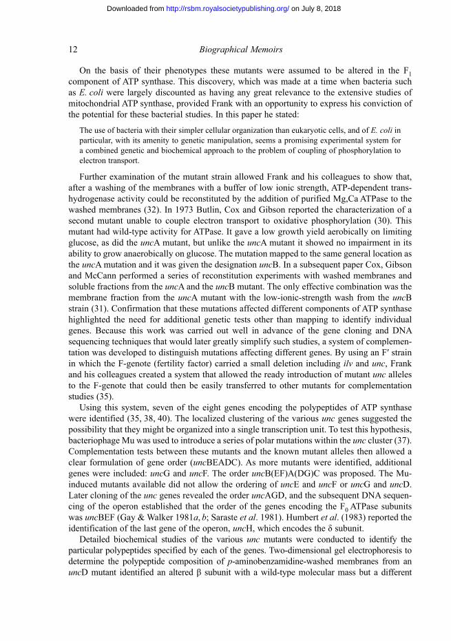
Further examination of the mutant strain allowed Frank and his colleagues to show that, after a washing of the membranes with a buffer of low ionic strength, atP-dependent trans-hydrogenase activity could be reconstituted by the addition of purified mg,Ca atPase to the washed membranes (32). in 1973 butlin, Cox and Gibson reported the characterization of a second mutant unable to couple electron transport to oxidative phosphorylation (30). this mutant had wild-type activity for atPase. it gave a low growth yield aerobically on limiting glucose, as did the unca mutant, but unlike the unca mutant it showed no impairment in its ability to grow anaerobically on glucose. the mutation mapped to the same general location as the unca mutation and it was given the designation uncb. in a subsequent paper Cox, Gibson and mcCann performed a series of reconstitution experiments with washed membranes and soluble fractions from the unca and the uncb mutant. the only effective combination was the membrane fraction from the unca mutant with the low-ionic-strength wash from the uncb strain (31). Confirmation that these mutations affected different components of atP synthase highlighted the need for additional genetic tests other than mapping to identify individual genes. because this work was carried out well in advance of the gene cloning and Dna sequencing techniques that would later greatly simplify such studies, a system of complemen-tation was developed to distinguish mutations affecting different genes. By using an F′ strain in which the F-genote (fertility factor) carried a small deletion including ilv and unc, Frank and his colleagues created a system that allowed the ready introduction of mutant unc alleles to the F-genote that could then be easily transferred to other mutants for complementation studies (3�).
Using this system, seven of the eight genes encoding the polypeptides of atP synthase were identified (3�, 38, 40). the localized clustering of the various unc genes suggested the possibility that they might be organized into a single transcription unit. to test this hypothesis, bacteriophage mu was used to introduce a series of polar mutations within the unc cluster (37). Complementation tests between these mutants and the known mutant alleles then allowed a clear formulation of gene order (uncbEaDC). as more mutants were identified, additional genes were included: uncG and uncF. the order uncb(EF)a(DG)C was proposed. the mu-induced mutants available did not allow the ordering of uncE and uncF or uncG and uncD. later cloning of the unc genes revealed the order uncaGD, and the subsequent Dna sequen-cing of the operon established that the order of the genes encoding the F0 atPase subunits was uncbEF (Gay & Walker 1981a, b; saraste et al. 1981). Humbert et al. (1983) reported the identification of the last gene of the operon, uncH, which encodes the δ subunit.
Detailed biochemical studies of the various unc mutants were conducted to identify the particular polypeptides specified by each of the genes. two-dimensional gel electrophoresis to determine the polypeptide composition of p-aminobenzamidine-washed membranes from an uncD mutant identified an altered β subunit with a wild-type molecular mass but a different
on July 8, 2018http://rsbm.royalsocietypublishing.org/Downloaded from
Frank William Ernest Gibson 13
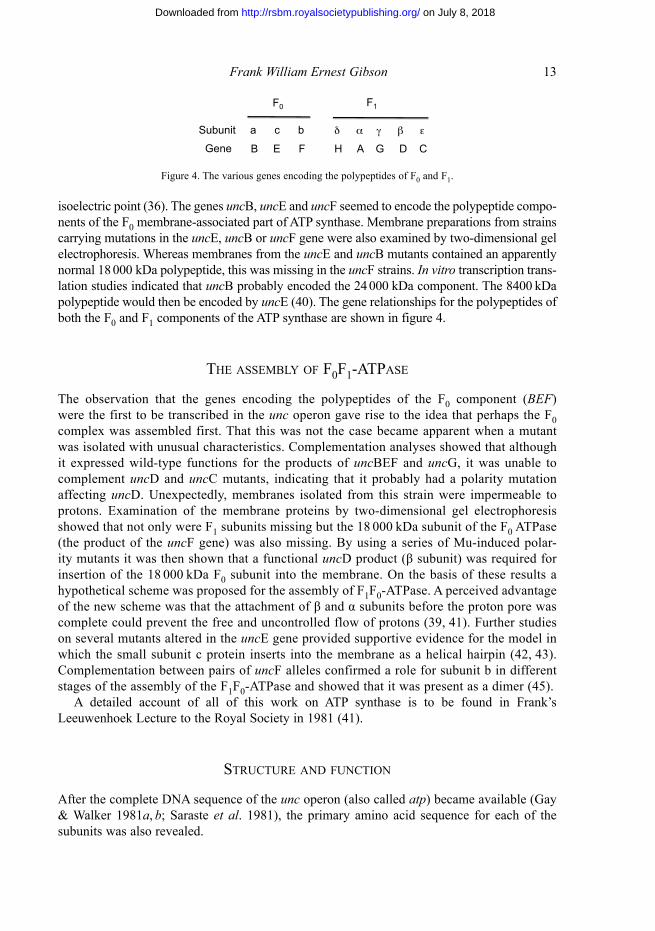
isoelectric point (36). the genes uncb, uncE and uncF seemed to encode the polypeptide compo-nents of the F0 membrane-associated part of atP synthase. membrane preparations from strains carrying mutations in the uncE, uncb or uncF gene were also examined by two-dimensional gel electrophoresis. Whereas membranes from the uncE and uncb mutants contained an apparently normal 18 000 kDa polypeptide, this was missing in the uncF strains. In vitro transcription trans-lation studies indicated that uncb probably encoded the 24 000 kDa component. the 8400 kDa polypeptide would then be encoded by uncE (40). the gene relationships for the polypeptides of both the F0 and F1 components of the atP synthase are shown in figure 4.
thE AssEMBly oF F0F1-AtPAsE
the observation that the genes encoding the polypeptides of the F0 component (BEF) were the first to be transcribed in the unc operon gave rise to the idea that perhaps the F0 complex was assembled first. that this was not the case became apparent when a mutant was isolated with unusual characteristics. Complementation analyses showed that although it expressed wild-type functions for the products of uncbEF and uncG, it was unable to complement uncD and uncC mutants, indicating that it probably had a polarity mutation affecting uncD. Unexpectedly, membranes isolated from this strain were impermeable to protons. Examination of the membrane proteins by two-dimensional gel electrophoresis showed that not only were F1 subunits missing but the 18 000 kDa subunit of the F0 atPase (the product of the uncF gene) was also missing. by using a series of mu-induced polar-ity mutants it was then shown that a functional uncD product (β subunit) was required for insertion of the 18 000 kDa F0 subunit into the membrane. on the basis of these results a hypothetical scheme was proposed for the assembly of F1F0-atPase. a perceived advantage of the new scheme was that the attachment of β and α subunits before the proton pore was complete could prevent the free and uncontrolled flow of protons (39, 41). Further studies on several mutants altered in the uncE gene provided supportive evidence for the model in which the small subunit c protein inserts into the membrane as a helical hairpin (42, 43). Complementation between pairs of uncF alleles confirmed a role for subunit b in different stages of the assembly of the F1F0-atPase and showed that it was present as a dimer (4�).
a detailed account of all of this work on atP synthase is to be found in Frank’s leeuwenhoek lecture to the royal society in 1981 (41).
struCturE And FunCtion
after the complete Dna sequence of the unc operon (also called atp) became available (Gay & Walker 1981a, b; saraste et al. 1981), the primary amino acid sequence for each of the subunits was also revealed.
Gene B E F
a c bSubunit
F0 F1
δ α γ β ε
H A G D C
Figure 4. the various genes encoding the polypeptides of F0 and F1.
on July 8, 2018http://rsbm.royalsocietypublishing.org/Downloaded from
14 Biographical Memoirs
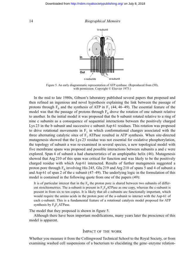
in the mid to late 1980s, Gibson’s laboratory published several papers that proposed and then refined an ingenious and novel hypothesis explaining the link between the passage of protons through F0 and the synthesis of atP in F1 (44, 46–49). the essential feature of the model was that the passage of protons through F0 drove the rotation of one subunit relative to another. in the initial model it was proposed that the b subunit rotated relative to a ring of nine c subunits as a consequence of sequential interactions between the positively charged lys 23 in the b subunit and successive c subunit asp 61 residues. this rotation was proposed to drive rotational movements in F1 in which conformational changes associated with the three alternating catalytic sites of F1 atPase resulted in atP synthesis. When site-directed mutagenesis showed that the lys 23 residue was not essential for oxidative phosphorylation, the topology of subunit a was re-examined in several species, a new topological model with five membrane spans was proposed and possible interactions between subunits a and c were explored. span 4 of subunit a had characteristics of an amphipathic helix (46). mutagenesis showed that arg 210 of this span was critical for function and was likely to be the positively charged residue with which asp 61 interacted. results of further mutagenesis suggested a proton pore through F0 involving His 24�, Glu 219 and arg 210 of spans � and 4 of subunit a and asp 61 of span 2 of the c subunit (47–49). the underlying logic in the formulation of this model is contained in the following quote from one of the papers (49):
it is of particular interest that in the F0 the proton pore is shared between two subunits of differ-ent stoichiometries. the a-subunit is present in F1F0atPase as one copy, whereas the c-subunit is present in from six to ten copies. it is likely that all c-subunits are functionally important, which would require the amino acids in the proton pore of the a-subunit to interact with the asp-61 of each c-subunit. this is a fundamental feature of a rotational catalysis model proposed for atP synthesis by F0F1atPase.
the model that they proposed is shown in figure �.although there have been important modifications, many years later the prescience of this
model is apparent.
iMPACt oF thE work
Whether you measure it from the Collingwood technical school to the royal society, or from examining washed cell suspensions of a bacterium to elucidating the gene–enzyme relation-
α β
β α δ γ ε
In
Out
c-subunits a-subunit
b-subunit
Figure �. an early diagrammatic representation of atP synthase. (reproduced from (�0), with permission. Copyright © Elsevier 1973.)
on July 8, 2018http://rsbm.royalsocietypublishing.org/Downloaded from
Frank William Ernest Gibson 1�
ships and the organization of a complex enzyme such as atP synthase, Frank Gibson’s life was an extraordinary journey. His research, driven by an enquiring mind, was original and pioneering. His discoveries dating from the identification of chorismic acid were pivotal to the development of new pathways of exploration. He never shrank from posing difficult ques-tions and was not influenced by prevailing norms that directed work into acceptable chan-nels. in the early days Frank said that he subscribed to the view that one experiment is worth a thousand expert opinions. He encouraged his students to get into the laboratory and test their hypotheses. During his lifetime he trained many young and aspiring graduate students. all of these, many of whom have progressed to distinguished positions in science, speak with great affection and respect for his role as a mentor and friend. His enthusiasm for the work, and the integrity and self-deprecating honesty that he always applied to research achievements, served as a model that was difficult not to adopt.
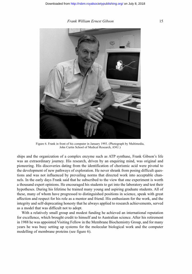
With a relatively small group and modest funding he achieved an international reputation for excellence, which brought credit to himself and to australian science. after his retirement in 1988 he was appointed Visiting Fellow in the membrane biochemistry Group, and for many years he was busy setting up systems for the molecular biological work and the computer modelling of membrane proteins (see figure 6).
Figure 6. Frank in front of his computer in January 1993. (Photograph by multimedia, John Curtin school of medical research, anU.)
on July 8, 2018http://rsbm.royalsocietypublishing.org/Downloaded from
16 Biographical Memoirs
distinCtions And AwArds
in 19�9 he was awarded a Carnegie Foundation travel Grant. in 1963 he received the David syme research Prize of the University of melbourne. in 1968 he was selected to give the first lemberg lecture to the annual meeting of the australian biochemical society. in 1971 he was elected a Fellow of the australian academy of science and in 1976 he was elected to the Fellowship of the royal society. He gave the s. D. rubbo memorial oration in 197� and was invited to give the leeuwenhook lecture to the royal society in london, manchester and Durham in 1981, the Hopkins memorial lecture in london in 1982 and the burnet lecture in Canberra in 1991. the australian biochemical society, the australian society of microbiology and University House at the anU all made him an Honorary life member. During his lifetime he was also an invited speaker at several international conferences. apart from three years as Director and Howard Florey Professor of medical research, John Curtin school of medical research, anU, Canberra, between 1977 and 1980, he was from 1967 to 1988 Professor and Head of the Division of biochemical sciences in the John Curtin school. in 1989 he was made an Emeritus Professor and a University Fellow at anU. as Visiting Fellow in the membrane biochemistry Group he continued his contributions to research in the new area of computer modelling of membrane proteins. in January 2004 he was appointed a member of the order of australia.
outsidE thE lABorAtory
outside the laboratory Frank exhibited a great enthusiasm for life, a willingness to take on all sorts of challenges, which he met with a quiet determination, a readiness to question anything that seemed false or overblown, and a quiet friendly persona that endeared him to many. He was also a highly competitive individual and was always very active in a variety of physical activities: in his early years in Queensland climbing mountains and surfing, back in Victoria cross-country skiing and scuba diving, followed by squash and tennis and, later, in Canberra swimming in the waters at Guerilla bay in all seasons and continuing to ski at every opportu-nity. He never let what he would regard as minor setbacks interfere with his plans. after suf-fering a major shoulder injury while skiing, he arrived at the tennis court the next day, with one arm tightly held in a sling, as ready as always for the morning battle. He maintained an active interest in political events, which he viewed with a well-honed cynicism. He was very attached to his children, to Frances and ruth from his first marriage and to mark from his second. He followed their activities with great interest and a certain amount of pride. He was greatly saddened when ruth died of breast cancer, and maintained an active involvement with the grandchildren. He and robin rollason were married in 1980, and in 1982 they travelled to the Uk and stayed in oxford, where Frank was appointed for a year as newton abrahams Visiting Professor. back in australia they built a holiday home at Guerilla bay, where close friends lloyd and margaret Evans had a house. many of Frank’s friends and associates spent wonder-fully enjoyable weekends with him and robin, walking, swimming and playing tennis.
on July 8, 2018http://rsbm.royalsocietypublishing.org/Downloaded from
Frank William Ernest Gibson 17
ACknowlEdGEMEnts
We would like to thank members of the family for reading the manuscript, and nancy millis and ian Young for help-ful comments. We should also mention an excellent autobiography written by Frank in Comprehensive Biochemistry (�0).
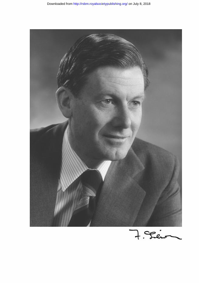
the frontispiece photograph was taken in 1976 by Godfrey argent and is reproduced with permission.
rEFErEnCEs to othEr Authors
brot, n., Goodwin, J. & Fales, H. 1966 in vivo and in vitro formation of 2,3-dihydroxybenzoylserine by Escherichia coli k12. Biochem. Biophys. Res. Commun. 25, 4�4–461.
Daves, G. D. Jr, Friis, P., olsen, r. k. & Folkers, k. 1966 the chemistry of ubiquinone. Vitamins Hormones 24, 427–439.
Friis, P., Daves, G. D. & Folkers, k. 1966 Complete sequence of biosynthesis from p-hydroxybenzoic acid to ubiquinone. J. Am. Chem. Soc. 88, 47�4–47�6.
Gay, n. J. & Walker, J. E. 1981a the atp operon: nucleotide sequence of the promoter and the genes for the membrane proteins, and the δ subunit of Escherichia coli ATP-synthase. Nucleic Acids Res. 9, 3919–3926.
Gay, n. J. & Walker, J. E. 1981b The atp operon: nucleotide sequence of the region encoding the α-subunit of Escherichia coli atP-synthase. Nucleic Acids Res. 9, 2187–2194.
Humbert, r., brusilow, a. W. s., Gunsalus, r. P., klionsky, D. J. & simoni, r. D. 1983 Escherichia coli mutants defective in the uncH gene. J. Bacteriol. 153, 416–422.
ito, t. & nielands, J. b. 19�8 Products of ‘low iron fermentation’ with Bacillus subtilis: isolation, characterization and synthesis of 2,3-dihydroxybenzoylglycine. J. Am. Chem. Soc. 80, 464�–4647.
Pollack, J. r. & neilands, J. b. 1970 Enterobactin, an iron transport compound from Salmonella typhimurium. Biochem. Biophys. Res. Commun. 38, 989–992.
saraste, m., Gay, n. J., Eberle, a., runswick, m. J. & Walker, J. E. 1981 the atp operon: nucleotide sequence of the genes for the γ, β, and ε subunits of Escherichia coli ATP synthase. Nucleic Acids Res 9, �287–�296.
sprinson, D. b. 1960 the biosynthesis of aromatic compounds from D-glucose. Adv. Carbohydr. Chem. 15, 23�–270.
BiBlioGrAPhy
the following publications are those referred to directly in the text. a full bibliography is available as electronic supplementary material at http://dx.doi.org/10.1098/rsbm.2009.0020 or via http://rsbm.royalsocietypublishing.org.
(1) 19�0 the inhibition of the oxidation of alanine by acridines and related compounds. Aust. J. Exp. Biol. Med. Sci. 28, 4�9–463.
(2) 19�9 (With C. H. Doy) 1-(o-carboxyphenylamino)-1-deoxyribulose: a compound formed by mutant strains of Aerobacter aerogenes and Escherichia coli blocked in the biosynthesis of tryptophan. Biochem. J. 72, �86–�97.
(3) 1960 (With D. D. Woods) the synthesis of methionine by suspensions of Escherichia coli. Biochem. J. 74, 160–172.
(4) 1960 (With C. Yanofsky) the partial purification and properties of indole-3-glycerol phosphate synthetase from Escherichia coli. Biochim. Biophys. Acta 43, 489–�00.
(�) 1961 (With a. J. Pittard & C. H. Doy) Phenolic compounds accumulated by washed cell suspensions of a tryptophan auxotroph of Aerobacter aerogenes. Biochim. Biophys. Acta 49, 48�–491.
(6) 1962 (With m. i. Gibson) a new intermediate in aromatic biosynthesis. Biochim. Biophys. Acta 65, 160–163.
on July 8, 2018http://rsbm.royalsocietypublishing.org/Downloaded from
18 Biographical Memoirs
(7) 1962 (With m. i. Gibson, C. H. Doy & P. morgan) the branchpoint in the synthesis of the aromatic amino acids. Nature 195, 1173–117�.
(8) 1963 (With l. m. Jackman) structure of chorismic acid, a new intermediate in aromatic biosynthesis. Nature 198, 388–389.
(9) 1963 (With P. n. morgan & m. i. Gibson) the conversion of shikimic acid into certain aromatic compounds by cell-free extracts of Aerobacter aerogenes and Escherichia coli. Biochem. J. 89, 229–239.
(10) 1964 (With G. b. Cox) biosynthesis of vitamin k and ubiquinone. relation to the shikimic acid pathway in Escherichia coli. Biochim. Biophys. Acta 93, 204–206.
(11) 1964 Chorismic acid: purification and some chemical and physical studies. Biochem. J. 90, 2�6–261.(12) 1964 (With m. Gibson & G. b. Cox) the biosynthesis of p-aminobenzoic acid from chorismic acid.
Biochim. Biophys. Acta 82, 637–638.(13) 1964 (With m. i. Gibson) Preliminary studies on the isolation of an intermediate in aromatic biosynthesis:
chorismic acid. Biochem. J. 90, 248–2�6.(14) 196� (With r. G. H. Cotton) the biosynthesis of phenylalanine and tyrosine; enzymes converting chorismic
acid into prephenic acid and their relationships to prephenate dehydratase and prephenate dehydrogen-ase. Biochim. Biophys. Acta 100, 76–88.
(1�) 1966 (With G. b. Cox) the role of shikimic acid in the biosynthesis of vitamin k2. Biochem. J. 100, 1–6.(16) 1967 (With G. b. Cox) 2,3-Dihydroxybenzoic acid, a new growth factor for multiple aromatic auxotrophs.
J. Bacteriol. 93, �02–�03.(17) 1967 (With i. G. Young & G. b. Cox) 2,3-Dihydroxybenzoate as a bacterial growth factor and its route of
biosynthesis. Biochim. Biophys. Acta 141, 319–331.(18) 1967 (With i. G. Young & l. m. Jackman) 2,3-Dihydro-2,3-dihydroxybenzoic acid: an intermediate in the
biosynthesis of 2,3-dihydroxybenzoic acid. Biochim. Biophys. Acta 148, 313–31�.(19) 1968 (With G. b. Cox & J. Pittard) mutant strains of Escherichia coli k-12 unable to form ubiquinone. J.
Bacteriol. 95, 1�91–1�98.(20) 1969 (With G. b. Cox, i. G. Young & l. m. mcCann) biosynthesis of ubiquinone in Escherichia coli k-12:
location of genes affecting the metabolism of 3-octaprenyl-4-hydroxybenzoic acid and 2-octaprenyl-phenol. J. Bacteriol. 99, 4�0–4�8.
(21) 1969 (With i. G. o’brien & G. b. Cox) 2,3-dihydroxy-N-benzoylserine: chemical synthesis and comparison with the natural product. Biochim. Biophys. Acta 177, 321–328.
(22) 1969 (With i. G. Young) regulation of the enzymes involved in the biosynthesis of 2,3-dihydroxybenzoic acid in Aerobacter aerogenes and Escherichia coli. Biochim. Biophys. Acta 177, 401–411.
(23) 1969 (With i. G. Young & t. batterham) isochorismic acid: a new intermediate in the biosynthesis of 2,3-dihydrobenzoic acid. Biochim. Biophys. Acta 165, �67–�68.
(24) 1970 (With m. Huang) the biosynthesis of 4-aminobenzoate in Escherichia coli. J. Bacteriol. 102, 767–773.
(2�) 1970 (With i. G. o’brien & G. b. Cox) biologically active compounds containing 2,3-dihydroxybenzoic acid and serine formed by Escherichia coli. Biochim. Biophys. Acta 201, 4�3–460.
(26) 1971 (With J. D. butlin & G. b. Cox) oxidative phosphorylation in Escherichia coli k12. mutations affect-ing magnesium ion- or calcium ion-stimulated adenosine triphosphatase. Biochem. J. 124, 7�–81.
(27) 1971 (With G. l. E. koch & D. C. shaw) Characterisation of the subunits of chorismate mutase–prephenate dehydrogenase from Escherichia coli k12. Biochim. Biophys. Acta 229, 80�–812.
(28) 1971 (With r. k. luke) location of three genes concerned with the conversion of 2,3-dihydroxybenzoate into enterochelin in Escherichia coli k-12. J. Bacteriol. 107, ��7–�62.
(29) 1971 (With i. G. Young, l. langman & r. k. luke) biosynthesis of the iron-transport compound entero-chelin: mutants of Escherichia coli unable to synthesize 2,3-dihydroxybenzoate. J. Bacteriol. 106, �1–�7.
(30) 1973 (With J. D. butlin & G. b. Cox) oxidative phosphorylation in Escherichia coli k-12: the genetic and biochemical characterisations of a strain carrying a mutation in the uncb gene. Biochim. Biophys. Acta 292, 366–37�.
(31) 1973 (With G. b. Cox & l. mcCann) reconstitution of oxidative phosphorylation and the adenosine tri-phosphate-dependent transhydrogenase activity by a combination of membrane fractions from uncA and uncB mutant strains of Escherichia coli k12. Biochem. J. 134, 101�–1021.
on July 8, 2018http://rsbm.royalsocietypublishing.org/Downloaded from
Frank William Ernest Gibson 19
(32) 1973 (With G. b. Cox, l. m. mcCann, J. D. butlin & F. l. Crane) reconstitution of the energy-linked trans-hydrogenase activity in membranes from a mutant strain of Escherichia coli k12 lacking magnesium ion- or calcium ion-stimulated adenosine triphosphatase. Biochem. J. 132, 689–69�.
(33) 1973 Chemical and genetic studies on the biosynthesis of ubiquinone by Escherichia coli. Biochem. Soc. Trans. 1, 316–326.
(34) 1973 (With i. G. Young, P. stroobant & C. G. macdonald) Pathway for ubiquinone biosynthesis in Escherichia coli k-12: gene–enzyme relationships and intermediates. J. Bacteriol. 114, 42–�2.
(3�) 1977 (With G. b. Cox, J. a. Downie & J. radik) Partial diploids of Escherichia coli carrying normal and mutant alleles affecting oxidative phosphorylation. Biochem. J. 162, 66�–670.
(36) 1978 (With D. r. Fayle, J. a. Downie, G. b. Cox & J. radik) Characterization of the mutant-uncD-gene product in a strain of Escherichia coli K12. An altered β-subunit of the magnesium ion-stimulated adenosine triphosphatase. Biochem. J. 172, �23–�31.
(37) 1978 (With J. a. Downie, G. b. Cox & J. radik) mu-induced polarity in the unc operon of Escherichia coli. J. Bacteriol. 134, 728–736.
(38) 1979 (With J. a. Downie, a. E. senior & G. b. Cox) a fifth gene (uncE) in the operon concerned with oxida-tive phosphorylation in Escherichia coli. J. Bacteriol. 137, 711–718.
(39) 1981 (With G. b. Cox, J. a. Downie, l. langman, a. E. senior, G. ash & D. r. Fayle) assembly of the adenosine triphosphatase complex in Escherichia coli: assembly of F0 is dependent on the formation of specific F1 subunits. J. Bacteriol. 148, 30–42.
(40) 1981 (With J. a. Downie, G. b. Cox, l. langman, G. ash & m. becker) three genes coding for subunits of the membrane sector (F0) of the Escherichia coli adenosine triphosphatase complex. J. Bacteriol. 145, 200–210.
(41) 1982 the leeuwenhoek lecture, 1981. the biochemical and genetic approach to the study of bioenergetics with the use of Escherichia coli: progress and prospects. Proc. R. Soc. Lond. B 215, 1–18.
(42) 1983 (With a. l. Fimmel, D. a. Jans, l. langman, l. b. James, G. r. ash, J. a. Downie, a. E. senior & G. b. Cox) the F1F0-atPase of Escherichia coli. substitution of proline by leucine at position 64 in the c-subunit causes loss of oxidative phosphorylation. Biochem. J. 213, 4�1–4�8.
(43) 1983 (With D. a. Jans, a. l. Fimmel, l. langman, l. b. James, J. a. Downie, a. E. senior, G. r. ash & G. b. Cox) mutations in the uncE gene affecting assembly of the c-subunit of the adenosine triphos-phatase of Escherichia coli. Biochem. J. 211, 717–726.
(44) 1984 (With G. b. Cox, D. a. Jans, a. l. Fimmel & l. Hatch) Hypothesis. the mechanism of atP synthase. Conformational change by rotation of the b-subunit. Biochim. Biophys. Acta 768, 201–208.
(4�) 198� (With D. a. Jans, l. Hatch, a. l. Fimmel & G. b. Cox) Complementation between uncF alleles affect-ing assembly of the F1F0-atPase complex of Escherichia coli. J. Bacteriol. 162, 420–426.
(46) 1986 (With G. b. Cox, a. l. Fimmel & l. Hatch) the mechanism of atP synthase: a reassessment of the functions of the b and a subunits. Biochim. Biophys. Acta 849, 62–69.
(47) 1987 (With r. n. lightowlers, s. m. Howitt, l. Hatch & G. b. Cox) the proton pore in the Escherichia coli F0F1-atPase: a requirement for arginine at position 210 of the a-subunit. Biochim. Biophys. Acta 894, 399–406.
(48) 1988 (With s. m. Howitt & G. b. Cox) the proton pore of the F0F1-atPase of Escherichia coli: ser-206 is not required for proton translocation. Biochim. Biophys. Acta 936, 74–80.
(49) 1988 (With r. n. lightowlers, s. m. Howitt, l. Hatch & G. Cox) the proton pore in the Escherichia coli F0F1-ATPase: substitution of glutamate by glutamine at position 219 of the α-subunit prevents F0-mediated proton permeability. Biochim. Biophys. Acta 933, 241–248.
(�0) 199� Chorismic acid and beyond. in Selected topics in the history of biochemistry: personal recollections, vol. 4 (ed. E. C. slater, r. Jaenicke & G. semenza) (Comprehens. Biochem. 38), pp. 2�9–301. Elsevier science and technology.
(�1) 1999 the elusive branch-point compound of aromatic amino acid biosynthesis. Trends Biochem. Sci. 24, 36–38.
on July 8, 2018http://rsbm.royalsocietypublishing.org/Downloaded from
CORRECTIONs
Biogr. Mems Fell. R. Soc. 56, 41–61 (2010; Published online 30 september 2010) (http://dx.doi.org/10.1098/rsbm.2010.0001)
GEOffREy DEaRNalEy22 June 1930 — 5 May 2009
By Marshall stonehaM1 Frs and Ian Buckley-Golder2
1London Centre for Nanotechnology, and Department of Physics and Astronomy, University College London, Gower Street, London WC1E 6BT, UK
2Chairman, Xceleron Ltd, and Visiting Professor in Physics, University College London, Gower Street, London WC1E 6BT, UK
We regret that on page 60, in the acknowledgements, we erroneously referred to Ms Carol ann Campbell as Ms Carol ann Cameron.
(http://dx.doi.org/10.1098/rsbm.2010.0023)
Biogr. Mems Fell. R. Soc. 56, 85–103 (2010; Published online 28 april 2010) (http://dx.doi.org/10.1098/rsbm.2009.0020)
fRaNk WIllIaM ERNEsT GIbsON22 July 1923 — 11 July 2008
By a. J. PIttard1 aM Faa and G. B. cox2 Faa
1Department of Microbiology and Immunology, University of Melbourne, Parkville, Victoria 3010, Australia
2The John Curtin School of Medical Research, GPO Box 334, Canberra City, ACT 2601, Australia
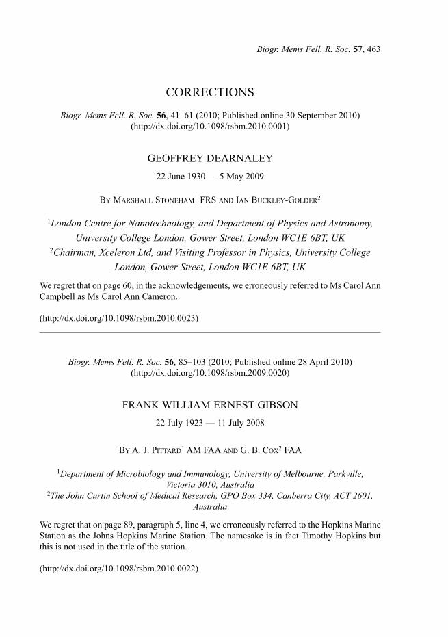
We regret that on page 89, paragraph 5, line 4, we erroneously referred to the Hopkins Marine station as the Johns Hopkins Marine station. The namesake is in fact Timothy Hopkins but this is not used in the title of the station.
(http://dx.doi.org/10.1098/rsbm.2010.0022)
Biogr. Mems Fell. R. Soc. 57, 463