Embed Size (px)
Citation preview

Francisco Pernas, MD – Presenter
Patricia Maeso, MD – Discussant The University of Texas Medical Branch
Department of Otolaryngology
Grand Rounds Presentation
May 29, 2009

Emil Zuckerkandl

Outline Definitions
Background and incidence
Anatomy and embryology
Patient evaluation
FESS Concepts of Surgery
Controversy in Sinus Surgery
Conclusion


Functional Endoscopic Sinus Surgery Replaced old practice of obliterating sinuses and
removing mucosa. Concept of irreversibly diseased mucosa refuted.
Functional aspect refers to:
Preserving normal structures
Removing only obstruction
Preserving mucosa
Attempt to restore function

Incidence Estimated at 14% of American population
$1.77 billion per year spent on rhinosinusitis
CRS ranks fifth compared to all diseases in frequency of antibiotic use associated with treatment.
CRS affects 32 million ppl/yr
Accounts for 11.6 million visits to physicians' offices.
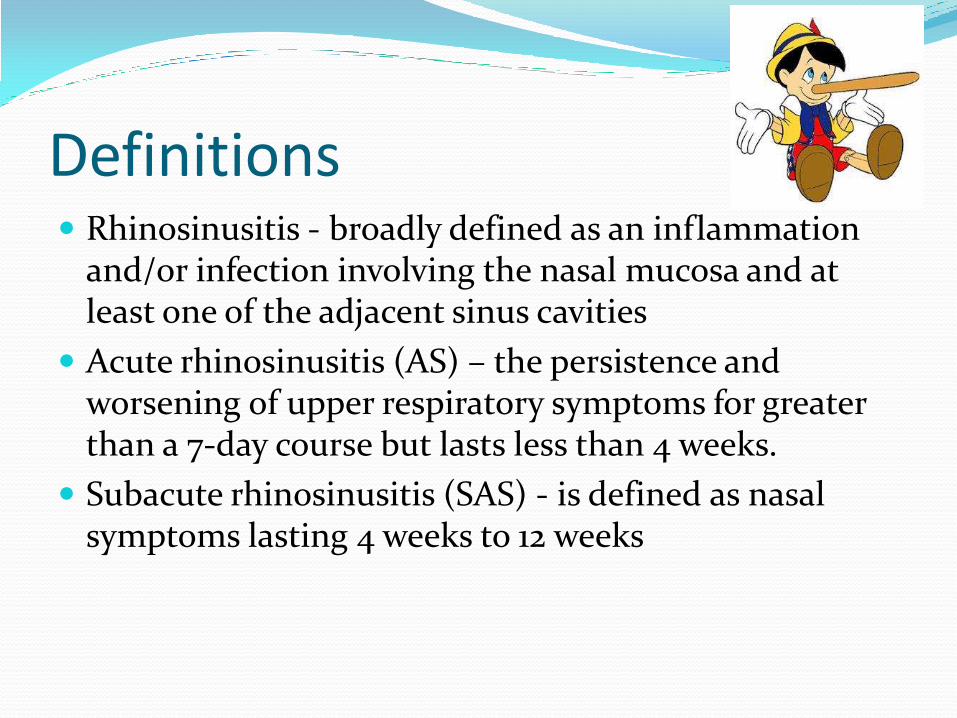
Definitions Rhinosinusitis - broadly defined as an inflammation
and/or infection involving the nasal mucosa and at least one of the adjacent sinus cavities
Acute rhinosinusitis (AS) – the persistence and worsening of upper respiratory symptoms for greater than a 7-day course but lasts less than 4 weeks.
Subacute rhinosinusitis (SAS) - is defined as nasal symptoms lasting 4 weeks to 12 weeks

Definitions Chronic Rhinosinusitis (CRS) – persistence mucosal
inflammation for > 12 consecutive weeks despite medical therapy or occurrence of more than four episodes of symptoms a year with persistent radiographic changes
Chronic Recurrent Rhinosinusitis (CRRS) - consists of multiple acute episodes with complete resolution of disease between episodes


Embryology Two processes:
Embryo head develops into a structure with two distinct nasal cavities
Lateral nasal walls invaginate to create complex folds known as turbinates

Embryology Development of sinuses – 6-8 weeks of gestation
6th week – Simple lateral nasal wall
7th week – Three axial furrows form, give rise to turbinates
10th week - Dev of maxillary sinus (invagination of the middle meatus) and uncinate process & the bulla ethmoidalis form a narrow groove known as the hiatus semilunaris

Embryology 14th week - the anterior ethmoidal cells appear as
several invaginations from the upper middle meatus and the posterior ethmoidal cells from the floor of the superior meatus.

Embryology
56th day

Embryology
60th Day

Embryology
63 days


Ethmoid anatomy is complex: Labyrinth
Lamellae
1st - Uncinate
2nd - Ethmoid bulla
3rd - Basal lamella of middle turbinate
4th - Superior turbinate
Ethmoid anatomy


Drainage Frontal, anterior
ethmoid & maxillary – OMC
Posterior Ethmoids – Superior meatus
Sphenoid sinus – Sphenoid-ethmoidal recess

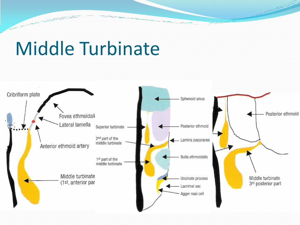
Middle Turbinate Three components
First – Anterior, oriented in a sagittal plane and attached to skull base
Second – Middle, oriented in a frontal plane and attached to lamina papyracea (AKA basal lamella and separates ant from post ethmoids)
Third – Posterior, oriented in a horizontal plane and attaches to perpendicular plate of palate (forms roof of middle meatus, anterior to sphenopalatine foramen)
Middle Turbinate


Ostiomeatal Complex (OMC) AKA – Anterior Ethmoid Middle Meatus Complex
Common drainage for frontal, maxillary and anterior ethmoid sinuses.

OMC

OMC Infundibulum – funnel shaped area whereby the
maxillary, ant ethmoid and frontal sinuses drains
Uncinate process– Sickle shaped bony ethmoidal structure
Hiatus Semilunaris – Half-moon shape opening of infundibulum

Uncinate Process
Attaches to the following structures:
1. Inf & far post. – To ethmoid process of inf. Turb

Uncinate Process 2. Ant & far sup. – To lamina
papyracea, skull base or mid turb

3. Laterally – Lamina papyracea and fontanelle area

Uncinate Process


Bulla Ethmoidalis Anterior ethmoid air cells
attached to lamina papyrcea and usually open into lateral sinus


Sinus Lateralis = Suprabullar recess and retrobullar recess

SBR
RBR
Sinus Lateralis
Middle turbinate: Horizontal and vertical basal lamella

*
*
SBR and RBR

Sphenoid Ostium Medial to posterior sup. turbinate
Located between nasal septum and inferior aspect of sup. turbinate
Located at the same level as the roof of the maxillary sinus
Located 4 microdebrider/suction tip breaths above the choanae
Located 7cm from nasal crest at 30°

Sphenoid Ostium

Sphenoid Sinus Relationships of important structures:
Optic nerve – superior-lateral
Carotid artery/cav sinus – mid-lateral
Vidian nerve and maxillary nerve – inferior-lateral
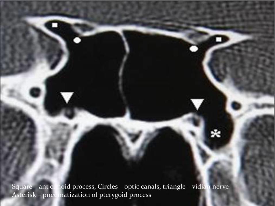
Square – ant clinoid process, Circles – optic canals, triangle – vidian nerve Asterisk – pneumatization of pterygoid process

Sphenoid Classification

Onodi Cells or Sphenoethmoid cells

Optic Canal in Onodi Cells

Keros classification Cribriform plate
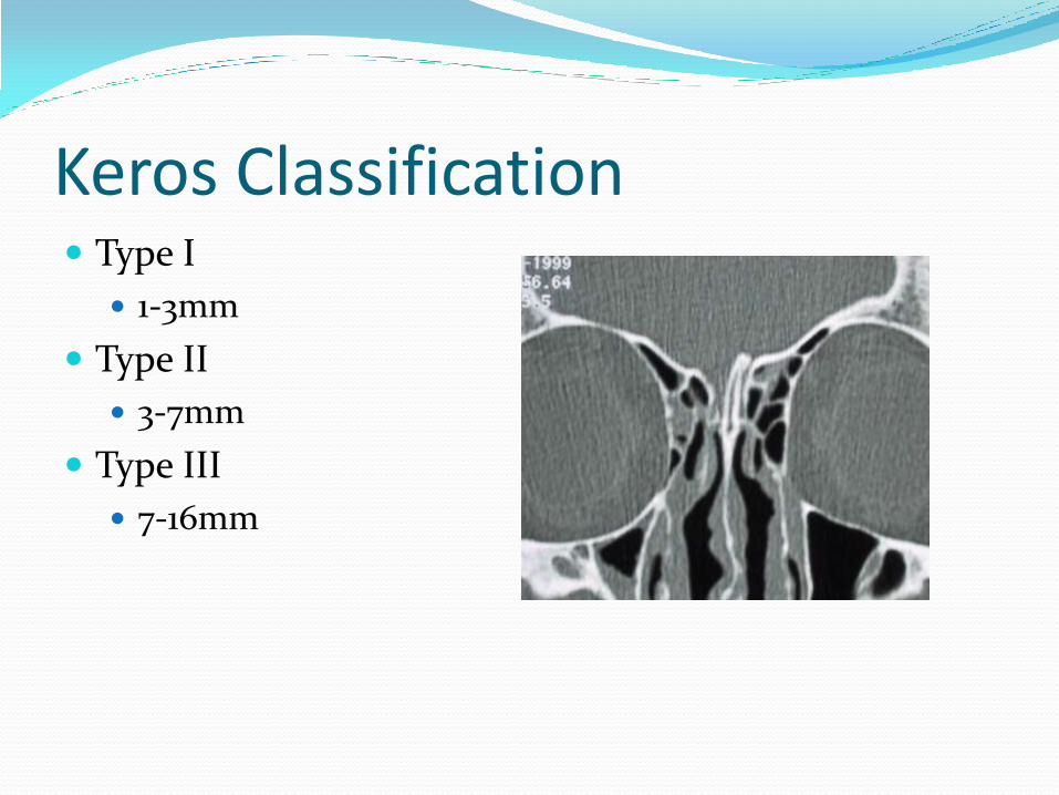
Keros Classification Type I
1-3mm
Type II
3-7mm
Type III
7-16mm

Frontal Cells

Frontal Recess Anatomic Boundries:
Ant – unicate process & agger nasi Post – bulla ethmoidalisand suprabullar lamella Lateral – lamina papyracea Medially – hiatus semilunaris or middle turb Inf – Ethmoid infundibulum Sup – Fovea ethmoidalis, supraorbital air cell, anterior ethmoid
artery and frontal ostium

Frontal Sinus


Patient evaluation Include in history:
Detailed CC
Allergy, asthma, asa sensitivity and polyps
For patients with CRS
Facial pain, congestion, nasal obstruction, drainage and hyposmia
Complete pmhx and pshx to identify co-morbidities
A review of the medical care a patient has received prior to evaluation is also important.

Patient evaluation Complete head and neck exam to include:
basic ocular examination
Visual fields, extraocular eye movement
anterior rhinoscopy
Evaluate septal deviations, character of mucosa, presence of polyps
nasal endoscopy (typically 30°)
Floor, nasopharynx, middle meatus, sphenoethmoidal recess,

Pre-op CT Evaluation CLOSE Technique
C – Cribriform
L – Lamina Papyracea
O – Orbits, onodi cell, Optic Nerve
S – Sphenoid, Skull Base
E – Ethmoid Arteries

C - Cribriform Assess the Keros type
Look for assymetry


L – Lamina Papyracea Check for dehiscence or pathologic fractures

O – Orbit, Optic Nerve, Onodi Cells Check for dehiscence
Assess for onodi cells (superior-lateral to sphenoid)
Orbital slope
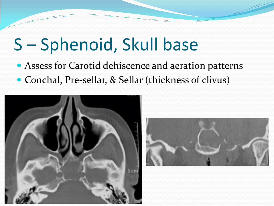
S – Sphenoid, Skull base Assess for Carotid dehiscence and aeration patterns
Conchal, Pre-sellar, & Sellar (thickness of clivus)

Skull base Assess slope of
skull base
Assess if roof of sphenoid is level with skull base

E – Ethmoid Artery


Concepts of surgery

Role of surgery Should be considered as adjunctive to medical therapy
CRS is an inflammatory and multifactorial disease
Underlying causes:
environmental, reactive airway disease, result from generalized host factors, or genetic
Institute medical therapy first prior to surgery unless impending complications
Continued medical therapy is required following surgery to avoid recurrence

Defined surgical substeps are defined according to specific pathophysiologic obstruction that exist based on microanatomy

Antrostomy Some speculate nitric oxide produced in maxillary
sinus has bacteriostatic properties, therefore better to keep antrostomy small
Uncinate must be completely removed, source of recurrence.
Mucociliary clearance remains t/o natural os
Antrostomy must include the natural osium and accessory osium if present

Recirculation

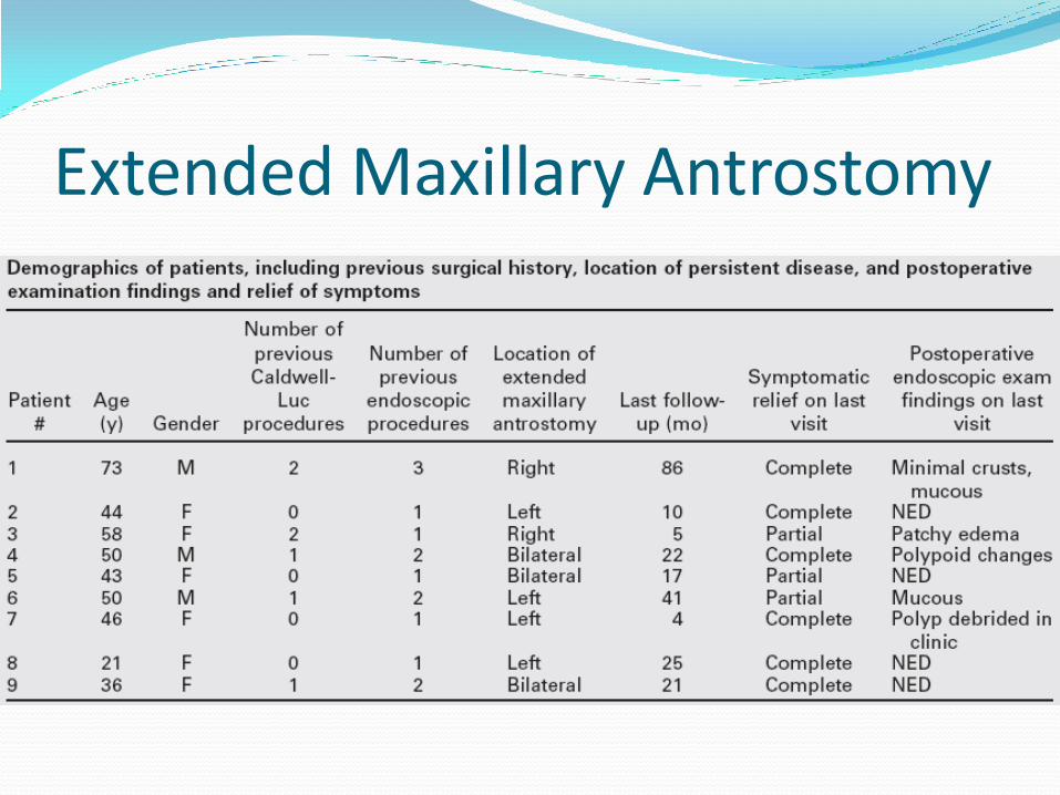
Extended Maxillary Antrostomy Advocated by some (R. Casiano) in refractory
maxillary disease
Middle meatal sinusotomy opened widely anteriorly (up to NLD), posteriorly to post wall of max sinus, superiorly to roof of max sinus and inferiorly to inferior turbinate.
Inferior maxillary antrostomy performed inferiorly into the inferior meatus, post to Hasner’s valve (lacrimal punctum).

Extended Maxillary Antrostomy
Extended Maxillary Antrostomy

Frontal Sinusotomy Question on to perform or not
Do as little as possible but as much as necessary Some advocate ethmoid dissection and monitor
Graduated approach to frontal sinuses
Should evaluate need with sagittal recons
Evaluate A-P and Mediolateral dimensions, asses neo-osteogenesis and pneumatization

Frontal Sinusotomy Common causes of Frontal sinus disease:
1. Infundibular disease obstructing frontal recess
2. Mucosal disease and expansion of the agger nasi air cells

Balloon Sinuplasty

Balloon Sinuplasty Developed in 2006
Different from prior french biliary catether in that new technique can fracture bones
Kennedy concludes that this technique may lead to bacterial introduction and subsequent osteitis, mucositis, and mucoceles.

Frontal Balloon Sinuplasty Bolger et. al. in ‘07 published results
24 week f/u
Exclusion criteria patients with extensive sinonasal polyps, prior surgery, CF
Enrolled 115 patients
f/u patency was 80.5%
Patency could not be assessed in 17.9% secondary to anatomy
Nonpatent 1.6%

Frontal Balloon Sinuplasty Revision was required in three sinuses (1%) and three
patients (2.75%)
SNOT-20 scores improved
Reported 9 cases of baceterial sinusitis, managed with oral abx
No other adverse events reported


References 1. Diseases of the Sinuses: Diagnosis and Management. Kennedy.
Chapters 1, 2, 3, 15, and 16 2. Head and Neck – Otolaryngology. Bailey. Chapters 21, 25, 26. 3. Endoscopic Sinus Surgery Dissection Manual With Cdrom.
Casiano 4. Endoscopic Anatomy of the lateral nasal wall, ostiomeatal
complex and anterior skull base, a step-by-step guide. Reda Kamel
5. Endoscopic diagnosis and surgery of the paranasal sinuses and the anterior skull base. Heinz Stammberger
6. Rhinology and Sinus Disease, a problem-oriented approach. Steven D. Schaefer
7. Nasal and Sinus Surgery. Steven Marks. Sections 1, 2, and 3.