Embed Size (px)
Citation preview
O
R
F
A
H
Tc1oati
aayo
1h
rthopaedics & Traumatology: Surgery & Research (2013) 99S, S160—S170
Available online at
www.sciencedirect.com
EVIEW ARTICLE
ractures in children younger than 18 months
. Fassier ∗, P. Gaucherand, R. Kohler
ospices civils de Lyon et université Lyon 1, hôpital Femme-Mère-Enfant, 69500 Bron cedex, France
Accepted: 5 November 2012
KEYWORDSObstetrical fracture;Toddler fracture;Non-accidentalfracture;Battered childsyndrome;Bone fragility
Summary Fractures in children younger than 18 months occur before the usual walking age.The prognosis is favourable across fracture types and circumstances of occurrence. The causeis obvious in obstetrical injuries, whose risk factors have been well documented. Diaphysealfractures are easy to recognise, whereas challenges may arise with the diagnosis of physealinjuries. Fractures occurring after the neonate is discharged home may be due to accidentalfalls related to clumsiness on the part of the carers. Other possibilities, however, are child abuseand abnormal bone fragility. Thus, the aetiological diagnosis has major medical, social, andlegal implications. Identifying the aetiology is often extremely difficult and benefits from the
involvement of a multidisciplinary team. The literature review presented herein is designed toassist orthopaedic surgeons in the diagnosis and management of children with fractures before18 months of age, in compliance with French legislation, which has undergone major changesover the last quarter century.© 2012 Elsevier Masson SAS. All rights reserved.nmc
O
Ft
he goal of this article is to review the circumstances thatan cause fractures in infants and toddlers younger than8 months of age. We searched the literature for data onbstetrical injuries, child abuse, bone fragility syndromes,nd accidental fractures due to clumsiness on the part ofhe carers. We will not discuss obstetrical brachial plexusnjury, which deserves a separate review.
We chose 18 months as the age cut-off, as the risk ofccidental injuries from falls while walking alone is greaterfter this age, whereas child abuse is most common in
ounger infants. In addition, rapid healing is the rule afterrthopaedic treatment of fractures before 18 months of age.∗ Corresponding author.E-mail address: [email protected] (A. Fassier).
a
E
Ita
877-0568/$ – see front matter © 2012 Elsevier Masson SAS. All rights rettp://dx.doi.org/10.1016/j.otsr.2012.11.004
The role of the orthopaedic surgeon is to diagnose theature of the lesion, treat the fracture and, above all, deter-ine whether the injury was accidental or due to another
ause.
bstetrical fractures
ew orthopaedic studies are available on obstetrical frac-ures. Most of the data come from studies by obstetriciansnd neonatologists.
pidemiology
n the US, the overall incidence of birth injury in single-on infants (including hematomas, lacerations, fractures,nd other injuries) was 2.45/1000 live births, with 9% of
served.
S161
Fa
ac
Fractures in children younger than 18 months
clavicular fractures, 4% of brachial plexus palsies, and 1.3%of other skeletal injuries [1]. In a university-hospital mater-nity unit in Paris, France, the incidence of birth injury was14.2/1000 live births in the 1970s [2].
At our institution, which provides care to women withhigh-risk pregnancies, among the 4293 neonates born alive in2011, seven had fractures, for an incidence of 1.63/1000. Allseven neonates had clavicular fractures; one neonate alsohad brachial plexus palsy and another facial palsy. They wereall delivered vaginally in the cephalic presentation; shoulderdystocia was reported for 4 of them.
Risk factors
Risk factors for long-bone fractures are caesarean delivery,breech presentation, and low birth weight [3]. The only frac-ture more common in vaginal deliveries is clavicular fracture[1]. Risk factors for femoral fractures are twin pregnancy,breech presentation, premature delivery, and osteoporosis.Dystocia is often reported in the medical charts [4].
Fracture types
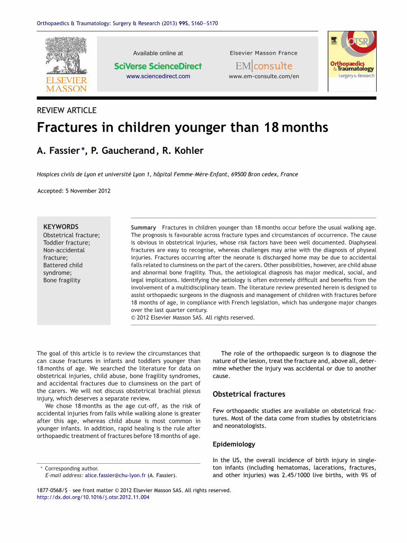
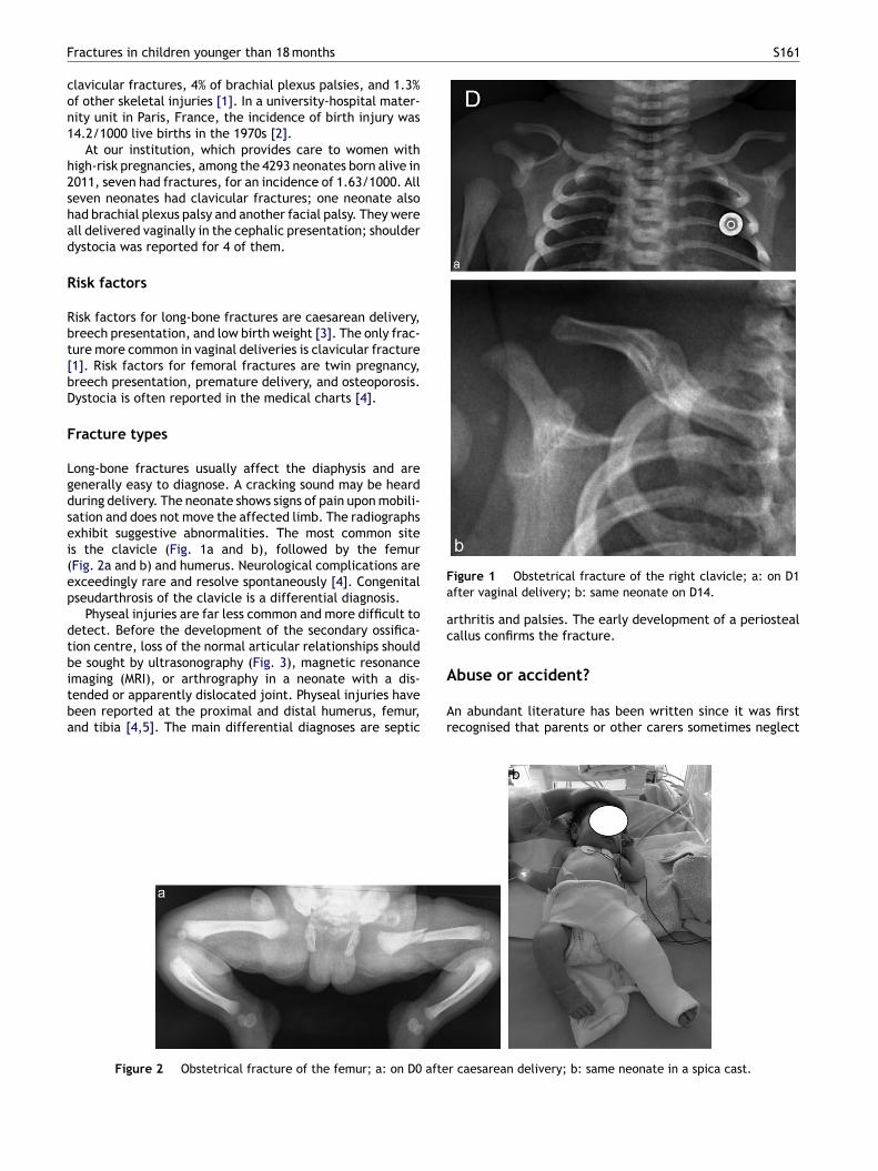
Long-bone fractures usually affect the diaphysis and aregenerally easy to diagnose. A cracking sound may be heardduring delivery. The neonate shows signs of pain upon mobili-sation and does not move the affected limb. The radiographsexhibit suggestive abnormalities. The most common siteis the clavicle (Fig. 1a and b), followed by the femur(Fig. 2a and b) and humerus. Neurological complications areexceedingly rare and resolve spontaneously [4]. Congenitalpseudarthrosis of the clavicle is a differential diagnosis.
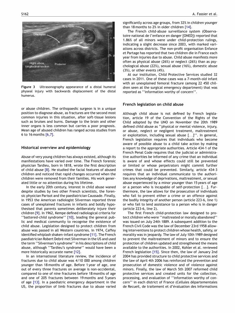
Physeal injuries are far less common and more difficult todetect. Before the development of the secondary ossifica-tion centre, loss of the normal articular relationships shouldbe sought by ultrasonography (Fig. 3), magnetic resonance
imaging (MRI), or arthrography in a neonate with a dis-tended or apparently dislocated joint. Physeal injuries havebeen reported at the proximal and distal humerus, femur,and tibia [4,5]. The main differential diagnoses are septicA
Ar
Figure 2 Obstetrical fracture of the femur; a: on D0 afte
igure 1 Obstetrical fracture of the right clavicle; a: on D1fter vaginal delivery; b: same neonate on D14.
rthritis and palsies. The early development of a periostealallus confirms the fracture.
buse or accident?
n abundant literature has been written since it was firstecognised that parents or other carers sometimes neglect
r caesarean delivery; b: same neonate in a spica cast.
S162
Figure 3 Ultrasonography appearance of a distal humeralph
opcsiM6
H
Ampocca
dsictc‘lcaiptam
fyocaoU
st
t1iaeyoc(
cwdr
F
AtCdooFaaFtiocrtaotwto(
twFimtpaF2tpmprotective services and created units for the collection,
hyseal injury with backwards displacement of the distalumerus.
r abuse children. The orthopaedic surgeon is in a uniqueosition to diagnose abuse, as fractures are the second mostommon injuries in this situation, after soft-tissue lesionsuch as bruises and burns. Damage to the brain and othernner organs is less common but carries a poor prognosis.ean age of abused children has ranged across studies from
to 16 months [6,7].
istorical overview and epidemiology
buse of very young children has always existed, although itsanifestations have varied over time. The French forensichysician Tardieu, born in 1818, wrote the first descriptionf child abuse [8]. He studied the facial features of abusedhildren and noticed that rapid changes occurred when thehildren were removed from their families. His work gener-ted little or no attention during his lifetime.
In the early 20th century, interest in child abuse wanedespite studies by two other French scientists, the foren-ic physician Parisot and the paediatrician Caussade. Finally,n 1953 the American radiologist Silverman reported threeases of unexplained fractures in infants and boldly hypo-hesised that parents sometimes deliberately injure theirhildren [9]. In 1962, Kempe defined radiological criteria for‘battered-child syndrome’’ [10], leading the general pub-ic and medical community to recognise the existence ofhild abuse. Legislation designed to protect children frombuse was passed in all Western countries. In 1974, Caffeydentified whiplash shaken-infant syndrome [11]. The Frenchaediatrician Robert Debré met Silverman in the US and usedhe term ‘‘Silverman’s syndrome’’ in his descriptions of childbuse, although ‘‘Tardieu’s syndrome’’ would have been aore historically accurate name [12].In an international literature review, the incidence of
ractures due to child abuse was 4/10 000 among childrenounger than 18 months of age. Before 1 year of age, oneut of every three fractures on average is non-accidental,ompared to one of nine fractures before 18 months of age
nd one of 205 fractures between 19 months and 5 yearsf age [13]. In a paediatric emergency department in theS, the proportion of limb fractures due to abuse variedpcd
A. Fassier et al.
ignificantly across age groups, from 32% in children youngerhan 18 months to 2% in older children [14].
The French child-abuse surveillance system (Observa-oire national de l’enfance en danger [ONED]) reported that.86% of all minors were under child-protection rulings,ndicating a slight decrease since 2003, with marked vari-tions across districts. The non-profit organisation Enfancet Partage has reported that two children die in France eachear from injuries due to abuse. Child abuse manifests moreften as physical abuse (26%) or neglect (26%) than as psy-hological abuse (22%), sexual abuse (16%), domestic abuse3%), or other events (4%).
At our institution, Child Protective Services studied 32ases in 2011. One of these cases was a 7-month-old infantith an unexplained femoral fracture (among 22 450 chil-ren seen at the surgical emergency department) that waseported as ‘‘information worthy of concern’’.
rench legislation on child abuse
lthough child abuse is not defined by French legisla-ion, article 19 of the Convention of the Rights of thehild adopted by the UNO on November the 20th 1989efined child abuse as ‘‘physical or mental violence, injury,r abuse, neglect or negligent treatment, maltreatmentr exploitation, including sexual abuse [. . .]’’. In general,rench legislation requires that individuals who becomeware of possible abuse to a child take action by making
report to the appropriate authorities. Article 434-1 of therench Penal Code requires that the judicial or administra-ive authorities be informed of any crime that an individuals aware of and whose effects could still be preventedr limited or whose perpetrators might commit furtherrimes that could be prevented. Similarly, article 434-3equires that an individual communicate to the authori-ies any knowledge of deprivations, maltreatment, or sexualbuse experienced by a minor younger than 15 years of ager a person who is incapable of self-protection [. . .]. Fur-hermore, the law allows for the prosecution of individualsho fail to prevent either a crime or an offence against
he bodily integrity of another person (article 223-6, line 1)r who fail to lend assistance to a person who is in dangerarticle 223-6, line 2).
The first French child-protection law designed to pro-ect children who were ‘‘maltreated or morally abandoned’’as issued on July 24th 1889. A subsequent addition to therench Civil Code was the law of December 23rd 1958 allow-ng interventions to protect children whose health, safety, ororality was in jeopardy. The law of July 10th 1989 designed
o prevent the maltreatment of minors and to ensure therotection of children updated and strengthened the meansvailable to the authorities. In 2002, Kohler et al. reviewedrench legislation [15]. Since then, the law of January 2nd004 has provided structure to child protective services andhe law of April 4th 2006 has reinforced the prevention androsecution of domestic violence and of violence againstinors. Finally, the law of March 5th 2007 reformed child
rocessing, and evaluation of ‘‘information worthy of con-ern’’ in each district of France (Cellules départementalese Recueil, de traitement et d’évaluation des Informations
S163
Fo
s(rm
oAmcptraa
esft(fm
R
Iftssla
Fractures in children younger than 18 months
Préoccupantes [CRIP]). This law replaces the critical terms‘‘abuse’’ and ‘‘maltreatment’’ by the term ‘‘child who is,or may be, in danger’’.
In the French code of medical deontology, articlesR.4127-43 and R.4127-44 of the public health code indi-cate that physicians should protect children whose healthseems misunderstood or inadequately preserved by the fam-ily: when a physician sees a minor who is the victim of abuseor deprivation, he or she must use the most appropriatemeans to protect the minor, while exercising caution andcircumspection. No punitive action can be taken as a resultof abuse or deprivation being reported by a physician tothe appropriate authorities. In this situation, article 226-14 of the French penal code specifies clearly that patientconfidentiality rules do not apply.
Risk factors, protective factors, and warning signs
These data are of statistical usefulness only, as any child inany environment can be the victim of abuse.
Risk factors are related to the family (difficult liv-ing conditions, social isolation, or very young parents),the perinatal period (unreported, closely spaced or mul-tiple pregnancy, early separation of the neonate from themother), or the child (unwanted pregnancy, preterm birth,disabled child, or child exhibiting challenging behaviours).
Protective factors are the factors related to the potentialof the parents and environment to ensure the protection ofthe child.
Warning signs include absence of regular health visits,developmental delay, and fearful or shy behaviour on thepart of the infant. The presence of more than one warningsign may indicate that the child is in danger.
Diagnosis
Fractures in children younger than 18 months are usuallydiagnosed in the emergency room. The child is brought inby the parents or another caregiver. Several items of theclinical interview can suggest a non-accidental injury:
• the stated reason for the visit may bear no relation to theinjury, which is discovered fortuitously;
• there may be a long time from symptom onset to the visit;• the mechanism of the injury may be unexplained, or
the explanations given may seen inconsistent with thefracture, in children who are too young to explain whathappened;
• the reported circumstances of the injury may vary acrossindividuals or over time;
• the child may have a history of injuries or of frequentemergency-room visits.
Clinical presentation
A thorough clinical evaluation should be performed by a mul-
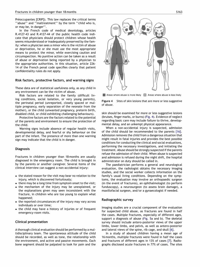
tidisciplinary team. The spontaneous attitude of the childshould be recorded, as well as tone, the relationship withthe environment, and active and passive movements. Eachbone segment should be palpated to look for pain and the1ag
igure 4 Sites of skin lesions that are more or less suggestivef abuse.
kin should be examined for more or less suggestive lesionsbruises, finger marks, or burns) (Fig. 4). Evidence of neglectegarding basic care may include failure to thrive, develop-ental delay, and an unkempt physical appearance.When a non-accidental injury is suspected, admission
f the child should be recommended to the parents [16].dmission removes the child from a dangerous situation thatight result in fatal injuries and provides the best possible
onditions for conducting the clinical and social evaluations,erforming the necessary investigations, and initiating thereatment. Abuse should be strongly suspected if the parentsefuse the admission of their child. When abuse is suspectednd admission is refused during the night shift, the hospitaldministrator on duty should be called in.
The paediatrician performs a general and neurologicalvaluation, the radiologist obtains the necessary imagingtudies, and the social worker collects information on theamily’s usual living conditions. Depending on the symp-oms, the evaluation may involve an orthopaedic surgeonin the event of fractures), an ophthalmologist (to performunduscopy), a neurosurgeon (to assess brain damage), aaxillofacial surgeon, and/or a gynaecologist if needed.
adiographic survey
maging studies are a crucial component of the evaluationor suspected child abuse, as fractures are found in halfhe cases. Multiple fractures, especially of different ages,upport a diagnosis of abuse (Fig. 5a and b). The skeletalurvey should include antero-posterior views of the upperimbs, lower limbs, and pelvis, as well as antero-posteriornd lateral views of the spine, rib cage, and skull [6].
In a study of abused children having a mean age of6 months, multiple fractures were found in half the casesnd fractures of different ages in 13% of cases [7]. Radio-raphs disclosed acute fractures in 77% of cases. The sites
S164
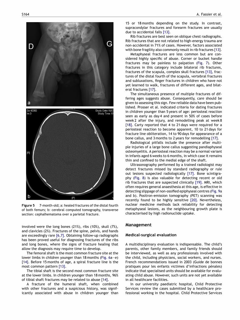
Figure 5 7-month-old; a: healed fractures of the distal fourthos
iaahaa
l[m
ao
wi
1sd
Rnw
sffftaye
fglisw[pfb
poit
doprodarnmc
M
M
ApbtFpiain all healthcare facilities.
f both femurs; b: cerebral computed tomography, transverseection: cephalhematoma over a parietal fracture.
nvolved were the long bones (21%), ribs (10%), skull (7%),nd clavicles (2%). Fractures of the spine, pelvis, and handsre exceedingly rare [6,7]. Obtaining follow-up radiographsas been proved useful for diagnosing fractures of the ribsnd long bones, where the signs of fracture healing thatllow the diagnosis may require time to develop.
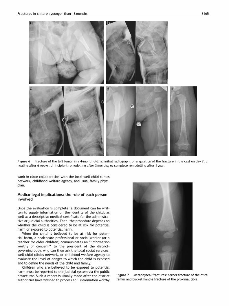
The femoral shaft is the most common fracture site at theower limbs in children younger than 18 months (Fig. 6a—e)14]. Before 15 months of age, a spiral fracture line is theost common pattern [13].The tibial shaft is the second most common fracture site
t the lower limbs. In children younger than 18 months, 96%f tibial shaft fractures may be related to abuse [14].
A fracture of the humeral shaft, when combinedith other fractures and a suspicious history, was signif-
cantly associated with abuse in children younger thanSf
A. Fassier et al.
5 or 18 months depending on the study. In contrast,upracondylar fractures and forearm fractures are usuallyue to accidental falls [13].
Rib fractures are best seen on oblique chest radiographs.ib fractures that are not related to high-energy trauma areon-accidental in 71% of cases. However, factors associatedith bone fragility also commonly result in rib fractures [13].
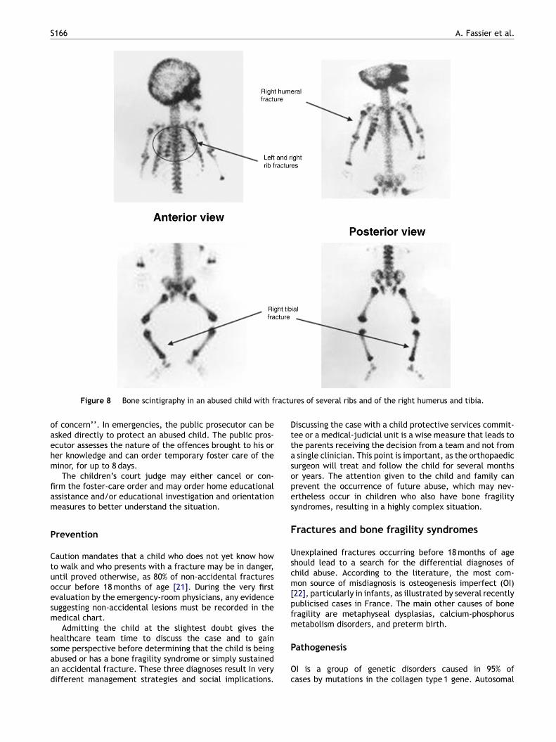
Metaphyseal fractures are less common but are con-idered highly specific of abuse. Corner or bucket handleractures may be painless to palpation (Fig. 7). Otherractures in this category include bilateral rib fractures,ractures of the scapula, complex skull fractures [13], frac-ures of the distal fourth of the scapula, vertebral fracturesnd subluxations, finger fractures in children who have notet learned to walk, fractures of different ages, and bilat-ral fractures [17].
The simultaneous presence of multiple fractures of dif-ering ages suggests abuse. Consequently, care should beiven to assessing this sign. Few reliable data have been pub-ished. Prosser et al. indicated criteria for dating fracturesn children younger than 5 years of age: periosteal reactioneen as early as day 4 and present in 50% of cases beforeeek 2 after the injury, and remodelling peak at week 8
18]. Carty reported that 4 to 21 days were required for aeriosteal reaction to become apparent, 10 to 21 days forracture line obliteration, 14 to 90 days for appearance of aone callus, and 3 months to 2 years for remodelling [17].
Radiological pitfalls include the presence after multi-le injuries of a large bone callus suggesting pandiaphysealsteomyelitis. A periosteal reaction may be a normal variantn infants aged 6 weeks to 6 months, in which case it remainshin and confined to the medial edge of the shaft.
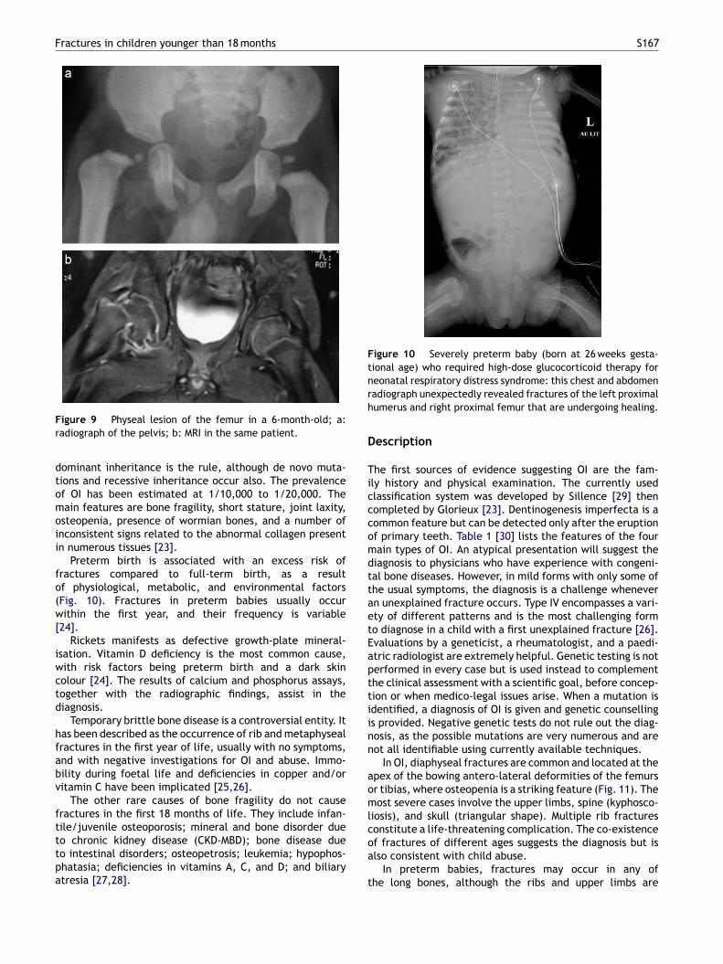
Ultrasonography performed by a trained radiologist mayetect fractures missed by standard radiography or ruleut lesions suspected radiologically [17]. Bone scintigra-hy (Fig. 8) is also valuable for detecting recent or oldib fractures that are suspected clinically [19]. MRI, whichften requires general anaesthesia at this age, is effective inetecting slippage of non-ossified epiphyseal centres (Fig. 9and b). Positron-emission tomography (PET) scanning wasecently found to be highly sensitive [20]. Nevertheless,uclear medicine methods lack reliability for detectingetaphyseal lesions, as the neighbouring growth plate is
haracterised by high radionuclide uptake.
anagement
edical-surgical evaluation
multidisciplinary evaluation is indispensable. The child’sarents, other family members, and family friends shoulde interviewed, as well as any professionals involved withhe child, including physicians, social workers, and nurses.rench recommendations issued in 2003 (Guide de bonnesratiques pour les enfants victimes d’infractions pénales)ndicate that specialised units should be available for evalu-ting child abuse. However, such units are not yet available
In our university paediatric hospital, Child Protectiveervices review the cases submitted by a healthcare pro-essional working in the hospital. Child Protective Services
Fractures in children younger than 18 months S165
al radiograph; b: angulation of the fracture in the cast on day 7; c:e: complete remodelling after 1 year.
Figure 6 Fracture of the left femur in a 4-month-old; a: initihealing after 6 weeks; d: incipient remodelling after 3 months;
work in close collaboration with the local well-child clinicsnetwork, childhood welfare agency, and usual family physi-cian.
Medico-legal implications: the role of each personinvolved
Once the evaluation is complete, a document can be writ-ten to supply information on the identity of the child, aswell as a descriptive medical certificate for the administra-tive or judicial authorities. Then, the procedure depends onwhether the child is considered to be at risk for potentialharm or exposed to potential harm.
When the child is believed to be at risk for poten-tial harm, a healthcare professional or social worker (or ateacher for older children) communicates an ‘‘informationworthy of concern’’ to the president of the district-governing body, who can then ask the local social services,well-child clinics network, or childhood welfare agency toevaluate the level of danger to which the child is exposedand to define the needs of the child and family.
Children who are believed to be exposed to potentialharm must be reported to the judicial system via the publicprosecutor. Such a report is usually made after the districtauthorities have finished to process an ‘‘information worthy
Figure 7 Metaphyseal fractures: corner fracture of the distalfemur and bucket handle fracture of the proximal tibia.
S166 A. Fassier et al.
ractu
oaehm
fiam
P
Ctuoesm
hsaad
Dttasopes
F
Uscm[pfm
Figure 8 Bone scintigraphy in an abused child with f
f concern’’. In emergencies, the public prosecutor can besked directly to protect an abused child. The public pros-cutor assesses the nature of the offences brought to his orer knowledge and can order temporary foster care of theinor, for up to 8 days.The children’s court judge may either cancel or con-
rm the foster-care order and may order home educationalssistance and/or educational investigation and orientationeasures to better understand the situation.
revention
aution mandates that a child who does not yet know howo walk and who presents with a fracture may be in danger,ntil proved otherwise, as 80% of non-accidental fracturesccur before 18 months of age [21]. During the very firstvaluation by the emergency-room physicians, any evidenceuggesting non-accidental lesions must be recorded in theedical chart.Admitting the child at the slightest doubt gives the
ealthcare team time to discuss the case and to gain
ome perspective before determining that the child is beingbused or has a bone fragility syndrome or simply sustainedn accidental fracture. These three diagnoses result in veryifferent management strategies and social implications.P
Oc
res of several ribs and of the right humerus and tibia.
iscussing the case with a child protective services commit-ee or a medical-judicial unit is a wise measure that leads tohe parents receiving the decision from a team and not from
single clinician. This point is important, as the orthopaedicurgeon will treat and follow the child for several monthsr years. The attention given to the child and family canrevent the occurrence of future abuse, which may nev-rtheless occur in children who also have bone fragilityyndromes, resulting in a highly complex situation.
ractures and bone fragility syndromes
nexplained fractures occurring before 18 months of agehould lead to a search for the differential diagnoses ofhild abuse. According to the literature, the most com-on source of misdiagnosis is osteogenesis imperfect (OI)
22], particularly in infants, as illustrated by several recentlyublicised cases in France. The main other causes of boneragility are metaphyseal dysplasias, calcium-phosphorusetabolism disorders, and preterm birth.
athogenesis
I is a group of genetic disorders caused in 95% ofases by mutations in the collagen type 1 gene. Autosomal
Fractures in children younger than 18 months S167
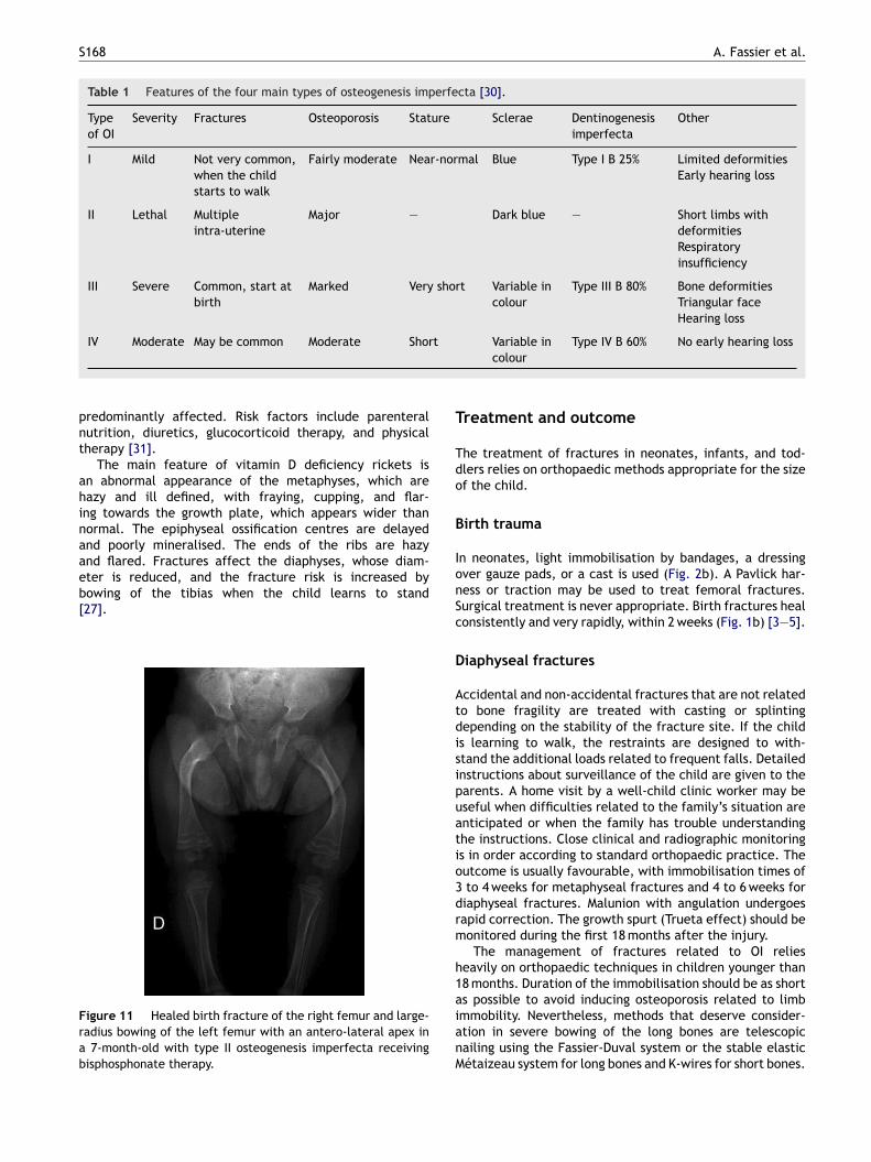
Figure 10 Severely preterm baby (born at 26 weeks gesta-tional age) who required high-dose glucocorticoid therapy forneonatal respiratory distress syndrome: this chest and abdomenrh
D
TicccomdttaetEapttiinn
aomlc
Figure 9 Physeal lesion of the femur in a 6-month-old; a:radiograph of the pelvis; b: MRI in the same patient.
dominant inheritance is the rule, although de novo muta-tions and recessive inheritance occur also. The prevalenceof OI has been estimated at 1/10,000 to 1/20,000. Themain features are bone fragility, short stature, joint laxity,osteopenia, presence of wormian bones, and a number ofinconsistent signs related to the abnormal collagen presentin numerous tissues [23].
Preterm birth is associated with an excess risk offractures compared to full-term birth, as a resultof physiological, metabolic, and environmental factors(Fig. 10). Fractures in preterm babies usually occurwithin the first year, and their frequency is variable[24].
Rickets manifests as defective growth-plate mineral-isation. Vitamin D deficiency is the most common cause,with risk factors being preterm birth and a dark skincolour [24]. The results of calcium and phosphorus assays,together with the radiographic findings, assist in thediagnosis.
Temporary brittle bone disease is a controversial entity. Ithas been described as the occurrence of rib and metaphysealfractures in the first year of life, usually with no symptoms,and with negative investigations for OI and abuse. Immo-bility during foetal life and deficiencies in copper and/orvitamin C have been implicated [25,26].
The other rare causes of bone fragility do not causefractures in the first 18 months of life. They include infan-tile/juvenile osteoporosis; mineral and bone disorder dueto chronic kidney disease (CKD-MBD); bone disease due
to intestinal disorders; osteopetrosis; leukemia; hypophos-phatasia; deficiencies in vitamins A, C, and D; and biliaryatresia [27,28].oa
t
adiograph unexpectedly revealed fractures of the left proximalumerus and right proximal femur that are undergoing healing.
escription
he first sources of evidence suggesting OI are the fam-ly history and physical examination. The currently usedlassification system was developed by Sillence [29] thenompleted by Glorieux [23]. Dentinogenesis imperfecta is aommon feature but can be detected only after the eruptionf primary teeth. Table 1 [30] lists the features of the fourain types of OI. An atypical presentation will suggest theiagnosis to physicians who have experience with congeni-al bone diseases. However, in mild forms with only some ofhe usual symptoms, the diagnosis is a challenge whenevern unexplained fracture occurs. Type IV encompasses a vari-ty of different patterns and is the most challenging formo diagnose in a child with a first unexplained fracture [26].valuations by a geneticist, a rheumatologist, and a paedi-tric radiologist are extremely helpful. Genetic testing is noterformed in every case but is used instead to complementhe clinical assessment with a scientific goal, before concep-ion or when medico-legal issues arise. When a mutation isdentified, a diagnosis of OI is given and genetic counsellings provided. Negative genetic tests do not rule out the diag-osis, as the possible mutations are very numerous and areot all identifiable using currently available techniques.
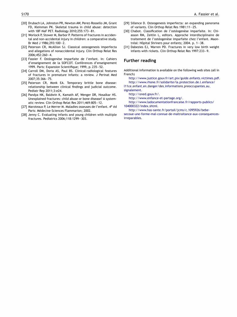
In OI, diaphyseal fractures are common and located at thepex of the bowing antero-lateral deformities of the femursr tibias, where osteopenia is a striking feature (Fig. 11). Theost severe cases involve the upper limbs, spine (kyphosco-
iosis), and skull (triangular shape). Multiple rib fracturesonstitute a life-threatening complication. The co-existence
f fractures of different ages suggests the diagnosis but islso consistent with child abuse.In preterm babies, fractures may occur in any ofhe long bones, although the ribs and upper limbs are
S168 A. Fassier et al.
Table 1 Features of the four main types of osteogenesis imperfecta [30].
Typeof OI
Severity Fractures Osteoporosis Stature Sclerae Dentinogenesisimperfecta
Other
I Mild Not very common,when the childstarts to walk
Fairly moderate Near-normal Blue Type I B 25% Limited deformitiesEarly hearing loss
II Lethal Multipleintra-uterine
Major — Dark blue — Short limbs withdeformitiesRespiratoryinsufficiency
III Severe Common, start atbirth
Marked Very short Variable incolour
Type III B 80% Bone deformitiesTriangular faceHearing loss
IV Moderate May be common Moderate Short Variable in Type IV B 60% No early hearing loss
predominantly affected. Risk factors include parenteralnutrition, diuretics, glucocorticoid therapy, and physicaltherapy [31].
The main feature of vitamin D deficiency rickets isan abnormal appearance of the metaphyses, which arehazy and ill defined, with fraying, cupping, and flar-ing towards the growth plate, which appears wider thannormal. The epiphyseal ossification centres are delayedand poorly mineralised. The ends of the ribs are hazyand flared. Fractures affect the diaphyses, whose diam-eter is reduced, and the fracture risk is increased bybowing of the tibias when the child learns to stand[27].
Figure 11 Healed birth fracture of the right femur and large-radius bowing of the left femur with an antero-lateral apex ina 7-month-old with type II osteogenesis imperfecta receivingbisphosphonate therapy.
T
Tdo
B
IonSc
D
Atdisipuatio3drm
h1aianM
colour
reatment and outcome
he treatment of fractures in neonates, infants, and tod-lers relies on orthopaedic methods appropriate for the sizef the child.
irth trauma
n neonates, light immobilisation by bandages, a dressingver gauze pads, or a cast is used (Fig. 2b). A Pavlick har-ess or traction may be used to treat femoral fractures.urgical treatment is never appropriate. Birth fractures healonsistently and very rapidly, within 2 weeks (Fig. 1b) [3—5].
iaphyseal fractures
ccidental and non-accidental fractures that are not relatedo bone fragility are treated with casting or splintingepending on the stability of the fracture site. If the childs learning to walk, the restraints are designed to with-tand the additional loads related to frequent falls. Detailednstructions about surveillance of the child are given to thearents. A home visit by a well-child clinic worker may beseful when difficulties related to the family’s situation arenticipated or when the family has trouble understandinghe instructions. Close clinical and radiographic monitorings in order according to standard orthopaedic practice. Theutcome is usually favourable, with immobilisation times of
to 4 weeks for metaphyseal fractures and 4 to 6 weeks foriaphyseal fractures. Malunion with angulation undergoesapid correction. The growth spurt (Trueta effect) should beonitored during the first 18 months after the injury.The management of fractures related to OI relies
eavily on orthopaedic techniques in children younger than8 months. Duration of the immobilisation should be as shorts possible to avoid inducing osteoporosis related to limb
mmobility. Nevertheless, methods that deserve consider-tion in severe bowing of the long bones are telescopicailing using the Fassier-Duval system or the stable elasticétaizeau system for long bones and K-wires for short bones.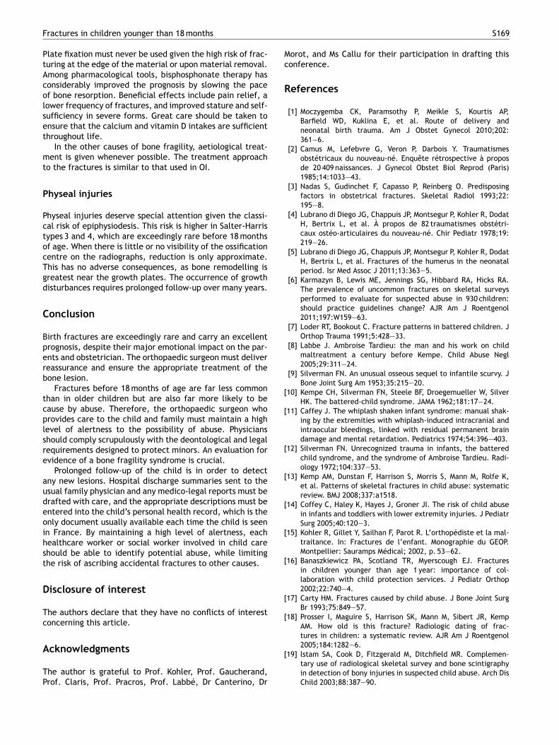
Mc
R
[
[
[
[
[
[
[
[
[
Fractures in children younger than 18 months
Plate fixation must never be used given the high risk of frac-turing at the edge of the material or upon material removal.Among pharmacological tools, bisphosphonate therapy hasconsiderably improved the prognosis by slowing the paceof bone resorption. Beneficial effects include pain relief, alower frequency of fractures, and improved stature and self-sufficiency in severe forms. Great care should be taken toensure that the calcium and vitamin D intakes are sufficientthroughout life.
In the other causes of bone fragility, aetiological treat-ment is given whenever possible. The treatment approachto the fractures is similar to that used in OI.
Physeal injuries
Physeal injuries deserve special attention given the classi-cal risk of epiphysiodesis. This risk is higher in Salter-Harristypes 3 and 4, which are exceedingly rare before 18 monthsof age. When there is little or no visibility of the ossificationcentre on the radiographs, reduction is only approximate.This has no adverse consequences, as bone remodelling isgreatest near the growth plates. The occurrence of growthdisturbances requires prolonged follow-up over many years.
Conclusion
Birth fractures are exceedingly rare and carry an excellentprognosis, despite their major emotional impact on the par-ents and obstetrician. The orthopaedic surgeon must deliverreassurance and ensure the appropriate treatment of thebone lesion.
Fractures before 18 months of age are far less commonthan in older children but are also far more likely to because by abuse. Therefore, the orthopaedic surgeon whoprovides care to the child and family must maintain a highlevel of alertness to the possibility of abuse. Physiciansshould comply scrupulously with the deontological and legalrequirements designed to protect minors. An evaluation forevidence of a bone fragility syndrome is crucial.
Prolonged follow-up of the child is in order to detectany new lesions. Hospital discharge summaries sent to theusual family physician and any medico-legal reports must bedrafted with care, and the appropriate descriptions must beentered into the child’s personal health record, which is theonly document usually available each time the child is seenin France. By maintaining a high level of alertness, eachhealthcare worker or social worker involved in child careshould be able to identify potential abuse, while limitingthe risk of ascribing accidental fractures to other causes.
Disclosure of interest
The authors declare that they have no conflicts of interestconcerning this article.
Acknowledgments
The author is grateful to Prof. Kohler, Prof. Gaucherand,Prof. Claris, Prof. Pracros, Prof. Labbé, Dr Canterino, Dr
[
S169
orot, and Ms Callu for their participation in drafting thisonference.
eferences
[1] Moczygemba CK, Paramsothy P, Meikle S, Kourtis AP,Barfield WD, Kuklina E, et al. Route of delivery andneonatal birth trauma. Am J Obstet Gynecol 2010;202:361—6.
[2] Camus M, Lefebvre G, Veron P, Darbois Y. Traumatismesobstétricaux du nouveau-né. Enquête rétrospective à proposde 20 409 naissances. J Gynecol Obstet Biol Reprod (Paris)1985;14:1033—43.
[3] Nadas S, Gudinchet F, Capasso P, Reinberg O. Predisposingfactors in obstetrical fractures. Skeletal Radiol 1993;22:195—8.
[4] Lubrano di Diego JG, Chappuis JP, Montsegur P, Kohler R, DodatH, Bertrix L, et al. À propos de 82 traumatismes obstétri-caux ostéo-articulaires du nouveau-né. Chir Pediatr 1978;19:219—26.
[5] Lubrano di Diego JG, Chappuis JP, Montsegur P, Kohler R, DodatH, Bertrix L, et al. Fractures of the humerus in the neonatalperiod. Isr Med Assoc J 2011;13:363—5.
[6] Karmazyn B, Lewis ME, Jennings SG, Hibbard RA, Hicks RA.The prevalence of uncommon fractures on skeletal surveysperformed to evaluate for suspected abuse in 930 children:should practice guidelines change? AJR Am J Roentgenol2011;197:W159—63.
[7] Loder RT, Bookout C. Fracture patterns in battered children. JOrthop Trauma 1991;5:428—33.
[8] Labbe J. Ambroise Tardieu: the man and his work on childmaltreatment a century before Kempe. Child Abuse Negl2005;29:311—24.
[9] Silverman FN. An unusual osseous sequel to infantile scurvy. JBone Joint Surg Am 1953;35:215—20.
10] Kempe CH, Silverman FN, Steele BF, Droegemueller W, SilverHK. The battered-child syndrome. JAMA 1962;181:17—24.
11] Caffey J. The whiplash shaken infant syndrome: manual shak-ing by the extremities with whiplash-induced intracranial andintraocular bleedings, linked with residual permanent braindamage and mental retardation. Pediatrics 1974;54:396—403.
12] Silverman FN. Unrecognized trauma in infants, the batteredchild syndrome, and the syndrome of Ambroise Tardieu. Radi-ology 1972;104:337—53.
13] Kemp AM, Dunstan F, Harrison S, Morris S, Mann M, Rolfe K,et al. Patterns of skeletal fractures in child abuse: systematicreview. BMJ 2008;337:a1518.
14] Coffey C, Haley K, Hayes J, Groner JI. The risk of child abusein infants and toddlers with lower extremity injuries. J PediatrSurg 2005;40:120—3.
15] Kohler R, Gillet Y, Sailhan F, Parot R. L’orthopédiste et la mal-traitance. In: Fractures de l’enfant. Monographie du GEOP.Montpellier: Sauramps Médical; 2002, p. 53—62.
16] Banaszkiewicz PA, Scotland TR, Myerscough EJ. Fracturesin children younger than age 1 year: importance of col-laboration with child protection services. J Pediatr Orthop2002;22:740—4.
17] Carty HM. Fractures caused by child abuse. J Bone Joint SurgBr 1993;75:849—57.
18] Prosser I, Maguire S, Harrison SK, Mann M, Sibert JR, KempAM. How old is this fracture? Radiologic dating of frac-tures in children: a systematic review. AJR Am J Roentgenol2005;184:1282—6.
19] lstam SA, Cook D, Fitzgerald M, Ditchfield MR. Complemen-tary use of radiological skeletal survey and bone scintigraphyin detection of bony injuries in suspected child abuse. Arch DisChild 2003;88:387—90.
S
[
[
[
[
[
[
[
[
[
[
[
[
F
AF
ls
170
20] Drubach LA, Johnston PR, Newton AW, Perez-Rossello JM, GrantFD, Kleinman PK. Skeletal trauma in child abuse: detectionwith 18F-NaF PET. Radiology 2010;255:173—81.
21] Worlock P, Stower M, Barbor P. Patterns of fractures in acciden-tal and non-accidental injury in children: a comparative study.Br Med J 1986;293:100—2.
22] Paterson CR, McAllion SJ. Classical osteogenesis imperfectaand allegations of nonaccidental injury. Clin Orthop Relat Res2006;452:260—4.
23] Fassier F. Ostéogenèse imparfaite de l’enfant. In: Cahiersd’enseignement de la SOFCOT. Conférences d’enseignement1999. Paris: Expansion Scientifique; 1999, p. 235—52.
24] Carroll DM, Doria AS, Paul BS. Clinical-radiological featuresof fractures in premature infants: a review. J Perinat Med2007;35:366—75.
25] Paterson CR, Monk EA. Temporary brittle bone disease:relationship between clinical findings and judicial outcome.Pediatr Rep 2011;3:e24.
26] Pandya NK, Baldwin K, Kamath AF, Wenger DR, Hosalkar HS.Unexplained fractures: child abuse or bone disease? A system-atic review. Clin Orthop Relat Res 2011;469:805—12.
27] Maroteaux P, Le Merrer M. Maladies osseuses de l’enfant. 4e édParis: Médecine-Sciences Flammarion; 2002.
28] Jenny C. Evaluating infants and young children with multiplefractures. Pediatrics 2006;118:1299—303.
1
si
A. Fassier et al.
29] Sillence D. Osteogenesis imperfecta: an expanding panoramaof variants. Clin Orthop Relat Res 1981:11—25.
30] Chabot. Classification de l’ostéogenèse imparfaite. In: Chi-asson RM, Zeitlin L, editors. Approche interdisciplinaire detraitement de l’ostéogenèse imparfaite chez l’enfant. Maon-tréal: Hôpital Shriners pour enfants; 2004. p. 1—38.
31] Dabezies EJ, Warren PD. Fractures in very low birth weightinfants with rickets. Clin Orthop Relat Res 1997:233—9.
urther reading
dditional information is available on the following web sites (all inrench)
http://www.justice.gouv.fr/art pix/guide enfants victimes.pdf.http://www.rhone.fr/solidarite/la protection de l enfance/
11ce enfant en danger/des informations preoccupantes auignalement.
http://oned.gouv.fr/.http://www.enfance-et-partage.org/.http://www.ladocumentationfrancaise.fr/rapports-publics/
04000322/index.shtml.http://www.has-sante.fr/portail/jcms/c 1095926/bebe-
ecoue-une-forme-mal-connue-de-maltraitance-aux-consequences-rreparables.