Embed Size (px)
Citation preview
Editorial
Case Report
Fracture of the Femur of A Newborn after Cesarean
Section for Breech Presentation and Fibroid Uterus :
A Case Report and Literature Review
Ibrahima Farikou , Ngo Nonga Bernadette , Handy Eone Daniel , Sosso Maurice Aurélien1 1 1 1
Abstract
Introduction:
Case Report:
Conclusion:
Keywords:
The practice of cesarean section is known to decrease the occurrence of long bone fractures. We
present here an unusual diaphyseal fracture of the femur of a newborn after cesarean section, the only case
observed in our 14 years of practice.
The patient was a 3.4-kg female child born at 38 weeks of gestation. The mother was a primipara
and aged 39 years. Ultrasound examination at 20th week revealed intrauterine fibroids with a breech
presentation. Therefore, elective cesarean section was indicated. There was no apparent bone disorder that
could predispose to sustain femur fracture. The fracture was treated successfully with a bilateral spica cast.
The cesarean section was indicated in an aged primipara, bearer of uterine fibroids, and breech presentation.
She had a good general health status, but her bone density was unknown since this examination is not
routinely performed in our clinical settings in Africa.
Elderly age, primipara status, presence of uterine fibroids, and breech presentation are usual
indications for cesarean section. However, there are not many reports on femur fracture after cesarean
section. Our present case suggests that despite the latest advances in delivery techniques, cesarean section
for breech presentation predisposes the neonate to femoral fractures.
Femur fracture; Cesarean section; Fibroid; Breech presentation; Africa.
Copyright © 2014 by Journal of Orthpaedic Case ReportsJournal of Orthopaedic Case Reports | pISSN 2250-0685 | eISSN | Available on www.jocr.co.in | doi:
This is an Open Access article distributed under the terms of the Creative Commons Attribution Non-Commercial License (http://creativecommons.org/licenses/by-nc/3.0) which permits unrestricted non-commercial use, distribution, and reproduction in any medium, provided the original work is properly cited.
2321-3817 10.13107/jocr.2250-0685.107
What to Learn from this Article?Rare presentation of femur fracture during Cesarian section.
Introduction
Cesarean delivery is designed to reduce the risks of mother and infant birth-related injuries in the newborn in women with a variety of health complications [1]. Jain et al [2] observed that the increase in cesarean section rate from 24.2% to 35.3% in a recent study in New Jersey from 1997 to 2005 led to a notable decrease in newborn injuries from 4.1% to 2.6% at the same time. However, there are few reports of long bone fractures that occurred during cesareans conducted to paradoxically avoid such complications. The hypotheses of risk factors for occurrence of such fractures remain conflicting. Matsubara et al. [3] correlated the occurrence of long bone fractures to only higher birth weight. Nadas et al. [4] reported the correlation between occurrence of bone fractures and cesarean section breech delivery with assistance and low birth weight. The present study reports the first case of cesarean-related femur injury in a neonate in our orthopedic clinics since 14 years of our clinical practice. Our goal is to feed the debate on the risks of fractures associated with cesareans. We also conducted a review of literature which reveals that certain complications in the mother predispose neonates to femur fractures by cesarean. Capobianco et al [5] argue that even if the cesarean helps reduce the risk of occurrence of traumatic
Journal of Orthopaedic Case Reports 2014 Jan-Mar;4(1): Page 18-20
1Department of Surgery and Surgical Specialties, Faculty of
Medicine and Biomedical Sciences, University of Yaoundé I,
Cameroon.
Address of Correspondence
Dr Ibrahima Farikou,
P.O. Box 14572, Yaoundé, Cameroon.
Email: [email protected] 18
Author’s Photo Gallery
Dr. Ibrahima
Farikou
Dr. Ngo Nonga
Bernadette
Dr. Handy Eone
Daniel
Pr Sosso Maurice
Aurélien
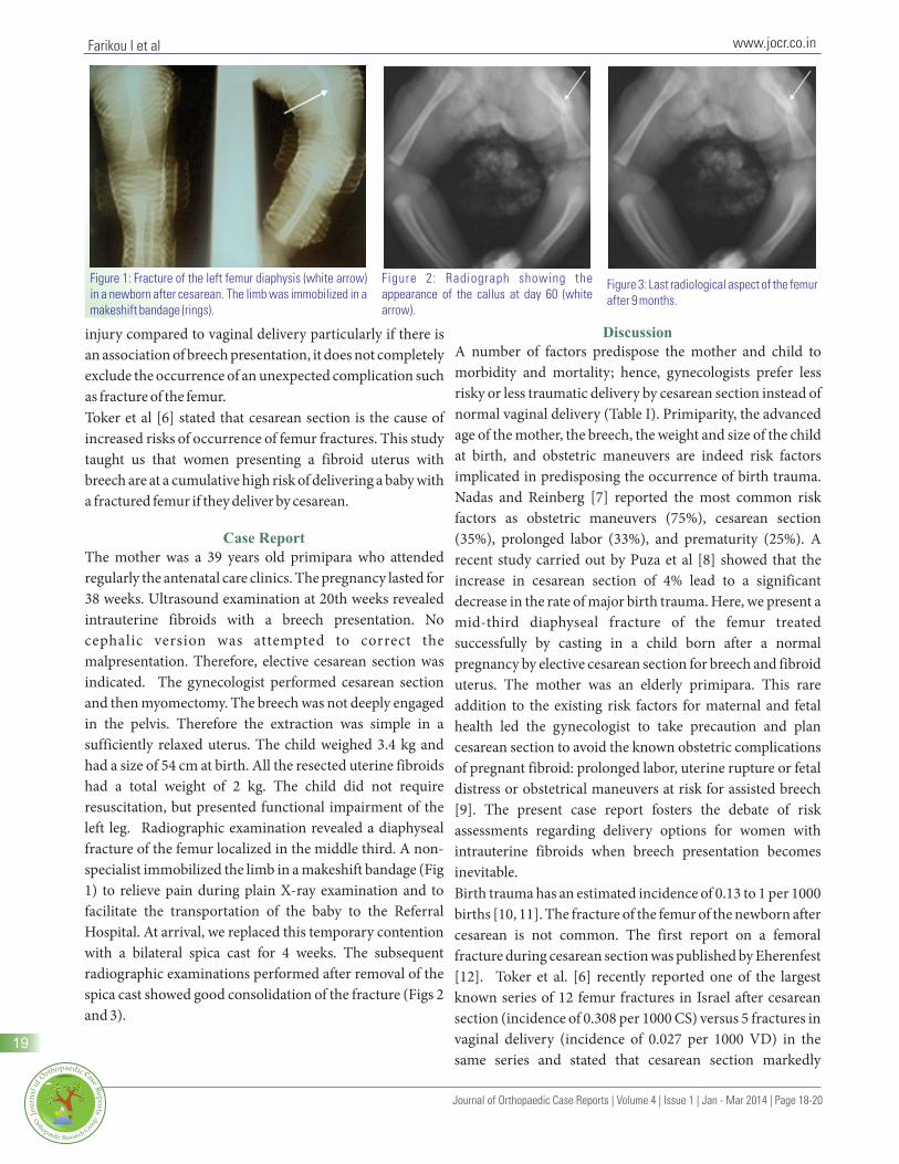
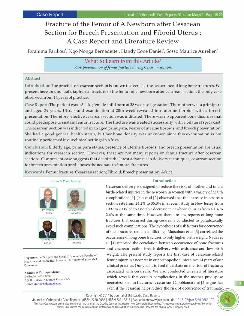
Figure 1: Fracture of the left femur diaphysis (white arrow) in a newborn after cesarean. The limb was immobilized in a makeshift bandage (rings).
Figure 2: Radiograph showing the appearance of the callus at day 60 (white arrow).
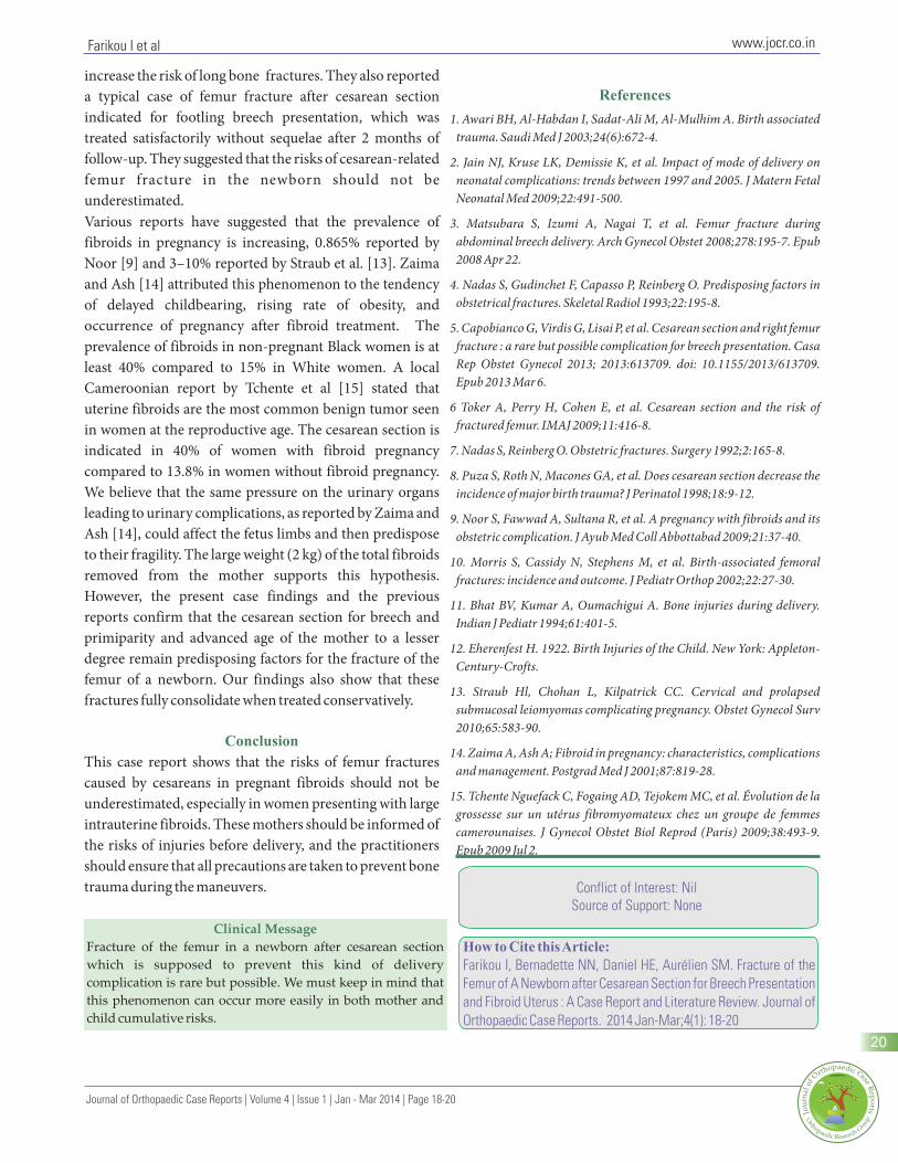
Figure 3: Last radiological aspect of the femur after 9 months.
www.jocr.co.in
injury compared to vaginal delivery particularly if there is A number of factors predispose the mother and child to an association of breech presentation, it does not completely morbidity and mortality; hence, gynecologists prefer less exclude the occurrence of an unexpected complication such risky or less traumatic delivery by cesarean section instead of as fracture of the femur.normal vaginal delivery (Table I). Primiparity, the advanced Toker et al [6] stated that cesarean section is the cause of age of the mother, the breech, the weight and size of the child increased risks of occurrence of femur fractures. This study at birth, and obstetric maneuvers are indeed risk factors taught us that women presenting a fibroid uterus with implicated in predisposing the occurrence of birth trauma. breech are at a cumulative high risk of delivering a baby with Nadas and Reinberg [7] reported the most common risk a fractured femur if they deliver by cesarean.factors as obstetric maneuvers (75%), cesarean section (35%), prolonged labor (33%), and prematurity (25%). A
The mother was a 39 years old primipara who attended recent study carried out by Puza et al [8] showed that the regularly the antenatal care clinics. The pregnancy lasted for increase in cesarean section of 4% lead to a significant 38 weeks. Ultrasound examination at 20th weeks revealed decrease in the rate of major birth trauma. Here, we present a intrauterine fibroids with a breech presentation. No mid-third diaphyseal fracture of the femur treated cephalic version was attempted to correct the successfully by casting in a child born after a normal malpresentation. Therefore, elective cesarean section was pregnancy by elective cesarean section for breech and fibroid indicated. The gynecologist performed cesarean section uterus. The mother was an elderly primipara. This rare and then myomectomy. The breech was not deeply engaged addition to the existing risk factors for maternal and fetal in the pelvis. Therefore the extraction was simple in a health led the gynecologist to take precaution and plan sufficiently relaxed uterus. The child weighed 3.4 kg and cesarean section to avoid the known obstetric complications had a size of 54 cm at birth. All the resected uterine fibroids of pregnant fibroid: prolonged labor, uterine rupture or fetal had a total weight of 2 kg. The child did not require distress or obstetrical maneuvers at risk for assisted breech resuscitation, but presented functional impairment of the [9]. The present case report fosters the debate of risk left leg. Radiographic examination revealed a diaphyseal assessments regarding delivery options for women with fracture of the femur localized in the middle third. A non- intrauterine fibroids when breech presentation becomes specialist immobilized the limb in a makeshift bandage (Fig inevitable.1) to relieve pain during plain X-ray examination and to Birth trauma has an estimated incidence of 0.13 to 1 per 1000 facilitate the transportation of the baby to the Referral births [10, 11]. The fracture of the femur of the newborn after Hospital. At arrival, we replaced this temporary contention cesarean is not common. The first report on a femoral with a bilateral spica cast for 4 weeks. The subsequent fracture during cesarean section was published by Eherenfest radiographic examinations performed after removal of the [12]. Toker et al. [6] recently reported one of the largest spica cast showed good consolidation of the fracture (Figs 2 known series of 12 femur fractures in Israel after cesarean and 3). section (incidence of 0.308 per 1000 CS) versus 5 fractures in
vaginal delivery (incidence of 0.027 per 1000 VD) in the same series and stated that cesarean section markedly
Discussion
Case Report
Farikou I et al
19
Journal of Orthopaedic Case Reports | Volume 4 | Issue 1 | Jan - Mar 2014 | Page 18-20
increase the risk of long bone fractures. They also reported a typical case of femur fracture after cesarean section indicated for footling breech presentation, which was treated satisfactorily without sequelae after 2 months of follow-up. They suggested that the risks of cesarean-related femur fracture in the newborn should not be underestimated.Various reports have suggested that the prevalence of fibroids in pregnancy is increasing, 0.865% reported by Noor [9] and 3–10% reported by Straub et al. [13]. Zaima and Ash [14] attributed this phenomenon to the tendency of delayed childbearing, rising rate of obesity, and occurrence of pregnancy after fibroid treatment. The prevalence of fibroids in non-pregnant Black women is at least 40% compared to 15% in White women. A local Cameroonian report by Tchente et al [15] stated that uterine fibroids are the most common benign tumor seen in women at the reproductive age. The cesarean section is indicated in 40% of women with fibroid pregnancy compared to 13.8% in women without fibroid pregnancy. We believe that the same pressure on the urinary organs leading to urinary complications, as reported by Zaima and Ash [14], could affect the fetus limbs and then predispose to their fragility. The large weight (2 kg) of the total fibroids removed from the mother supports this hypothesis. However, the present case findings and the previous reports confirm that the cesarean section for breech and primiparity and advanced age of the mother to a lesser degree remain predisposing factors for the fracture of the femur of a newborn. Our findings also show that these fractures fully consolidate when treated conservatively.
This case report shows that the risks of femur fractures caused by cesareans in pregnant fibroids should not be underestimated, especially in women presenting with large intrauterine fibroids. These mothers should be informed of the risks of injuries before delivery, and the practitioners should ensure that all precautions are taken to prevent bone trauma during the maneuvers.
References
Conclusion
1. Awari BH, Al-Habdan I, Sadat-Ali M, Al-Mulhim A. Birth associated trauma. Saudi Med J 2003;24(6):672-4.
2. Jain NJ, Kruse LK, Demissie K, et al. Impact of mode of delivery on neonatal complications: trends between 1997 and 2005. J Matern Fetal Neonatal Med 2009;22:491-500.
3. Matsubara S, Izumi A, Nagai T, et al. Femur fracture during abdominal breech delivery. Arch Gynecol Obstet 2008;278:195-7. Epub 2008 Apr 22.
4. Nadas S, Gudinchet F, Capasso P, Reinberg O. Predisposing factors in obstetrical fractures. Skeletal Radiol 1993;22:195-8.
5. Capobianco G, Virdis G, Lisai P, et al. Cesarean section and right femur fracture : a rare but possible complication for breech presentation. Casa Rep Obstet Gynecol 2013; 2013:613709. doi: 10.1155/2013/613709. Epub 2013 Mar 6.
6 Toker A, Perry H, Cohen E, et al. Cesarean section and the risk of fractured femur. IMAJ 2009;11:416-8.
7. Nadas S, Reinberg O. Obstetric fractures. Surgery 1992;2:165-8.
8. Puza S, Roth N, Macones GA, et al. Does cesarean section decrease the incidence of major birth trauma? J Perinatol 1998;18:9-12.
9. Noor S, Fawwad A, Sultana R, et al. A pregnancy with fibroids and its obstetric complication. J Ayub Med Coll Abbottabad 2009;21:37-40.
10. Morris S, Cassidy N, Stephens M, et al. Birth-associated femoral fractures: incidence and outcome. J Pediatr Orthop 2002;22:27-30.
11. Bhat BV, Kumar A, Oumachigui A. Bone injuries during delivery. Indian J Pediatr 1994;61:401-5.
12. Eherenfest H. 1922. Birth Injuries of the Child. New York: Appleton-Century-Crofts.
13. Straub Hl, Chohan L, Kilpatrick CC. Cervical and prolapsed submucosal leiomyomas complicating pregnancy. Obstet Gynecol Surv 2010;65:583-90.
14. Zaima A, Ash A; Fibroid in pregnancy: characteristics, complications and management. Postgrad Med J 2001;87:819-28.
15. Tchente Nguefack C, Fogaing AD, Tejokem MC, et al. Évolution de la grossesse sur un utérus fibromyomateux chez un groupe de femmes camerounaises. J Gynecol Obstet Biol Reprod (Paris) 2009;38:493-9. Epub 2009 Jul 2.
20
www.jocr.co.in
Conflict of Interest: Nil Source of Support: None
How to Cite this Article:
Farikou I, Bernadette NN, Daniel HE, Aurélien SM. Fracture of the
Femur of A Newborn after Cesarean Section for Breech Presentation
and Fibroid Uterus : A Case Report and Literature Review. Journal of
Orthopaedic Case Reports. 2014 Jan-Mar;4(1): 18-20
Clinical Message
Fracture of the femur in a newborn after cesarean section
which is supposed to prevent this kind of delivery
complication is rare but possible. We must keep in mind that
this phenomenon can occur more easily in both mother and
child cumulative risks.
Farikou I et al
Journal of Orthopaedic Case Reports | Volume 4 | Issue 1 | Jan - Mar 2014 | Page 18-20