Embed Size (px)
Citation preview

141
ficial to a fracture. Therefore, the en-tire foot and ankle has to be examined initially and for a few weeks after the event. Why does the ankle have such a high incidence of fracture blisters? The factors attributed to this are: sparse or no sweat glands, no hair follicles, thin skin with no subcutaneous fat (thus having prominent osseous structures), no deep venous system, and the ar-borization of veins perimalleolarly. The epidermis and dermis lack the above-mentioned structures to stabilize these levels, not only to one another but to the underlying subcutaneous tissue.3 For calcaneal fractures, in ad-dition to similar anatomic findings of the ankle, the mechanism of injury en-
As a growing cadre of foot and ankle surgeons come in contact with more trau-ma cases of the foot and ankle, they may encoun-
ter a sequela—namely a fracture blister. In some instances, although rare, these blisters may even appear after more involved elective surgery, for exam-ple, even after correction of a hallux abducto valgus deformity. It is incum-bent upon us that we recognize frac-ture blisters and treat them accordingly, being mindful that the underlying frac-ture has to be dealt with eventually.
Etiology, Incidence Fracture blisters have also been called by the following names: trau-ma blisters, epidermal necrosis, epi-
dermolysis, and avascular necrosis of the skin.1 Conventionally, however, the more common name is fracture blister. That will be the term used throughout this discussion. Fracture blisters have an incidence of 2.9% of all fractures. For ankle frac-tures, their incidence is 4.2%, and 10.9% of calcaneal fractures.2 The high-er the energy with components of ei-ther shear and/or torque during the traumatic event, the more one is pre-disposed to the formation of a fracture blister. The underlying fracture most likely will resemble a high-energy pat-tern. Fracture blisters can be singular or multiple in number. They can be distal or proximal to the fracture itself. They do not necessarily form directly super-
Welcome to Podiatry Management’s CME Instructional program. Our journal has been approved as a sponsor of Con-tinuing Medical Education by the Council on Podiatric Medical Education. You may enroll: 1) on a per issue basis (at $26.00 per topic) or 2) per year, for the special rate of $210 (you save $50). You may submit the answer sheet, along with the other information requested, via mail, fax, or phone. You can also take this and other exams on the Internet at www.podiatrym.com/cme. If you correctly answer seventy (70%) of the questions correctly, you will receive a certificate attesting to your earned credits. You will also receive a record of any incorrectly answered questions. If you score less than 70%, you can retake the test at no additional cost. A list of states currently honoring CPME approved credits is listed on pg. 146. Other than those entities currently accepting CPME-approved credit, Podiatry Management cannot guarantee that these CME credits will be acceptable by any state licensing agency, hospital, managed care organization or other entity. PM will, however, use its best efforts to ensure the widest acceptance of this program possible. This instructional CME program is designed to supplement, NOT replace, existing CME seminars. The goal of this program is to advance the knowledge of practicing podiatrists. We will endeavor to publish high quality manuscripts by noted authors and researchers. If you have any questions or comments about this program, you can write or call us at: Podiatry Management, P.O. Box 490, East Islip, NY 11730, (631) 563-1604 or e-mail us at [email protected]. Following this article, an answer sheet and full set of instructions are provided (pg. 146).—Editor
Continued on page 142
Objectives
After completion of this CME, the reader will:
1) Understand the etiology of fracture blisters.
2) Understand the difference between serous and hemor-rhagic fracture blisters.
3) Gain information on the various treatment options for the blisters.
4) Understand ways to pre-vent fracture blisters.
Fracture BlistersHere’s how to recognize and treat
this complication.
By GeorGe F. Wallace, DPM, MBa
www.podiatrym.com JANUARY 2017 | PODIATRY MANAGEMENT
Continuing MeDical eDucation

compassing a high-energy event increases the incidence (Figure 1, Figure 2).4
Fracture blisters form when the traumatic edema, which is part of the normal injury cascade, caus-es an interstitial pressure increase. Vascular congestion ensues. The epidermis and dermis lose their cohesion, and there is resultant separation. Fluid collects at the separated areas, yielding a fracture blister. Epidermal necrosis occurs and its extent will be predicated on the amount of energy generated to cause the injury. On average, formation of frac-ture blisters occurs 2.5 days after injury. Appearance can be as early as six hours post-injury.5 However, they can occur up to three weeks after injury.4
One has to remember that in the presence of an open frac-ture, fracture blisters can still form (Figure 3). Where present in con-junction with an open fracture, the Gustilo Anderson classifica-tion and treatment protocols are initiated along with concomitant fracture blister care.6
Compartment Syndrome With a compartment syn-drome, the appearance of a frac-ture blister over time does de-crease compartment pressures.7 However, one should not wait for fracture blisters to appear as a way to avoid surgical decompres-sion of the compartments. Where one has a high index of suspicion that the trauma could predispose the foot to raised pressures, com-partment pressures have to be monitored with or without frac-ture blisters in those cases. Once the pressures are elevated and the diagnosis of a compartment syn-drome is made, then the appropri-ate fasciotomies are performed in an emergent manner. The Podiatry Service at Uni-versity Hospital conducted a brief retrospective analysis of foot and ankle trauma over a two-year peri-od. The sole purpose was to quan-
www.podiatrym.comJANUARY 2017 | PODIATRY MANAGEMENT
142
contin
uing
Medica
l edu
cation
cMe
Fracture (from page 141)
Continued on page 143
Figure 1: Ankle fracture
Figure 2: Calcaneal fracture
Figure 3: open fracture 1st meta-tarsal
Figure 4: Serous fracture blister
Figure 7: Fracture blisters secondary to a talar neck fracture. this was consulted for a “vesicular skin condition”.
Figure 5: Hemorrhagic fracture blister
Figure 6: External fixator in place until definitive surgery

then collapses, which serves as a biological dressing. A non-ad-herent gauze is applied, usually with an antibiotic ointment and dress-ing. After the roof desiccates in a few days, it is then debrided. 2) Hemorrhagic fracture blister: The roof in this type is thinner and flaccid versus the serous one. The entire roof is removed and the fluid gently wiped from the area. Silver sul-
fadiazine (SSD) with a non-adherent gauze is applied. Dressings are changed every few days until re-epithelialization of the base occurs, which may take up-wards of a week or two. Hemorrhagic blisters will take longer than the se-rous ones to heal. In some instances, both types may be present simultaneously. The above protocols are then followed for each. Should a fracture blister not be easily classified, it would be better to treat it as the one which represents a more severe injury, the hemorrhagic type.
Surgery and Fracture Blisters The literature is replete with salient questions regarding surgery around and through fracture blisters. Pertinent questions which can be raised are: 1) Should surgery be done if any are present? 2) Should an incision be made through one? 3) How close can an incision be to a fracture blister? Just like the discussion regarding treatment above, there is no clear con-sensus although statements are more forceful, if you will, as to what not to do. The rate of infection can be dou-bled and wound dehiscence can occur if the incision traverses the base prior to healing/re-epithelialization.5 Those aforementioned consequences are more common with the hemorrhagic type. Let’s look at University Hospital’s surgical fracture blister protocol in hopes of answering the above ques-tions. It is a distillation of the various
tify the number of fracture blisters per anatomical location. The results follow. Total traumatic cases over that period: 31 ankle fractures 5 calcaneal fractures 5 talar fractures 11 Lisfranc fracture/dislocations
Appearance of fracture blisters with the following pathology: 2 bimalleolar fractures 2 trimalleolar fractures 0 calcaneal fractures 1 Lisfranc fracture/dislocation 1 post Kalish osteotomy 1 post Lapidus fusion3
Note the Kalish and Lapidus proce-dures with the fracture blisters above. Both cases were serous and over the second metatarsophalangeal joints. The fracture blisters were treated per pro-tocol, which follows, and the patients had an uneventful recovery. In both cases, these lesions appeared at the first dressing change, which was day eight.
Anatomy and Types of Fracture Blisters Fracture blisters share the following characteristics: all are sub-epidermal, and any fluid contained within this blis-ter is sterile and has the characteristics of serum. From a histological perspec-tive, fracture blisters resemble a second degree burn.3
Giordano, et al., in 1994, wrote the seminal article regarding fracture blisters.8 Through their observations, they were able to devise a classification based on appearance and characteris-tics. Thus, there are two types of frac-ture blisters: serous and hemorrhagic. (Figure 4, Figure 5) The serous or clear fracture blis-ter has a tense roof. The fluid within the blister is clear. This represents a partial separation of the epidermis from the dermis.8
The second type of fracture blister is the hemorrhagic. The roof is flac-cid. The fluid is crimson. The blood tinge may be from a papillary vas-cular injury. A complete separation has occurred between the epidermis and dermis. Therefore, these blisters represent a higher energy event and are more devastating than their se-
rous counterpart. Hemorrhagic frac-ture blisters take longer to heal than the serous ones. Although rare, both types may be present simultaneously.
Prevention of Fracture Blisters Although we can never totally pre-vent fracture blisters, nonetheless there are maneuvers which can be followed to mitigate their formation. The gold standard of rest, ice, com-
pression, and elevation (RICE) are still pertinent to any trauma. When apply-ing compressive dressings or devices, bony prominences need to be well-pad-ded. Rapid swelling may necessitate bi-valving the dressing/cast prophylac-tically. If not, the extremity is examined frequently to monitor pain, neurovascu-lar status, and movement. Nelson advocates early reduction and stabilization of the fracture to re-duce the incidence of a fracture blis-ter.5 Secondly, open reduction internal fixation if performed within the first 24 hours of injury has demonstrated a decrease in fracture blister formation. The ability to perform this surgery at that time is contingent upon the soft tissue envelope and whether the patient is medically optimized and ultimately cleared for surgery. The AO Founda-tion advocates postponing any surgery which can’t be completed within the initial six to eight hours until seven to ten days have elapsed.9 Hopefully, by that time, the soft tissue envelope will be ready for surgical intervention.
Treatment of Fracture Blisters Although Giordano10 and others,3,
11-14 have described the treatment of fracture blisters based on type, the treatments are varied. No clear con-sensus has been promulgated in the literature.3
After review of the various treat-ment plans, the following are Universi-ty Hospital’s protocols for each type: 1) Serous fracture blister: The blis-ter is aspirated by incising the base with an 11 blade in two to four loca-tions. The fluid is extruded. The roof
143
continuing
Medical education
cMe
Fracture (from page 142)
Continued on page 144
www.podiatrym.com JANUARY 2017 | PODIATRY MANAGEMENT
From a histological perspective, fracture blisters resemble a second degree burn.
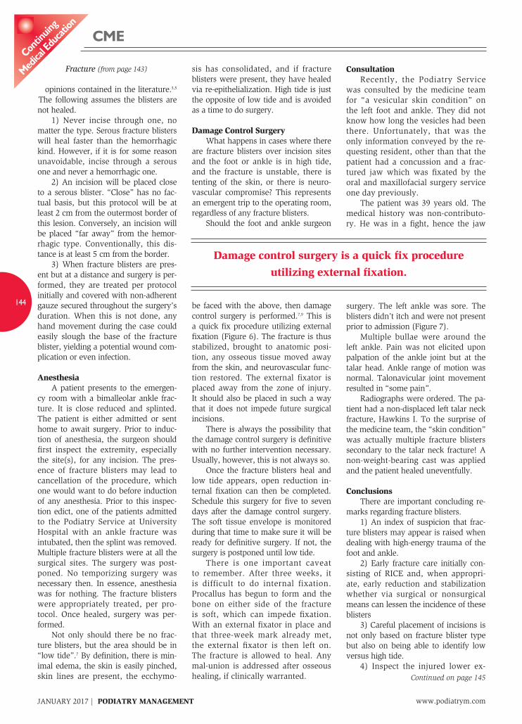
Consultation Recently, the Podiatry Service was consulted by the medicine team for “a vesicular skin condition” on the left foot and ankle. They did not know how long the vesicles had been there. Unfortunately, that was the only information conveyed by the re-questing resident, other than that the patient had a concussion and a frac-tured jaw which was fixated by the oral and maxillofacial surgery service one day previously. The patient was 39 years old. The medical history was non-contributo-ry. He was in a fight, hence the jaw
surgery. The left ankle was sore. The blisters didn’t itch and were not present prior to admission (Figure 7). Multiple bullae were around the left ankle. Pain was not elicited upon palpation of the ankle joint but at the talar head. Ankle range of motion was normal. Talonavicular joint movement resulted in “some pain”. Radiographs were ordered. The pa-tient had a non-displaced left talar neck fracture, Hawkins I. To the surprise of the medicine team, the “skin condition” was actually multiple fracture blisters secondary to the talar neck fracture! A non-weight-bearing cast was applied and the patient healed uneventfully.
Conclusions There are important concluding re-marks regarding fracture blisters. 1) An index of suspicion that frac-ture blisters may appear is raised when dealing with high-energy trauma of the foot and ankle. 2) Early fracture care initially con-sisting of RICE and, when appropri-ate, early reduction and stabilization whether via surgical or nonsurgical means can lessen the incidence of these blisters 3) Careful placement of incisions is not only based on fracture blister type but also on being able to identify low versus high tide. 4) Inspect the injured lower ex-
opinions contained in the literature.3,5 The following assumes the blisters are not healed. 1) Never incise through one, no matter the type. Serous fracture blisters will heal faster than the hemorrhagic kind. However, if it is for some reason unavoidable, incise through a serous one and never a hemorrhagic one. 2) An incision will be placed close to a serous blister. “Close” has no fac-tual basis, but this protocol will be at least 2 cm from the outermost border of this lesion. Conversely, an incision will be placed “far away” from the hemor-rhagic type. Conventionally, this dis-tance is at least 5 cm from the border. 3) When fracture blisters are pres-ent but at a distance and surgery is per-formed, they are treated per protocol initially and covered with non-adherent gauze secured throughout the surgery’s duration. When this is not done, any hand movement during the case could easily slough the base of the fracture blister, yielding a potential wound com-plication or even infection.
Anesthesia A patient presents to the emergen-cy room with a bimalleolar ankle frac-ture. It is close reduced and splinted. The patient is either admitted or sent home to await surgery. Prior to induc-tion of anesthesia, the surgeon should first inspect the extremity, especially the site(s), for any incision. The pres-ence of fracture blisters may lead to cancellation of the procedure, which one would want to do before induction of any anesthesia. Prior to this inspec-tion edict, one of the patients admitted to the Podiatry Service at University Hospital with an ankle fracture was intubated, then the splint was removed. Multiple fracture blisters were at all the surgical sites. The surgery was post-poned. No temporizing surgery was necessary then. In essence, anesthesia was for nothing. The fracture blisters were appropriately treated, per pro-tocol. Once healed, surgery was per-formed. Not only should there be no frac-ture blisters, but the area should be in “low tide”.7 By definition, there is min-imal edema, the skin is easily pinched, skin lines are present, the ecchymo-
sis has consolidated, and if fracture blisters were present, they have healed via re-epithelialization. High tide is just the opposite of low tide and is avoided as a time to do surgery.
Damage Control Surgery What happens in cases where there are fracture blisters over incision sites and the foot or ankle is in high tide, and the fracture is unstable, there is tenting of the skin, or there is neuro-vascular compromise? This represents an emergent trip to the operating room, regardless of any fracture blisters. Should the foot and ankle surgeon
be faced with the above, then damage control surgery is performed.7,9 This is a quick fix procedure utilizing external fixation (Figure 6). The fracture is thus stabilized, brought to anatomic posi-tion, any osseous tissue moved away from the skin, and neurovascular func-tion restored. The external fixator is placed away from the zone of injury. It should also be placed in such a way that it does not impede future surgical incisions. There is always the possibility that the damage control surgery is definitive with no further intervention necessary. Usually, however, this is not always so. Once the fracture blisters heal and low tide appears, open reduction in-ternal fixation can then be completed. Schedule this surgery for five to seven days after the damage control surgery. The soft tissue envelope is monitored during that time to make sure it will be ready for definitive surgery. If not, the surgery is postponed until low tide. There is one important caveat to remember. After three weeks, it is difficult to do internal fixation. Procallus has begun to form and the bone on either side of the fracture is soft, which can impede fixation. With an external fixator in place and that three-week mark already met, the external fixator is then left on. The fracture is allowed to heal. Any mal-union is addressed after osseous healing, if clinically warranted.
www.podiatrym.comJANUARY 2017 | PODIATRY MANAGEMENT
144
contin
uing
Medica
l edu
cation
cMe
Fracture (from page 143)
Continued on page 145
Damage control surgery is a quick fix procedure utilizing external fixation.

7 Wallace GF, Pachuda NM, Gumann G. Open frac-tures in Fractures of the Foot and Ankle. Gumann G, ed. Elsevier, Philadelphia 2004: 1. 8 Giordano CP, Koval KJ, Zuckerman JD, et al. Fracture blisters. Clin Orthop 307:214, 1994. 9 Stover MD, Kellam JF. Articular fractures: principles in AO Principles of Fracture Management 2nd ed, Ruedi TP, Buckley RE, Moran CG eds. Thieme, NY 2007: 139. 10 Giordano CP, Koval KS. Treatment of fracture blisters. J Or-thop Trauma 9: 171, 1995. 11 Bong M, Koval KJ et al. Blisters associated with lower extrem-ity fractures: Results of a prospective treatment protocol. J Orthop Trauma 20: 618, 2006. 12 Tolpinrud WL, Pebolledo BJ, Lorich DG. J Am Acad Derm 3: 132, 2015. 13 Gumann G. Ankle fractures in Foot and Ankle Trauma 2nd ed, Scurran BL, ed. Churchill Livingstone, NY 1996: 731. 14 Manway J, Highlander P. Open fractures of the foot and ankle: An evidence-based review. Foot Ankle Specialist. 8: 59, 2015.
continuing
Medical education
145
www.podiatrym.com JANUARY 2017 | PODIATRY MANAGEMENT
cMe
tremity prior to any anesthesia, preferably in the pre-opera-tive holding area versus on the operating table. 5) Damage control surgery may be necessary. 6) Be able to ascertain the mechanism of injury and whether the fracture was the result of a high-energy event. 7) De-roof hemorrhagic blisters and evacuate serous blis-ter fluid as treatments. 8) Be conversant with fracture and trauma classifica-tions, general principles, and treatment algorithms. They are important as discussion points when conversing with the emergency room physician who consulted your team. PM
References 1 Kenzora JE, Burgess AR. The neglected foot and ankle in poly-trauma. Advances Ortho Surg 7:89, 1983. 2 Varela CD, Vaughan TK, Carr JB, et al. Fracture blisters: Clini-cal and pathological aspects. J Orthrop Trauma 7:417, 1993. 3 Wallace GF, Sullivan J. Fracture blisters. Clinics Pod Med Surg 12:801, 1995. 4 Lim E, Leung JPF. Complications of intraarticular calcaneal fractures. Clin Ortho Related Research 391:7, 2001. 5 Nelson RD. General principles in Simon’s Emergency Orthope-dics 7th ed, Sherman S, ed. McGraw Hill, NY 2015: 3. 6 Gustilo RB, Anderson JT. Prevention of infection in the treatment of one thousand and twenty-five open fractures of long bones: retrospective and prospective analyses. JBJS(A) 58:453, 1976.
Fracture (from page 144)
1) Fracture blisters are also known as: A) Epidermal necrosis B) AVN of skin C) Epidermolysis d) All of the above
2) There are two types of fracture blisters: A) Closed and open B) Serous and hemorrhagic C) Simple and complex d) Infected and non-infected
3) In foot and ankle trauma, the highest incidence of fracture blisters occurs in what type of fracture: A) Metatarsal B) Talar C) Calcaneal d) Ankle
4) On average, formation of fracture blisters oc-curs how many days after injury: A) 1 B) 2.5 C) 4 d) 7
5) Fracture blisters are akin to what type of burn: A) First degree B) Second degree C) Third degree d) None of the above
6) After trauma a patient presents with edema, fracture blisters, and ecchymosis. This represents: A) “Low tide”
Dr. Wallace is the Director of the Podiatry Service and Medical Director of Ambulatory Care Services at university Hospital in new-ark, nJ. the hospital is a Level-i trauma cen-ter and the main teaching hospital of Rutgers new Jersey Medical School. there are six podiatric residents in the three-year program at university Hospital.
CME eXaMination
See anSwer Sheet on page 147.
Continued on page 146

JANUARY 2017 | PODIATRY MANAGEMENT
146
Continued on page 146
PM’scMe Program
Welcome to the innovative Continuing Education Program brought to you by Podiatry Management Magazine. our journal has been approved as a sponsor of Continuing Medical Education by the Council on Podiatric Medical Education.
now it’s even easier and more convenient to enroll in PM’s ce program! You can now enroll at any time during the year and submit eligible exams at any time during your enrollment period. PM enrollees are entitled to submit ten exams published during their consecutive, twelve–month enrollment period. Your enrollment period begins with the month payment is received. For example, if your payment is received on november 1, 2014, your enrollment is valid through october 31, 2015.if you’re not enrolled, you may also submit any exam(s) published in PM magazine within the past twelve months. cMe articles and examination questions from past issues of Podiatry Manage-ment can be found on the internet at http://www.podiatrym.com/cme. Each lesson is ap-proved for 1.5 hours continuing education contact hours. Please read the testing, grading and payment instructions to decide which method of participa-tion is best for you. Please call (631) 563-1604 if you have any ques-tions. A personal operator will be happy to assist you. Each of the 10 lessons will count as 1.5 credits; thus a maximum of 15 CME credits may be earned during any 12-month period. You may select any 10 in a 24-month period.
The Podiatry Management Magazine CME program is approved by the Council on Podiatric Education in all states where credits in instructional media are accepted. This article is approved for 1.5 Continuing Education Contact Hours (or 0.15 CEU’s) for each examination successfully completed.
Home Study cMe credits now accepted in Pennsylvania
$
See anSwer Sheet on page 147.
CME eXaMinationcon
tinuin
g
Medica
l edu
cation
B) “High tide” C) An abnormal response d) Cellulitis
7) Which type of fracture blister should never be the site of an incision: A) Serous B) Hemorrhagic C) Infected d) Non-infected
8) A patient presents with a trimalleolar ankle fracture. Fracture blisters are every-where around the ankle. You decide to apply an external fixator and will wait until the fracture blisters heal; then you will do inter-nal fixation. This is an example of: A) damage control surgery B) Low tide C) Compartment syndrome d) don’t wait. do the internal fixation
immediately.
9) Fracture blisters can occur with which of the following: A) Open fracture B) Compartment Syndrome C) Elective surgery (i.e., HAV surgery) d) All of the above
10) What is the treatment for fracture blisters? A) Biopsy all; then cover with gauze B) There is no clear consensus C) Prescribe an oral NSAId d) Inject an antibiotic solution into them

Please print clearly...Certificate will be issued from information below.
name _______________________________________________________________________ Soc. Sec. #______________________________Please Print: FiRSt Mi LASt
Address_____________________________________________________________________________________________________________
City__________________________________________________ State_______________________ Zip________________________________
Charge to: _____Visa _____ MasterCard _____ American Express
Card #________________________________________________Exp. Date____________________
note: credit card is the only method of payment. checks are no longer accepted.
Signature__________________________________ Soc. Sec.#______________________ Daytime Phone_____________________________
State License(s)___________________________ is this a new address? Yes________ no________
check one: ______ i am currently enrolled. (if faxing or phoning in your answer form please note that $2.50 will be charged to your credit card.)
______ i am not enrolled. Enclosed is my credit card information. Please charge my credit card $26.00 for each exam submitted. (plus $2.50 for each exam if submitting by fax or phone).
______ i am not enrolled and i wish to enroll for 10 courses at $210.00 (thus saving me $50 over the cost of 10 individual exam fees). i understand there will be an additional fee of $2.50 for any exam i wish to submit via fax or phone.
note: if you are mailing your answer sheet, you must complete all info. on the front and back of this page and mail with your credit card information to: Podiatry Management, P.o. Box 490, east islip, ny 11730.
teStinG, GraDinG anD PayMent inStructionS (1) Each participant achieving a passing grade of 70% or higher on any examination will receive an official computer form stating the number of CE credits earned. this form should be safeguarded and may be used as documentation of credits earned. (2) Participants receiving a failing grade on any exam will be noti-fied and permitted to take one re-examination at no extra cost. (3) All answers should be recorded on the answer form below. For each question, decide which choice is the best answer, and circle the letter representing your choice. (4) Complete all other information on the front and back of this page. (5) Choose one out of the 3 options for testgrading: mail-in, fax, or phone. to select the type of service that best suits your needs, please read the following section, “test grading options”.
teSt GraDinG oPtionS Mail-In Grading to receive your CME certificate, complete all information and mail with your credit card information to:
Podiatry ManagementP.o. Box 490, east islip, ny 11730
PleaSe Do not SenD WitH SiGnature reQuireD, aS tHeSe Will not Be accePteD. there is no charge for the mail-in service if you have already en-
enrollMent ForM & anSWer SHeet
$
rolled in the annual exam CME program, and we receive this exam during your current enrollment period. if you are not enrolled, please send $26.00 per exam, or $210 to cover all 10 exams (thus saving $50 over the cost of 10 individual exam fees).
Facsimile Grading to receive your CME certificate, complete all information and fax 24 hours a day to 1-631-563-1907. Your CME certificate will be dated and mailed within 48 hours. this service is available for $2.50 per exam if you are currently enrolled in the annual 10-exam CME program (and this exam falls within your enrollment period), and can be charged to your Visa, MasterCard, or American Express. if you are not enrolled in the annual 10-exam CME program, the fee is $26 per exam.
Phone-In Grading You may also complete your exam by using the toll-free service. Call 1-800-232-4422 from 10 a.m. to 5 p.m. ESt, Monday through Friday. Your CME certificate will be dated the same day you call and mailed within 48 hours. there is a $2.50 charge for this service if you are currently enrolled in the annual 10-exam CME program (and this exam falls within your enrollment period), and this fee can be charged to your Visa, Mastercard, American Express, or Discover. if you are not current-ly enrolled, the fee is $26 per exam. When you call, please have ready: 1. Program number (Month and Year) 2. the answers to the test 3. Your social security number 4. Credit card information
in the event you require additional CME information, please contact PMS, inc., at 1-631-563-1604.
Over, please
continuing
Medical education
enrollment/testing informationand answer Sheet
147
www.podiatrym.com JANUARY 2017 | PODIATRY MANAGEMENT

148
www.podiatrym.comJANUARY 2017 | PODIATRY MANAGEMENT
contin
uing
Medica
l edu
cation
enrollMent ForM & anSWer SHeet (continued)
Medical education lesson evaluation
Strongly Strongly agree Agree neutral Disagree disagree [5] [4] [3] [2] [1]
1) this CME lesson was helpful to my practice ____
2) the educational objectives were accomplished ____
3) i will apply the knowledge i learned from this lesson ____
4) i will makes changes in my practice behavior based on this lesson ____
5) this lesson presented quality information with adequate current references ____
6) What overall grade would you assign this lesson?A B C D
How long did it take you to complete this lesson?
______hour ______minutes
What topics would you like to see in future CME lessons ? Please list :
__________________________________________________
__________________________________________________
__________________________________________________
__________________________________________________
__________________________________________________
__________________________________________________
__________________________________________________
1. a B c D
2. a B c D
3. a B c D
4. a B c D
5. a B c D
6. a B c D
7. a B c D
8. a B c D
9. a B c D
10. a B c D
circle:
eXaM #1/17Fracture Blisters
(Wallace)
$