Embed Size (px)
Citation preview
OPEN ACCESSHuman & Veterinary MedicineInternational Journal of the Bioflux Society Research Article
Volume 5 | Issue 2 Page 45 HVM Bioflux
http://www.hvm.bioflux.com.ro/
Fractal and lacunarity analysis of human retinal vessel arborisation in normal and amblyopic eyes
1Ştefan Ţălu, 2Cristina Vlăduţiu, 3Livia A. Popescu, 4Carmen A. Lupaşcu, 5Ştefan C. Vesa, 2Simona D. Ţălu
1 The Technical University of Cluj-Napoca, Faculty of Mechanical Engineering, Department of AET, Discipline of Descriptive Geometry and Engineering Graphics, Cluj-Napoca, Romania; 2 ”Iuliu Haţieganu” University of Medicine and Pharmacy Cluj-Napoca, Faculty of Medicine, Department of Surgical Specialties and Medical Imaging, Discipline of Ophthalmology, Cluj-Napoca, Romania; 3 County Emergency Hospital Cluj-Napoca, Ophthalmology Department, Cluj-Napoca, Romania; 4 Università degli Studi di Palermo, Dipartimento di Matematica e Informatica, Palermo, Italy; 5 “Iuliu Haţieganu” University of Medicine and Pharmacy Cluj-Napoca, Faculty of Medicine, Department of Pharmacology, Cluj-Napoca, Romania.
IntroductionAmblyopia occurs in approximately 2 % of the general popula-tion and is the most common cause of decreased vision in child-hood. Amblyopia is caused by abnormal visual stimulation dur-ing visual development, resulting in abnormalities in the visual centers of the brain. Children are susceptible to amblyopia be-tween birth and 7 years old (Wright et al 2006).Investigating the human retinal vascular network, as a part of an in-vivo analysis, is helpful in detection and diagnose of reti-nal disorders (Ţălu 2005a; Ţălu 2005b; Ryan et al 2006; ; Holz & Spaide 2010; Ţălu et al 2009; Ţălu 2011a; Ţălu et al 2011; Ţălu & Ţălu 2012; Ţălu 2013a).The human retinal vascular network has a branching pattern and it is considered to be a fractal structure at low resolution, in a “scaling window” which normally ranges in two to three orders of magnitude (Kyriacos et al 1997; Losa et al 2005). It can be observed in its natural living state using a retinal cam-era (Masters 2004).The fractal and multifractal analysis of human retinal vascu-lar network depends on the experimental and methodological
parameters involved as: diversity of subjects, image acquisition, type of image, image processing, fractal methods, including the algorithm and specific calculation used etc. (Kyriacos et al 1997; Mendonça et al 2007; Ţălu 2011b; Ţălu & Giovanzana 2011; Fraz et al 2012; Ţălu & Giovanzana 2012; Ţălu 2012a; Ţălu 2012b; Ţălu et al 2012; Ţălu 2012c; Ţălu 2012d; Ţălu et al 2013; Ţălu 2013b; Ţălu & Ţǎlu 2013).
Material and methodsFractal dimensionThe fractal analysis of fundus photographs may allow a quanti-tative measure of complexity of the retinal vessel branching. In fractal analysis, box-counting is one of the most widely used and practical methods to determine the fractal dimension of a figure (Falconer 2003; Lopes & Betrouni 2009; Napolitano et al 2012).Let’s consider a fractal object recorded into a digital image. Let A be any nonempty bounded subset of Rn (n-dimensional Euclidian space Rn). The fractal dimension gives the scaling between the smallest number of n-dimensional ε boxes needed to cover the
Abstract. Objective: The purpose of this paper is to determine a global assessment of the human retinal vascular network for patients with amblyopia. Fractal geometry and lacunarity parameters are used in this study. Materials and methods: A set of 12 segmented and skeletonized human retinal images, corresponding to both normal (6 images) and amblyopia states of the retina (6 images), was analyzed using the Image J software with box-counting method. Statistical analyses were performed for these groups using Microsoft Office Excel 2003 and GraphPad InStat software. Results: The human retinal vascular network architecture can be estimated using the fractal geometry. The average of fractal dimensions D for the amblyopia images (segmented and skeletonized versions) is slightly higher than the corresponding values for normal im-ages (segmented and skeletonized versions). However, the average of lacunarity parameter Λ for the amblyopia images (segmented and skel-etonized versions) is slightly lower than the corresponding values for normal images (segmented and skeletonized versions). Conclusions:The fractal and lacunarity analysis may be used for an early diagnosis of patients with amblyopia.
Key Words: retina, retinal vessels, amblyopia, strabismus, fractal dimension
Copyright: This is an open-access article distributed under the terms of the Creative Commons Attribution License, which permits unrestricted use, distribution, and reproduction in any medium, provided the original author and source are credited.
Corresponding Author: S. Ţălu, [email protected]
Ţălu et al 2013
Volume 5 | Issue 2 Page 46 HVM Bioflux
http://www.hvm.bioflux.com.ro/
set A completely, and the boxes’ size ε. The box-counting di-mension of A is expressed by (Falconer 2003);
(1)
In equation (1) the zero limit cannot be applied to biological images and DB(A) can be estimated by means of next equation (Grizzi et al 2005):
(2)
where D is the slope of the regression line for the log-log plot of the scanning box size and the count from a box counting scan. The “count” usually refers to the number of grid boxes that contained pixels in a box counting scan. The slope of the linear region of the plot is (-D), where D is the box-counting dimension that corresponds to the fractal dimension. The fractal dimension D of retinal vascular network quantifies the global measure of complexity of the vascular branching pat-tern (Ţǎlu 2011; Ţălu & Giovanzana 2011; Ţălu & Giovanzana 2012; Ţălu 2012a; Ţălu 2012b; Ţălu et al 2012).(Kyriacos et al 1997; Masters 2004; Ţălu 2011b; Ţălu & Giovanzana 2012). Generally, arterioles have a lower fractal dimension than venules (Patton et al 2006).Some investigators (Azemin et al 2012) observed a significant decrease in the fractal dimensions of human retinal vascular network with aging, consistent with observations from other human organ systems.Investigators have also found different values in the fractal di-mensions associated with the retinal pathological status (Avakian et al 2002; Lakshminanarayanan et al 2003; Kunicki et al 2009; Liberek et al 2010; Olujić et al 2011; Ţălu et al 2012).
Lacunarity analysisLacunarity analysis is a multiscale method for describing pat-terns of spatial dispersion. It can be considered a scale-depend-ent measure of heterogeneity or texture of an object, whether or not it is a fractal. Lacunarity measures the deviation of a geo-metric structure from translational invariance, or gappiness of geometric structure. It can be used with both binary and quan-titative data in one, two, and three dimensions (Plotnick et al 1996; Ţălu & Giovanzana 2011; Ţălu & Giovanzana 2012; Ţălu 2012a; Ţălu 2012b).Lacunarity can be evaluated both for fractal and non-fractal sets. Lacunarity may characterize different texture appearances, which may have the same fractal dimension value. Lacunarity is complementary to the fractal dimension, as shapes with the same fractal dimension may have different lacunarity values. If the fractal object is dense the lacunarity is small, the lacunar-ity increases with coarseness (Ţălu & Giovanzana 2011; Ţălu & Giovanzana 2012; Ţălu 2012a; Ţălu 2012b).Various algorithms have been proposed to calculate and quan-tify lacunarity of an image. Among them, the gliding-box al-gorithm developed by Allain and Cloitre (1991) is one of the most widely used. The lacunarity λ was evaluated using FracLac. The mean lacu-narity (or Λ) was expressed as (Image J software; FracLac V 2.0f for Image J software):
(3)
where σ is the standard deviation; μ is the mean for pixels per box at this size, ε, in a box count at this orientation, g; and n is the number of box sizes.
Segmentation methodAn automatic unsupervised method for the segmentation of retinal vessels was applied before fractal analysis. Finally the segmented images were post-processed, by eliminating small connected components in order to remove noisy pixels and to improve in this way the accuracy of the segmentation (Lupaşcu et al 2009; Lupaşcu et al 2010; Lupaşcu & Tegolo 2011).
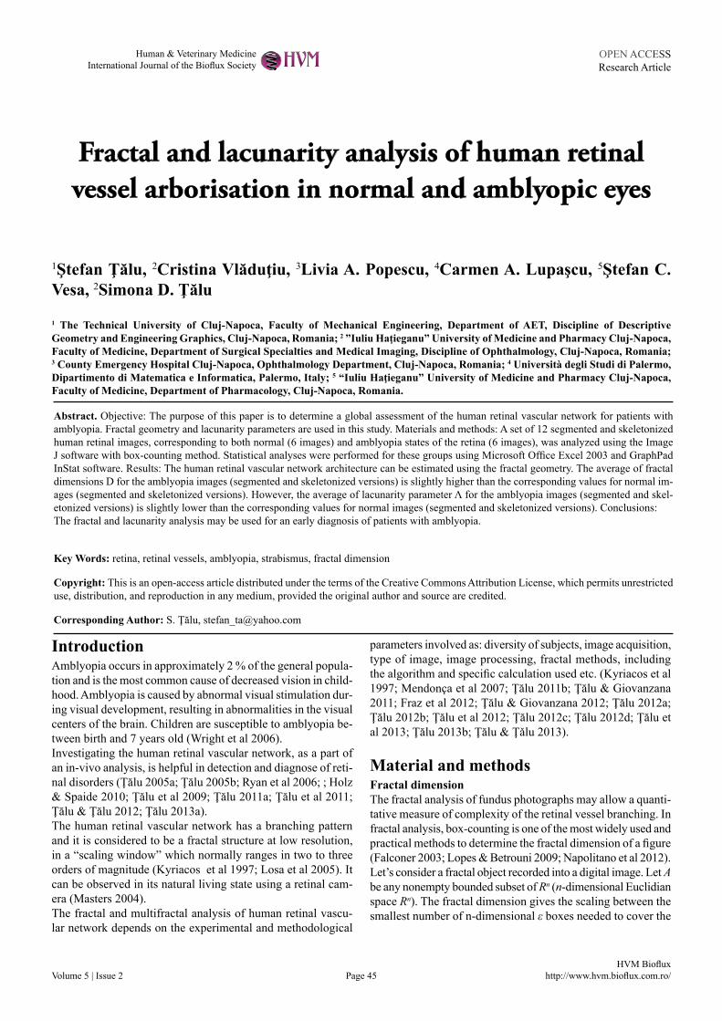
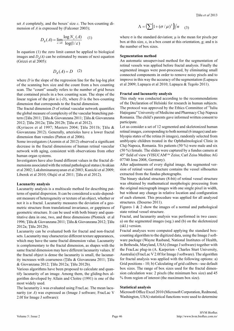
Fractal and lacunarity analysisThis study was conducted according to the recommendations of the Declaration of Helsinki for research in human subjects. The protocol was approved by the Ethics Committee of ”Iuliu Haţieganu” University of Medicine and Pharmacy Cluj-Napoca Romania. The child’s parents gave informed written consent to participate.Let us consider a set of 12 segmented and skeletonized human retinal images, corresponding to both normal (6 images) and am-blyopia states of the retina (6 images), randomly selected from amblyopic children treated in the Ophthalmological Clinic in Cluj-Napoca, Romania. Six patients (50 %) were male and six (50 %) female. The slides were captured by a fundus camera at 45° field of view (VISUCAMLite Zeiss; Carl Zeiss Meditec AG 07740 Jena 2008, Germany).After adjustments of every digital image, the segmented ver-sion of retinal vessel structure contains the vessel silhouettes extracted from the fundus photographs.The binary skeletal structure (8-bit) of retinal vessel structure was obtained by mathematical morphologic processing from the original micrograph images with one single pixel in width, but without any change in relative location and configuration of each element. This procedure was applied for all analyzed structures. (Deserno 2011).Figures 1 & 2 show the images of a normal and pathological state retinal vessel structure.Fractal, and lacunarity analysis was performed in two cases: a) on the segmented images (seg.) and (b) on the skeletonized (skl.) version.Fractal analyses were computed applying the standard box-counting algorithm to the digitized data, using the Image J soft-ware package (Wayne Rasband, National Institutes of Health, in Bethesda, Maryland, USA) (Image J software) together with the FracLac plug-in (A. Karperien - Charles Sturt University, Australia) (FracLac V 2.0f for Image J software). The algorithm for fractal analysis was applied with the following options: a) Grid positions - 10; b) Calculating of grid calibers - use default box sizes. The range of box sizes used for the fractal dimen-sion calculation was: 2 pixels (the minimum box size) and 45 % from region of interest (the maximum box size).
Statistical analysisMicrosoft Office Excel 2010 (Microsoft Corporation, Redmond, Washington, USA) statistical functions were used to determine
[ ]( ) n∑ +=Λ 2)/(1 µσ
)/1log()(loglim)(
0 εε
ε
ANADB →=
DADB =)(
Ţălu et al 2013
Volume 5 | Issue 2 Page 47 HVM Bioflux
http://www.hvm.bioflux.com.ro/
(average ± standard deviation) of the fractal dimensions D and lacunarity parameters Λ obtained with Image J software.The statistical processing of the results obtained with Image J software (Table 1) was done using GraphPad InStat software program, version 3.20 (GraphPad, San Diego, CA, USA).Normal distribution of variables was previously assessed by means of the Kolmogorov-Smirnov test. It was found that fractal dimensions and lacunarity parameters Λ of the vascular trees followed a normal distribution. An analysis of variance (ANOVA) was performed to see whether significant differences were found between different diseased states. Differences with a P value of 0.05 or less were considered statistically signifi-cant. The average results were expressed as mean value and standard deviation.
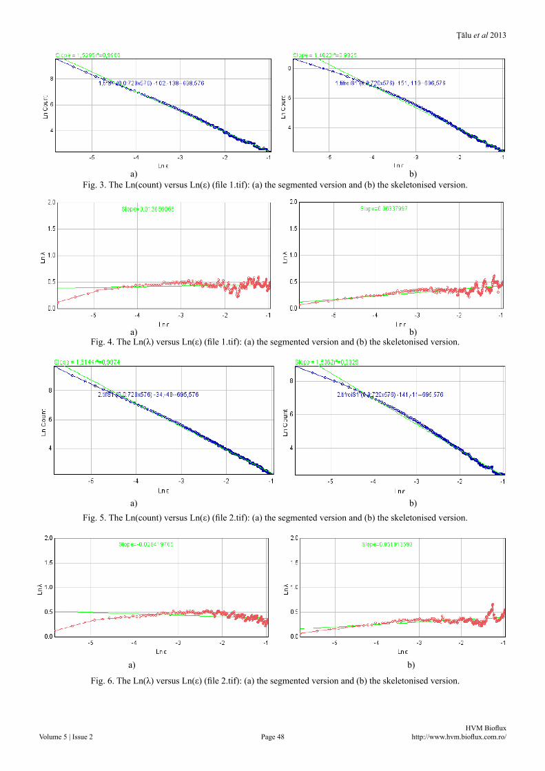
ResultsResults of fractal and lacunarity analysisFor all analyzed cases (Table 1), the coefficients of correlation (R2) associated with fractal dimensions D were greater than 0.9925 representing a good linear correlation. An (R2) of 1.0 indicates that the regression line perfectly fits the data.In Figs. 3-4 are shown the results of fractal analyses for retinal digital image (file 1.tif – normal status) analyzed with the Image J software, in segmented and skeletonised variants.
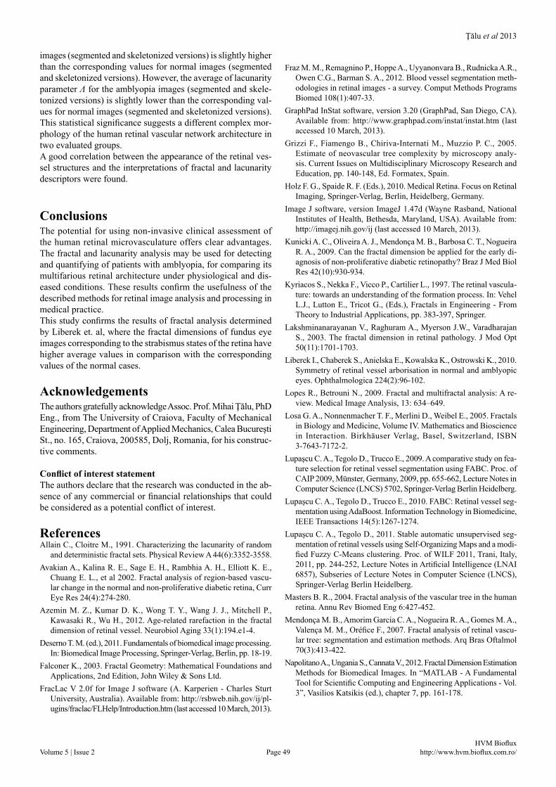
In Figs. 5-6 are shown the results of fractal analyses for reti-nal digital image (file 2.tif – pathological status) analyzed with the Image J software, in segmented and skeletonised variants.
Table 1. The fractal dimensions and lacunarity parameter Λ with average ± standard deviation, of 12 analyzed images, for normal (N) and pathological (P) status, in segmented and skel-etonized variants.
DiscussionThe retinal diseases modify the human retinal vessel arborisa-tion. The complex-dynamical fractal structure development of human retinal vascular network architecture can be estimated using the fractal geometry. The fractal dimension (as a quantita-tive estimator of the spatial complexity) and lacunarity param-eter may be markers of subtle changes in retinal vascular archi-tecture. The average of fractal dimensions D for the amblyopia
a) b) c)
Fig. 1. Image of a normal state retinal vessel network, right eye (file 1.tif): (a) the color image version, (b) the segmented ver-sion and (c) the skeletonized version.
a) b) c)
Fig. 2. Image of an amblyopia state retinal vessel network, left eye (file 2.tif): (a) the color image version, (b) the segmented ver-sion and (c) the skeletonized version.
Status Type Value of fractal dimension (D)
Lacunarity parameter Λ
N Seg. 1.5295 ± 0.0240 0.6766 ± 0.1136N Skl. 1.4544 ± 0.0407 0.5820 ± 0.0928P Seg. 1.6132 ± 0.0539 0.5069 ± 0.0659P Skl. 1.5380 ± 0.0671 0.4348 ± 0.0319
Ţălu et al 2013
Volume 5 | Issue 2 Page 48 HVM Bioflux
http://www.hvm.bioflux.com.ro/
a) b) Fig. 3. The Ln(count) versus Ln(ε) (file 1.tif): (a) the segmented version and (b) the skeletonised version.
a) b) Fig. 4. The Ln(λ) versus Ln(ε) (file 1.tif): (a) the segmented version and (b) the skeletonised version.
a) b) Fig. 5. The Ln(count) versus Ln(ε) (file 2.tif): (a) the segmented version and (b) the skeletonised version.
a) b)
Fig. 6. The Ln(λ) versus Ln(ε) (file 2.tif): (a) the segmented version and (b) the skeletonised version.
Ţălu et al 2013
Volume 5 | Issue 2 Page 49 HVM Bioflux
http://www.hvm.bioflux.com.ro/
images (segmented and skeletonized versions) is slightly higher than the corresponding values for normal images (segmented and skeletonized versions). However, the average of lacunarity parameter Λ for the amblyopia images (segmented and skele-tonized versions) is slightly lower than the corresponding val-ues for normal images (segmented and skeletonized versions).This statistical significance suggests a different complex mor-phology of the human retinal vascular network architecture in two evaluated groups.A good correlation between the appearance of the retinal ves-sel structures and the interpretations of fractal and lacunarity descriptors were found.
ConclusionsThe potential for using non-invasive clinical assessment of the human retinal microvasculature offers clear advantages. The fractal and lacunarity analysis may be used for detecting and quantifying of patients with amblyopia, for comparing its multifarious retinal architecture under physiological and dis-eased conditions. These results confirm the usefulness of the described methods for retinal image analysis and processing in medical practice.This study confirms the results of fractal analysis determined by Liberek et. al, where the fractal dimensions of fundus eye images corresponding to the strabismus states of the retina have higher average values in comparison with the corresponding values of the normal cases.
AcknowledgementsThe authors gratefully acknowledge Assoc. Prof. Mihai Ţălu, PhD Eng., from The University of Craiova, Faculty of Mechanical Engineering, Department of Applied Mechanics, Calea Bucureşti St., no. 165, Craiova, 200585, Dolj, Romania, for his construc-tive comments.
Conflict of interest statementThe authors declare that the research was conducted in the ab-sence of any commercial or financial relationships that could be considered as a potential conflict of interest.
ReferencesAllain C., Cloitre M., 1991. Characterizing the lacunarity of random
and deterministic fractal sets. Physical Review A 44(6):3352-3558.Avakian A., Kalina R. E., Sage E. H., Rambhia A. H., Elliott K. E.,
Chuang E. L., et al 2002. Fractal analysis of region-based vascu-lar change in the normal and non-proliferative diabetic retina, Curr Eye Res 24(4):274-280.
Azemin M. Z., Kumar D. K., Wong T. Y., Wang J. J., Mitchell P., Kawasaki R., Wu H., 2012. Age-related rarefaction in the fractal dimension of retinal vessel. Neurobiol Aging 33(1):194.e1-4.
Deserno T. M. (ed.), 2011. Fundamentals of biomedical image processing. In: Biomedical Image Processing, Springer-Verlag, Berlin, pp. 18-19.
Falconer K., 2003. Fractal Geometry: Mathematical Foundations and Applications, 2nd Edition, John Wiley & Sons Ltd.
FracLac V 2.0f for Image J software (A. Karperien - Charles Sturt University, Australia). Available from: http://rsbweb.nih.gov/ij/pl-ugins/fraclac/FLHelp/Introduction.htm (last accessed 10 March, 2013).
Fraz M. M., Remagnino P., Hoppe A., Uyyanonvara B., Rudnicka A.R., Owen C.G., Barman S. A., 2012. Blood vessel segmentation meth-odologies in retinal images - a survey. Comput Methods Programs Biomed 108(1):407-33.
GraphPad InStat software, version 3.20 (GraphPad, San Diego, CA). Available from: http://www.graphpad.com/instat/instat.htm (last accessed 10 March, 2013).
Grizzi F., Fiamengo B., Chiriva-Internati M., Muzzio P. C., 2005.Estimate of neovascular tree complexity by microscopy analy-sis. Current Issues on Multidisciplinary Microscopy Research and Education, pp. 140-148, Ed. Formatex, Spain.
Holz F. G., Spaide R. F. (Eds.), 2010. Medical Retina. Focus on Retinal Imaging, Springer-Verlag, Berlin, Heidelberg, Germany.
Image J software, version ImageJ 1.47d (Wayne Rasband, National Institutes of Health, Bethesda, Maryland, USA). Available from: http://imagej.nih.gov/ij (last accessed 10 March, 2013).
Kunicki A. C., Oliveira A. J., Mendonça M. B., Barbosa C. T., Nogueira R. A., 2009. Can the fractal dimension be applied for the early di-agnosis of non-proliferative diabetic retinopathy? Braz J Med Biol Res 42(10):930-934.
Kyriacos S., Nekka F., Vicco P., Cartilier L., 1997. The retinal vascula-ture: towards an understanding of the formation process. In: Vehel L.J., Lutton E., Tricot G., (Eds.), Fractals in Engineering - From Theory to Industrial Applications, pp. 383-397, Springer.
Lakshminanarayanan V., Raghuram A., Myerson J.W., Varadharajan S., 2003. The fractal dimension in retinal pathology. J Mod Opt 50(11):1701-1703.
Liberek I., Chaberek S., Anielska E., Kowalska K., Ostrowski K., 2010. Symmetry of retinal vessel arborisation in normal and amblyopic eyes. Ophthalmologica 224(2):96-102.
Lopes R., Betrouni N., 2009. Fractal and multifractal analysis: A re-view. Medical Image Analysis, 13: 634–649.
Losa G. A., Nonnenmacher T. F., Merlini D., Weibel E., 2005. Fractals in Biology and Medicine, Volume IV. Mathematics and Bioscience in Interaction. Birkhäuser Verlag, Basel, Switzerland, ISBN 3-7643-7172-2.
Lupaşcu C. A., Tegolo D., Trucco E., 2009. A comparative study on fea-ture selection for retinal vessel segmentation using FABC. Proc. of CAIP 2009, Münster, Germany, 2009, pp. 655-662, Lecture Notes in Computer Science (LNCS) 5702, Springer-Verlag Berlin Heidelberg.
Lupaşcu C. A., Tegolo D., Trucco E., 2010. FABC: Retinal vessel seg-mentation using AdaBoost. Information Technology in Biomedicine, IEEE Transactions 14(5):1267-1274.
Lupaşcu C. A., Tegolo D., 2011. Stable automatic unsupervised seg-mentation of retinal vessels using Self-Organizing Maps and a modi-fied Fuzzy C-Means clustering. Proc. of WILF 2011, Trani, Italy, 2011, pp. 244-252, Lecture Notes in Artificial Intelligence (LNAI 6857), Subseries of Lecture Notes in Computer Science (LNCS), Springer-Verlag Berlin Heidelberg.
Masters B. R., 2004. Fractal analysis of the vascular tree in the human retina. Annu Rev Biomed Eng 6:427-452.
Mendonça M. B., Amorim Garcia C. A., Nogueira R. A., Gomes M. A., Valença M. M., Oréfice F., 2007. Fractal analysis of retinal vascu-lar tree: segmentation and estimation methods. Arq Bras Oftalmol 70(3):413-422.
Napolitano A., Ungania S., Cannata V., 2012. Fractal Dimension Estimation Methods for Biomedical Images. In “MATLAB - A Fundamental Tool for Scientific Computing and Engineering Applications - Vol. 3”, Vasilios Katsikis (ed.), chapter 7, pp. 161-178.

Ţălu et al 2013
Volume 5 | Issue 2 Page 50 HVM Bioflux
http://www.hvm.bioflux.com.ro/
Olujić M., Milošević N.T., Oros A., Jelinek H. F., 2011. Aggressive pos-terior retinopathy of prematurity: fractal analysis of images before and after laser surgery. In: Dumitrache I. (Ed.) Proceedings CSCS-18, vol. 2, pp. 877-881, Politehnica Press, Bucharest.
Patton N., Aslam T. M., MacGillivray T., Deary I. J., Dhillon B., Eikelboom R. H., Yogesan K., Constable I. J., 2006. Retinal im-age analysis: concepts, applications and potential. Prog Retin Eye Res. 25(1):99-127.
Plotnick R. E., Gardner R. H., Hargrove W. W., Prestegaard K., Perlmutter M., 1996. Lacunarity analysis: A general technique for the analysis of spatial patterns. Physical Review E 53(5):5461-8.
Ryan S. J., Schachat A. P., Wilkinson C. P., Hinton D. R., Sadda S., Wiedemann P. (Eds.) 2006. Retina, 5th edition, Elsevier.
Stosic T., Stosic B., 2006. Multifractal analysis of human retinal ves-sels. IEEE Trans Med Imaging 25(8):1101-1107.
Ţălu S. D., 2005a. Ophtalmologie - Cours, Medical publishing house “Iuliu Haţieganu”, Cluj-Napoca, Romania.
Ţălu S. D., 2005b. Ophtalmologie - Travaux pratiques, Medical pub-lishing house “Iuliu Haţieganu”, Cluj-Napoca, Romania.
Ţǎlu Ş., Baltă F., Ţălu S. D., Merticariu A., Ţălu M., 2009.Fourier Domain - Optical Coherence Tomography in diagnosing and moni-toring of retinal diseases. IFMBE Proceedings MEDITECH, Cluj-Napoca, Romania, 26:261-266.
Ţǎlu Ş., Ţălu M., Giovanzana S., Shah R., 2011. The history and use of optical coherence tomography in ophthalmology. HVM Bioflux 3(1):29-32.
Ţălu Ş., Giovanzana S., 2011. Fractal and multifractal analysis of hu-man retinal vascular network: a review. HVM Bioflux 3(3):205-212.
Ţălu Ş., 2011a. Mathematical models of human retina. Oftalmologia 55(3):74-81.
Ţălu Ş., 2011b. Fractal analysis of normal retinal vascular network. Oftalmologia 55(4):11-16.
Ţălu Ş., Giovanzana S., 2012. Image analysis of the normal human reti-nal vasculature using fractal geometry. HVM Bioflux, 4(1):14-18.
Ţălu Ş., Ţălu S. D., Giovanzana S., Ţǎlu M., Petrescu-Mag I. V., 2012. Monofractal and multifractal analysis in human retinal pathology. In: 6th EOS Topical Meeting on Visual and Physiological Optics (EMVPO 2012), 20 - 29 August 2012, University College Dublin, Dublin, Ireland, p. 77-78. ISBN 978-3-9815022-3-7.
Ţălu S. D., Ţălu Ş., 2012. Use of OCT Imaging in the diagnosis and monitoring of Age Related Macular Degeneration. In “Age Related Macular Degeneration - The Recent Advances in Basic Research and Clinical Care”, Gui-Shuang Y. (ed.) part 2, chapter 13, pp. 253-272.
Ţălu Ş., 2012a. The influence of the retinal blood vessels segmentation algorithm on the monofractal dimension. Oftalmologia 56(3):73-83.
Ţălu Ş., 2012b. Mathematical methods used in monofractal and mul-tifractal analysis for the processing of biological and medical data and images. ABAH Bioflux 4(1):1-4.
Ţălu Ş., 2012c. Texture analysis methods for the characterisation of biological and medical images. ELBA Bioflux, 4(1): 8-12.
Ţălu Ş., 2012d. Multifractal characterization of human retinal blood vessels. Oftalmologia. 56(2):63-71.
Ţălu S. D., 2013a. Optical coherence tomography in the diagnosis and monitoring of retinal diseases, ISRN Biomedical Imaging, vol. 2013, Article ID 910641, 13 pages, doi:10.1155/2013/910641.
Ţălu Ş., 2013b. Multifractal geometry in analysis and processing of digi-tal retinal photographs for early diagnosis of human diabetic macular edema, Current Eye Research, doi:10.3109/02713683.2013.779722.
Ţǎlu Ş., Fazekas Z., Ţǎlu M., Giovanzana S., 2013. Analysis of human peripapillary atrophy using computerised image analysis. In: The 9th Conference of the Hungarian Association for Image Processing and Pattern Recognition (KÉPAF 2013), 29 January - 1 February 2013, Bakonybél, Hungary, p. 427-438.
Ţălu Ş., Ţǎlu M., 2013. Influence of image processing on the meas-urement of the human retinal microvascular fractal dimension. In: International Conference of Mechanical Engineering (ICOME 2013), 16–17 May 2013, Craiova, Romania, Proceedings, Tome 1, Universitaria Publishing House Craiova, pp. 105-112. ISBN 978-606-14-0692-0.
Wright K.W., Spiegel P. H., Thompson L. S. (Eds.) 2006. Handbook of pediatric strabismus and amblyopia, Springer.
Authors•Ştefan Ţălu, Technical University of Cluj-Napoca, Faculty of Mechanical Engineering, Department of AET, Discipline of Descriptive Geometry and Engineering Graphics, 103-105th B-dul Muncii St., 400641, Cluj-Napoca, Cluj, Romania, e-mail: [email protected]
•Cristina Vlăduţiu, ”Iuliu Haţieganu” University of Medicine and Pharmacy Cluj-Napoca, Faculty of Medicine, Department of Surgical Specialties and Medical Imaging, Discipline of Ophthalmology, 8th Victor Babeş St., 400012, Cluj-Napoca, Cluj, Romania, e-mail: [email protected]
•Livia Alina Popescu, County Emergency Hospital Cluj-Napoca, Ophthalmology Department, 3-5 Clinicilor St., Cluj-Napoca 400006, Romania.
•Carmen Alina Lupaşcu, Università degli Studi di Palermo, Dipartimento di Matematica e Informatica, Via Archirafi, 34, Palermo 90123, Italy, e-mail: [email protected]
•Ştefan Cristian Vesa, “Iuliu Haţieganu” University of Medicine and Pharmacy Cluj-Napoca, Faculty of Medicine, Department of Pharmacology, 6 Pasteur St., Cluj-Napoca 400349, Cluj, Romania, e-mail: [email protected]
•Simona-Delia Ţălu, ”Iuliu Haţieganu” University of Medicine and Pharmacy Cluj-Napoca, Faculty of Medicine, Department of Surgical Specialties and Medical Imaging, Discipline of Ophthalmology, 8th Victor Babeş St., 400012, Cluj-Napoca, Cluj, Romania, e-mail: [email protected]

Ţălu et al 2013
Volume 5 | Issue 2 Page 51 HVM Bioflux
http://www.hvm.bioflux.com.ro/
CitationŢălu, Ş., Vlăduţiu, C., Popescu, A. L., Lupaşcu, C. A.,Vesa, Ş. C., Ţălu S. D., 2013. Fractal and lacunarity analysis of human retinal vessel arborisation in normal and amblyopic eyes. HVM Bioflux 5(2):45-51.
Editor I. Valentin Petrescu-MagReceived 24 March 2013Accepted 24 April 2013
Published Online 21 May 2013Funding None reported
Conflicts/ Competing
InterestsNone reported