Embed Size (px)
Citation preview
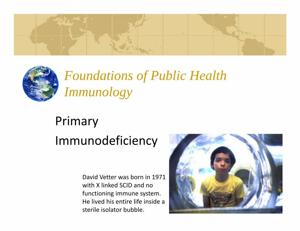
Foundations of Public Health Immunology
Primary Immunodeficiency
David Vetter was born in 1971 with X linked SCID and no functioning immune system. He lived his entire life inside a sterile isolator bubble.
Objectives• Describe the difference between primary & secondary immunodeficiencies
• Identify signs/symptoms of primary immunodeficiency• Identify SCID deficiencies, mutations in specific genes• Describe the difference between X linked & autosomal recessive inheritance
• Identify specific defects that result in different primary immunodeficiency disorders• Adaptive & Innate/Other
• Identify treatment options for primary immunodeficiency
• Identify examples of secondary immunodeficiency
Two Types of Immunodeficiency• Primary (Congenital) Immunodeficiency
• Diseases caused by genetic defects in the immune system
• Diseases are not contagious
• Secondary (Acquired) Immunodeficiency• Diseases caused by other factors that
compromise the immune system• Infection (HIV/AIDS), malnutrition, chemotherapy for
cancer, removal of spleen, etc.

Primary Immunodeficiency (PI)• Group of single‐gene disorders of the immune system
• Single‐gene defects may lead to a missing enzyme or structural component, developmental arrest at a specific stage of immune development, or nonfunctional proteins
• Nearly 100 separate primary diseases have been described• Only ~20 diseases cause the vast majority of PI cases
• Estimates indicate 1 in 500 people in US & Europe have a primary immunodeficiency• 80% of people affected are younger than 20 years old
• Diseases often inherited in X‐linked recessive fashion• 70% of cases occur among males
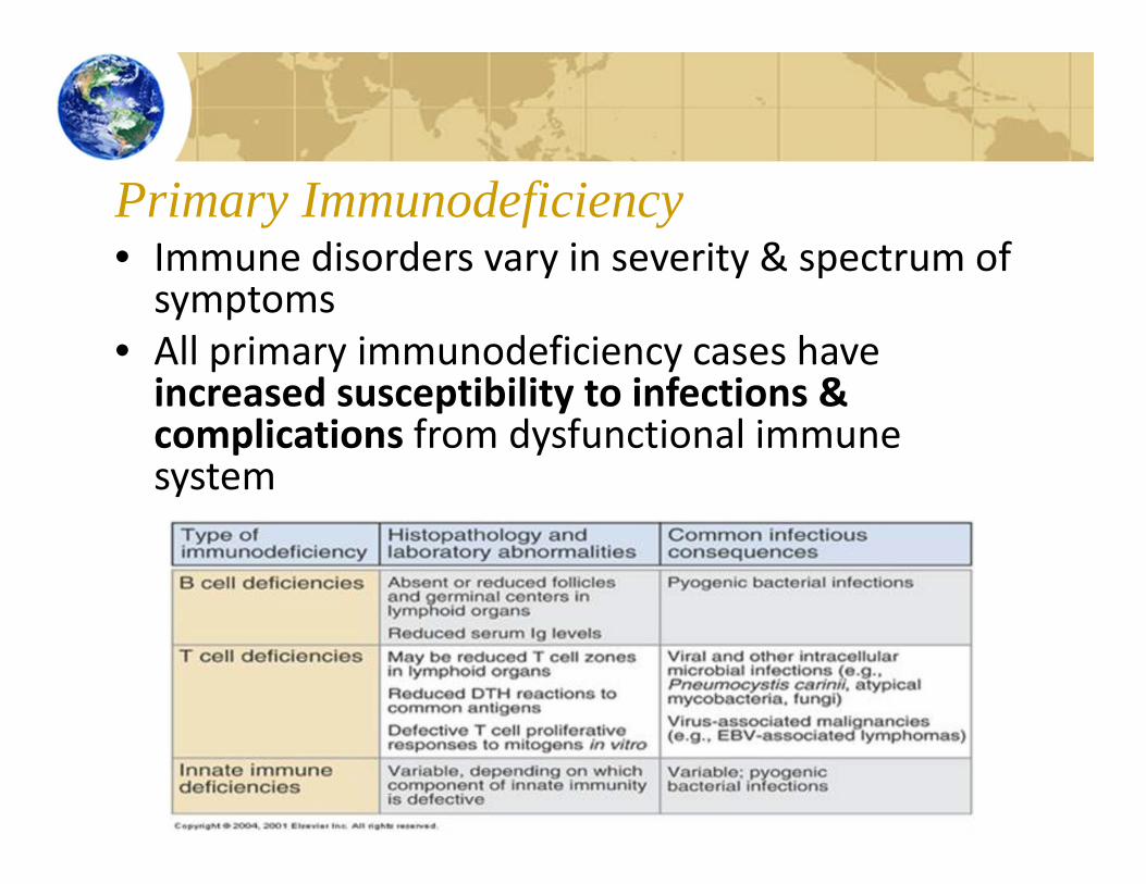
Primary Immunodeficiency• Immune disorders vary in severity & spectrum of symptoms
• All primary immunodeficiency cases have increased susceptibility to infections & complications from dysfunctional immune system
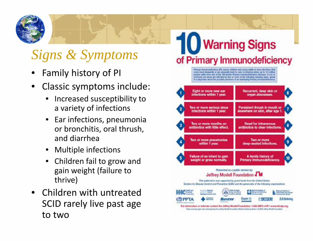
Signs & Symptoms• Family history of PI• Classic symptoms include:
• Increased susceptibility to a variety of infections
• Ear infections, pneumonia or bronchitis, oral thrush, and diarrhea
• Multiple infections• Children fail to grow and
gain weight (failure to thrive)
• Children with untreated SCID rarely live past age to two
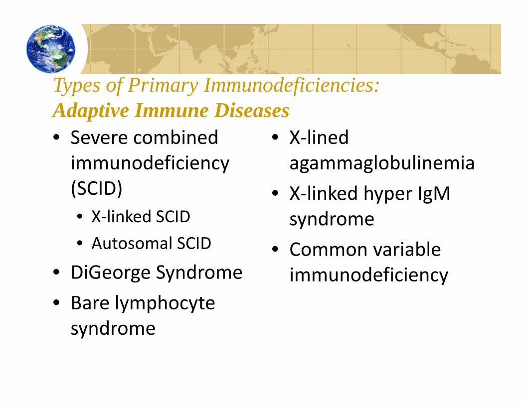
Types of Primary Immunodeficiencies:Adaptive Immune Diseases• Severe combined immunodeficiency (SCID)• X‐linked SCID• Autosomal SCID
• DiGeorge Syndrome• Bare lymphocyte syndrome
• X‐lined agammaglobulinemia
• X‐linked hyper IgMsyndrome
• Common variable immunodeficiency

Severe Combined Immunodeficiency• Combined B cell and T cell immunodeficiencies constitute
20% of PI diseases• Most serious forms of primary immunodeficiency
• Survival beyond first year of life rare without early immune reconstitution through stem cell transplantation (or gene therapy)
• Early diagnosis critical to improve prognosis for infants who have not had severe opportunistic infections
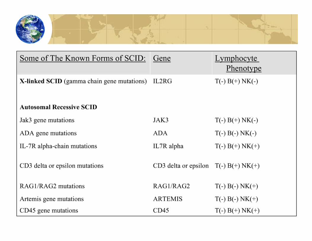
• Caused by mutations in 8 different genes
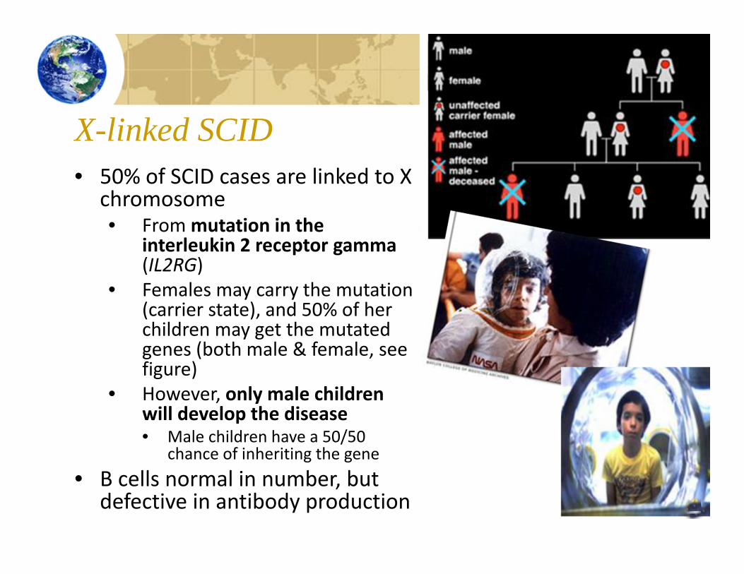
X-linked SCID• 50% of SCID cases are linked to X
chromosome• From mutation in the
interleukin 2 receptor gamma(IL2RG)
• Females may carry the mutation (carrier state), and 50% of her children may get the mutated genes (both male & female, see figure)
• However, only male children will develop the disease• Male children have a 50/50
chance of inheriting the gene• B cells normal in number, but
defective in antibody production
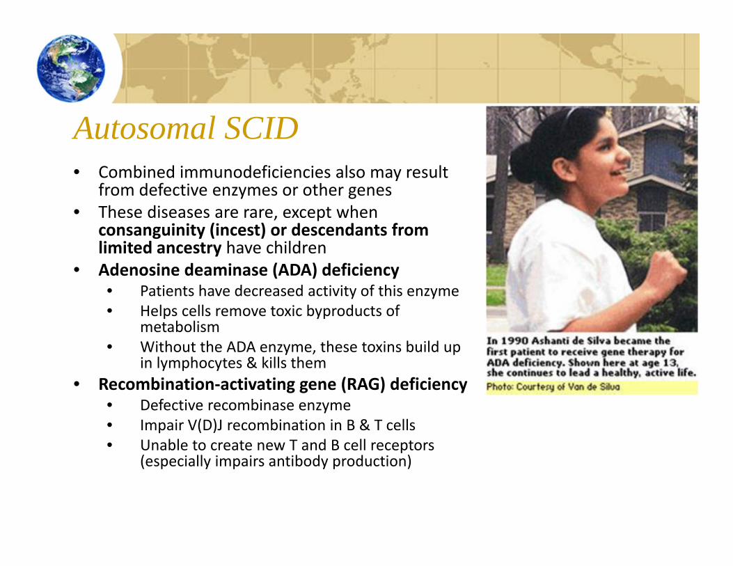
Autosomal SCID• Combined immunodeficiencies also may result
from defective enzymes or other genes• These diseases are rare, except when
consanguinity (incest) or descendants from limited ancestry have children
• Adenosine deaminase (ADA) deficiency• Patients have decreased activity of this enzyme• Helps cells remove toxic byproducts of
metabolism• Without the ADA enzyme, these toxins build up
in lymphocytes & kills them• Recombination‐activating gene (RAG) deficiency
• Defective recombinase enzyme• Impair V(D)J recombination in B & T cells• Unable to create new T and B cell receptors
(especially impairs antibody production)
DiGeorge Syndrome• Rare congenital disease• Caused by large deletion from chromosome 22
• DGS gene required for normal development of thymus and related glands
• Thymus is absent in these patients• Difficult to medically counteract loss of this gene
• Symptoms vary greatly between individuals but usually include recurrent infections, heart defects, and characteristic facial features• Heart defects and some of speech impairments
often treated either surgically or therapeutically• Loss of T‐cells (produced by the thymus) is very
difficult to treat
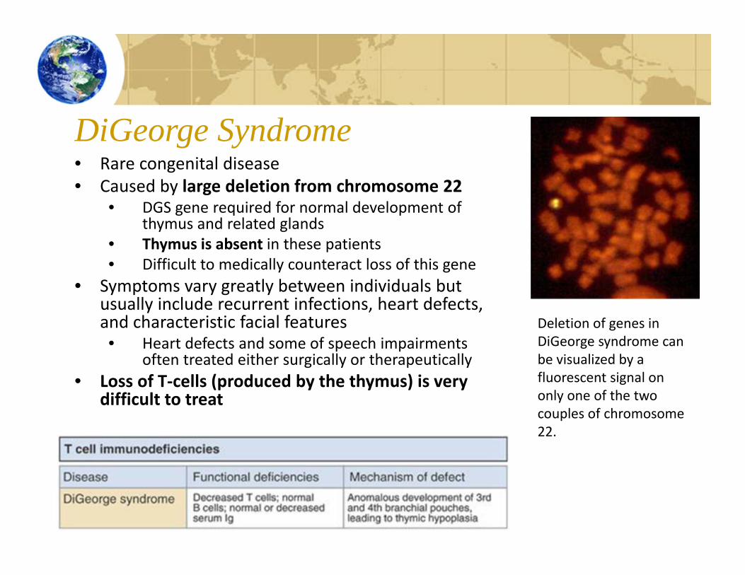
Deletion of genes in DiGeorge syndrome can be visualized by a fluorescent signal on only one of the two couples of chromosome 22.
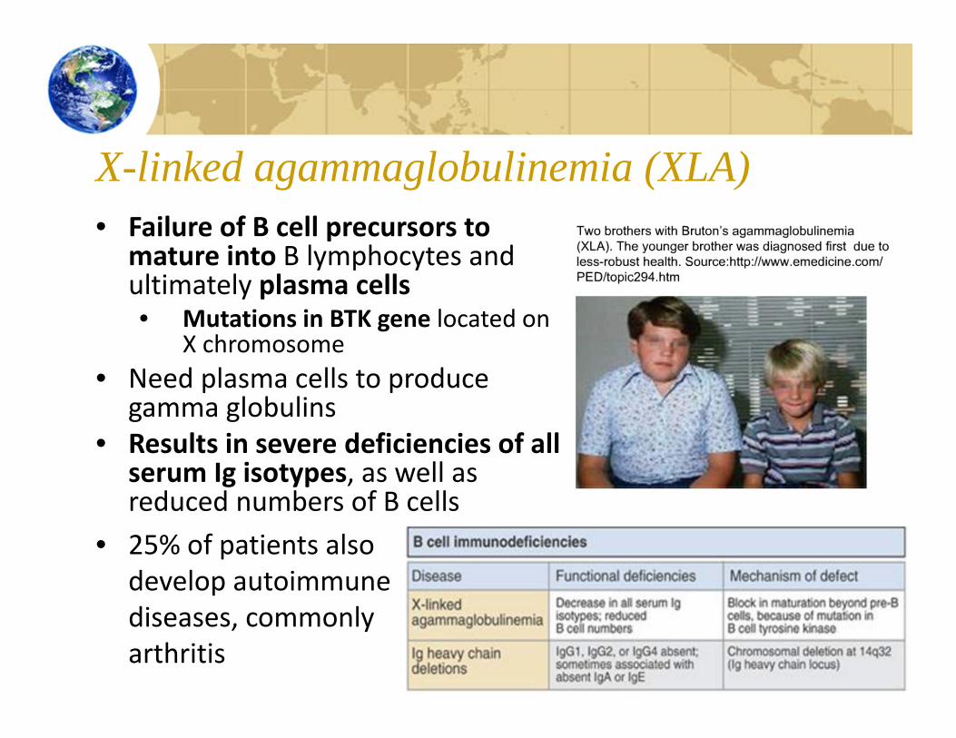
X-linked agammaglobulinemia (XLA)• Failure of B cell precursors to
mature into B lymphocytes and ultimately plasma cells• Mutations in BTK gene located on
X chromosome• Need plasma cells to produce
gamma globulins• Results in severe deficiencies of all
serum Ig isotypes, as well as reduced numbers of B cells
• 25% of patients also develop autoimmune diseases, commonly arthritis
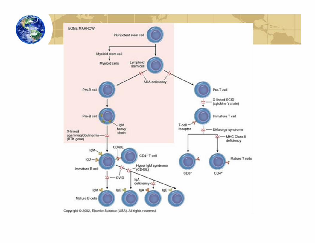
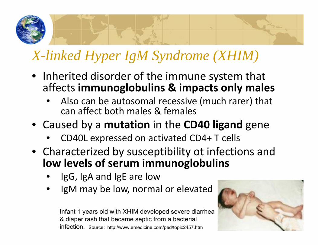
X-linked Hyper IgM Syndrome (XHIM)• Inherited disorder of the immune system that affects immunoglobulins & impacts only males• Also can be autosomal recessive (much rarer) that
can affect both males & females• Caused by a mutation in the CD40 ligand gene
• CD40L expressed on activated CD4+ T cells• Characterized by susceptibility ot infections and low levels of serum immunoglobulins• IgG, IgA and IgE are low• IgM may be low, normal or elevated

• Focus on defects related to impaired helper TcRfunction
• These defects impair B cell, macrophage activation
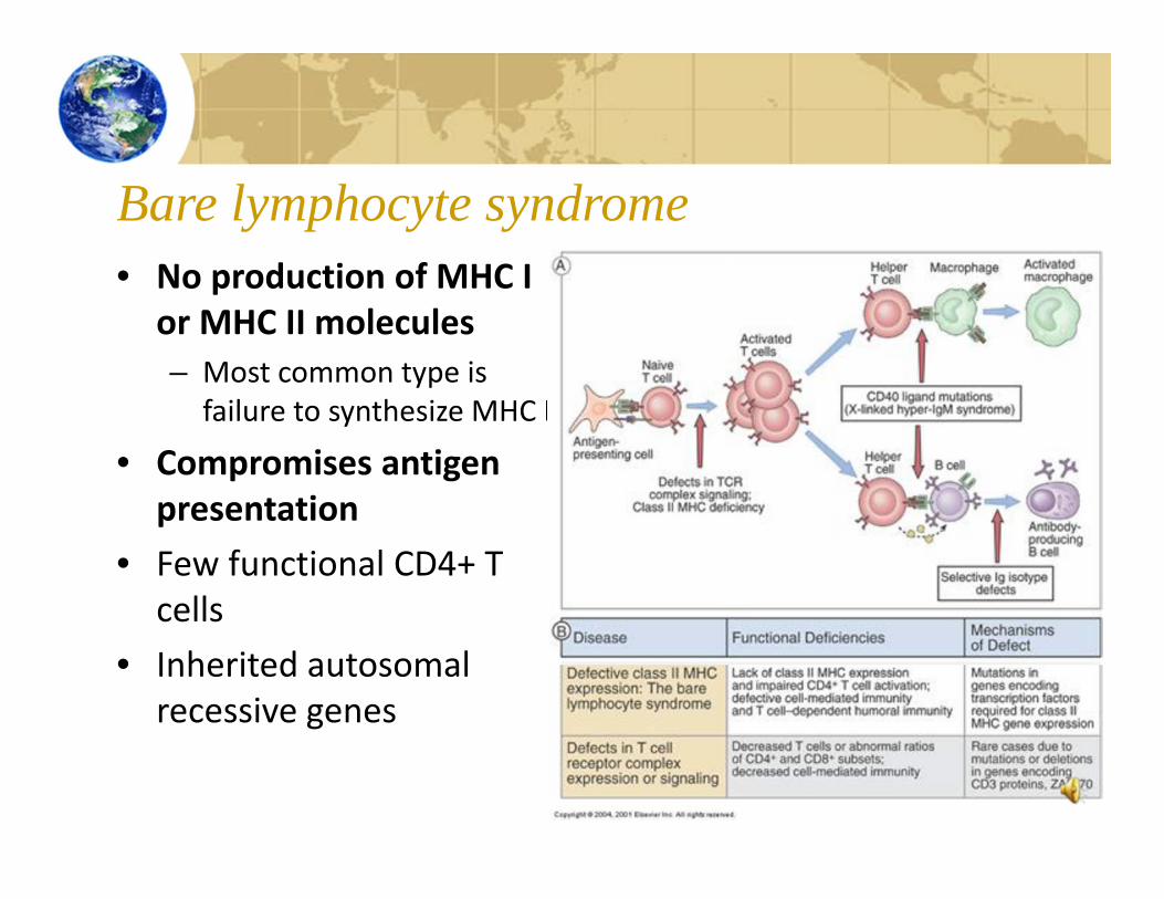
Bare lymphocyte syndrome• No production of MHC I
or MHC II molecules– Most common type is
failure to synthesize MHC II
• Compromises antigen presentation
• Few functional CD4+ T cells
• Inherited autosomal recessive genes

Common Variable Immunodeficiency• Group of disorders that form most common primary
immunodeficiency• Exact cause is unknown, and clinical symptoms vary by
patient• Characterized by low levels of serum immunoglobins,
increased susceptibility to infections• Most patients have normal numbers of B cells, but fail to
undergo normal maturation into plasma cells• Results in poor antibody responses and reduced serum levels
of IgG, IgA, and IgM• Some patients have defects in helper T cell function• Another group of patients have excesive numbers of
cytotoxic T cells• Complication include lung damage, enlarged lymph nodes
& spleen, arthritis and cancer
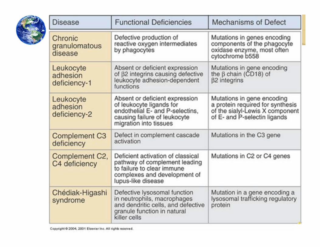
Types of Primary Immunodeficiency:Innate Immune & Other Disorders• Chronic granulomatous disease
• Leukocyte adhesion deficiency
• Complement deficiencies
• Chediak – Higashi syndrome
• Wiskott ‐ Aldrich syndrome
• Ataxia ‐telangiectasia
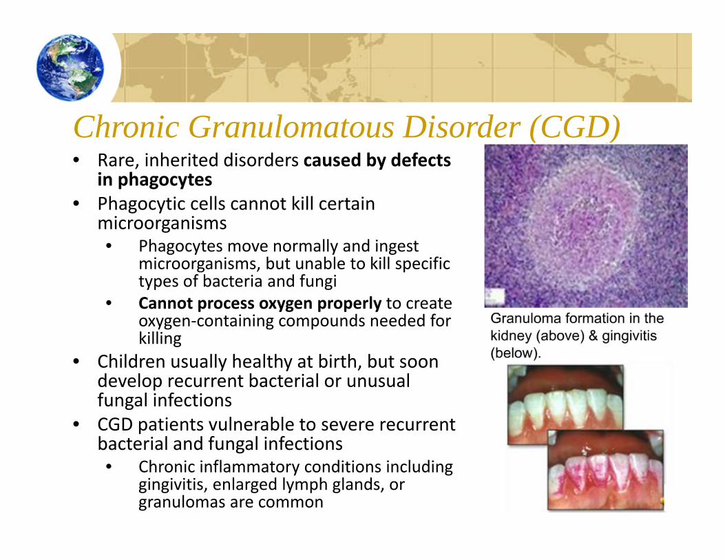
Chronic Granulomatous Disorder (CGD)• Rare, inherited disorders caused by defects
in phagocytes• Phagocytic cells cannot kill certain
microorganisms• Phagocytes move normally and ingest
microorganisms, but unable to kill specific types of bacteria and fungi
• Cannot process oxygen properly to create oxygen‐containing compounds needed for killing
• Children usually healthy at birth, but soon develop recurrent bacterial or unusual fungal infections
• CGD patients vulnerable to severe recurrent bacterial and fungal infections• Chronic inflammatory conditions including
gingivitis, enlarged lymph glands, or granulomas are common
Leukocyte Adhesion Deficiency (LAD)• Very rare disease with fewer than 200 patients reported• Characterized by leukocytosis and localized bacterial infection
• Difficult to detect until infections have progressed to life‐threatening level
• Disorder results when patient cannot produce CD18 protein• CD18 is necessary for leukocytes to travel to the site of an infection
• Leukocyte adhesion deficiency type I (LAD I)• Failure to express the CD18 integrin, a receptor for C3b on myeloid, lymphoid cells• No CD18 on lymphocytes, macrophages, and neutrophils• Patients succumb to infection [mostly bacterial], commonly when younger than 2
years
• Leukocyte adhesion deficiency type II (LAD II)• More rare than type I• Defect in expression of ligands for E and P selectins (remember those?)• Patients have leukocytosis, recurrent infections, severe growth and mental retardation• Usually do not die from infection, but also may have neurologic impairment, and
short stature
Chediak Higashi Syndrome (CHS)• Rare childhood autosomal recessive disorder
that affects multiple systems of body• Hypopigmentation of skin, eyes, and hair• Prolonged bleeding, bruise easily, and recurrent
infections• Mutation in CHS gene affects synthesis of
storage/secretory granules in various types of cells• Abnormal natural killer cell function• Defective lysosomal function in macs, dendritic
cells & neutrophils• Often fatal in childhood as a result of infection
or an accelerated lymphomalike phase• Few patients live to adulthood
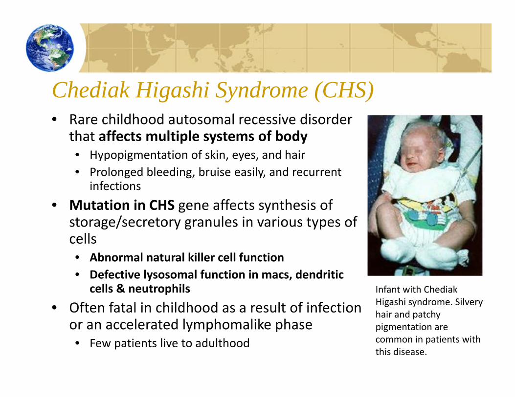
Infant with ChediakHigashi syndrome. Silvery hair and patchy pigmentation are common in patients with this disease.
Wiskott-Aldrich Syndrome (WAS)• X‐linked recessive genetic condition,
found almost exclusively in males• Disorder causes persistent
thrombocytopenia, IgM deficiency• Reduced number of platelets, eczema,
combined immunodeficiency, and higher rist of developing autoimmune diseases
• Results from defect in protein called Wiskott‐Aldrich syndrome protein (WASp)
• WAS protein important for migration and mortality of immune cells• Platelets and leukocytes are smaller, do
not develop properly, & fail to migrate
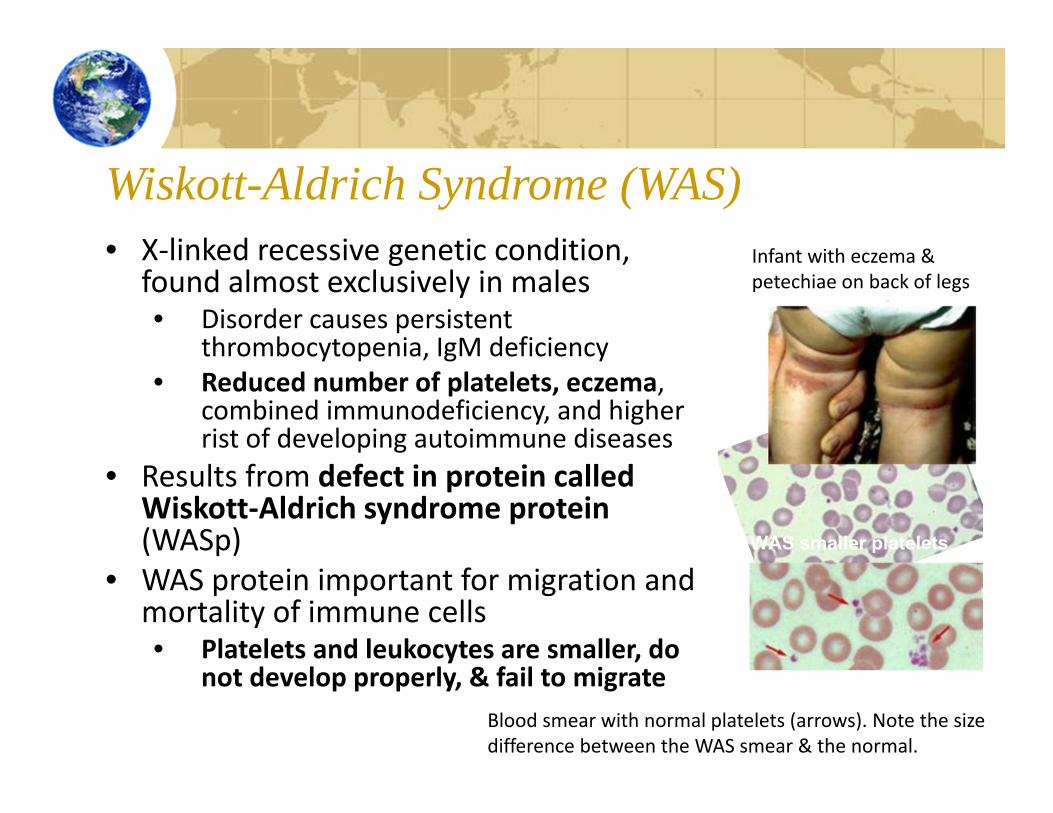
Infant with eczema & petechiae on back of legs
Blood smear with normal platelets (arrows). Note the size difference between the WAS smear & the normal.
Ataxia telangiectasia (AT)• Autosomal recessive disorder is a multi‐system
disease• Characterized by gait abnormalities (ataxia) &
vascular malformations (telangiectasia)• Affects brain, skin & immune system
• Mutation in AT gene impairs DNA repair during recombination of antigen receptor genes• Compromises T cell maturation & function
• AT patients may have defective isotypeswitching, from dysregulation of immunoglobulin gene superfamily
• AT protein also controls cell cycle & mutation of this gene on chromosome 11 may explain immunologic & neurologic symptoms
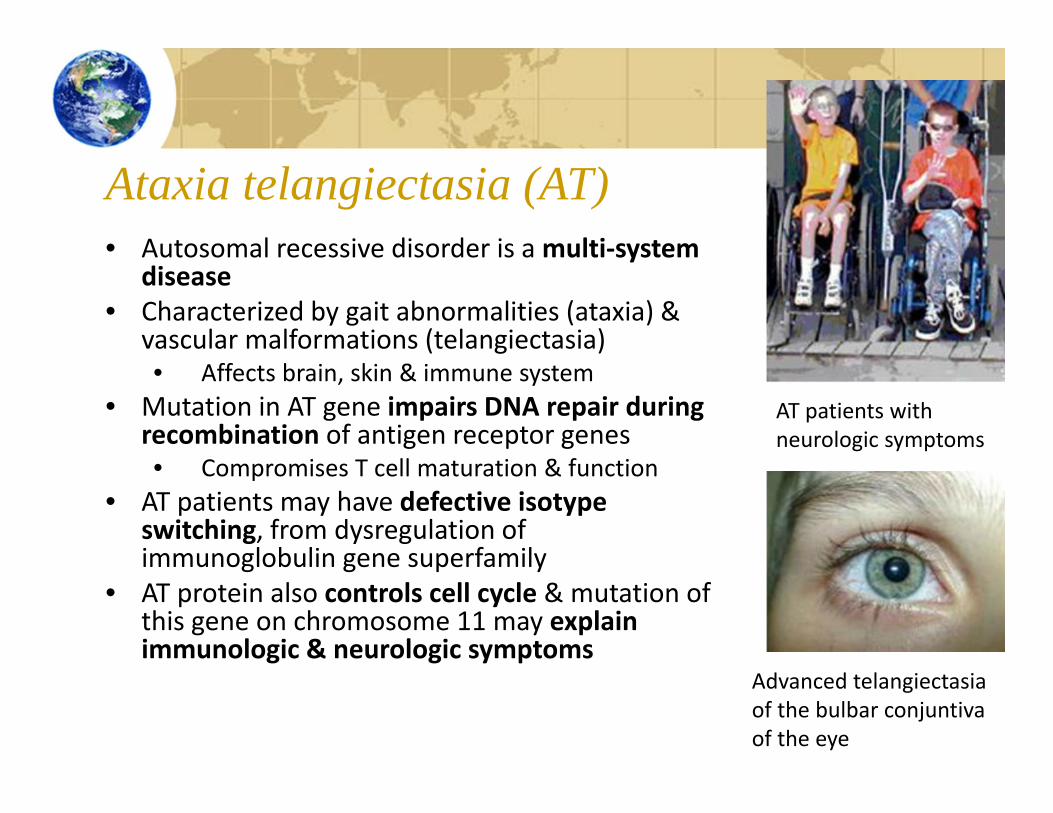
AT patients with neurologic symptoms
Advanced telangiectasia of the bulbar conjuntivaof the eye
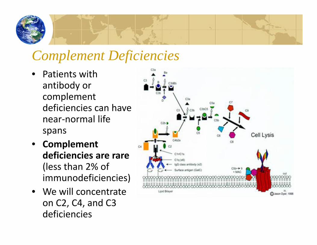
Complement Deficiencies• Patients with
antibody or complement deficiencies can have near‐normal life spans
• Complement deficiencies are rare (less than 2% of immunodeficiencies)
• We will concentrate on C2, C4, and C3 deficiencies

C2 & C4 Deficiencies• Associated with recurrent infections by encapsulated bacteria (antibodies,
complement and neutrophils required for proper clearance)• C2 is most widely reported deficiency of all the components in the
complement pathways• Immune complex disorders are main problem with C2 deficiency
• Skin and joint manifestations are common• Frequently found in patients with SLE, Henoch‐Schonlein vasculitis,
polymyositis, and recurrent pyogenic infection• Most individuals with C2 deficiency are asymptomatic (until disease
development)• Almost all patients with complete C4 deficiency have discoid or systemic
lupus erythematosus (with or without associated glomerulonephritis)• Need classical pathway to eliminate immune complexes• Classical pathway is impaired in C2 & C4 deficiency
• Not susceptible to infection (like C3 deficiencies) because alternative pathway still available to protect host defenses

C3 Deficiencies• C3 deficiency may be due to a primary defect in the C3 gene or expression of the C3 protein
• Deficiencies predisposes person to frequent bouts of pyogenic bacterial infections (especially Gram‐negative bacteria such as meningococciand pneumococci) and immune complex disease• Approximately, 78% of patients with C3 deficiency
have repeated infections and 79% of patients experience autoimmune disorders (such as arthralgia and vasculitic rashes, lupuslike syndrome, and membranoproliferative glomerulonephritis)

Primary Immunodeficiency Treatment• Need effective and early treatment
• Untreated primary deficiencies characterized by frequent life‐threatening infection, debilitating illnesses
• Usually fatal if untreated• Medical advances in treatment allow patients to survive childhood & live almost normal lives• Requires life long therapy including IV gamma
globulin infusions, antibiotic therapy, or bone marrow transplanatation

Treatment Options• Bone Marrow transplantation
• Undifferentiated stem cells taken from healthy bone marrow are injected into SCID patients
• Stem cells can then differentiate into healthy immune cells
• Antibiotics• Patients often treated with IV
antibiotics for bacterial infections• Also as a prohylactic method to
prevent recurrent infections• Antibody replacement therapy
• Intravenous (IV) infusion of plasma with protective IgG antibodies in large doses
• Helps reduce severity and frequency of infections
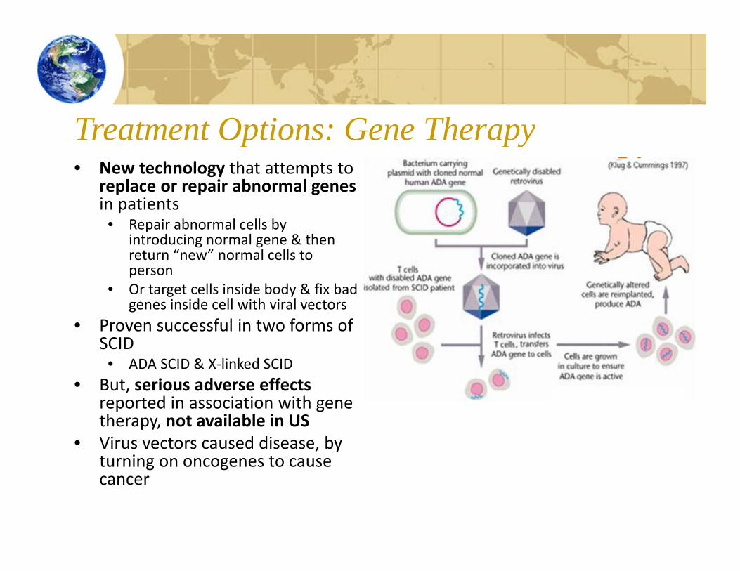
Treatment Options: Gene Therapy• New technology that attempts to
replace or repair abnormal genesin patients• Repair abnormal cells by
introducing normal gene & then return “new” normal cells to person
• Or target cells inside body & fix bad genes inside cell with viral vectors
• Proven successful in two forms of SCID• ADA SCID & X‐linked SCID
• But, serious adverse effects reported in association with gene therapy, not available in US
• Virus vectors caused disease, by turning on oncogenes to cause cancer

Secondary Immunodeficiency• Acquired immunodeficiency• More common than primary deficiencies• Causes include non‐immune disorders (diabetes, malnutrition) and
immunosuppressive treatment• Prolonged serious illness may also lead to impaired immune
response• Impairment is often reversible
In Summary• Understand the
difference between primary & secondary immunodeficiencies
• Identify SCID deficiencies, mutations in specific genes
• Understand the difference between X linked & autosomal recessive inheritance
• Identify specific defects that result in different primary immunodeficiency disorders
• Adaptive & Innate/Other• Identify treatment
options for primary immunodeficiency
• Identify examples of immunodeficiency
Self-Test Questions• Describe the difference between a primary & secondary
immunodeficiency. Name 3 examples of each type.• Describe how the patterns of inheritance (X linked &
autosomal recessive) are different.• What is SCID? How does this impact the immune response?
What genetic defect causes X linked SCID?• What is consanguinity? Which PI diseases are linked to
this?• What defect causes DiGeorge’s Syndrome? CGD? Chediak
Higashi Syndrome? WAS? AT? Describe the phenotype (problems) that occurs in each of these patients.
• Identify and describe the 4 different treatment options available for primary immunodeficiency.