Embed Size (px)
Citation preview
1
Fossil diatoms (Bacillariophyta) as potential paleoenvironmental indicators at St. Catherines Island, Georgia. DAVID M. JARZEN Paleobotany and Palynology Laboratory Florida Museum of Natural History University of Florida Gainesville Florida 32611-7800 U.S.A. SUSAN A. JARZEN Development Office Florida Museum of Natural History University of Florida Gainesville Florida 32611-7800 U.S.A. IRVY R. QUITMYER Environmental Archaeology Florida Museum of Natural History University of Florida Gainesville Florida 32611-7800 U.S.A.
e-mail: [email protected]; [email protected]; [email protected]
Contents
Abstract ……………………………………………………………………………… p. 2 Introduction………………………………………………………………………….. p. 3 Methods and Materials……………………………………………………………... p. 4 Diatom Systematics ………………………………………………………………… p. 8 Other palynomorphs……………………………………………………………….. p. 22 Results and Conclusions………………………………………………………….. p. 24 Acknowledgements ………………………………………………………………… p. 28 References Cited……………………………………………………………………. p. 30 Appendix I …………………………………………………………………………… p. 36 Text-figure 1 Map Eastern USA showing location of St. Catherines Is………… p. 38 Text-figure 2 Map of St. Catherines Island Recent Collection Sites ……………. p. 39 Text-figure 3 Collection and Processing of Shellfish Gut Samples……………. p. 40 Text-figure 4 Locality map of AMNH Archeological Sites………………………. p. 41 Table 1. pH and Salinity of the East and West Water Samples……………….. p. 42 Table 2. Taxa of Shellfish Identified from the Gut and Water Samles………… p. 42
2
Table 3. Collection Data for the AMNH Archeological Samples………………. p. 43 Table 4. Distribution of Diatom Taxa Through Four Seasons West Side…….. p. 44 Table 5. Distribution of Diatom Taxa Through Four Seasons East Side…….. p. 45 Plate 1. Description of Palynomorphs Identified ……………………………….. p. 46 Plate 2. Description of Palynomorphs Identified……………………………….. p. 47 Plate 3. Description of Palynomorphs Identified……………………………….. p. 49 Abstract
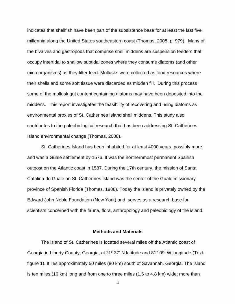
In this research we identify diatoms contained in the guts of three living, filter-feeding
mollusks that are commonly identified in the shell middens of St. Catherines Island,
Georgia, USA: Eastern Oyster (Crassostrea virginica); hard clam (Mercenaria sp.); and
Atlantic ribbed mussel (Geukensia demissa). These taxa, along with water samples
were collected during each season between 2009 -2010 from two locations on St.
Catherines Island. The resulting modern assemblage of diatoms forms a comparative
collection that might be expected to be identified in St. Catherines Island archeological
sites. Thirty-six sediment samples were excavated and evaluated for their palynological
content. Diatom recovery from archeological samples collected from two shell midden
sites on St. Catherines Island, Georgia was poor, probably due to the alkaline nature of
the shell midden sediments. Diatoms, if recovered from areas outside the shell middens,
where the sediment pH is more acidic, may provide the assemblages needed to
reconstruct paleoenvironmental conditions at the time of midden formation. Recovery of
diatoms and other microorganisms as pollen, spores, and fungal elements from the gut
contents of three taxa of living shellfish provides a systematic treatment and description
of 27 diatoms which represent the first recorded and systematically described diatoms
from the coastal waters of Georgia. These descriptions may facilitate the identification of
fossil diatoms recovered from sediments associated with shell middens elsewhere on
the island.

3
Key words: Palynology; archaeology; diatoms; pollen; spores; mollusca; St. Catherines
Island; Georgia
Introduction
“Salt marshes…are glorious places, bugs and all. With their green and brown grasses producing nutrients for the sea,
they are among the richest places and most productive environments on earth. Able to withstand salt water, the grasses stand eternally
as a buffer between the murky estuaries and bays and the high green forests, exuding life and energy. – Jack Rudloe, The Wilderness Coast, 1988
The salt marshes surrounding the islands of the coast of southeastern United
States harbor a rich and diverse biota. In these waters are contained an assemblage of
planktonic and benthic organisms, key of which are the diatoms. Diatoms
(Bacillariophyta) are single-celled benthic or planktonic algae that form durable, biogenic
silica cell walls (frustules). There are about 200 genera and more than 100,000 species
of diatoms worldwide (Round et al., 1990). The frustule shape and morphological details
may be used to identify genera or species in recent habitats and subrecent lithological
deposits (Round et al., 1990). The diatoms represent a poorly understood resource in
the field of environmental archaeology and paleobiology along the Georgia Coast, and
the study of extant diatoms along the Atlantic and Gulf Coasts of Florida and Georgia
are for the most part, non-existent (Evelyn Gaiser, written communication, 2008).
Diatoms, having specific salinity and pH requirements, may be useful in reconstructing
paleohabitats represented in the shell middens found on St. Catherines Island (Vos and
Wolf, 1993; Stoermer and Smol, 1999).
Shell middens are anthropogenic deposits that account for nearly 5000 years of
human and environmental history of St. Catherines Island. The zooarchaeological record
4
indicates that shellfish have been part of the subsistence base for at least the last five
millennia along the United States southeastern coast (Thomas, 2008, p. 979). Many of
the bivalves and gastropods that comprise shell middens are suspension feeders that
occupy intertidal to shallow subtidal zones where they consume diatoms (and other
microorganisms) as they filter feed. Mollusks were collected as food resources where
their shells and some soft tissue were discarded as midden fill. During this process
some of the mollusk gut content containing diatoms may have been deposited into the
middens. This report investigates the feasibility of recovering and using diatoms as
environmental proxies of St. Catherines Island shell middens. This study also
contributes to the paleobiological research that has been addressing St. Catherines
Island environmental change (Thomas, 2008).
St. Catherines Island has been inhabited for at least 4000 years, possibly more,
and was a Guale settlement by 1576. It was the northernmost permanent Spanish
outpost on the Atlantic coast in 1587. During the 17th century, the mission of Santa
Catalina de Guale on St. Catherines Island was the center of the Guale missionary
province of Spanish Florida (Thomas, 1988). Today the island is privately owned by the
Edward John Noble Foundation (New York) and serves as a research base for
scientists concerned with the fauna, flora, anthropology and paleobiology of the island.
Methods and Materials
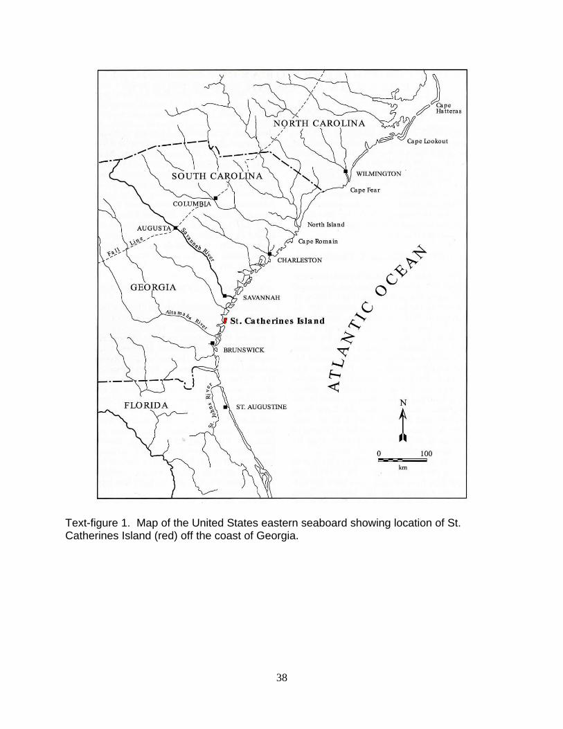
The island of St. Catherines is located several miles off the Atlantic coast of
Georgia in Liberty County, Georgia, at 31° 37’ N latitude and 81° 09’ W longitude (Text-
figure 1). It lies approximately 50 miles (80 km) south of Savannah, Georgia. The island
is ten miles (16 km) long and from one to three miles (1.6 to 4.8 km) wide; more than
5
half of its 14,640 acres (59 km²) are tidal marsh and wetlands, reached only by boat.
Extant shellfish specimens of three genera from the east and west sides of the island
were collected during all seasons, as modern comparative specimens. Subrecent fossil
and soil samples were collected from the St. Catherines shell midden and McQueen
shell midden sites on the island.
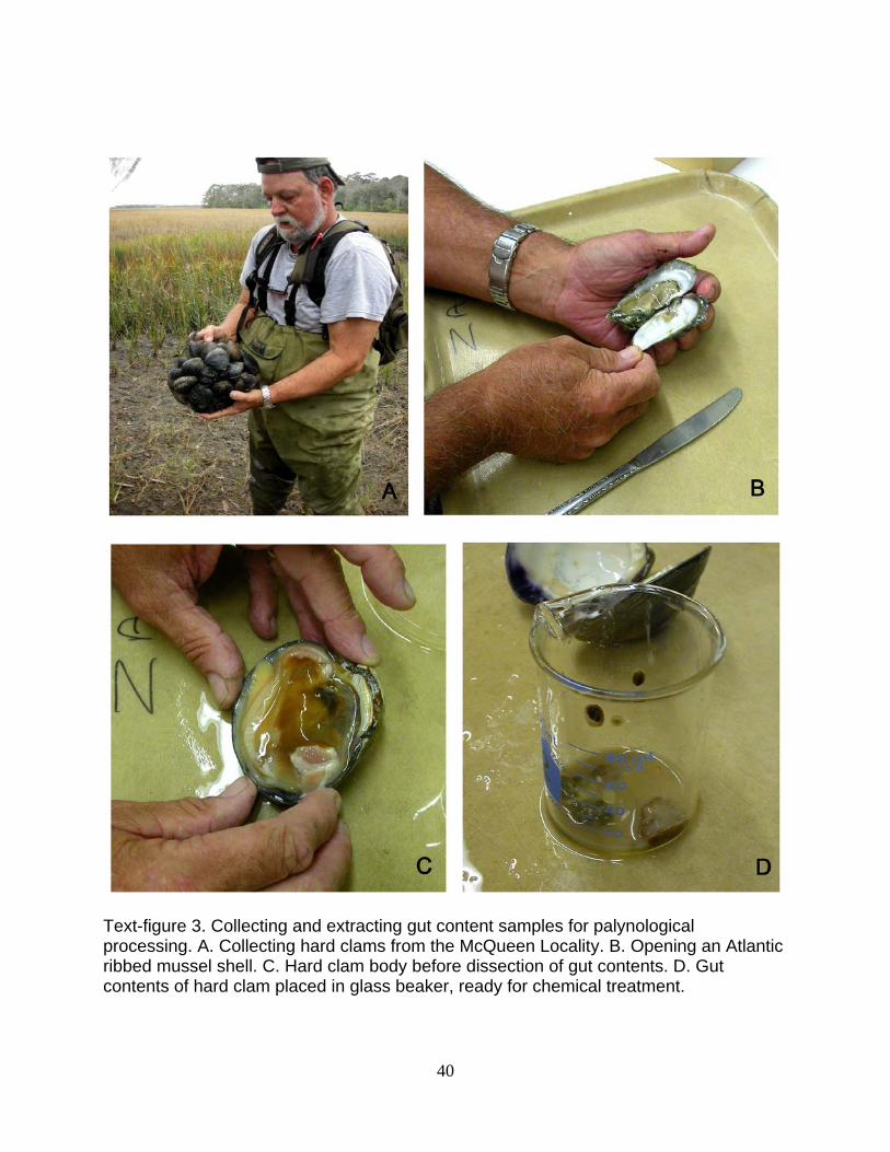
Collection and processing of extant shellfish
Collections of living, filter-feeding mollusks that are commonly identified in the
shell middens of St. Catherines Island were made from the island on four separate
occasions to represent the four seasons of the year. At each season, four specimens
each of the Eastern oyster [Crassostrea virginica (Gmelin 1791)], hard clam
(Mercenaria spp.) and, Atlantic ribbed mussel [Geukensia demissa (Dillwyn 1817)] were
collected. Collections from the East side of the island (Atlantic Ocean side) and from the
West side of the island (landward side) were made of each of the three shellfish taxa. A
water sample was also collected from each site, to record pH and salinity (Table I) and
to concentrate the planktonic organisms from which a comparative slide was prepared.
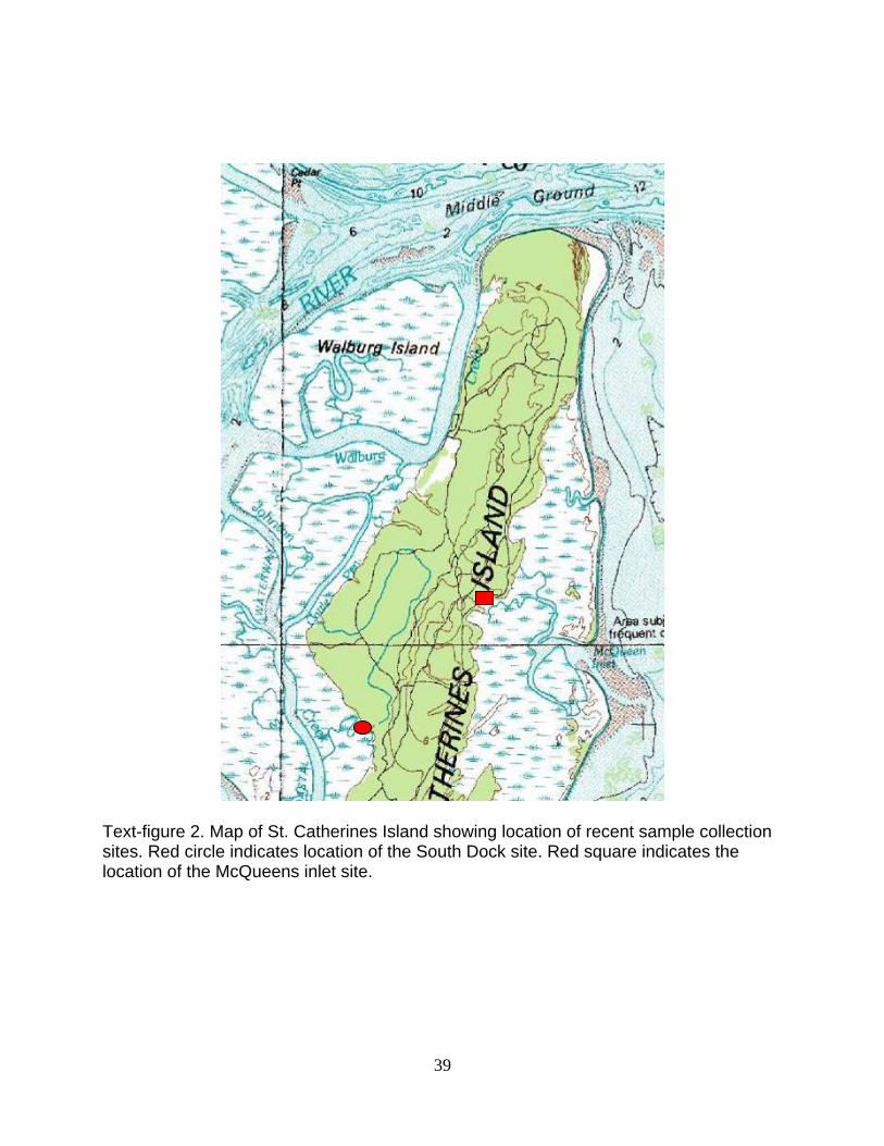
The West Side locality is at the South End Dock area (GPS N 31° 36.403’ W 081°
10.646’) along an adjacent stream bottom. The East Side locality is at the Kings New
Ground site (GPS N 31° 38.519’ W 081° 09.206’), along a stream channel (Text-figure
2). The collection dates for the extant comparative specimens were August 18, 2009
(summer), November 14, 2009 (autumn), February 21, 2010 (winter), and April 24,
2010 (spring).
The diatom flora contained within the gut contents of these mollusks serves as a
comparative collection for both sides of the island through the four seasons of the year.
6
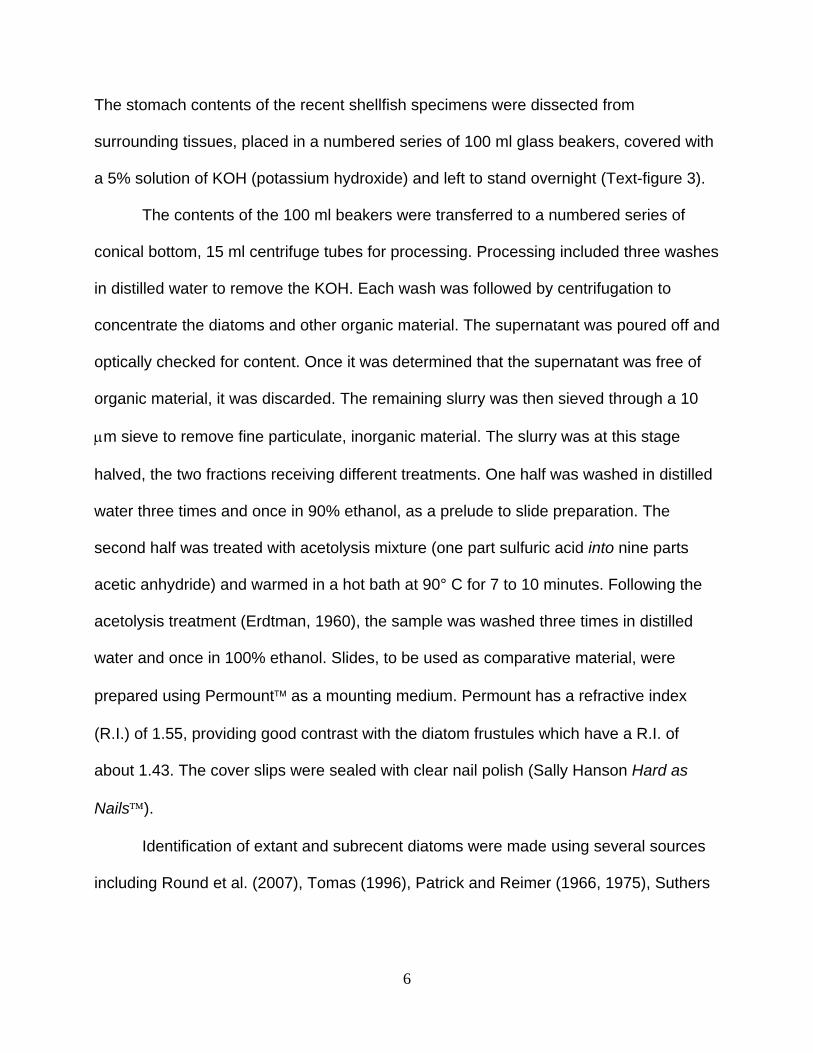
The stomach contents of the recent shellfish specimens were dissected from
surrounding tissues, placed in a numbered series of 100 ml glass beakers, covered with
a 5% solution of KOH (potassium hydroxide) and left to stand overnight (Text-figure 3).
The contents of the 100 ml beakers were transferred to a numbered series of
conical bottom, 15 ml centrifuge tubes for processing. Processing included three washes
in distilled water to remove the KOH. Each wash was followed by centrifugation to
concentrate the diatoms and other organic material. The supernatant was poured off and
optically checked for content. Once it was determined that the supernatant was free of
organic material, it was discarded. The remaining slurry was then sieved through a 10
μm sieve to remove fine particulate, inorganic material. The slurry was at this stage
halved, the two fractions receiving different treatments. One half was washed in distilled
water three times and once in 90% ethanol, as a prelude to slide preparation. The
second half was treated with acetolysis mixture (one part sulfuric acid into nine parts
acetic anhydride) and warmed in a hot bath at 90° C for 7 to 10 minutes. Following the
acetolysis treatment (Erdtman, 1960), the sample was washed three times in distilled
water and once in 100% ethanol. Slides, to be used as comparative material, were
prepared using Permount™ as a mounting medium. Permount has a refractive index
(R.I.) of 1.55, providing good contrast with the diatom frustules which have a R.I. of
about 1.43. The cover slips were sealed with clear nail polish (Sally Hanson Hard as
Nails™).
Identification of extant and subrecent diatoms were made using several sources
including Round et al. (2007), Tomas (1996), Patrick and Reimer (1966, 1975), Suthers
7
and Rissik (2009), and through discussions with M. J. Sullivan (St. Andrews Episcopal
School, Ridgeland, MS).
Collection and processing of fossil/subrecent samples
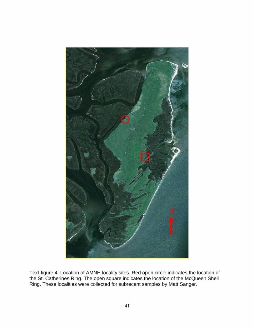
Thirty-six subrecent samples were collected by Matt Sanger from two
archeological sites previously excavated (Text-figure 4) by the American Museum of
Natural History (AMNH), Division of Anthropology (Thomas, 2008). Ten (10) soil
samples were collected from the AMNH Site 504 (= St. Catherines Shell Ring, south wall
feature). Eighteen (18) samples of loosely consolidated soil and shell material were
collected from AMNH 504, T281 (= St. Catherines Shell Ring, north wall of trench 281),
and eight (8), carbonaceous, shell and soil samples were collected from AMNH 696 (=
McQueens Shell Ring, east wall). All samples were collected on May 27, 2009, and
transported to the Florida Museum of Natural History (FLMNH), Paleobotany and
Palynology Laboratory for processing. Table 2 provides the sample collection data.
Processing of the subrecent soil and shell midden samples followed for the most
part standard palynological processing techniques as outlined by Bryant (2009) and
Jarzen (2006). The use of hydrofluoric acid (HF) was omitted in order to retrieve the
siliceous frustules of the diatom flora. As a result of not using HF to remove the silicates,
the resulting residue is often laden with crystals of silica making observation and
photography difficult. Two slides were prepared for each residue fraction using Glycerin
jelly, Cellosize™ and Lucite™ or Cellosize and Permount™ as the mounting media
(medium noted on slide label). Pollen and spores are better observed and photographed
8
using either glycerin jelly or Lucite as the mounting medium, while diatoms fare better
using Permount. The cover slips were sealed with clear nail polish (Sally Hanson Hard
as Nails™). Slides of both extant comparative material and subrecent archaeological
samples, and the remaining residues and unused sediment samples, are curated at the
Paleobotany and Palynology Laboratory, FLMNH, Gainesville, Florida, 32611, USA as
Localities UF 19272, UF 19273, and UF 19274. Access to these collections may be
made through Dr. Hongshan Wang ([email protected]).
Photographs of the palynomorphs (diatoms, algal cysts, pollen, spores, etc.)
were made using a Nikon Coolpix 4500™ camera mounted on a Leitz Dialux 20™
research microscope. Specimens are located by stage coordinates marking an x
(horizontal) and y (vertical) axis (with printed label to the left), location for the Leitz
Dialux 20 microscope (Serial Number 513467). Specimens may also be located using
the England Finder Slide locator. For details of the nature and instructions for use of the
England Finder Slide, see: (http://www.2spi.com/catalog/magnifiers/england-finder-
graticule-instructions.html.)
Diatom Systematics
“Taxonomy (the science of classification) is often undervalued as a glorified form of filing—with each species in its folder, like a stamp in its prescribed place in an album; but taxonomy is a
fundamental and dynamic science, dedicated to exploring the causes of relationships and similarities among organisms.
Classifications are theories about the basis of natural order, not dull catalogues compiled only to avoid chaos.”
Stephen Jay Gould, Wonderful Life (1989, p. 98)
The systematic section covers the palynomorphs (any organism or part thereof
including pollen, spores, diatoms, silicoflagellates, or other organic material) recovered
9
from the living shellfish and water samples. The diatom classification follows that of
Round, Crawford and Mann (2007).
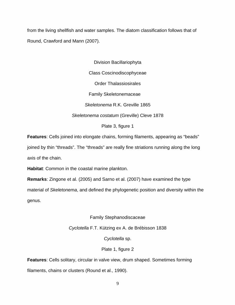
Division Bacillariophyta
Class Coscinodiscophyceae
Order Thalassiosirales
Family Skeletonemaceae
Skeletonema R.K. Greville 1865
Skeletonema costatum (Greville) Cleve 1878
Plate 3, figure 1
Features: Cells joined into elongate chains, forming filaments, appearing as “beads”
joined by thin “threads”. The “threads” are really fine striations running along the long
axis of the chain.
Habitat: Common in the coastal marine plankton.
Remarks: Zingone et al. (2005) and Sarno et al. (2007) have examined the type
material of Skeletonema, and defined the phylogenetic position and diversity within the
genus.
Family Stephanodiscaceae
Cyclotella F.T. Kützing ex A. de Brébisson 1838
Cyclotella sp.
Plate 1, figure 2
Features: Cells solitary, circular in valve view, drum shaped. Sometimes forming
filaments, chains or clusters (Round et al., 1990).
10
Habitat: Mainly freshwater, but invading brackish coastal waters (Round et al., 1990).
This form was common in the water samples and most of the gut samples.
Remarks: Prasad and Nienow (2006) discuss and describe the centric diatom genus
Cyclotella from the Florida Bay region. The genus has about 100 species. A similar
genus is Stephanodiscus which differs from Cyclotella in lacking a distinct ring of spines
as seen in valve view (Round et al., 1990).
Order Melosirales
Family Melosiraceae
Melosira C.A. Agardh 1824
Melosira sp.
(not illustrated)
Features: Cells are cylindrical, to subspherical and united in chains or filaments.
Habitat: Common in marine and freshwater epibenthic (living on the surface of
sediment) habitats. Melosira was rarely found in the water and mollusk gut samples
collected from St. Catherines Island.
Remarks: Melosira prefers, though not restricted to, olighaline water, or waters with low
salinity (0.5 to 5.0 ppt). Hasle and Syvertsen (1996) discuss the distribution and
identification of species of Melosira.
Order Paraliales
Family Paraliaceae
Paralia P.A.C. Heiberg 1863
Paralia marina (W. Smith) Heiberg.
11
Plate 1, figure 7
Features: Cells are cylindrical, united to form linked chains of several cells. Those
observed here are free, circular in valve view, giving the impression in valve view of a
“toothed gear.” Diameter of the valve is variable from 8 – 130 µm (Tomas, 1996).
Habitat: Common on near shore plankton, especially on sandy sediments. The genus is
common in many of the gut samples collected from St. Catherines Island (see Tables 4
and 5).
Remarks: This is a small genus of only two (possibly three) species, P. marina and the
fossil taxon, P. sulcata (Round et. al., 2007). A detailed account of the genus may be
found in Crawford (1979).
Order Coscinodiscales
Family Coscinodiscaceae
Coscinodiscus C.G. Ehrenberg 1838
Coscinodiscus sp.
Plate 1, figures 3, 4; Plate 2, figure 2
Features: Centric, discoid, thin to thicker and more barrel-shaped.
Habitat: Marine free living in the plankton. Common in coastal bays and estuaries.
Common in the St. Catherines material.
Remarks: This genus has many species. Some of which have a documented fossil
record (Round et al., 1990).
Family Hemidiscaceae
Actinocyclus C.G. Ehrenberg 1837
12
Actinocyclus sp.
Plate 2, figure 1
Features: The centric cells are barrel-shaped. The “pseudonodulus” is characteristic of
the genus. This feature, a small pore-like structure, is often difficult to observe. The
diameter of the frustrule ranges from 25 to 95 μm.
Habitat: Mostly epiphytic on seagrass (e.g. Thalassia), but also found in the nearshore
plankton and in localities with mangroves.
Remarks: The pseudonodulus is often very difficult to observe under light microscopy
(personal communication M. J. Sullivan, 2009)
Hemidiscus G.C. Wallich 1860
Hemidiscus sp.
Plate 3, figure 2
Features: Valves are cuneiforme (wedge shaped). Surface covered with closely spaced
areolae.
Habitat: Widely distributed in marine warmer waters, carried to temperate waters on
ocean currents.
Remarks: There are nine species within the genus. Blooms of Hemidiscus spp. are
reported as the casual agent in mass mortality of fish and invertebrates in some areas
(Subramanian and Purushothaman, 1985).
Family Heliopeltaceae
Actinoptychus C.G. Ehrenberg 1843
13
Actinoptychus sp.
Plate 1, figure 1
Features: Solitary, centric diatoms, cells circular in valve view. The valve is sectioned
into six segments (but may be up to 20 segments), as in the slices of a pie. These
sections alternate in height, providing an undulate surface.
Habitat: This diatom is common in neritic assemblages, free or attached to other algae
on coastal sediments (Round et al., 1990).
Remarks: There may be as many as 150 validly published species (Round et al., 1990),
making identification to the species level difficult for the non-specialist.
Order Triceratiales
Family Triceratiaceae
Odontella C.A. Agardh 1832
Odontella aurita (Lyngbye) C. A. Agardh
Plate 3, figure 3
Features: Solitary cells, oblong in girdle view, ornamented with elongated spines or
apical elevations Cells may be united in chains.
Habitat: Marine planktonic or epiphytic, abundant in all ocean waters (Round et al.,
1990).
Remarks: Odontella is a diatom monitored among toxic algae blooms by NOAA's
Phytoplankton Monitoring Network (http://odontella.blogspot.com/).
Odontella mobiliensis (J. W. Bailey) Grunow
Plate 3, figure 4
14
Features: Solitary or in chains. Cells large ranging from 60 to 130 μm longest dimension
(Johnson and Allen, 2005).
Habitat: Common in marine waters, preferring higher salinities.
Remarks: Species of Odontella are most abundant in north temperate regions, but are
truly a cosmopolitan taxon.
Order Biddulphiales
Family Biddulphiaceae
Trigonium P.T. Cleave 1868
Trigonium sp.
Plate 2, figure 5
Features: Cells triangular in valve view, rectangular in girdle view. Occurring singly or in
chains. Surface of cells pitted.
Habitat: A cosmopolitan, marine form attached to seagrasses.
Remarks: This form has a fossil record extending from the Late Eocene to the present
(Round et al., 1990; Lautour, 1889)
Order Cymatosirales
Family Cymatosiraceae
Cymatosira A. Grunow 1862
Cymatosira belgica Grunow
(not illustrated)
15
Features: Valves rectangular to pointed, united in chains by interlocking teeth. The
valves are heterovalvar, i.e. having slightly different morphology, especially at the ends
of the valves.
Habitat: Marine benthic. Occurs in the episammon, sandy beaches and salt marshes
(Round et al., 1990).
Remarks: Seen only rarely in the water samples.
Order Chaetocerotales
Family Chaetocerotaceae
Chaetoceros C.G. Ehrenberg 1844
Chaetoceros sp.
Plate 3, figure 5
Features: Cells rarely occur singly, mostly in chains, curved or coiled filaments, of a few
to many cells. Each cell provided with elongated processes (setae), which serve to unite
the cells. The setae may be up to several micrometers in length.
Habitat: A dominant form in marine waters, with a few forms found in fresh water.
Remarks: Rines and Hargraves (1988) have studied extensively the Chaetoceros of
Narragansett Bay, Rhode Island.
Bacteriastrum G. Shadbolt 1854
Bacteriastrum sp.
Plate 3, figure 6
Features: Cells cylindrical, united in chains, each cell with several long, radiating,
bifurcating, processes.
16
Habitat: Marine plankton, widely distributed.
Remarks: Round et al. (1990) note that Bacteriastrun is always associated with
Chaetoceros spp. See Tables 4 and 5 for confirmation of this observation in the gut
samples of the shellfish collected at St. Catherines Island.
Class Fragilariophyceae
Order Fragilariales
Family Fragilariaceae
Tabularia (Kützing) D.M. Williams & F. E. Round 1986
Tabularia sp.
(not illustrated)
Features: Cells elongate or “needle-like”, with tapered, rounded ends.
Habitat: World-wide marine to brackish water epiphytic and epilithic genus of diatoms.
Remarks: Very similar morphology to the freshwater genus Synedra. Only observed in
the water samples, not in the gut samples collected from St. Catherines Island.
Order Rhaphoneidales
Family Rhaphoneidaceae
Rhaphoneis C.G. Ehrenberg 1844
Rhaphoneis sp.
Plate 1, figures 5,6; Plate 2, figure 3
Features: Cells usually solitary. Broadly elliptical in valve view, with rounded, pointed
ends. Surface with closely spaced, aligned areolae.
17
Habitat: Common with a wide distribution in shallow marine habitats. Often found on
sand grains (Round et al., 1990)
Remarks: This taxon has an excellent fossil record (Andrews, 1975).
Delphineis G. W. Andrews 1977
Delphineis surirella (Ehrenberg) G. W. Andrews
(not illustrated)
Features: Cell morphology similar to Rhaphoneis, but differs in the arrangement of the
areolae.
Habitat: Episammon (sandy substrate) marine environments.
Remarks: This genus has an extensive fossil record (Round et al., 1990)
Family Thalassionemataceae
Thalassionema A. Grunow ex F. Hustedt 1932 in Rabenhorst
Thalassionema nitzschioides (Grunow) Grunow ex Hustedt
(not illustrated)
Features: Individual cells are linear elongated, forming zigzag, or stellate colonies. Size
2-3.5 µm wide, by 10-80 µm long.
Habitat: A common form found in the marine plankton.
Remarks: This is a small genus of only three species (Round et al., 1990).
Class Bacillariophyceae
Order Naviculales
Family Diploneidaceae
18
Diploneis C.G. Ehrenberg ex Cleve. 1894
Diploneis sp.
Plate 1, figure 10
Features: Cells peanut-shaped in valve view. Rounded elliptical with a midway, or
nearly midway, constriction. Valves linear to elliptical with broadly rounded apices.
Thickened canals are present on either side of the raphe slit and usually have a pattern
of ornamentation that differs from the marginal striae (often seen as longitudinal lines
either side of the axial area). Cells often robust in appearance (See: Linkletter, 1977).
Habitat: Mostly a marine form, but with a few freshwater forms.
Remarks: Diploneis is a common inhabitant of the supratidal area, tidal levees and in
pools in the back levee marshes which are periodically dry (Vos and de Wolf, 1988)
Family Naviculaceae
Navicula J.B. M. Bory de St.-Vincent 1822
Navicula sp.
Plate 3, figure 7
Features: Cells solitary, naviculoid (boat-shaped), often narrow, elongated.
Habitat: Extremely common forms in freshwater and marine waters, cosmopolitan.
Usually living on or in fine muddy sediments (epipelon).
Remarks: The genus is a large one, inasmuch as it has become a “dump” for many
bilaterally symmetrical, raphid diatoms whose morphological details are difficult to
discern. Patrick and Reimer (1966) have provided a comprehensive systematic
treatment of the genus Navicula of the United States. There are several forms in the
19
water and gut samples collected from St. Catherines Island. Identification to species of
the many forms observed, will require the work of a specialist.
Family Pleurosigmataceae
Pleurosigma W. Smith 1852
Pleurosigma sp.
Plate 3, figure 11
Features: Cells solitary, valves sigmoid, elongate, narrow. Surface with very fine small,
areolae. This taxon may obtain large sizes, up to 300 μm (Tomas, 1996).
Habitat: Usually marine to brackish waters, on sand or planktonic. Sometimes in
freshwater.
Remarks: A large genus of perhaps as many as 250 species (Tomas, 1996). The most
common species, distributed from the tropics to the polar oceans is P. normanii as a part
of the plankton.
Gyrosigma Hassall. 1845
Gyrosigma fasciola (Ehrenberg) J.W. Griffith & Henfrey
(not illustrated)
Features: Cells solitary, sigmoid in valve view. Similar to Pleurosigma, but differs in the
structure of the areolae, which is difficult to observe using light microscopy.
Habitat: Common in brackish habitats, but extending into the marine environment
(Round et al., 1990). A few species are commonly found in freshwater.
20
Remarks: Stidolph (1994) studied the genus and described the salient features of the
frustrule in great detail.
Order Bacillariales
Family Bacillariaceae
Psammodictyon D. G. Mann 1990
Psammodictyon panduriforme (W. Gregory) D. G. Mann
var. continua
(not illustrated)
Features: Cells solitary, peanut-shaped in valve view, pointed to rounded ends.
Habitat: Marine epipelic form, widespread on sandy substrates.
Remarks: Sullivan (1977) reported the occurrence of this species (as Nitzschia
panduriformis var. continua) associated with the grass Spartina alterniflora Loisel. From
the Bay St. Louis, Mississippi. Rare in the St. Catherines samples.
Tryblionella W. Smith 1853
Trylbionella apiculata Gregory
Plate 3, figure 8
Features: Cells solitary, usually observed in valve view. Valves linear, slightly
constricted centrally, bluntly rounded with ends terminating in a rounded point. Surface
with parallel, closely spaced striations running perpendicular to the long axis.
Habitat: A large epipelic genus, but not too common in brackish and marine sediments.
21
Remarks: Also common in high-conductivity fresh water. The genus is close to
Psammodictyon and Nitzschia (Round et al., 1990)
Tryblionella sp. cf. T. gracilis W. Smith
(not illustrated)
Features: Cells solitary, usually observed in valve view. Valves linear, slightly
constricted centrally, bluntly rounded with ends terminating in a rounded point. Surface
with parallel, closely spaced striations running perpendicular to the long axis.
Habitat: A large epipelic genus, but not too common in brackish and marine sediments.
Remarks: The identification of this species is tentative as the differences between this
and the previous species are at best minor.
Nitzschia Hassall 1845
Nitzschia aurariae Cholnoky
Plate 3, figure 10
Features: Cells usually solitary, but sometimes linked into chains or stellate colonies.
Frustules elongated, linear, with rounded tips. Striate surface, striae running
perpendicular to the long axis. Surface between striae pitted.
Habitat: The many species of this genus are marine to freshwater in distribution, and
common as epipelic or planktonic (Round et al., 1990)
Remarks: Round et al. (1990) note that Nitzschia is a large and difficult genus, that has
been split into several sections by some workers. There are probably several species of
Nitzschia present in the St. Catherines material, separation of which is beyond the scope
of this report.
22
Nitzschia sigma (Kützing) W. Smith
(not illustrated)
Features: Cells usually solitary, but sometimes linked into chains or stellate colonies.
Frustules elongated, linear, with rounded tips. Striate surface, striae running
perpendicular to the long axis. Surface between striae pitted
Habitat: Marine to freshwater.
Remarks: See comments for N. aurariae, above.
Other Palynomorphs
In addition to the diatoms recovered from the shellfish gut contents and water
samples, several other palynomorphs were recovered and identified. These include
microforaminiferal test linings (Plate 2, fig. 12). These amoeboid protists are marine,
free-floating plankton that form mineralized multi-chambered tests which rain down on
the sediment in great numbers and are ingested by shellfish. Mathison and Chmura
(1995) have demonstrated the value of using microforaminiferal test linings in the
determination of salinity zones.
Fungal spores (Plate 1, fig. 14) and fructifications were recovered from nearly all
samples from the three sites. Identification of the dispersed spores is frequently difficult,
as many spores are only known through proper identification of their associated host
organism. The work of Kalgutkar and Jansonius (2000) may be used to identify some of
the spores or other fungal structures recovered. Further, more detailed work, by a
trained mycologist, may allow for more identifications and environmental interpretations
(See for example the work of Jarzen and Elsik, 1986) .
23
The Silicoflagellates Dictyocha fibula Ehrenberg was frequently encountered in
the scanning of the shellfish gut contents slides. This amoeboid, flagellate organism is
common in the upper water layers in all oceans and saltwater regions. The structure
seen in Plate 1, fig. 8 is the inner, silicate, skeleton of the living organism.
Phytoliths (Plate 1, fig. 15) are silicate-based inorganic structures recovered in
the shellfish gut contents. They are the result of biological processes within the cells of
some higher angiosperms (notably palms and grasses) which deposit silica in a soluble
state absorbed from groundwater within and between plant cells (Piperno, 2006). After
death of the plant, the phytoliths may be released into the surrounding water (as is the
case for aquatic plants) or into the sediments (as is the case for terrestrial plants).
Pollen grains (Plate 1, figs. 11-13 and Plate 2, figs. 6-11) recovered from the
three sites include pollen of Pinus (perhaps several species of pine), Quercus (oak),
Carya glabra (Pignut hickory), Gramineae (grass family), Myrica-type (probably wax
myrtle), Alnus (Alder), Chenopodium/Amaranthaceae-type, Compositae (aster family),
Juniperus virginiana L. (Red Cedar), Liquidambar styraciflua L. (Sweet gum), and
several as yet undetermined tricolpate and tricolporate angiosperm pollen types. All of
the identified pollen forms represent plants that occur on St. Catherines Island (Cole and
Jones, 1988), and are a part of the “pollen rain” flora.
Trilete and monolete spores were recovered from the shellfish gut contents. Cole
and Jones (1988) list only six pteridophytes as growing on the island, although it is
possible that the spores recovered from the gut contents were transported from the
mainland, and represent taxa not listed by Cole and Jones (1988).
24
Further identification of the pollen and spores collected from the gut contents of
the shellfish, especially the numerous tricolporate forms, will require detailed study and
possibly the use of scanning electron microscopy (SEM).
Results and Conclusions
The 36 archaeological samples collected from three sites (AMNH 504, AMNH
504, T281, and AMNH 696) on St. Catherines Island, Georgia, were barren of diatoms
and pollen. Sparse fungal spores are present, and plant tissue is abundant indicating
that the processing techniques, especially the oxidation methods used were not
responsible for the barren nature of the samples. It is suggested that the alkalinity of the
shell middens is responsible for the lack of palynomorphs. Dimbleby (1995) has noted
that the pH of sediments is critical for the preservation of palynomorphs, especially
pollen and spores.
Diatoms are composed of biogenic silica (BSi, hydrated silica, SiO2•nH2O), which
is comparable to opal and amorphous opaline silica. BSi is essential to the growth and
functions of many plants and animals. Flower (1993) has discussed the preservation and
disolution of living and fossil diatoms. The process is a complex one that involves
chemical processes, the details of which are beyond the scope of this report. Basically
biogenic silica is easily disolved in alkaline sediments. Inasmuch as the middens, and
indeed the samples processed for this experiment, were largely composed of shell
fragments the pH would be, as expected, more alkaline than acidic.
Even though the midden samples proved barren of identifiable organic remains,
this report serves another, arguably scientifically valuable purpose. The diatoms and
other microorganisms recovered from the gut contents of three taxa of living shellfish,
25
intended to serve as a comparison assemblage for the diatoms recovered from the
archaeological sites, are within themselves a unique systematic collection. These
diatoms collected from the gut contents of the Eastern oyster (Crassostrea virginica), the
hard clam (Mercenaria spp.) and, Atlantic ribbed mussel (Geukensia demissa), from
both sides of St. Catherines Island, during the four seasons of the year, provide the first
documented, seasonal, diatom flora from the coastal waters of Georgia. Although
perhaps somewhat incomplete and preliminary, these data, documented in the
systematic section and in Tables 4 and 5, may be useful in the determination of
seasonal variations in the feeding habits of the shellfish, and the seasonal diversity of
the diatom species identified.
A total of 27 diatom taxa were identified from the gut samples. This number is
certainly preliminary as identification of species and even genera requires the expertise
of specialists. We were fortunate to have a diatomist, Dr. Michael Sullivan, assist us in
the early stages of the identification of some of the taxa. Sullivan noted that more
detailed study of the samples, both water and the gut content samples would be needed
to fully understand the diatom flora at St. Catherines Island. With this caveat in mind,
this report will treat only the 27 taxa thus far identified.
A cursory examination of Tables 4 and 5, will quickly indicate that some diatom
species recovered from the water sample are rarely or never recovered from the gut
samples of the three shellfish during any season of the year. This is no doubt a real
absence and not a sampling or identification error as this absence is consistent. Note for
example the absence of Gyrosigma fasciula, Hemidiscus, Melosira, Skeletonema
costatum, Tabularia and Thalasionema. The significance of these absences is unknown
to us, yet, may in part be due to selective filtering by the three shellfish species. This
26
selective filtering has been shown to be of significance in some shellfish. Cognie et al.
(2001) have documented the selective feeding of the oyster, Crassostrea gigas (Pacific
Oyster) on microbenthic organisms from estuarine environments at Bourgneuf Bay,
France. These authors showed that under natural conditions four species of diatoms
accounted for 95% of the diet of the oysters. Although detailed counts were not recorded
for the various taxa recovered from the gut contents of Crassostrea virginica or from the
other shellfish species investigated from St. Catherines Island, it was observed that
certain diatom species were more abundant than others in these samples. Among the
most frequently occurring diatoms are species of Cyclotella , Coscinodiscus,
Actinocyclus and Actinotycus.
The near barren nature of the gut contents of Crassostrea and Mercenaria from
the Summer 2009 collection from both the East and West sides of the island may in part
be due to the as yet untested processing techniques employed during the early stages
of this project. As the collection and processing techniques of the gut contents became
routine, the recovery results improved. Future studies looking at the gut contents of
shellfish should refine the processing techniques on test samples before the actual study
begins. This lack of diatoms during this time of the year may also in part be due to the
salinity of the water. Fritz, et al. (1999) have show that for saline lakes, the diversity and
abundance of diatoms decreases with increased salinity. They were able to use fossil
diatom assemblages to estimate the salinity range for ancient saline lakes. This however
does not hold for the salinity of the waters around St. Catherines Island, as Table 1
clearly shows and increase in salinity later in the year and not necessarily during the
summer of 2009. Increases in diatom diversity are not here linked to increases in
27
salinity. Salinity factors change with tides, rainfall and runoff, human activity, and time of
day, so conclusions about salinity and diatom abundance are premature for this study.
Suggestions for Further Research
1. Work with professional diatom expert(s) to confirm or emend the identifications
supplied in this report. This diatomist may also add additional taxa not observed
or identified herein.
2. Recollect additional palynological samples inside and outside the shell ring
deposits. Record soil pH at these collection sites. If possible invite a trained
palynologist to assist in the collection of these samples. The amount and nature
of the samples needed for palynological analysis varies from locality to locality
and by sediment type. The amount of sample needed will also depend on several
factors, prime of which is the type of sediment being collected and the nature of
the kinds of palynomorphs being investigated.
3. Reprocess using preparation techniques not employed in this analysis, but
following those of Bryant and Hall (1993) and Charles Stapleton as provided in
Appendix I. These alternative processing techniques may provide final residues
with better recovery of various palynomorphs. No one technique is better than all
others.
4. Experimentation with various mounting media including Hyrax™, Arodor™,
Naphrax™ and Pleurax™. These mounting media, specially manufactured with
high refractive indices, may improve the optical properties of the diatoms thereby
improving the photographic results (Hanna, 1930; Hanna, Penn and Ruedrich,
1929; Flemming, 1943, 1954).
28
5. Counts of all diatoms through all seasons, for the east and west localities should
be made in order to determine the relative percentages of each taxon recovered.
This may indicate the feeding preferences for selected shellfish during the year at
each location. See: Cognie et al. (2001). With these counts various statistical
methods could be employed to determine the significance of the presence or
absence of species, species dominance, and recurrent species groups of the
diatom species identified.
6. These data may be used to evaluate and supplement data obtained from
geophysical samples already collected from the island.
The potential exists for several studies to complement the present work. Can
diatoms be used to indicate past environmental conditions? The answer is obviously
yes, pending the adequate recovery of material from ancient human occupied sites, and
the proper processing and identification of the material. This report offers a beginning
that may allow further research to fully understand the important role of diatoms as
indicators of past environments and the nature of early, human, site occupation.
Acknowledgements
We express our sincere appreciation to the Trustees of the St. Catherines Island
and Edward John Noble Foundation for their support of the research necessary to
complete this preliminary study. We also extend our thanks to Mr. Royce H. Hayes
(Superintendent of St. Catherines Island) who facilitated the palynological research and
extended his courtesy to us on our several visits to the island. Additionally, we thank
Matt Sanger (American Museum of Natural History) for collection of the soil and shell
29
midden samples. Our friend, Dr. Fred Rich (Georgia Southern University), offered
advice on techniques and supplied published papers relevant to the palynology of St.
Catherines Island. Professor Vaughn Bryant (Texas A&M University) likewise supplied
advice on processing procedures, and provided valuable literature sources. Mr. Charles
Stapleton, III (University of South Alabama), shared his unpublished diatom processing
procedures with us for which we thank him. We especially wish to thank Dr. Michael J.
Sullivan (St. Andrews Episcopal School, Ridgeland, MS) for his consul and identification
of many of the diatoms recovered from the gut samples and water samples we studied.
David Hurst Thomas (American Museum of Natural History) was instrumental in
supplying literature sources, samples and discussions from which we and this report
have benefitted.
30
References Cited
ANDREWS, G.W.
1975 Taxonomy and stratigraphic occurrence of the marine diatom genus
Raphoneis. Nova Hedwigia, 53: 193-222.
BOOTH, R.K., BISHOP, G.A., and RICH, F.J.
1999 Palynology and depositional history of Late Pleistocene and Holocene
coastal sediments from St. Catherines Island, Georgia, U.S.A. Palynology,
23: 67-86.
BRYANT, V.M. JR.
2009 Pollen analysis of samples from Madera Quemada Pueblo. In: Miller, M.R.,
and Graves, T.B. (eds.), Madera Quemada: archaeological investigations
of a fourteenth Century Jornada Mongollon Pueblo. Fort Bliss Cultural
Resources Report No. 03-12: 273-290.
BRYANT, V.M. JR., and HALL, S.A.
1993 Archaeological palynology in the United States: A critique. American
Antiquity 58(2): 277-286.
COGNIE, B., BARILLÉ, L., AND RINCÉ, Y.
2001 Selective feeding of the oyster Crassostrea gigas fed on a natural
microphytobenthos assemblage. Estuaries 24(1): 126-131.
COLE, N.C., and JONES, S.B.
1988 Checklist of the vascular flora of St. Catherines Island, Georgia. American
Museum Novitates, Number 2920, pp. 1-14.
CRAWFORD, R.M.
31
1979 Taxonomy and frustular structure of the marine centric diatom Paralia
sulcata. Journal of Phycology, 15: 200-210.
ERDTMAN, G.
1960 The acetolysis method. Svensk Botanisk Tidskrift, 54: 561-564.
FLEMMING, W. D.
1943. Synthetic mounting medium of high refractive index. Journal of the
Royal Microscopical Society. Soc. 63:34.
FLEMMING, W. D.
1954. Naphrax: a synthetic mounting medium of high refractive index. New and
improved methods of preparation.ved. Journal of the Royal Microscopical
Society 74:42.
FLOWER, R.J.
1993 Diatom preservation: experiments and observations on dissolution and
breakage in modern and fossil material. Hydrobiologia 269-270: 473-484.
FRITZ, S.C., CUMMING, B.F., GASSE, F., and LAIRD, K.R.
1999 Diatoms as indicators of hydrologic and climatic change in saline lakes. In:
Stoermer, E.F. and Smol, J.P. The Diatoms: Applications for the
Environmental and Earth Sciences. Cambridge University Press, 469 pp.
GOULD, S.J.
1989 Wonderful Life. W.W. Norton and Company, New York, 347 pp.
HANNA, G.D.
1930 Hyrax, a new mounting medium for diatoms. Journal of the Royal
Micrcroscopical Society 50: 424-426.
HANNA, G. D., PENN, L.A., and RUEDRICH, P.
32
1929. Another synthetic resin useful in microscopy. Science 70:16-17.
HASLE, G.R., and SYVERTSEN, E.E.
1996 Marine Diatoms. In: Tomas, C.R. (ed.) Identifying Marine Diatoms and
Dinoflagellates. Academic Press, Inc., San Diego, p. 89.
JARZEN, D.M.
2006 Guide to the operations and procedures adopted in the Palynology
Laboratory, Florida Museum of Natural History, University of Florida,
Gainesville, FL, USA. Unpublished Report, 40 p.
JARZEN, D.M., and ELSIK, W.C.
1986 Fungal palynomorphs recovered from recent river deposits, Luangwa
Valley, Zambia. Palynology, 10:35-60.
JOHNSON, W.S., and ALLEN, D.M.
2005 Zooplankton of the Atlantic and Gulf coasts: a guide to their identification
and ecology. The Johns Hopkins University Press, Baltimore, MD, 165 pp.
KALGUTKAR, R. M., and JANSONIUS, J. 2000 Synopsis of fossil fungal spores, mycelia and fructifications. American
Association of Stratigraphic Palynologists Foundation, Contributions
Series, No. 39, 421 p.
LAUTOUR, H.A.
1889 On the fossil marine diatomaceous deposit near Oamaru.
Transactions of the New Zealand Institute, 21: 293-311.
LINKLETTER, L.E.
1977 A checklist of marine fauna and flora of the Bay of Fundy. Huntsman
Marine Laboratory, St. Andrews, N.B. 68 p.
33
MATHISON, S.C., and CHUMRA, G.L.
1995 Utility of microfroaminifera test linings in palynological preparations.
Palynology 19: 77-84.
PATRICK, R., and REIMER, C.W.
1966 The Diatoms of the United States. Exclusive of Alaska and Hawaii, Volume
1. Monographs of the Academy of Natural Sciences of Philadelphia
Number 13: 1-688.
1975 The Diatoms of the United States. Exclusive of Alaska and Hawaii, Volume
2, Part 1. Monographs of the Academy of Natural Sciences of Philadelphia
Number 13: 1-213.
PIPERNO, D.R.
2006 Phytoliths. A comprehensive guide for archaeologists and paleoecologists.
Altamira Press, A division of Rowman & Littlefield Publishers, Inc., New
York, 238 pp.
PRASAD, A.K.S.K., AND NIENOW, J.A.
2006. The centric diatom genus Cyclotella, (Stephanodiscaceae: Bacillariophyta)
from Florida Bay, USA, with special reference to Cyclotella
choctawhatcheeana and Cyclotella desikacharyi, a new marine species
related to the Cyclotella striata complex. Phycologia 45(2): 127-140.
RINES, J.E.B., and HARGRAVES, P.E.
1988 The Chaetoceros Ehrenberg (Bacillariophyceae) flora of Narragansett Bay,
Rhode Island, U.S.A. Bibliotheca Phycologica 79, 196pp.
ROUND, F.E., CRAWFORD, R.M., and MANN, D.G.
34
1990 The Diatoms. Biology & Morphology of the Genera. Cambridge University
Press, Cambridge, 747 p.
SARNO, D., KOOISTRA, W.H.C.F., BALZANO, S., HARGRAVES, P.E. and, ZINGONE,
A.
2007 Diversity in the genus Skeletonema (Bacillariophyceae): III. Phylogenetic
position and morphological variability of Skeletonema costatum and
Skeletonema grevillei, with the description of Skeletonema ardens sp. nov.
Journal of Phycology, 43: 156-170.
STIDOLPH, S.R.
1994 Observations and remarks on the morphology and taxonomy of the diatom
genera Gyrosigma Hassall and Pleurosigma W. Smith. IV. Gyrosigma
fogedii sp. nov., and some diatoms similar to G. fasciola (Ehrenb.) Griffith
& Henfrey. Diatom Research 9:1213–224.
STOERMER, E.F., and SMOL, J.P.
1999 The Diatoms: Applications for the Environmental and Earth Sciences.
Cambridge University Press, 469 pp.
SUBRAMANIAN, A., and PURUSHOTHAMAN, A.
1985 Mass mortality of fish and invertebrates associated with a bloom of
Hemidiscus hardmannianus (Bacillariophyceae) in Parangipettai (Southern
India). Limnology and Oceanography, 30: 910-911.
SULLIVAN, M.J.
1977 The distribution of diatoms in Mississippi salt marshes. Water Resources
Institute, Mississippi State University, Mississippi, 68pp.
SUTHERS, I.M., and RISSIK, D. (eds.)
35
2009 Plankton: a guide to their ecology and monitoring for water quality. CSIRO
Publishing, Australia, 256 pp.
THOMAS, D.H.
1988 St. Catherines: An Island in Time. Georgia Endowment for the Humanities,
Atlanta, 83 p.
2008 Native American Landscapes of St. Catherines Island, Georgia.
Anthropological Papers of the American Museum of Natural History 88: 1-
1136.
TOMAS, C.R.
1996 Identifying Marine Diatoms and Dinoflagellates. Academic Press, Inc., San
Diego, 598 pp.
VOS, P.C., and WOLF, HEIN DE
1988 Methodological aspects of paleo-ecological diatom research in coastal
areas of the Netherlands. Geologie en Mijnbouw, 67: 31-40.
1993 Diatoms as a tool for reconstructing sedimentary environments in coastal
wetlands; methodological aspects. Hydrobiologia 269/270:285-296.
ZINGONE, A., PERCOPO, I., SIMS, P.A. and, SARNO, D.
2005 Diversity in the genus Skeletonema (Bacillariophyceae): I. A
reexamination of the type material of S. costatum with the description of S.
grevillei sp. nov. Journal of Phycology, 41: 140-150.
36
APPENDIX I
Processing of Diatoms using the methods of Charles A. Stapleton, III Department of Marine Sciences LSCB 25, Univ. of South Alabama Mobile, Alabama 36688 USA
Approximately 1 teaspoon of sodium pyrophosphate was added to each jar (fill
with water) to ensure disaggregation of the clumps of sediment. The samples sat for
twenty four hours on a hot pad (rubber lab hot pads) with occasional swirling. Siphon
down to ~200ml using a siphon tube (see below). H2O2 (~200 ml) (I used Claroxide™-
40% which I got at a beauty supply store for a couple of dollars for 750ml) was added to
each jar. Water was added to approximately one half (~500 ml) the jar. The jars were
placed on a heating pad and swirled occasionally for at least 48 hours to ensure the
removal of all organic material.
The samples were subjected to a lengthy settling procedure to remove clay
particles. The jars were filled to a certain mark (~ 750ml) and after three hours were
siphoned down to approximately 2 cm (~200 ml) above the settled sediment. Siphon
with a 3/8 inch clear plastic tubing with a portion of the wide end of a glass disposable
pipette inserted in the tube to decrease the siphon’s vacuum effect. Repeat until the
water above the sediment is clear (usually 10 to 12 iterations).
The sediment was transferred to 50cc test tubes as follows: Jars were swirled
and a portion of the contents were decanted into test tubes. After two hours for settling,
excess water in the tubes was siphoned (use ¼ inch flexible clear plastic tubing with a
glass disposable pipette with approx 1cm of the thin portion of the pipette remaining)
down to 50cc/ml. Ensure enough water is left over the sediment to prevent the siphon
37
from sucking up sediment. Approximately, four iterations will be necessary to transfer
the contents of the jars into the test tubes. Test tubes were centrifuged for approximately
ten minutes at lower speeds and all excess water decanted.
Five ml of heavy liquid (Zn Br2 with a specific gravity of 2.3) was added and the
samples were mixed with a vortex mixer. At this specific gravity biogenic silica (including
diatoms) float while the clastic silica sinks. The mixture was centrifuged for five minutes
at higher speeds and the biogenic silica decanted into a clean tube. This procedure was
repeated to ensure all biogenic silica was removed from the sample. Water was added
to the ZnBr2/biogenic silica mixture to reduce the specific gravity, which causes the
biogenic silica to sink. The ZnBr2 was recycled by filtration and re-concentration. The
biogenic silica was “washed” (using the above settling method in the test tube) with
distilled water until all ZnBr2 was removed.
Slide preparation: Water was added to the samples until a certain level of opacity
was achieved (approximately ¾ of the tube). Sub-samples were transferred to cover-
slips with disposable pipettes. The cover-slips were covered with the water/sample and
allowed to dry overnight before mounting on a slide using Naphrax™.
38
Text-figure 1. Map of the United States eastern seaboard showing location of St. Catherines Island (red) off the coast of Georgia.
39
Text-figure 2. Map of St. Catherines Island showing location of recent sample collection sites. Red circle indicates location of the South Dock site. Red square indicates the location of the McQueens inlet site.
40
Text-figure 3. Collecting and extracting gut content samples for palynological processing. A. Collecting hard clams from the McQueen Locality. B. Opening an Atlantic ribbed mussel shell. C. Hard clam body before dissection of gut contents. D. Gut contents of hard clam placed in glass beaker, ready for chemical treatment.
41
Text-figure 4. Location of AMNH locality sites. Red open circle indicates the location of the St. Catherines Ring. The open square indicates the location of the McQueen Shell Ring. These localities were collected for subrecent samples by Matt Sanger.
N
42
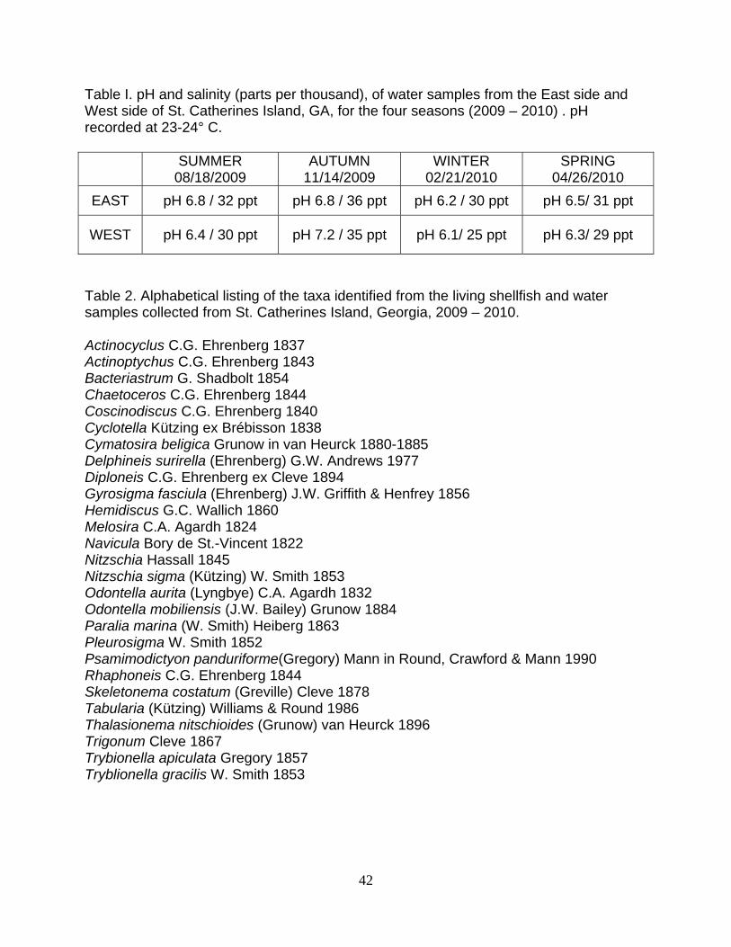
Table I. pH and salinity (parts per thousand), of water samples from the East side and West side of St. Catherines Island, GA, for the four seasons (2009 – 2010) . pH recorded at 23-24° C.
SUMMER 08/18/2009
AUTUMN 11/14/2009
WINTER 02/21/2010
SPRING 04/26/2010
EAST pH 6.8 / 32 ppt pH 6.8 / 36 ppt pH 6.2 / 30 ppt pH 6.5/ 31 ppt
WEST pH 6.4 / 30 ppt pH 7.2 / 35 ppt pH 6.1/ 25 ppt pH 6.3/ 29 ppt
Table 2. Alphabetical listing of the taxa identified from the living shellfish and water samples collected from St. Catherines Island, Georgia, 2009 – 2010. Actinocyclus C.G. Ehrenberg 1837 Actinoptychus C.G. Ehrenberg 1843 Bacteriastrum G. Shadbolt 1854 Chaetoceros C.G. Ehrenberg 1844 Coscinodiscus C.G. Ehrenberg 1840 Cyclotella Kützing ex Brébisson 1838 Cymatosira beligica Grunow in van Heurck 1880-1885 Delphineis surirella (Ehrenberg) G.W. Andrews 1977 Diploneis C.G. Ehrenberg ex Cleve 1894 Gyrosigma fasciula (Ehrenberg) J.W. Griffith & Henfrey 1856 Hemidiscus G.C. Wallich 1860 Melosira C.A. Agardh 1824 Navicula Bory de St.-Vincent 1822 Nitzschia Hassall 1845 Nitzschia sigma (Kützing) W. Smith 1853 Odontella aurita (Lyngbye) C.A. Agardh 1832 Odontella mobiliensis (J.W. Bailey) Grunow 1884 Paralia marina (W. Smith) Heiberg 1863 Pleurosigma W. Smith 1852 Psamimodictyon panduriforme(Gregory) Mann in Round, Crawford & Mann 1990 Rhaphoneis C.G. Ehrenberg 1844 Skeletonema costatum (Greville) Cleve 1878 Tabularia (Kützing) Williams & Round 1986 Thalasionema nitschioides (Grunow) van Heurck 1896 Trigonum Cleve 1867 Trybionella apiculata Gregory 1857 Tryblionella gracilis W. Smith 1853
43
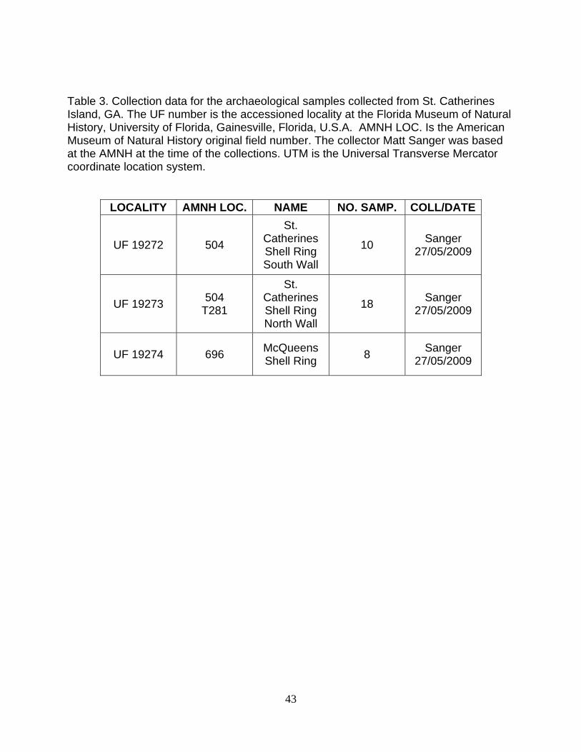
Table 3. Collection data for the archaeological samples collected from St. Catherines Island, GA. The UF number is the accessioned locality at the Florida Museum of Natural History, University of Florida, Gainesville, Florida, U.S.A. AMNH LOC. Is the American Museum of Natural History original field number. The collector Matt Sanger was based at the AMNH at the time of the collections. UTM is the Universal Transverse Mercator coordinate location system.
LOCALITY AMNH LOC. NAME NO. SAMP. COLL/DATE
UF 19272 504
St. Catherines Shell Ring South Wall
10 Sanger 27/05/2009
UF 19273 504 T281
St. Catherines Shell Ring North Wall
18 Sanger 27/05/2009
UF 19274 696 McQueens Shell Ring 8 Sanger
27/05/2009
44
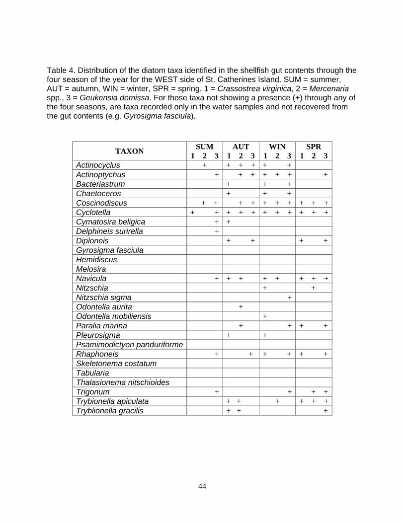
Table 4. Distribution of the diatom taxa identified in the shellfish gut contents through the four season of the year for the WEST side of St. Catherines Island. SUM = summer, AUT = autumn, WIN = winter, SPR = spring. 1 = Crassostrea virginica, 2 = Mercenaria spp., 3 = Geukensia demissa. For those taxa not showing a presence (+) through any of the four seasons, are taxa recorded only in the water samples and not recovered from the gut contents (e.g. Gyrosigma fasciula).
TAXON SUM 1 2 3
AUT 1 2 3
WIN 1 2 3
SPR 1 2 3
Actinocyclus + + + + + + Actinoptychus + + + + + + +Bacteriastrum + + + Chaetoceros + + + Coscinodiscus + + + + + + + + + +Cyclotella + + + + + + + + + + +Cymatosira beligica + + Delphineis surirella + Diploneis + + + +Gyrosigma fasciula Hemidiscus Melosira Navicula + + + + + + + +Nitzschia + + Nitzschia sigma + Odontella aurita + Odontella mobiliensis + Paralia marina + + + +Pleurosigma + + Psamimodictyon panduriforme Rhaphoneis + + + + + + Skeletonema costatum Tabularia Thalasionema nitschioides Trigonum + + + +Trybionella apiculata + + + + + +Tryblionella gracilis + + +
45
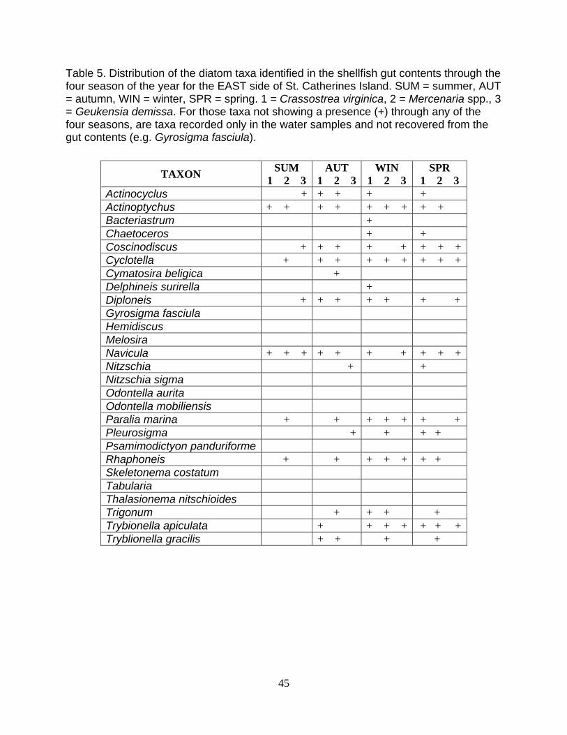
Table 5. Distribution of the diatom taxa identified in the shellfish gut contents through the four season of the year for the EAST side of St. Catherines Island. SUM = summer, AUT = autumn, WIN = winter, SPR = spring. 1 = Crassostrea virginica, 2 = Mercenaria spp., 3 = Geukensia demissa. For those taxa not showing a presence (+) through any of the four seasons, are taxa recorded only in the water samples and not recovered from the gut contents (e.g. Gyrosigma fasciula).
TAXON SUM 1 2 3
AUT 1 2 3
WIN 1 2 3
SPR 1 2 3
Actinocyclus + + + + + Actinoptychus + + + + + + + + + Bacteriastrum + Chaetoceros + + Coscinodiscus + + + + + + + +Cyclotella + + + + + + + + +Cymatosira beligica + Delphineis surirella + Diploneis + + + + + + + Gyrosigma fasciula Hemidiscus Melosira Navicula + + + + + + + + + +Nitzschia + + Nitzschia sigma Odontella aurita Odontella mobiliensis Paralia marina + + + + + + + Pleurosigma + + + + Psamimodictyon panduriforme Rhaphoneis + + + + + + + Skeletonema costatum Tabularia Thalasionema nitschioides Trigonum + + + + Trybionella apiculata + + + + + + +Tryblionella gracilis + + + +
46
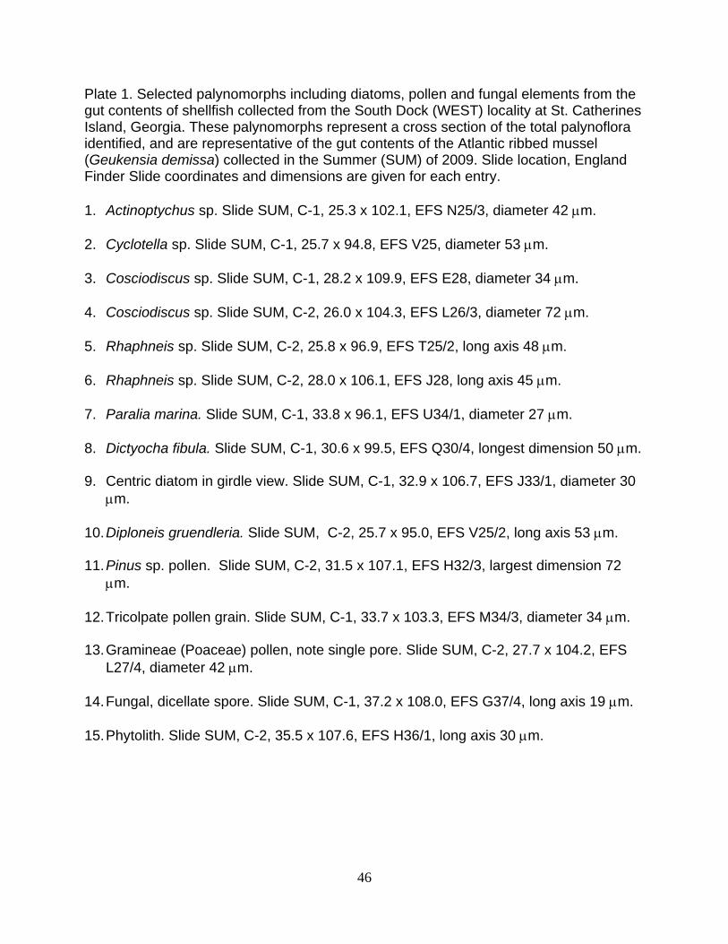
Plate 1. Selected palynomorphs including diatoms, pollen and fungal elements from the gut contents of shellfish collected from the South Dock (WEST) locality at St. Catherines Island, Georgia. These palynomorphs represent a cross section of the total palynoflora identified, and are representative of the gut contents of the Atlantic ribbed mussel (Geukensia demissa) collected in the Summer (SUM) of 2009. Slide location, England Finder Slide coordinates and dimensions are given for each entry. 1. Actinoptychus sp. Slide SUM, C-1, 25.3 x 102.1, EFS N25/3, diameter 42 μm. 2. Cyclotella sp. Slide SUM, C-1, 25.7 x 94.8, EFS V25, diameter 53 μm. 3. Cosciodiscus sp. Slide SUM, C-1, 28.2 x 109.9, EFS E28, diameter 34 μm. 4. Cosciodiscus sp. Slide SUM, C-2, 26.0 x 104.3, EFS L26/3, diameter 72 μm. 5. Rhaphneis sp. Slide SUM, C-2, 25.8 x 96.9, EFS T25/2, long axis 48 μm. 6. Rhaphneis sp. Slide SUM, C-2, 28.0 x 106.1, EFS J28, long axis 45 μm. 7. Paralia marina. Slide SUM, C-1, 33.8 x 96.1, EFS U34/1, diameter 27 μm. 8. Dictyocha fibula. Slide SUM, C-1, 30.6 x 99.5, EFS Q30/4, longest dimension 50 μm. 9. Centric diatom in girdle view. Slide SUM, C-1, 32.9 x 106.7, EFS J33/1, diameter 30
μm. 10. Diploneis gruendleria. Slide SUM, C-2, 25.7 x 95.0, EFS V25/2, long axis 53 μm. 11. Pinus sp. pollen. Slide SUM, C-2, 31.5 x 107.1, EFS H32/3, largest dimension 72
μm. 12. Tricolpate pollen grain. Slide SUM, C-1, 33.7 x 103.3, EFS M34/3, diameter 34 μm. 13. Gramineae (Poaceae) pollen, note single pore. Slide SUM, C-2, 27.7 x 104.2, EFS
L27/4, diameter 42 μm. 14. Fungal, dicellate spore. Slide SUM, C-1, 37.2 x 108.0, EFS G37/4, long axis 19 μm. 15. Phytolith. Slide SUM, C-2, 35.5 x 107.6, EFS H36/1, long axis 30 μm.
47
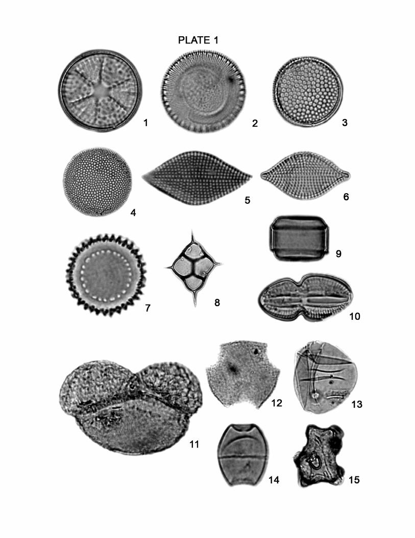
Plate 2. Selected palynomorphs including diatoms and pollen from the gut contents of shellfish collected from the McQueens Inlet (EAST) locality at St. Catherines Island, Georgia. These palynomorphs represent a cross section of the total palynoflora identified, and are representative of the gut contents of the Eastern oyster (Crassostrea virginica) or Hard clam (Mercenaria spp.) collected in the Autumn (AUT) of 2009. Slide location, England Finder Slide coordinates and dimensions are given for each entry.
1. Actinocyclus sp. in Crassostrea virginica. Slide AUT, A-1, 23.1 x 105.4, EFS K23,
diameter 53 μm.
2. Coscinodiscus sp. in Crassostrea virginica. Slide AUT, A-1, 25.2 x 100.0, EFS
Q25/1, diameter 61μm.
3. Rhaphoneis sp. in Crassostrea virginica. Slide AUT, A-1, 32.2 x 100.1, EFS
P32/4, long axis 60 μm.
4a. Pennate diatom (probably Navicula sp.) in Crassostrea virginica. Slide AUT, A-1,
26.7 x 99.1, EFS Q37/3 long axis 76x μm.
4b. As above in different focal plane.
5. Trigonium sp. in Mercenaria sp. Slide AUT, A-1, 33.2 x 105.9, EFS J33/4, longest
dimension 110 μm.
6. Quercus pollen in Mercenaria sp. Slide AUT, A-1, 27.3 x 111.0, EFS E27,
diameter 21μm.
7. Myrica sp. in Mercenaria sp. Slide AUT, A-1, 30.1 x 103.9, EFS M30/2, diameter
30.5 μm.
8. Unknown spore (algal?) in Crassostrea virginica. Slide AUT, A-1, 32.4 x 106.8,
EFS J32/2, diameter 61μm.
9. Periporate pollen (possibly Chenopodiaceae) in Mercenaria sp. Slide AUT, A-1,
34.8 x 101.2, EFS O35/3, diameter 23 μm.

48
10. Carya sp. pollen in Mercenaria sp. Slide AUT, A-1, 41.4 x 94.3, EFS W42/1,
diameter 50 μm.
11. Pinus sp. pollen in Mercenaria sp. Slide AUT, A-1, 26.1 x 108.2, EFS G26, long
axis 85 μm.
12. Miroforaminifera test lining in Mercenaria sp. Slide AUT, A-1, 33.1 x 97.7, EFS
S33, diameter 65 μm.

49
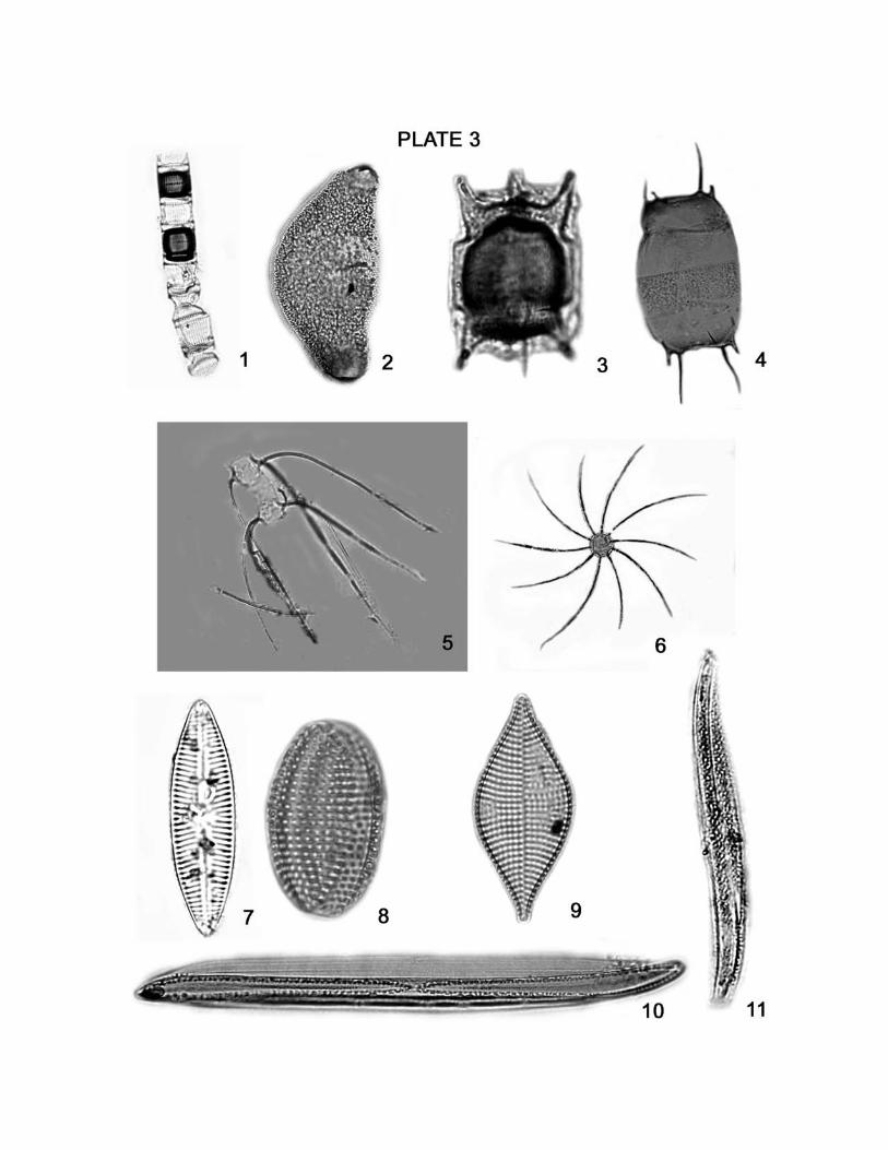
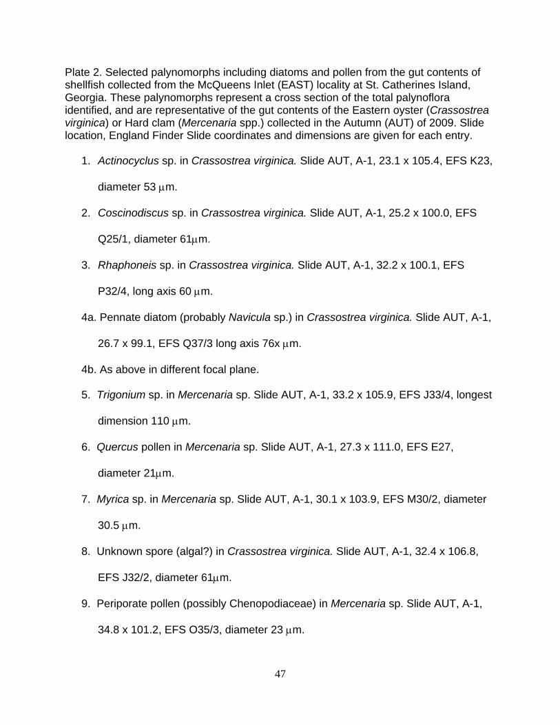
Plate 3. Selected diatoms from the water samples (WS) collected from the McQueens Inlet (EAST) and South Dock (WEST) localities at St. Catherines Island, Georgia. Slide location, England Finder Slide coordinates and dimensions are given for each entry.
1. Skeletonema costatum (chain of several cells). WS EAST, Summer, A-1, 23.1 x
95.3, EFS U23/3, length 135 μm.
2. Hemidiscus sp. WS WEST, Summer, A-1, 27.1 x 100.4, EFS P27, length 106 μm.
3. Odontella aurita. WS WEST, Summer, A-1, 20.1 x 108.7, EFS G20/1, long axis
34 μm.
4. Odontella mobiliensis. WS WEST, Summer, A-1, 25.3 x 97.0, EFS T25/2, long
axis 225 μm.
5. Chaetoceros sp. WS EAST, Summer, A-1, 31.7 x 94.9, EFS V32/1, overall size
about 100 μm.
6. Bacteriastrum sp. WS EAST, Summer, A-1, 24.7 x 107.4, EFS H24/2, diameter
60 μm.
7. Navicula sp. WS WEST, Autumn, A-1, 32.3 x 101.1, EFS O32/4, Long axis 76
µm.
8. Tryblionella granulate. WS EAST, Summer, A-1 32.7 x 107.0, EFS H33/3, long
axis 38 µm.
9. Rhaphoneis sp. WS EAST, Summer, A-1 32.5 x 94.1, EFS W32/2, long axis 59
µm .
10. Nitzschia scalpaliformis. WS EAST, Summer, A-1, 27.4 x 106.1, EFS J27/4,
Long axis 230 µm .
11. Pleurosigma sp. WS WEST, Autumn, A-1, 22.3 x 98.1, EFS S22/1, long axis 115
µm.