Embed Size (px)
Citation preview

____________________________________________________________________________________________
*Corresponding author: Email: [email protected];
British Journal of Pharmaceutical Research4(4): 490-512, 2014
SCIENCEDOMAIN internationalwww.sciencedomain.org
Formulation and Characterization of Nystatin-loaded Nanostructured Lipid Carriers for
Topical Delivery against Cutaneous Candidiasis
Rawia M. Khalil1*, A. Abd- Elbary2, Mahfouz A. Kassem1,Mohamed S. El Ridy1, Mona M. Abou Samra1, Ghada E. A. Awad3
and Soheir S. Mansy4
1Department of Pharmaceutical Technology, National Research Centre, Dokki, CAiro, Egypt.2Faculty of Pharmacy, Cairo University, Cairo, Egypt.
3Chemistry of Natural and Microbial Product Department, National Research Centre, Dokki,Cairo, Egypt.
4Electron Microscopy Research department Theodor bilharz Research Institute, Cairo,Egypt.
Authors’ contributions
This work was carried out in collaboration between all authors. Author RMK designed thestudy, wrote the protocol, managed the analyses of the study and reviewed the manuscript.
Author MMAS carried out the experimental work, performed the statistical analysis and wrotethe first draft of the manuscript. Authors MAK, AAE and MSER checked the revised
manuscript thoroughly and confirmed all the data given in manuscript and managed theliterature. Author GEAA carried out the microbiological experiments. Author SSM performedthe histopathologic examinations and the discussion of the pathological findings. All authors
read and approved the final manuscript.
Received 23rd September 2013Accepted 29th November 2013Published 24th December 2013
ABSTRACT
Aims: The objective of the current study was to formulate nystatin (Nyst) intonanostructured lipid carriers (NLCs) to enhance its antifungal activity.Place and Duration of Study: Department of pharmaceutical technology, nationalresearch centre, Egypt, between mars 2011 to april 2013Methodology: Nyst loaded NLCs (NYST-NLCs) were prepared by the hothomogenization and ultrasonication method followed by evaluation of its topical effect on
Original Research Article

British Journal of Pharmaceutical Research, 4(4): 490-512, 2014
491
the cutaneous candidiasis. The prepared Nyst-NLCs were characterized for entrapmentefficiency, particle size, zeta potential, morphology (transmission electron microscopy),thermal characterisation (differential scanning calorimetry) and in vitro drug release. Thestudy design involves the investigation of the effect of three independent variables namelyliquid lipid type (Miglyol 812 and Squalene), liquid lipid concentration (30 and 50%) andNyst concentration (0.125 and 0.25%). A stability study for 6 months was performed. Amicrobiological study was conducted in male rats infected with Candida albicans.Results: NLC dispersions were spherical in shape with particle size ranging from68.06±6.56 to 141.8±3.33 nm. The entrapment efficiencies ranged from 45.50±2.34 to92.73±0.33% with zeta potential ranging from -26.2 to -39.2 mV. The stability studiesdone for 6 months indicated that Nyst-NLCs were stable for more than 6 monthes.themicrobiological studies showed A least number of colonies forming units (cfu/ml) wererecorded for the selected Nyst-NLCs compared to the drug solution and the Nystatin®cream present in the market.Conclusion: It can be fulfilled from this work that NLCs represent promising carrier fortopical delivery of Nyst offering good physical stability, high entrapment efficiency andcontrolled drug release. Nyst-NLCs are a good candidate for cutaneous treatment offungal infection.
Keywords: Nanostructured lipid carriers; characterization; Nystatin; fungal infection;Candida albicans.
1. INTRODUCTION
Solid lipid nanoparticles (SLNs) combining the advantages of colloidal carriers, had attractedattention as a drug delivery system when it was introduced in 1991 [1]. Several advantagesof SLNs including biocompatibility, drug targeting, modified release and ease of large scaleproduction have been demonstrated [2]. However, depending on the drug, some potentialproblems can occur, such as drug leakage during storage and insufficient total drug loading.To overcome the limitations of SLNs, nanostructured lipid carriers (NLCs) have beendeveloped [3]. NLCs are produced by mixing the solid lipid with the liquid lipid. This leads tospecial nanostructures with improved properties for drug loading, modulation of the drugrelease profile and stability [3,4]. Nystatin (Nyst) is a polyene antibiotic produced byStreptomyces noursei; used topically in treatment of infections due to Candida albicans,Aspergillus species, yeasts and some dermatophytes. It is often used in large doses whichvary from 100,000 units (for oral infections) to 1 million (for intestinal ones) at risk of fungalinfections such as AIDS patients and patients receiving chemotherapy. Trials to improve theeffectiveness of Nyst or decrease its dose are ongoing. Recently, topical formulation ofnystatin was developed, Quinones et al, prepared a Nyst gel for topical delivery [5].Melkoumov et al. succeeded to prepare Nyst nanosuspension by wet media milling [6].Other researches were done to study the effect of silver nanoparticles in combination withNyst against candida albicans [7,8]. The aim of the current study is to develop lipidnanoparticulate formulations of Nyst in an attempt to increase its efficacy for topicalapplication. Its formulation in nanostructured lipid carriers may promote its lethal effect as itsmode of action depends on its binding to ergosterol, a major component of the fungal cellmembrane forming pores in the membrane that lead to potassium leakage and death of thefungus.

British Journal of Pharmaceutical Research, 4(4): 490-512, 2014
492
2. MATERIALS AND METHODS
2.1 Materials
Nystatin, kindly supplied by Rameda Pharmaceuticals, Sixth of October, Egypt. Compritol888 ATO (glyceryl behenate), kindly donated by Gattefossé, France. Nystatin® creampurchased from the Egyptian market. Poloxamer 188 (Pluronic F68; a triblock copolymer ofpolyoxyethylene- polyoxypropylene), PVA(polyvinyl alcohol), Dialysis tubing cellulosemembrane (molecular weight cut-off 12,000-14,000 g/mole) and Methanol were purchasedfrom Sigma-Aldrich Chemical Company., St.louis, USA. Miglyol 812 and Squalene werepurchased from Sigma-Aldrich Chemical Company., St.louis, USA. Potassium dihydrogenorthophosphate and citric acid were of analytical grade.
2.2 Methods
2.2.1 Formulation technique
Nanostructured lipid carriers were prepared by the modified high shear homogenization andultrasonication method [9,10,11]. The lipid was melted to approximately 5ºC above itsmelting point(74.09ºC), replacing 30 %w/w and 50%w/w of the solid lipid matrix by Miglyol®812 or Squalene (liquid lipid). Nyst was dispersed in the melted Compritol 888 ATO. Anaqueous phase was prepared by dissolving the surfactant (poloxamer 188 or PVA) indistilled water and heating up to the same temperature of the molten lipid. The hot lipidphase was poured on the aqueous phase and homogenization was carried out at 25000 rpmfor 5 minutes using Heidolph homogenizer. The resulted O/W emulsion was sonicated for 30minutes (water bath sonicator). The dispersion thus obtained was allowed to cool to roomtemperature, forming lipid nanoparticles by recrystallization of the dispersed lipid [12]. Theproduced Nyst-NLCs were kept at 4ºC for 24 hours before centrifugation at 9000 rpm for 30minutes and separation. Different formulations are presented in Tables 1 and 2.
Table 1. Composition of Nyst-NLCs using Miglyol 812® as liquid lipid
Formulations Surfactants Lipid conc. Drug conc.(w/w)Type Conc.(%w/w) Compritol Miglyol
NLC1
Poloxamer 188
1 70 30 0.25NLC2 1 50 50 0.25NLC3 2.5 70 30 0.125NLC4 2.5 50 50 0.125NLC5 2.5 70 30 0.25NLC6 2.5 50 50 0.25NLC7 5 70 30 0.25NLC8 5 50 50 0.25NLC9
PVA
1 70 30 0.25NLC10 1 50 50 0.25NLC11 2.5 70 30 0.125NLC12 2.5 50 50 0.125NLC13 2.5 70 30 0.25NLC14 2.5 50 50 0.25NLC15 5 70 30 0.25NLC16 5 50 50 0.25
British Journal of Pharmaceutical Research, 4(4): 490-512, 2014
493
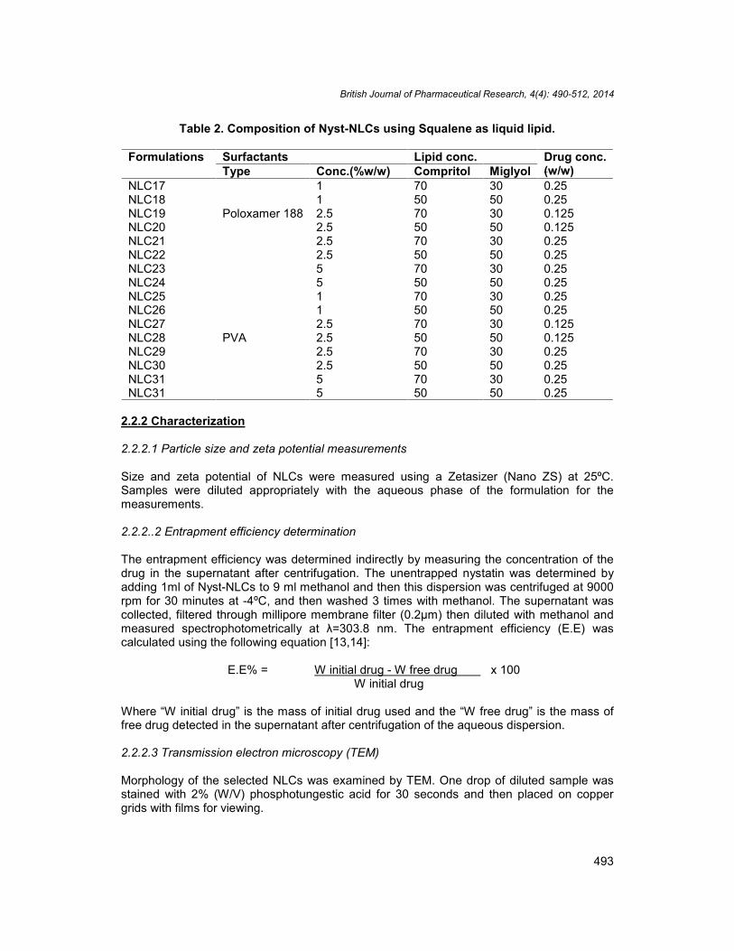
Table 2. Composition of Nyst-NLCs using Squalene as liquid lipid.
Formulations Surfactants Lipid conc. Drug conc.(w/w)Type Conc.(%w/w) Compritol Miglyol
NLC17
Poloxamer 188
1 70 30 0.25NLC18 1 50 50 0.25NLC19 2.5 70 30 0.125NLC20 2.5 50 50 0.125NLC21 2.5 70 30 0.25NLC22 2.5 50 50 0.25NLC23 5 70 30 0.25NLC24 5 50 50 0.25NLC25
PVA
1 70 30 0.25NLC26 1 50 50 0.25NLC27 2.5 70 30 0.125NLC28 2.5 50 50 0.125NLC29 2.5 70 30 0.25NLC30 2.5 50 50 0.25NLC31 5 70 30 0.25NLC31 5 50 50 0.25
2.2.2 Characterization
2.2.2.1 Particle size and zeta potential measurements
Size and zeta potential of NLCs were measured using a Zetasizer (Nano ZS) at 25ºC.Samples were diluted appropriately with the aqueous phase of the formulation for themeasurements.
2.2.2..2 Entrapment efficiency determination
The entrapment efficiency was determined indirectly by measuring the concentration of thedrug in the supernatant after centrifugation. The unentrapped nystatin was determined byadding 1ml of Nyst-NLCs to 9 ml methanol and then this dispersion was centrifuged at 9000rpm for 30 minutes at -4ºC, and then washed 3 times with methanol. The supernatant wascollected, filtered through millipore membrane filter (0.2µm) then diluted with methanol andmeasured spectrophotometrically at λ=303.8 nm. The entrapment efficiency (E.E) wascalculated using the following equation [13,14]:
E.E% = W initial drug - W free drug x 100W initial drug
Where “W initial drug” is the mass of initial drug used and the “W free drug” is the mass offree drug detected in the supernatant after centrifugation of the aqueous dispersion.
2.2.2.3 Transmission electron microscopy (TEM)
Morphology of the selected NLCs was examined by TEM. One drop of diluted sample wasstained with 2% (W/V) phosphotungestic acid for 30 seconds and then placed on coppergrids with films for viewing.

British Journal of Pharmaceutical Research, 4(4): 490-512, 2014
494
2.2.2.4 Differential scanning calorimetry (DSC)
The thermal characteristics of selected batches of lipid nanoparticles were determined bydifferential scanning calorimetry (DSC)-50, Kyoto, japan. Samples containing 10 mgnanoparticle dispersions were weighed accurately into standard aluminium pans using anempty pan as a reference. DSC scans were recorded at a heating and cooling rate of10ºC/min. The samples were heated from 30-300ºC and cooled from 300-30ºC.
2.2.3 Drug release study
The in-vitro release of Nyst from different NLCs was evaluated by the dialysis bag diffusiontechnique reported by Yang et al. [15]. The release studies of Nyst from lipid nanoparticleswere performed in phosphate buffer of pH5.5 and methanol (70:30). The nanostructured lipidcarriers of Nyst equivalent to 2 mg of Nyst were suspended in the buffer solution (pH 5.5)and placed in a dialysis bag (donor compartment) and sealed at both ends. The dialysis bagwas immersed in the receptor compartment containing 50ml of the dissolution medium,which was stirred at 100 rpm and maintained at 32±2ºC. The receptor compartment wascovered to prevent evaporation of the dissolution medium. Samples (2 ml) were taken fromthe receptor compartment and the same amount of fresh dissolution medium was added tokeep a constant volume at fixed time intervals (0.5, 1, 2,3,4,5,6,7,8 and 24 h). Nyst in thesamples was measured spectrophotometrically at λ=303.8 nm. The release studies werecarried out in triplicate for all formulations and the results were expressed as the meanvalues ±SD.
2.2.4 Stability study
The selected NLC formulations were stored in a sealed amber colored glass vials atrefrigerator temperature (2-4ºC) in a dark environment. The physical appearance wasassessed and the formulations were analyzed with respect to particle size, drug entrapmentefficiency and zeta potential after 6 months of storage and compared with fresh formulations.In addition, the drug release properties of the Nyst-NLCs stored for 6 months were evaluatedand compared with those of fresh formulations. The experiment was performed in triplicate.Shelf life values were calculated as follows [16]:
The values of Log E.E. % were plotted against the time (days of storage) and the slopes (m)were calculated by linear regression. The slopes (m) were then substituted into the followingequation for the determination of K values [17] :
K= m x 2.303.
As reported by Wells [18], the shelf life values (the time for 10% loss, t90) were thencalculated by the following equation:
t90 =0.105/K.

British Journal of Pharmaceutical Research, 4(4): 490-512, 2014
495
2.2.5 Antifungal activity
2.2.5.1 Preparation of the animals
Male albino rats (120-150g) purchased from the animal house of the National ResearchCentre were used in the experiment. These rats were divided into five groups; six animalswere used for each group. Group 1 served as control, group 2 was treated with the plaindrug solution, group 3 received topically an equivalent dose of Nystatin® cream present inthe market and group 4 and 5 received topically the NLC3 and NLC27 formulae respectively.The rats were housed in individual cages and received food and water ad libitum. Theexperimental protocol of the study was reviewed and approved by the Animal EthicsCommittee of the National Research Centre. Experiments were carried out in accordancewith the guidelines laid down by the National Research Centre regarding the care and use ofanimals for experimental procedures and in accordance with local laws and regulations.
2.2.5.2 Preparation of microorganism
Clinical isolate of Candida albicans was used to infect the animals. A working culture ofCandida was grown for 24 h at 37ºC on Potato Dextrose Broth (PDB). The culture wasdiluted with a sterile saline solution to reach a final concentration of 1.1 X10⁶ colony formingunit/ ml (cfu/ml) according to the modified method of Maebashi et al. [19].
2.2.5.3 Cutaneous infection
Each animal’s back was shaved; approximately 5.0cm2 area was marked on each animal’sback. The marked area was infected with 1.1 X10⁶ cfu/ml suspension by gently rubbing ontothe skin with the help of a sterile cotton-tipped swab until no more visible fluid was observed[19]. Infection was produced under an occlusive dressing and the infected area was coveredwith a sterile adhesive bandage, held in place with extra-adherent tape for 24h beforetreatment began. Control animals were infected in the same manner; however, they did notreceive any Nyst formulation.
2.2.5.4 Treatment of the infection
Treatment began 24h after the infection was induced, and test formulations were topicallyapplied twice daily for two consecutive days. After 48 h of treatment swabs were taken fromeach infected area into sterile tubes containing 5 ml of (PDB). Serial dilutions were done andthen one ml of each dilution was inoculated into Petri dishes containing 10 ml of PotatoDextrose Agar (PDA) .The inoculated plates were incubated for 24h at 37ºC after which thecolonies were counted.
2.2.5.5 Histopathological examination
After taking the skin swab in a way the skin is completely cleaned out from the infectedorganism, the rats are sacrificed. The defined part of the shaved skin is excised and fixed in10% formalin. The fixed skin tissues were processed until embedded in paraffin [20]. Twosections of 4µm thickness were performed from each skin embedded in paraffin block usingmicrotome. One section was stained with Haematoxylin and eosin as well as the other wasstained with Masson trichrome [20].

British Journal of Pharmaceutical Research, 4(4): 490-512, 2014
496
2.2.6 Stastical analysis
All the results were expressed as mean±S.D. Statistical analysis of the results wasperformed using SPSS program with help of one way analysis of variance (ANOVA),followed by post hoc multiple comparisons and least significant difference (LSD). Theresults are significant when P<0.05.
3. RESULTS AND DISCUSSION
In this study, Compritol as solid lipid (5%), Miglyol 812 or squalene as liquid lipid(30 and 50%), Poloxamer 188 or PVA as stabilizers (1%, 2.5% and 5%) and Nyst (0.125 and0.25%) were used to produce NLCs (Table1&2).
3.1 Characterization of the Prepared Nyst-NLCs
3.1.1 Particle size analysis
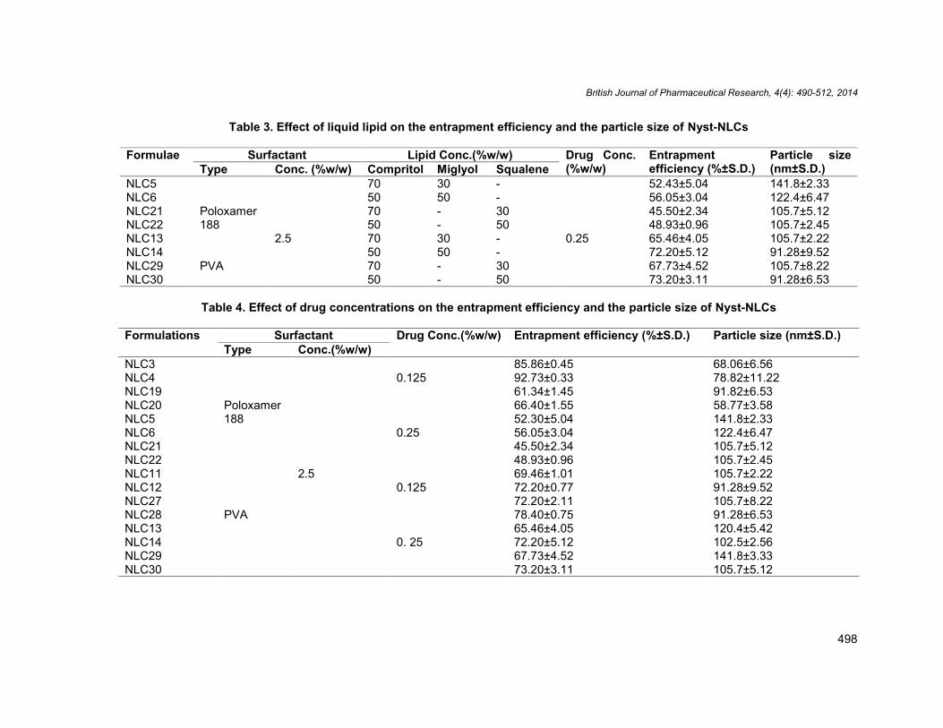
Table 3 shows the effect of increasing Miglyol® 812 or Squalene percentage in the lipidmatrix from 30% to 50% with constant surfactant concentration at 2.5 %w/w and constantdrug concentration at 0.25%w/w. The results show that increasing the concentration of liquidlipid is accompanied with a decrease in particle size. Based upon previous studies; theaddition of Miglyol® 812 to Compritol tends to promote the formation of small particlepopulation, which might be due to a higher molecular mobility of the matrix after liquid oiladdition [21,22]. Also, the effect of oil content on particle size was in agreement with thework performed by Chen et al. who found that increasing the concentration of Squalene toPrecirol from 0-80% resulted in significant decrease in particle size from 286.5±3.7 nm to210.4±5.6 nm [23]. Similar observation was reported by Khalil et al. [24] who found thatincreasing Miglyol concentration in meloxicam NLCs resulted in a significant decrease inparticle size.
The drug concentration has also a marked role on the particle size. The results show that theparticle size increases with increasing drug concentration from 0.125 %(w/w) to 0.25%(w/w)(Table 4).This can be explained as the lipid has a certain loading capacity, the addition ofexcess drug may lead to aggregation and the formation of large sized particles [25].Considering the effect of surfactants on the particle size of different Nyst-NLCs, it wasnoticed that the change in surfactant types and concentrations doesn’t show a remarkableeffect.
3.1.2 Entrapment efficiency (E.E)
The entrapment efficiency of Nyst within the different prepared nanostructured lipid carrierformulaions is shown in Tables 3 and 4 and Figs. 1 and 2. Increasing surfactantconcentrations from 1 to 5% w/w was accompanied with increase in the E.E.%, (NLC2,NLC6 & NLC8) and (NLC10, NLC14 & NLC16) for poloxamer 188 and PVA respectively(Figs. 1 and 2) . This might be due to the efficient loading and retention of drug moleculeswithin the nanoparticle matrix or nanoparticle surface at higher surfactant concentration [22].Concerning the effect of surfactant type, it was noticed that the effect of PVA is higher thanpoloxamer 188 on the entrapment efficiencies. This increase in the E.E% may be also due tothe increase in the solubility of the drug in the lipid on increasing the concentration of thesurfactant [26,27]. The results reveal that replacing solid lipid by increasing percent of liquid

British Journal of Pharmaceutical Research, 4(4): 490-512, 2014
497
lipid leads to a gradual increase in E.E% (Table 3). In general, drug solubility is higher inliquid lipid than in solid lipid, which increases the E.E% [28]. Incorporation of liquid lipids intosolid lipids leads to massive crystal order disturbance. Greater imperfections in the crystallattice leave enough space to hold drug molecules, which ultimately improved drug loadingcapacity and drug entrapment efficiency [29]. Increasing the amount of drug, while keepingthe emulsifier level constant, as shown in table 4 is found to decrease the E.E%. As the lipidhas certain drug loading capacity, addition of excess drug led to increase of unencapsulated-drug (i.e. decrease in E.E.%), which signifies that E.E.% reaches its maximum at 0.125%drug concentration [25] .
Fig. 1. Effect of type and concentration of surfactants on the entrapment efficiency ofNyst-NLCs in presence of Miglyol
Fig. 2. Effect of type and concentration of surfactants on the entrapment efficiency ofNyst-NLCs in presence of Squalene
British Journal of Pharmaceutical Research, 4(4): 490-512, 2014
498
Table 3. Effect of liquid lipid on the entrapment efficiency and the particle size of Nyst-NLCs
Formulae Surfactant Lipid Conc.(%w/w) Drug Conc.(%w/w)
Entrapmentefficiency (%±S.D.)
Particle size(nm±S.D.)Type Conc. (%w/w) Compritol Miglyol Squalene
NLC5
Poloxamer188
2.5
70 30 -
0.25
52.43±5.04 141.8±2.33NLC6 50 50 - 56.05±3.04 122.4±6.47NLC21 70 - 30 45.50±2.34 105.7±5.12NLC22 50 - 50 48.93±0.96 105.7±2.45NLC13
PVA
70 30 - 65.46±4.05 105.7±2.22NLC14 50 50 - 72.20±5.12 91.28±9.52NLC29 70 - 30 67.73±4.52 105.7±8.22NLC30 50 - 50 73.20±3.11 91.28±6.53
Table 4. Effect of drug concentrations on the entrapment efficiency and the particle size of Nyst-NLCs
Formulations Surfactant Drug Conc.(%w/w) Entrapment efficiency (%±S.D.) Particle size (nm±S.D.)Type Conc.(%w/w)
NLC3
Poloxamer188
2.5
0.12585.86±0.45 68.06±6.56
NLC4 92.73±0.33 78.82±11.22NLC19 61.34±1.45 91.82±6.53NLC20 66.40±1.55 58.77±3.58NLC5
0.2552.30±5.04 141.8±2.33
NLC6 56.05±3.04 122.4±6.47NLC21 45.50±2.34 105.7±5.12NLC22 48.93±0.96 105.7±2.45NLC11
PVA
0.12569.46±1.01 105.7±2.22
NLC12 72.20±0.77 91.28±9.52NLC27 72.20±2.11 105.7±8.22NLC28 78.40±0.75 91.28±6.53NLC13
0. 2565.46±4.05 120.4±5.42
NLC14 72.20±5.12 102.5±2.56NLC29 67.73±4.52 141.8±3.33NLC30 73.20±3.11 105.7±5.12
British Journal of Pharmaceutical Research, 4(4): 490-512, 2014
499
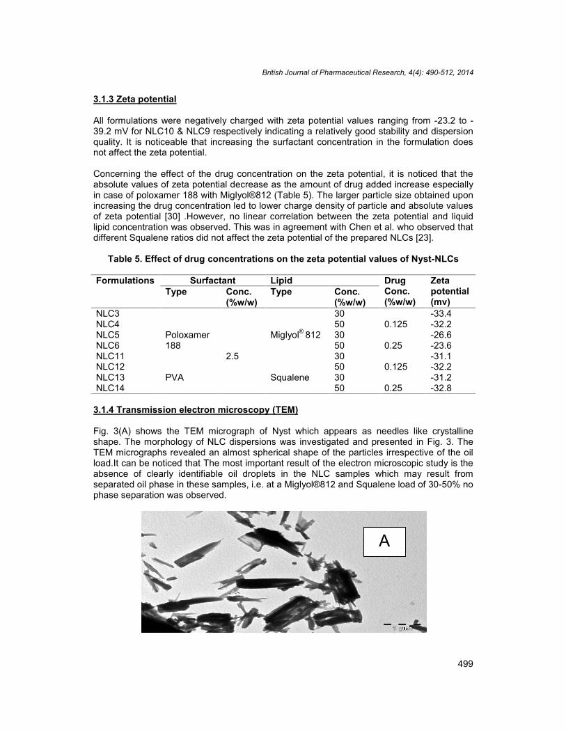
3.1.3 Zeta potential
All formulations were negatively charged with zeta potential values ranging from -23.2 to -39.2 mV for NLC10 & NLC9 respectively indicating a relatively good stability and dispersionquality. It is noticeable that increasing the surfactant concentration in the formulation doesnot affect the zeta potential.
Concerning the effect of the drug concentration on the zeta potential, it is noticed that theabsolute values of zeta potential decrease as the amount of drug added increase especiallyin case of poloxamer 188 with Miglyol®812 (Table 5). The larger particle size obtained uponincreasing the drug concentration led to lower charge density of particle and absolute valuesof zeta potential [30] .However, no linear correlation between the zeta potential and liquidlipid concentration was observed. This was in agreement with Chen et al. who observed thatdifferent Squalene ratios did not affect the zeta potential of the prepared NLCs [23].
Table 5. Effect of drug concentrations on the zeta potential values of Nyst-NLCs
Formulations Surfactant Lipid DrugConc.(%w/w)
Zetapotential(mv)
Type Conc.(%w/w)
Type Conc.(%w/w)
NLC3
Poloxamer188
2.5
Miglyol® 812
300.125
-33.4NLC4 50 -32.2NLC5 30
0.25-26.6
NLC6 50 -23.6NLC11
PVA Squalene
300.125
-31.1NLC12 50 -32.2NLC13 30
0.25-31.2
NLC14 50 -32.8
3.1.4 Transmission electron microscopy (TEM)
Fig. 3(A) shows the TEM micrograph of Nyst which appears as needles like crystallineshape. The morphology of NLC dispersions was investigated and presented in Fig. 3. TheTEM micrographs revealed an almost spherical shape of the particles irrespective of the oilload.It can be noticed that The most important result of the electron microscopic study is theabsence of clearly identifiable oil droplets in the NLC samples which may result fromseparated oil phase in these samples, i.e. at a Miglyol®812 and Squalene load of 30-50% nophase separation was observed.
A

British Journal of Pharmaceutical Research, 4(4): 490-512, 2014
500
NLC11 NLC3
NLC19 NLC27
Fig. 3. Transmission electron micrographs of free Nyst (A) & micrograph of differentNyst-NLCs formulations
3.1.5 Differential scanning calorimetry
The DSC data of Nyst, bulk lipid and Nyst- NLCs are presented in figure 4. Nystatin andcompritol showed sharp endothermic peaks at 167.11ºC and 74.09ºC, respectively. Miglyol®812 and Squalene were not examined by DSC because liquids cannot be registered usingthe described temperatures and analytical conditions.The data clearly show that the meltingendothermic of Nyst (167.11 ºC) was not recorded indicating the complete solubilization ofNyst in the lipid matrix. It was noticed that the addition of a liquid lipid (Miglyol ®812 orSqualene) to solid lipid (compritol) induced a shift of the melting point to lower temperaturesfrom 74.09 to 66.84ºC, 66.92ºC, 67.32ºC and 68.31ºC in case of NLC3, NLC11, NLC19 andNLC27, respectively. A sharp reduction in melting enthalpy (required to melt the lipid matrix)indicates an interaction of liquid oil with the crystalline lipid matrix, creating a more massivecrystal order disturbance (lattice defects) which could allow enough space to accommodateNyst molecules [31] .

British Journal of Pharmaceutical Research, 4(4): 490-512, 2014
501
Nystatin
CompritolNLC3
NLC11
NLC19
NLC27
Fig. 4. DSC thermograms of pure Nyst, compritol and different Nyst-NLCsformulations.
3.2 Drug Release Study
Figure 5 shows the effect of lipid concentration when using 2.5%w/w of different surfactantsand 0.25%w/w of the drug. The increase in the oil concentration from 30%w/w to 50%w/w ofthe lipid is accompanied with a decrease of the percentage of Nyst released. All the formulaein which 50% of the lipid was replaced by Miglyol or Squalene show the lowest percent ofdrug released compared to the formulae containing 30% of the liquid oil. This may be due tothat increasing the oil concentration may lead to increase in the viscosity of the systemleading to suppression of the release. Another reason, that after lipid crystallization, thesolubility of oil in solid lipid exceeded; hence, oil precipitates leading to formation of finedroplets of oil incorporated in solid lipid thereby providing prolonged release [32]. The resultsalso pointed to the effect of drug concentration on the release of Nyst from NLCs releaseprofile. Figs. 6 and 7 show that upon increasing the drug concentration from 0.125%w/w to0.25% w/w there is a significant decrease in the percentage released of Nyst from NLCsformulations.
As discussed before, Nyst is released more quickly when used in lower concentrationbecause of the drug-enriched shell model [33,34]. The release data are analyzed according

British Journal of Pharmaceutical Research, 4(4): 490-512, 2014
502
to zero, first order and Higuchi equations which are widely used in determining the releasekinetics of lipid nanoparticles. The amount of Nyst released from all the NLCs formulationsstudied shows a linear relationship with the square root of time, therefore, the release rate ofNyst is expressed following the theoretical model by Higuchi [35].
Fig. 5. Effect of liquid lipid on the in vitro release of Nyst from NLCs
Fig. 6. Effect of drug concentration on the in-vitro release of Nyst from NLCsin presence of Miglyol.
0
10
20
30
40
50
60
0 5 10 15 20 25 30
Cum
ulat
ive
perc
ent o
f nys
tatin
rele
ased
(±S.
D.)
Time (hr)
NLC5NLC6NLC21NLC22NLC13NLC14NLC29NLC30
0
20
40
60
80
100
120
0 5 10 15 20 25 30
Cum
mul
ativ
e pe
rcen
t of d
rug
rele
ased
(±S.
D.)
Time (hr)
NLC3
NLC4
NLC5
NLC6
NLC11
NLC12
NLC13
NLC14

British Journal of Pharmaceutical Research, 4(4): 490-512, 2014
503
Fig. 7. Effect of drug concentration on the in-vitro release of Nyst from NLCsin presence of Squalene
3.3 Stability Study
Stability was performed for 2 formulations which are, NLC3 and NLC27 as they show thehighest cumulative percent of Nyst released. The physical stability of Miglyol 812 andSqualene NLCs formulations was suggested by the absence of visible phase separation andall dispersions remained in a homogenous state upon storage at 2-4ºC for 6 months. Themean of the entrapment efficiency and particle size of the prepared NLCs after storage areshown in Figs. 8 and 9. A non significant increase was observed in the entrapment efficiencyand the particle size for the stored samples. Fig. 10 shows the values of the zeta potentialwhich decrease with time but still indicating stability of the formulations. The in-vitro releaseprofile of Nyst from the nanostructured lipid carriers (NLC3 & NLC27) was investigated over24 h. The results are shown in Fig. 11. The drug release rate of NLC 3 & NLC27 after 2, 4 &6 months shows no significant increase compared to freshly prepared ones. The t90 valuesfor NLC3 and NLC27 were eleven and eight months respectively. In this respect it can behypothesized that the stability of Nyst is better controlled in NLC3.
0
10
20
30
40
50
60
70
80
90
0 5 10 15 20 25 30
Cum
mul
ativ
e pe
rcen
t of d
rug
rele
ased
(±S.
D.)
Time (hr)
NLC19
NLC20
NLC21
NLC22
NLC27
NLC28
NLC29
NLC30

British Journal of Pharmaceutical Research, 4(4): 490-512, 2014
504
Fig. 8. Effect of storage time on the entrapment efficiency of different Nyst –NLCS
Fig. 9. Effect of storage time on the particle size of different Nyst-NLCs
Fig. 10. Effect of storage time on the zeta potential of different Nyst-NLCs
0102030405060708090
100
Zero time 2 months 4 months 6 months
Entr
apm
ent e
ffici
ency
(±S.
D.)
NLC3
NLC27
0
20
40
60
80
100
120
Zero time 2 months 4 months 6 months
Parti
cle
size
(±S.
D.)
NLC3
NLC27
-35
-30
-25
-20
-15
-10
-5
0
Zero time 2 months 4 months 6 months
Zeta
pot
entia
l
NLC3
NLC27

British Journal of Pharmaceutical Research, 4(4): 490-512, 2014
505
Fig. 11. Effect of storage time on the in-vitro release of Nyst from NLCs.
3.4 Antifungal Activity
After two days of treatment with plain drug solution, Nystatin® cream and the tested formulae(NLC3 and NLC27), the animals treated with the tested formulae demonstrated a low colonycount significantly (p˂0.05) less than that for those animals treated with plain drug solutionand Nystatin® cream (Fig. 12). The higher therapeutic efficacy in the case of NLCformulations may be expected due to penetration into the skin, followed by drug carrieraccumulation in different strata of the skin resulting a reservoir effect with higher level oflocalization [36,37]. These findings also confirmed that lipid nanoparticles provoke theaccumulation of the embedded drug into the upper skin layers [38].
It is clear from the histopathology (Figs. 13-18) of the skin biopsies stained with Massontrichrome & Haematoxylin and eosin that the skin of the normal control rat revealskeratinized squamous epithelium with intact basal cell layer and basal lamina. The basal partof the epidermis is folded to form the dermal papillae. Hair follicles were clearly identifiedwith their different constituents extending into the dermis. The dermis formed the thickconnective tissue layer in which the hair follicles were seen with sebaceous glands. Thishistological findings agree with the previously reported data in the literature containing theconstituent of normal skin (Fig. 13) [39]. However, Masson trichrome stained skin of Candidaalbicans infected control rat reveals erosion or ulceration of the epidermis, edematousdermis and increased dermal vascularities. Also complete loss of the hair follicles withapparent sweat glands is observed (Fig. 14). Masson trichrome and Haematoxylin stainedskin sections of rat infected with Candida albicans and treated with the available drugsolution and Nystatin® cream (Figs. 15 and 16) reveal a smaller area of skin erosion, minimalulceration and no characteristic hair follicles.
0
20
40
60
80
100
120
0 5 10 15 20 25 30
Cum
ulat
ive
perc
ent o
f nys
tatin
rele
ased
(±S.
D.)
Time (hr)
NLC3 at zero days
NLC3 after 2 months
NLC3 after 4 months
NLC3 after 6 months
NLC27 at zero days
NLC27 after 2 months
NLC27 after 4 months
NLC27 after 6 months

British Journal of Pharmaceutical Research, 4(4): 490-512, 2014
506
The Candida albicans infected skin and treated with the prepared formula NLC27 revealedareas of ulceration or incomplete loss of epidermis. Minimal polymorph leucocyte infiltrationwas evident. Laminated keratin was noticed. Also, minimal primitive hair follicles and manysebaceous glands were detected in the subepidermal area (Fig. 17). However, the skin ofthe group of rats infected with Candida albicans and treated with NLC3 revealed impressiveimprovement. No skin ulceration was detected in the examined sections (Fig.18). Therecuperation of skin characteristic constituent was noticed in some sections, with theformation of primitive hair follicles and appearance of fully developed one. Also mildpolymorphnuclear leucocytes in the subepidermal region were evident.
This improvement may be related to the better diffusion of the small sized particles of thisformula in comparison to the other tested formulae. This suggestion is supported by thefinding of Shuaidong et al. who proved that the smaller sized nanoparticles maximize theireffective accumulation in tumor tissue [40]. Also similar results were obtained by Melkoumovet al. who found that Nyst nanosuspension has a higher antifungal effect on the Candidaalbicans compared to Nyst suspension [6].
The results obtained in this study prove that formula NLC3 can be employed as promisingtopical formulation for the treatment of skin fungal infection and can be subjected to furtherclinical studies after following the ethical applied steps.
Fig. 12. Antifungal efficacy of Nystatin formulations expressed as colony forming unit(CFU) after treatment.
0
0.5
1
1.5
2
2.5
3
3.5
4
Control Plainnystatinsolution
Nystatincream
NLC3 NLC27
Con
cent
ratio
n of
Can
dida
alb
ican
s(C
FU/m
l)

British Journal of Pharmaceutical Research, 4(4): 490-512, 2014
507
(A) (B)
Fig. 13. (A) and (B) show normal skin stained with Hamatoxylin & eosin and MassonTrichrome respectively. Both sections reveal intact keratinized epidermis. The hairfollicles (arrow) extend from the epidermis down into the dermis. Notice the thick
dermal layer.
(A) (B)
Fig. 14. Haematoxylin & eosin (A) and Masson trichrome (B) stained skin sectionsfrom control rat infected with Candida albicans reveal discontinuous ulcerated
epidermis (thick arrow) in the vicinity of incomplete loss of other part or erosion.Many new vessels formation are disclosed in the upper part of the dermis (thin
arrows). Note the absence of hair follicles and increased sweat glands (head of arrow)in the dermis. (x100).

British Journal of Pharmaceutical Research, 4(4): 490-512, 2014
508
(A) (B)
Fig. 15. Photomicrographs showing sections of rats skin treated with plain drugsolution,(A) section stained with Haematoxylin and Eosin, (B) section stained with
Masson trichrome. The sections reveal area of skin erosion (arrow), minimalulceration (thick arrow) and no characteristic hair follicles.
(A) (B)
Fig. 16. Photomicrographs showing sections of rats skin treated with Nystatin®
cream,(A) section stained with Haematoxylin and Eosin, (B) section stained withMasson trichrome. The sections reveal area of skin erosion (arrow), minimal
ulceration (thick arrow) and no characteristic hair follicles.
British Journal of Pharmaceutical Research, 4(4): 490-512, 2014
509
(A) (B)
Fig. 17. Photomicrographs showing sections of rat skin treated with Nyst-NLC27,(A) &(B) sections stained with Haematoxylin and Masson trichrome. Sections reveal
discontinuity of the skin. The intact epidermis (B) shows laminated keratin or stratumcorneum. Note the sebaseous glands in the dermis and the primitive hair follicle
formation (thin arrow).
(A) (B)
Fig. 18. Photomicrographs showing sections of the rat skin treated with Nyst-NLC3,(A) section stained with Haematoxylin and Eosin, (B) section stained with
Masson trichrome. Improvement of the lesion with reformed skin layer evident insection (B) . Also in these sections primitive or well formed hair follicles are seen.
British Journal of Pharmaceutical Research, 4(4): 490-512, 2014
510
4. CONCLUSION
Homogenization followed by ultrasonication method is suitable to produce Nyst-NLCs. Thelipophilic drug, Nyst, can be loaded in compritol using poloxamer 188 or PVA as surfactantsand Miglyol 812 or Squalene as liquid lipids. The loading of nystatin proved to follow thedrug-enriched-shell theory. DSC analysis showed amorphous state of Nyst. Nystatin isreleased more quickly when used in lower concentration. Stability studies proved that the 2formulae suggested NLC3 and NLC27 are stable upon storage .Nyst-NLC3 formula testedfor antifungal activity showed better results compared to the drug solution and the Nystatin®
cream present in the market.
COMPETING INTERESTS
Authors have declared that no competing interests exist.
REFERENCES
1. Muller RH, Mader K, Gohla S. Solid lipid nanoparticles (SLN) for controlled drugdelivery- A review of the state of the art. Eur J Pharm Biopharm. 2000;50:161-177.
2. Mehnert W, Mader K. Solid lipid nanoparticles: production, characterization andapplication. Adv. Drug Deliv. Rev. 2001;47:165-196.
3. Muller RH, Radtke M, Wissing SA. Solid lipid nanoparticles (SLN) and nanostructuredlipid carriers (NLC) in cosmetic and dermatological preparations. Adv. Drug Deliv. Rev.2002;1:131-55.
4. Radtke M, Souto EB, Muller RH. Nanostructured lipid carriers: a novel generation ofsolid lipid drug carriers. Pharm. Technol. Eur. 2005;17:45-50.
5. Quinones D, Evone MS, Ghaly S. Formulation and characterization of nystatin gel. –PRHSJ. 2008;27:62.
6. Melkoumov A, Goupil M, Louhichi F, Raymond M, Repentigny L, Leclair G. Nystatinnanosizing enhances in vitro and in vivo ntifungal activity against Candida albicans. JAntimicrob Chemother. 2013;68(9):2099-105.
7. Manteiro DR, Silva S, Negri M, Gorup LF, de Camargo ER, Oliveira R, Barbosa DBand Henriques M, Antifungal activity of silver nanoparticles in combination withnystatin and chlorhexidine digluconate against Candida albicans and Candida glabratabiofilms. Mycoses, 2013;56:672-680.
8. Silva S, Pires P, Monteiro DR, Negri M, Gorup LF, de Camargo ER, Oliveira R,Williams DW, Henriques M, Azerdo J. The effect of silver nanoparticles and nystatinon mixed biofilms of Candida glabrata and candida albicans on acrylic.
9. Sandri G, Bonferoni MC, Gokce EH, Ferrari F, Rossi S, Patrini M, Caramella C.Chitosan associated SLN: in vitro and ex vivo characterization of cyclosporine Aloaded ophthalmic systems. J Of Microencapsulation. 2010;27:735-746.
10. Sharma A, Jindal M, Aggarwal G, Jain S. Development of a novel method forfabrication of solid lipid nanoparticles: using high shear homogenization andultrasonication. RJPBCS. 2010;1:266.
11. Ekambaram PA, Abdul Hassan Sathali K, Priyanka. Solid lipid nanoparticles: A review.Scientific Reviews & Chemical Communications. 2012;2:82-85.
12. Gohla SH, Dingler A. Scaling up feasibility of the production of solid lipid nanoparticles(SLN). Pharmazie. 2001;56:61-63.

British Journal of Pharmaceutical Research, 4(4): 490-512, 2014
511
13. Souto EB, Wissing SA, Barbosa CM, Muller RH. Development of a controlled releaseformulation based on SLN and NLC for topical clotrimazole delivery. Int J Pharm.2004;278:71.
14. Jifu H, Xinsheng F, Yanfang Z, Jianzhu W, Fengguang G, Fei L, Xinsheng P.Development and optimization of solid lipid nanoparticle formulation for ophthalmicdelivery of chloramphenicol using a Box-Behnken design. International journal ofnanomedecine. 2011;6:683-692.
15. Yang SC, Lu LF, Cai Y, Zhu JB, Liang BW, Yang CZ. Body distribution in mice ofintravenously injected camptothecin solid lipid nanoparticles and targeting effect onbrain. J Control Release. 1999;59:299.
16. Esposito E, Bortolotti F, Menegatti E, Cortesi R. Amphiphilic association systems foramphoterecin B delivery. Int J Pharm. 2003;260:249-260.
17. Ferrer J. Vaginal candidosis: epidemiological and etiological factors. Int J GynaecolObstet. 2000;71:21-27.
18. Wells JI. Pharmaceutical formulations: The physicochemical properties of drugsubstances.England: Ellis Hortwood, Chichester; 1988.
19. Maebashi K, Toyama T, Uchida K, Yamaguchi H. A novel model of cutaneouscandidiasis produced in prednisolone treated guinea pigs. J Med Vet Mycol.1995;19:390-392.
20. Masson P, Some histological methods. Trichrome staining and their preliminarytechnique. Bulletin of the International Association of Medicine. 1929;12:75.
21. Puglia C, Blasi P, Rizza L, Schoubben A, Bonina F, Rossi C, Ricci M. Lipidnanoparticles for prolonged topical delivery: an in vitro and in vivo investigation. Int. J.Pharm. 2008;357:295-304.
22. Patlolla RR, Chougule M, Patel AR, Jackson T, Tata PNV, Singh M. Formulation,characterization and pulmonary deposition of nebulized celecoxib encapsulatednanostructured lipid carriers. J. Control. Release. 2010;144:233-241.
23. Chen CC, Tsai TH, Huang ZR, Fang JY. Effects of lipophilic emulsifiers on the oraladministration of lovastatin from nanostructured lipid carriers: Physicochemicalcharacterization and pharmacokinetics. Eur. J. Pharm. Biopharm. 2010;74:474-482.
24. Khalil RM, Abd El-Bary A, Kassem MA, Ghorab MM, Basha M. Nanostructured lipidcarriers (NLCs) versus solid lipid nanoparticles (SLNs) for topical delivery ofmeloxicam. Pharm. Dev. Technol; 2013.Available: http://informahealthcare.com/phd
25. Das, S., Ng, W.K., Tan, RBH. Are nanostructured lipid carriers (NLCs) better thansolid lipid nanoparticles (SLNs): Development, characterizations and comparativeevaluations of clotrimazole-loaded SLNs and NLCs? Europ J Pharm Sci. 2012;47:139-151.
26. Ekambaram P, Hasan SA. Formulation and evaluation of solid lipid nanoparticles oframipril. J Young Pharm. 2001;3:216-220.
27. Abdelbary G, Fahmy RH. Diazepam loaded solid lipid nanoparticles: design andcharacterization. AAPS Pharm Sci Tech. 2009;10:215.
28. Radtke M, Souto EB, Muller RH. Nanostructured lipid carriers: a novel generation ofsolid lipid drug carriers. Pharm. Technol. Eur. 2005;17:45-50.
29. Patel D, Dasgupta S, Dey S, Ramani YR, Ray S, Mazumder B. Nanostructured lipidcarriers (NLC)- based gel for the topical delivery of aceclofenac: Preparation,characterization and in vivo evaluation. Scientia Pharmaceutica. 2012;80:749-764.
30. Sheikh FA, Barakat NAM, Kanjwal MA, Aryal S, Khil MS, Kim HY. Novel self-assembled amphiphilic poly (Ɛ-caprolactone)-grafted poly (vinyl alcohol) nanoparticles:hydrophobic and hydrophilic drugs carrier nanoparticles. J. Master Sci. Mater Med.2009;20:821-831.

British Journal of Pharmaceutical Research, 4(4): 490-512, 2014
512
31. Fang JY, Fang CL, Liu CH, Su YH. Lipid nanoparticles as vehicles for topical psoralendelivery: solid lipid nanoparticles (SLN) versus nanostructured lipid carriers (NLC).Eur. J. Pharm. Biopharm. 2008;70:633–640.
32. Yuan H, Chen J, Du YZ, Hu FQ, Zeng S, Zhao HL. Studies on oral absorption ofstearic acid SLN by a novel fluorometric method. Colloids Surf. B.: Biointerfaces.2007;58:157-164.
33. Heiati H, Tawashi R, Shivers RR. Solid lipid nanoparticles as drug carriers I.Incorporation and retention of the lipophilic prodrug 3’-azido-3’deaxythymidinepalmitate. Int. J. Pharm. 1997;146:123-31.
34. Muller RH, Radtke M, Wissing SA. Solid lipid nanoparticles (SLN) and nanostructuredlipid carriers (NLC) in cosmetic and dermatological preparations. Adv. Drug Deliv. Rev.2002;1:131-55.
35. Higuchi WI. Analysis of data on the medicament release from ointments. J. Pharm.Sci. 1962;51:802-804.
36. Fang JY, Fang CL, Liu CH, SU YH. Lipid nanoparticles as vehicles for topical psoralendelivery: solid lipid nanoparticles (SLN) versus nanostructured lipid carriers (NLC). EurJ Pharm Biopharm. 2008;70:633-640.
37. Sivaramakrishnan R, Nakamura C, Mehnert W, Korting HC, Kramer KD, Schafer-Korting M. Glucocorticoid entrapment into lipid carriers-characterization by parelectricspectroscopy and influence on dermal uptake. J Control Release. 2004;97:493-502.
38. Gupta M,Vyas SP, Development, characterization and in vivo assessment of effectivelipidic nanoparticles for dermal delivery of fluconazole against cutaneous candidiasis.Chemistry and Physics of Lipids. 2012;165:460.
39. Amany MM. Histological and immunohistochemical study on the effect of tretinoin onthe skin of adult male albino rat. Egypt. J. Histol. 2008;31:208-219.
40. Shuaidong H, Huili M, Keyang H, Juan L, Tuo W, Shubin J, Jinchao Z, Shengtai H,Xing-jie L. Superior penetration and retention behavior of 50 nm gold nanoparticles intumors. Cancer Res. 2012;73:1-12.
_________________________________________________________________________© 2014 Rawia et al.; This is an Open Access article distributed under the terms of the Creative CommonsAttribution License (http://creativecommons.org/licenses/by/3.0), which permits unrestricted use, distribution, andreproduction in any medium, provided the original work is properly cited.
Peer-review history:The peer review history for this paper can be accessed here:
http://www.sciencedomain.org/review-history.php?iid=374&id=14&aid=2834