Embed Size (px)
Citation preview

FORMULATION AND CHARACTERIZATION OF GELATIN LOADED ROSUVASTATIN NANOPARTICLES BY
TWO STEP DESOLVATION METHOD
Dissertation Submitted to The Tamil Nadu Dr. M.G.R. Medical University
Chennai – 600032
In partial fulfillment for the award of Degree of MASTER OF PHARMACY
(Pharmaceutics)
Submitted by M.UDHAYA KUMAR Register No .26106010
Under the Guidance of
Prof. K. SUNDARAMOORTHY, B.Sc., M. Pharm., Department of Pharmaceutics
ADHIPARASAKTHI COLLEGE OF PHARMACY
(Accredited by “NAAC” with a CGPA of 2.74 on a four point scale at “B”-Grade)
MELMARUVATHUR - 603319 MAY- 2012

CERTIFICATE
This is to certify that the research work entitled“FORMULATION AND CHARACTERIZATION
OF GELATIN LOADED ROSUVASTATIN NANOPARTICLES BY TWO STEP
DESOLVATION METHOD” submitted to The Tamil Nadu Dr. M.G.R. Medical University,
Chennai in partial fulfillment for the award of the Degree of the Master of Pharmacy (Pharmaceutics)
was carried out by M.UDHAYAKUMAR (Register No.26106010) in the Department of
Pharmaceutics under my direct guidance and supervision during the academic year 2011-2012.
Place: Melmaruvathur Prof. K. SUNDARAMOORTHY, B.Sc., M. Pharm.,
Date: Department of Pharmaceutics,
Adhiparasakthi College of Pharmacy,
Melmaruvathur - 603 319,
Tamilnadu.
CERTIFICATE
This is to certify that the dissertation entitle “FORMULATION AND CHARACTERIZATION OF
GELATIN LOADED ROSUVASTATIN NANOPARTICLES BY TWO STEP DESOLVATION
METHOD”.The bonafide research work carried out by M.UDHAYA KUMAR (Register
No.26106010) in the Department of Pharmaceutics, Adhiparasakthi College of Pharmacy,
Melmaruvathur which is affiliated to The Tamil Nadu Dr. M.G.R. Medical University, Chennai under
theguidance of Prof. K. SUNDARAMOORTHY, B. Sc., M. Pharm., Department of Pharmaceutics,
Adhiparasakthi College of Pharmacy, during the academic year
2011-2012.
Place: Melmaruvathur Prof. Dr. T. VETRICHELVAN, M. Pharm., Ph.D.,
Date: Principal,
Adhiparasakthi College of Pharmacy,
Melmaruvathur - 603 319,
Tamilnadu.

ACKNOWLEDGEMENT
First and foremost, I wish to express my deep sense of gratitude to his Holiness ARULTHIRU
AMMA, President, ACMEC Trust, Melmaruvathurfor his ever growing Blessings in each step of the
study.
With great respect and honour, I extend my thanks to THIRUMATHI LAKSHMI
BANGARU ADIGALAR, Vice President, ACMEC Trust, Melmaruvathur for given me an
opportunity and encouragement all the way in completing the study. Her excellence in providing
skillful and compassionate spirit of unstinted support to our department for carrying out research work
in the topic“FORMULATION AND CHARACTERIZATION OF GELATIN LOADED
ROSUVASTATIN NANOPARTICLES BY TWO STEP DESOLVATION METHOD”.
I concede my thanks to Prof. K. SUNDARAMOORTHY,B.Sc.,M.Pharm., Department
of Pharmaceutics, Adhiparasakthi College of Pharmacy for the active guidance, a source of
inspiration where the real treasure of my work.
Its my privilege to express my sincere gratitude and heartful thanks to
Prof. Dr.T. VETRICHELVAN, M. Pharm., Ph.D., Principal, Adhiparasakthi College of
Pharmacy, without his encouragement and supervision it would have been absolutely impossible
to bring out the work in this manner.

I thank to Dr.S.SHANMUGAM, M.Pharm., Ph.D., Assistant Professor,
Mr.T.AYYAPPAN, M.Pharm., SeniorLecturer, Mr.A. UMARFARUKSHA,M. Pharm.,
Lecturer, Department of pharmaceutics and Mr. K. ANANDHAKUMAR, M. Pharm.,
Assistant Professor,Department of Pharmaceutical Analysis.
My sincere thanks to our lab technicians Mrs.S.KARPAGAVALLI,D.Pharm.,B.B.A. and
Mr. M. GOMATHI SHANKAR, D. Pharm., for their kind help throughout this work.
I am indeed very much thankful to the librarian Mr. M. SURESH, M.L.I.S., for providing all
reference books for the completion of this project.
I am thankful to all my class friends, seniors and my friends for their support and suggestion
during my work.
My personal thanks to my friend Mr.S.NAVEEN KUMAR who helped me in completing the
dissertation.
I must be lavishly awesome to my Parents who helped me in getting this dissertation
better than I ever imagined in terms of finance and encouragementMr.K.MAYAKRISHNAN,
Mrs.M.SELVI, my brother Mr.M.SEKAR and my sister Mrs.M.KALAIVANI and
Ms.V.NAGAVALLI for their frequent prayers, which has sustained me a lot in the successful
completion of my project work.
Above all I dedicate myself and my work to Almighty, who is the source of knowledge and for
showering all his blessings and grace upon me.
UDHAYA KUMAR.M

DeDicateD to
My BeloveD faMily
&
all My frienDs...

CONTENTS
CHAPTER CONTENT PAGE No.
1. INTRODUCTION 1
1.1. Novel drug delivery system 1
1.2. Nanotechnology 5
1.2.1. Characterization of Nanoparticles 10
1.2.2. Classification of Nanoparticles 11
1.2.3. Polymers employed for Nanoparticles 20
1.2.4. Preparation techniques of Nanoparticles 24
1.2.5. Main methods for the purification of Nanoparticles 29
1.3. Antilipedimic drugs 33
1.3.1.Lipid structure 36
1.4. HMG-CoA reductase inhibitors (statins) 39
1.4.1.Other statins drugs 43
2. NEED AND OBJECTIVES 47
3. PLAN OF WORK 48
4. LITERATURE REVIEW 49
5. DRUG AND POLYMER PROFILE 54
5.1.Drug profile 54
5.2.Polymer profile 59
CHAPTER CONTENT PAGE No.
6. MATERIALS AND EQUIPMENTS 66
6.1.Raw material 66
6.2. Equipments 67
7. EXPERIMENTAL WORK 68
7.1. Preformulation studies 68
7.2 .Preparation of Nanoparticles 73
7.3. Characterization of Nanoparticles 74
7.4. Release kinetics data 76
7.5. Stability studies 78
7.6. Zeta potential 79
8. RESULTS AND DISCUSSION 80
8.1. Preformulation parameters 80
8.2 Characterization of Nanoparticles 94
8.3 Stability studies 108
8.4 Zeta potential 110
9. SUMMARY AND CONCLUSION 111
10. FUTURE PROSPECTS 113
11. BIBLIOGRAPHY 114
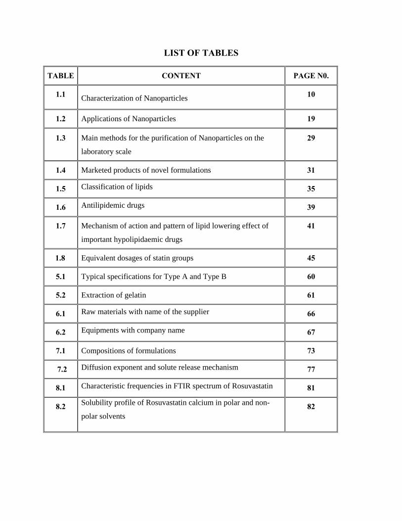
LIST OF TABLES
TABLE CONTENT PAGE N0.
1.1 Characterization of Nanoparticles 10
1.2 Applications of Nanoparticles 19
1.3 Main methods for the purification of Nanoparticles on the
laboratory scale
29
1.4 Marketed products of novel formulations 31
1.5 Classification of lipids 35
1.6 Antilipidemic drugs 39
1.7 Mechanism of action and pattern of lipid lowering effect of
important hypolipidaemic drugs
41
1.8 Equivalent dosages of statin groups 45
5.1 Typical specifications for Type A and Type B 60
5.2 Extraction of gelatin 61
6.1 Raw materials with name of the supplier 66
6.2 Equipments with company name 67
7.1 Compositions of formulations 73
7.2 Diffusion exponent and solute release mechanism 77
8.1 Characteristic frequencies in FTIR spectrum of Rosuvastatin 81
8.2 Solubility profile of Rosuvastatin calcium in polar and non-
polar solvents 82

TABLE CONTENT PAGE N0.
8.3 Percentage loss on drying for Rosuvastatin 83
8.4 Data of concentration and absorbance for Rosuvastatin in
0.1N methanol 85
8.5 Data for calibration curve parameters for 0.1 methanol 86
8.6 Calibration curve of Rosuvastatin calcium in methanol 240 nm 87
8.7 Data for calibration curve parameters for in 6.8 phosphate buffer 88
8.8 Percentage purity of drug 88
8.9 Interpretation of FTIR spectrum 91
8.10 % LE and % LC of Rosuvastatin Nanoparticles with gelatin and gelatin B
94
8.11 Percentage in vitro drug released of formulations F1-F8 98
8.12 Release kinetics of in-vitro drug release gelatin A and gelatin B 104
8.13 Stability studies of optimized formulation (F1) 109
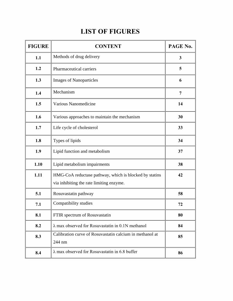
LIST OF FIGURES
FIGURE CONTENT PAGE No.
1.1 Methods of drug delivery 3
1.2 Pharmaceutical carriers 5
1.3 Images of Nanoparticles 6
1.4 Mechanism 7
1.5 Various Nanomedicine 14
1.6 Various approaches to maintain the mechanism 30
1.7 Life cycle of cholesterol 33
1.8 Types of lipids 34
1.9 Lipid function and metabolism 37
1.10 Lipid metabolism impairments 38
1.11 HMG-CoA reductase pathway, which is blocked by statins
via inhibiting the rate limiting enzyme.
42
5.1 Rosuvastatin pathway 58
7.1 Compatibility studies 72
8.1 FTIR spectrum of Rosuvastatin 80
8.2 λ max observed for Rosuvastatin in 0.1N methanol 84
8.3 Calibration curve of Rosuvastatin calcium in methanol at
244 nm 85
8.4 λ max observed for Rosuvastatin in 6.8 buffer 86
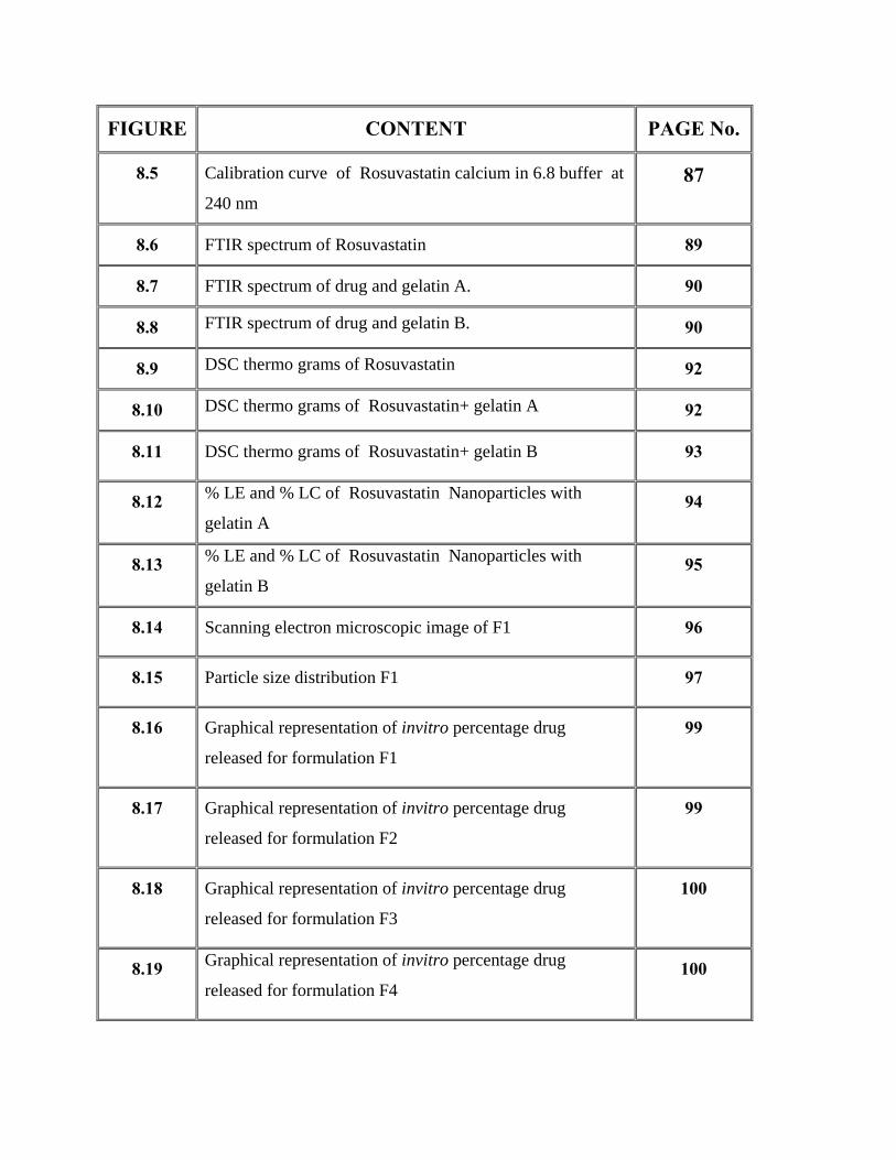
FIGURE CONTENT PAGE No.
8.5 Calibration curve of Rosuvastatin calcium in 6.8 buffer at
240 nm 87
8.6 FTIR spectrum of Rosuvastatin 89
8.7 FTIR spectrum of drug and gelatin A. 90
8.8 FTIR spectrum of drug and gelatin B. 90
8.9 DSC thermo grams of Rosuvastatin 92
8.10 DSC thermo grams of Rosuvastatin+ gelatin A 92
8.11 DSC thermo grams of Rosuvastatin+ gelatin B 93
8.12 % LE and % LC of Rosuvastatin Nanoparticles with
gelatin A 94
8.13 % LE and % LC of Rosuvastatin Nanoparticles with
gelatin B 95
8.14 Scanning electron microscopic image of F1 96
8.15 Particle size distribution F1 97
8.16 Graphical representation of invitro percentage drug
released for formulation F1
99
8.17 Graphical representation of invitro percentage drug
released for formulation F2
99
8.18 Graphical representation of invitro percentage drug
released for formulation F3
100
8.19 Graphical representation of invitro percentage drug
released for formulation F4 100

FIGURE CONTENT PAGE No.
8.20 Graphical representation of invitro percentage drug
released for formulation F5 101
8.21 Graphical representation of invitro percentage drug
released for formulation F6 101
8.22 Graphical representation of invitro percentage drug
released for formulation F7 102
8.23 Graphical representation of invitro percentage drug
released for formulation F8
102
8.24 Graphical representation of comprehensive invitro
percentage drug released for formulation F1-F8
103
8.25 Best fit model (zero order) of formulation F1 104
8.26 Best fit model (zero order) of formulation F2 105
8.27 Best fit model (zero order) of formulation F3 105
8.28 Best fit model (zero order) of formulation F4 106
8.29 Best fit model (zero order) of formulation F5 106
8.30 Best fit model (zero order) of formulation F6 107
8.31 Best fit model (zero order) of formulation F7 107
8.32 Best fit model (zero order) of formulation F8 108
8.33 Zeta potential of F1 formulation 110
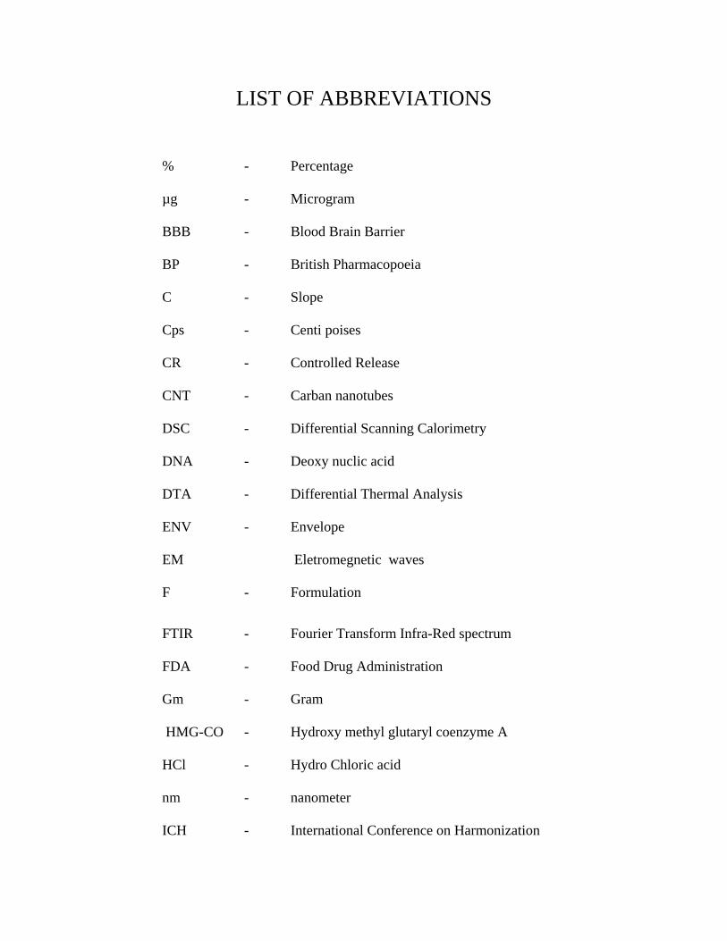
LIST OF ABBREVIATIONS
% - Percentage
µg - Microgram
BBB - Blood Brain Barrier
BP
C
-
-
British Pharmacopoeia
Slope
Cps - Centi poises
CR
CNT
-
-
Controlled Release
Carban nanotubes
DSC
DNA
DTA
ENV
EM
-
-
-
-
Differential Scanning Calorimetry
Deoxy nuclic acid
Differential Thermal Analysis
Envelope
Eletromegnetic waves
F - Formulation
FTIR
FDA
-
-
Fourier Transform Infra-Red spectrum
Food Drug Administration
Gm
HMG-CO
HCl
nm
ICH
-
-
-
-
-
Gram
Hydroxy methyl glutaryl coenzyme A
Hydro Chloric acid
nanometer
International Conference on Harmonization
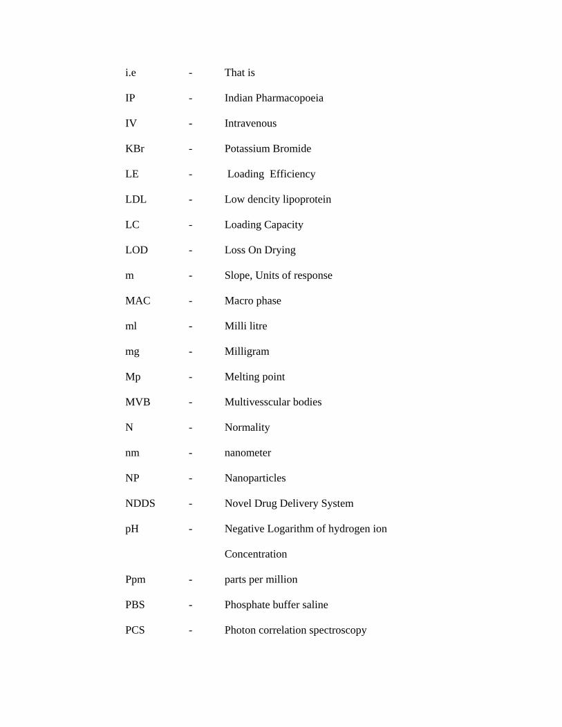
i.e
IP
IV
KBr
-
-
-
-
That is
Indian Pharmacopoeia
Intravenous
Potassium Bromide
LE
LDL
LC
-
-
-
Loading Efficiency
Low dencity lipoprotein
Loading Capacity
LOD - Loss On Drying
m
MAC
-
-
Slope, Units of response
Macro phase
ml
mg
-
-
Milli litre
Milligram
Mp
MVB
-
-
Melting point
Multivesscular bodies
N
nm
NP
-
-
-
Normality
nanometer
Nanoparticles
NDDS - Novel Drug Delivery System
pH - Negative Logarithm of hydrogen ion
Concentration
Ppm
PBS
PCS
-
-
-
parts per million
Phosphate buffer saline
Photon correlation spectroscopy
PACA
PLA
PLGA
PMA
PMMA
-
-
-
-
-
Poly Alkyl-Cyanoacrylate
Poly Lactide
Poly Lactide Co Glycolide
Polymethacrylate
Polymethyl methacrylate
PVA
PVD
QIA
-
-
-
Polyvinyl alcohol
Physical Vapour Deposition
Quality improvement activity
RH - Relative Humidity
rpm
RES
Sol
-
-
-
revolutions per minute
Reticulo Endothelial System
Solution
S.D.
SEM
SLN
TEM
-
-
-
-
Standard Deviation
Scanning Electron Microscopy
Solid Lipid Nanoparticles
Transmission electron Microscopy
t - Time
t1/2 - Biological half-life
USP - United State Pharmacopoeia
UV-VIS
VLDL
-
-
Ultraviolet-Visible Spectroscopy
Very low dencity lipoprotein
λ max - Absorption maximum

INTRODUCTION

Gelatin Loaded Rosuvastatin Nanoparticles Introduction
Adhiparasakthi College of Pharmacy, Melmaruvathur. Page 1
1.1. Novel drug delivery system (Shoba Rani., 2008)
Recently, several technical advancements have resulted in the development of new
techniques for drug delivery. These techniques are capable of controlling the rate of drug
delivery, sustaining the duration of therapeutic activity and or targeting the delivery of
drug to a tissue. These are referred to as novel drug delivery systems. And they have
revolutionized the method of medication, provides a number of therapeutic benefits.
The method by which a drug is delivered can have a significant effect on its efficacy.
Some drugs have an optimum concentration range within which maximum benefit is
derived, and concentrations above or below this range can be toxic or produce no
therapeutic benefit at all. From this, new ideas on controlling the pharmacokinetics,
pharmacodynamics, non-specific toxicity, immunogenicity, biorecognition, and efficacy
of drugs were generated. These new strategies, often called drug delivery systems (DDS),
are based on interdisciplinary approaches that combine polymer science, pharmaceutics,
bioconjugate chemistry, and molecular biology.
Controlled drug release and subsequent biodegradation are important for developing
successful formulations. Potential release mechanisms involve.
• Desorption of surface-bound /adsorbed drugs;
• Diffusion through the carrier matrix;
• Diffusion (in the case of nanocapsules) through the carrier wall;
• Carrier matrix erosion.
• A combined erosion /diffusion process.
1.INTRODUCTION

Gelatin Loaded Rosuvastatin Nanoparticles Introduction
Adhiparasakthi College of Pharmacy, Melmaruvathur. Page 2
Advantages of novel drug delivery systems.(Illinois., 2004)
• Improve therapy by increasing the duration of action and reducing side effects.
• Increase patient compliance through decreased dosing frequency and
convenient routes of administration
• Achieved targeting of to a specific site to reduce unwanted side effects and
obtain maximum efficacy.
• Lead to reduction in dose and thus reduction side effects of drugs.
• Decreased toxicity/side effects and increased convenience.
• Shorter hospitalization and better patient compliance.
• Viable treatments for previously in arable disease.
• Potential prophylactic application
The mode of delivery can be the difference between a drug’s success and failure, as
the choice of a drug is often influenced by the way the medicine is administered. Sustained
(or continuous) release of a drug involves polymers that release the drug at a controlled rate
due to diffusion. It is achieved by using drug-carrying polymers that respond to specific
stimuli (e.g., exposure to light, changes in pH or temperature).
Novel drug delivery system is a system that offers multiple drug delivery solutions
such as.
Oral Drug Delivery Systems and Materials
Parenteral and Implant Drug Delivery Systems
Pulmonary and Nasal Drug Delivery
Transmucosal Drug Delivery
Transdermal and Topical Drug Delivery
Delivery of Proteins and Peptides
Drug Delivery Pipelines

Gelatin Loaded Rosuvastatin Nanoparticles Introduction
Adhiparasakthi College of Pharmacy, Melmaruvathur. Page 3
Nanoparticles for drug delivery.
Metal-based nanoparticles
Lipid-based nanoparticles
Polymer-based nanoparticles
Biological nanoparticles
1.1.3. Methods of drug delivery.(ChayaVenkat., et al., 2003)
Figure 1.1: Methods of drug delivery

Gelatin Loaded Rosuvastatin Nanoparticles Introduction
Adhiparasakthi College of Pharmacy, Melmaruvathur. Page 4
Types of novel drug delivery systems.( Costas Kaparissides., et al., 2005)
• Hydrogels
• Colloids
• Microspores
• Liposomes
• Nanoparticles
• Mucoadhesives
• Transdermal
Hydrogels.
Hydrogels are three-dimensional, hydrophilic, polymeric networks capable of
imbibing large amounts of water or biological fluids. The networks are composed of
homopolymers or copolymers, and are insoluble due to the presence of chemical crosslinks
(tie-points, junctions), or physical crosslinks, such as entanglements or crystallites. They are
used to regulate drug release in reservoir-based, controlled release systems or as carriers in
swellable and swelling-controlled release devices.
Liposomes.
Liposomes are a form of vesicles that consist either of many, few or just one
phospholipid bilayers. The polar character of the liposomal core enables polar drug molecules
to be encapsulated. Amphiphilic and lipophilic molecules are solubilized within the
phospholipid bilayer according to their affinity towards the phospholipids.
Nanoparticles.
Nanoparticles(including nanospores and nanocapsules of size 10-200 nm) can adsorb
or encapsulated a drug,thus protecting it from chemical & enzymatic degradation.
Nanoparticles as drug carriers can be formed from both biodegradable and non-
biodegradable polymer.

Gelatin Loaded Rosuvastatin Nanoparticles Introduction
Adhiparasakthi College of Pharmacy, Melmaruvathur. Page 5
Drug delivery carriers.
Colloidal drug carrier systems such as micellar solutions, vesicle and liquid crystal
dispersions, as well as nanoparticle dispersions consisting of small particles of 10–400 nm.
Figure 1.2: Pharmaceutical carriers
1.2. Nanotechnology. (Shoba Rani., 2008)
Definition of Nanoparticles.
Nanoparticles are solid, colloidal particles ranging 10-1000nm (1µm) in size. They
consist of macromolecular materials in which the active ingredient (drug or biologically
active material) is dissolved, entrapped or encapsulated, and or absorbed or attached.
Nanoparticles are often defined as particles of less than 100nm in diameter.
Nanoparticles can be also defined as particles less than 100nm in diameter that exhibit new or
enhanced size-dependent properties compared with larger particles of the same material.
Gelatin Loaded Rosuvastatin Nanoparticles Introduction
Adhiparasakthi College of Pharmacy, Melmaruvathur. Page 6
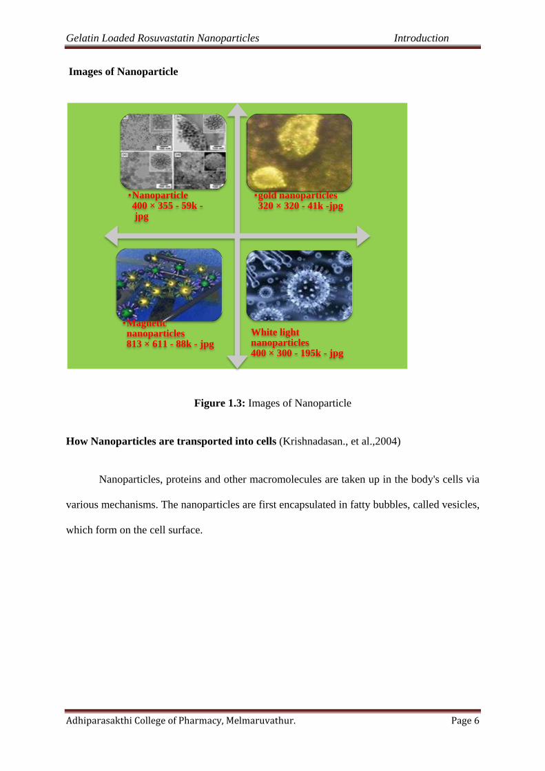
Images of Nanoparticle
Figure 1.3: Images of Nanoparticle
How Nanoparticles are transported into cells (Krishnadasan., et al.,2004)
Nanoparticles, proteins and other macromolecules are taken up in the body's cells via
various mechanisms. The nanoparticles are first encapsulated in fatty bubbles, called vesicles,
which form on the cell surface.
•Nanoparticle400 × 355 - 59k -jpg
•gold nanoparticles320 × 320 - 41k -jpg
•Magnetic nanoparticles813 × 611 - 88k - jpg
White light nanoparticles400 × 300 - 195k - jpg
Gelatin Loaded Rosuvastatin Nanoparticles Introduction
Adhiparasakthi College of Pharmacy, Melmaruvathur. Page 7
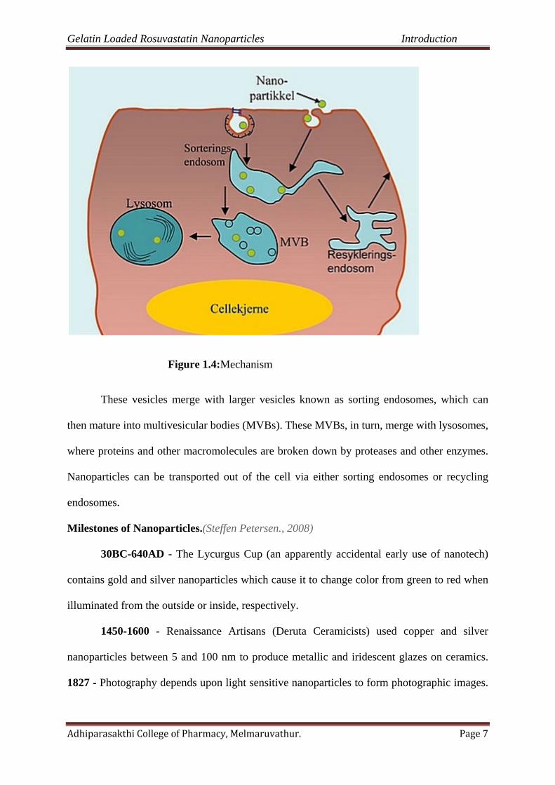
Figure 1.4:Mechanism
These vesicles merge with larger vesicles known as sorting endosomes, which can
then mature into multivesicular bodies (MVBs). These MVBs, in turn, merge with lysosomes,
where proteins and other macromolecules are broken down by proteases and other enzymes.
Nanoparticles can be transported out of the cell via either sorting endosomes or recycling
endosomes.
Milestones of Nanoparticles.(Steffen Petersen., 2008)
30BC-640AD - The Lycurgus Cup (an apparently accidental early use of nanotech)
contains gold and silver nanoparticles which cause it to change color from green to red when
illuminated from the outside or inside, respectively.
1450-1600 - Renaissance Artisans (Deruta Ceramicists) used copper and silver
nanoparticles between 5 and 100 nm to produce metallic and iridescent glazes on ceramics.
1827 - Photography depends upon light sensitive nanoparticles to form photographic images.

Gelatin Loaded Rosuvastatin Nanoparticles Introduction
Adhiparasakthi College of Pharmacy, Melmaruvathur. Page 8
1856 - Researcher, Michael Faraday discovers/prepares the first metallic colloids. His gold
colloids (fine particles suspended in solution) had unique optical/electronic properties.
1959 - Richard Feynman delivers famous speech, "There's Plenty of Room at the Bottom"
1960 - William McLellan constructs the first 250 microgram, 2000 rpm motor out of 13
individual parts, as large as the period at the end of this sentence.
1974 - Professor Norio Taniguchi becomes the first to formulate and use the term Nano-
technology.
1985 - Fullerenes (commonly called buckyballs) are discovered by researchers at Rice
University.
1986 - Gerd Binnig, Christopher Gerber and Calvin F. Quate invent the Atomic Force
Microscope
1986 - Eric Drexler, an American Engineer and the founder of Foresight Nanotech Institute,
writes Engines of Creation, introducing nanotechnology to the world.
1989 - The first commercially available Atomic Force Microscope is introduced.
1991 - Carbon nanotubes (CNT) are discovered by SumioIijima. Nanotubes are essentially
rolled sheets of graphene which can be single or multi-walled. Nanotubes have potential
applications in electronics, composite materials, the space elevator, and drug delivery.
1994 - US science advisor, Dr. Jack Gibbons talks at the White House about nanotechnology
and calls for increased .
1997 - Zyvex, the first company to research nanotechnology, is founded.
1999 - Consumer nanotech-based products start appearing on the global marketplace.
2003 - Congress enacts the 21st Century Nanotechnology Research and Development Act.
Gelatin Loaded Rosuvastatin Nanoparticles Introduction
Adhiparasakthi College of Pharmacy, Melmaruvathur. Page 9
2009 - The outlines a new research and regulatory strategy to better control the production
and use of engineered nanomaterials.
2010 - The House of Lords Science and Technology Committee, United Kingdom, warns its
country's food industry against hiding its use of nanotechnology.
Ideal properties of polymeric based Nanoparticles(Deepak Thassu.,et al., 2007)
• Natural or synthetic polymer
• Inexpensive
• Nontoxic
• Biodegradable
• Nonthrombogenic
• Nonimmunogenic
Nanoparticle recovery & drug incorporation efficiency
(Leroueli Le Verger M., et al., 1998)
Concentration of drug in Nanoparticles
Nanoparticles recovery % = X100
Concentration of Nanoparticles recovered
Drug incorporation efficiency has been expressed both as drug content (%w/w), also
referred to as drug loading, & drug entrapment (%) represented by the following equation.
Drug incorporation Amount of drug entrapped in Nanoparticles
efficiency in Nanoparticles =
Total amount of drug added

Gelatin Loaded Rosuvastatin Nanoparticles Introduction
Adhiparasakthi College of Pharmacy, Melmaruvathur. Page 10
1.2.1. Characterization of Nanoparticles. (Rakesh P. Patel.,et al., 2008)
Table 1.1:Characterization of Nanoparticles
Parameters Characterization methods
Particle size and size
distribution
photon correlation spectroscopy, Scanning electron microscopy
(SEM), Transmission electron microscopy (TEM), Atomic force
microscopy (AFM), Mercury porositometry, Laser defractrometry
Charge determination Laser droplet anemometry, Zeta potentiometer
Surface hydrophobicity Water contact angle measurements, rose bangle (dye) binding,
hydrophobic interaction chromatography, X-ray photoelectron
spectroscopy
Chemical analysis of
surface
Static secondary ion mass spectrometry, sorptometer
Carrier drug interaction Differential scanning calorimetry
Nanoparticle dispersion
stability
Critical flocculation temperature(CFT)
Release profile In-vitro release characteristic under physiologic & sink condition
Drug stability Bioassay of drug extracted from nanoparticle, chemical analysis.
Gelatin Loaded Rosuvastatin Nanoparticles Introduction
Adhiparasakthi College of Pharmacy, Melmaruvathur. Page 11
1.2.2. Classification of Nanoparticles.(Rakesh P. Patel., et al., 2008)
1. In one dimensions (Thin surface coatings)
One-dimensional systems, such as thin films or manufactured surfaces.
2. In Two dimensions.
a) Carbon Nanotubes
Carbon nanotubes are a new form of carbon molecule. Wound in a hexagonal network
of carbon atoms, these hollow cylinders can have diameters as small as 0.7 nm and reach
several millimeters in length. Each end can be opened or closed by a fullerene half-molecule.
These nanotubes can have a single layer (like a straw) or several layers (like a poster rolled in
a tube) of coaxial cylinders of increasing diameters in a common axis.
3. In three dimensions.
a) Fullerenes (Carbon 60)
Fullerenes are spherical cages containing from 28 to more than 100 carbon atoms
displaying unique physical properties. They can be subjected to extreme pressures and regain
their original shape when the pressure is released.
b) Dendrimers
Dendrimers represent a new class of controlled-structure polymers with nanometric
dimensions. They are considered to be basic elements for large-scale synthesis of organic and
inorganic nanostructures with dimensions of 1 to 100 nm, displaying unique properties.
Compatible with organic structures such as DNA, they can also be fabricated to interact with
metallic nanocrystals and nanotubes or to possess an encapsulation capacity
Gelatin Loaded Rosuvastatin Nanoparticles Introduction
Adhiparasakthi College of Pharmacy, Melmaruvathur. Page 12
c) Quantum dots
It represents a special form of spherical nanocrystals from 1 to 10 nm in diameter.
They have been developed in the form of semiconductors, insulators, metals, magnetic
materials or metallic oxides.
Types of Nanoparticles
• Quantum Dots
• Nanocrystalline silicon
• Photonic
• Liposome
• Gliadin Nanoparticles
• Polymeric Nanoparticles
• Solid Lipid Quantum Nanoparticles
• Others-Gold.Carbon, Silver.etc.
1. Quantum dots
A quantum dot is a semiconductor nanostructure that confines the motion of conduction
band electrons, valence band holes, or excitons (pairs of conduction band electrons and
valence band holes)in all three spatial directions. A quantum dothas a discrete quantized
energy spectrum. A quantum dot4 contains a small integer number of the order of 1-100) of
conduction band electrons, valence band holes, or excitons
2. Nanocrystalline silicon.
Nanocrystalline silicon is anallotropic form of silicon – is similar to amorphous silicon is
sometimes also known as microcrystalline silicon. One of the most important advantages of

Gelatin Loaded Rosuvastatin Nanoparticles Introduction
Adhiparasakthi College of Pharmacy, Melmaruvathur. Page 13
Nanocrystalline silicon, it has increasdstabilithovara one of the reasons being because of its
lower hydrogen concentration.
3.Photonic crystals.
Photonic crystals are periodic dielectric or metallo-dielectric (nano) structures that are
designed to affect the propagation of electromagnetic waves (EM) in the same way as the
periodic in a semiconductor crystal affects the electron motion by defining allowed and
forbidden electronic energy bands. Photonic crystals are the attractive optical materials for
controlling and manipulating the flow of light. They are of great interest for both fundamental
& applied research, & are expected to find commercial applications soon.
4.Liposomes.
A Liposome is a spherical vesicle with a membrane composed of phospholipids bilayer
used to deliver drugs or genetic material into a cell. Liposomes can be composed of naturally
derived phospholipids wit mixed lipid chains(like egg, phosphatidylethanolamine), or of pure
components .
The use of liposomes for transformation or transfection of DNA into a host cell is known
as lipofection . Liposomes can be created by sonicating phospholipids in water.
5.Gliadin Nanoparticles.
To improve bioavailability anti-H.pylori effects of antibiotics,
mucoadhesivegliadin.Neutral amino acid can promote hydrogen bonding interaction with the
mucosa whereas the lipophilic components can interact within biological tissue by
hydrophilic interaction.

Gelatin Loaded Rosuvastatin Nanoparticles Introduction
Adhiparasakthi College of Pharmacy, Melmaruvathur. Page 14
6.Polymeric Nanoparticles.
Polymeric Nanoparticles have been invented by Speiser et al. They represent interesting
alternative as drug delivery systems to liposomes. They usually exhibit a long shelf life & a
good stability on storage. Nanoparticles can be prepared either from preformed polymers,
such as polyesters (i.e.polylactic acid), or from a monometer during its polymerization, as in
the case of alkylcyanoacrylates.
Figure 1.5: Various Nanomedicine
7.Solid lipid Nanoparticles.
Solid Lipid Nanoparticles have been developed as alternative delivery system to
conventional polymeric nanoparticles. SLNs are sub-micron colloidal carriers.
Advantages.
Avoidance of coalescence leads to enhanced physical stability
Reduced mobility of incorporated drug molecules leads of drug leakage
Static interface solid/liquid facilitates surface modification.
Gelatin Loaded Rosuvastatin Nanoparticles Introduction
Adhiparasakthi College of Pharmacy, Melmaruvathur. Page 15
8.Others
Gold nanoparticles stabilized by thiol functionally are extraordinarily stable. A common
synthesis involves the reduction of a gold salt in the presence of capping agent molecules
such as thiols, citrates or phosphines. The synthesis of gold nanoparticles with a polymer-
thiol monolayer involves the mechanism of particle formation in the presence of bulky
ligands.
Gold nanoparticles are very good at scattering and absorbing light. It doesn’t stick as
well to noncancerous cell. The results can be seen with a simple microscope. In the study,
researchers found that the gold nanoparticles have 600 percent greater affinity
Advantages of Nanoparticles
Smaller dosage form (i.e., smaller tablet)
Decreased toxicity
Stable dosage forms of drugs which are either unstable or have unacceptable low
bioavailability in non-nanoparticulate dosage forms.
Increased active agent surface area results in a faster dissolution of the active agent in
an aqueous environment, such as the human body. Faster dissolution generally
equates with greater bioavailability, smaller drug doses, less toxicity.
Reduction in fed/fasted variability.
They are suitable for different routes of administration
Carrying capacity of nanoparticles is high
Shelf-stability of drug increases
Ability to sustain and control drug release patterns
Suitable for combination therapy where two or more drug can be co-deliverd
Both hydrophobic and hydrophilic drug can be incorporated
Gelatin Loaded Rosuvastatin Nanoparticles Introduction
Adhiparasakthi College of Pharmacy, Melmaruvathur. Page 16
System increases the bioavailability of drugs
Imaging studies can be done by utilizing them
It is used for targeted drug delivery of drugs
Development of new medicines
Toxicity and adverse drug interactions are reduced to a possible extent
E.g. polymethacrylic nanoparticles for targeting anticancer drug of doxorubicin to reduce
liver toxicity.Nanoparticles posses’ better stability as compared to leptosomes which make it
more important for many modes of targeting.
Nanoparticles formulated as amorphous offer solubility than standard crystalline
formulations, thus improving the poor aqueous solubility of the drug and hence the
bioavailability.
The methods of preparation are simple, easier and reproducible.
A high degree of patient compliance can be achieved.
Wide range of polymer can be used depends on the nature of the drug and usage i.e.
biodegradable polymer for shorter periods, and non-biodegradable for longer periods.
It can easily pass through syringe needle and exhibit good rheological properties.
Disadvantages of Nanoparticles.(Bshsagar., et al., 2010)
The manufacturing costs of nanoparticle are high which result in overall product cost
Solvents are toxic in nature which is used in the preparation process
Can start immune response and allergic reactions in body
Extensive use of poly (vinyl alcohol) as stabilizer may toxicity issues
According to a discovery, silver nanoparticles used in socks to fight foot odor if
released in water can prove detrimental to the purity of water.
Gelatin Loaded Rosuvastatin Nanoparticles Introduction
Adhiparasakthi College of Pharmacy, Melmaruvathur. Page 17
Silver nanoparticles are bacteriostatic, by which we mean that they limit the growth of
bacteria. This may result in the destruction of bacteria that help in breaking down the
organic matter in water treatment plants.
The process of manufacturing nanomaterials results in the release of certain waste
products. This waste can float in air or even penetrate animal and plant cells.
Nanoparticles have large surfaces. This makes them susceptible to get absorbed by
macromolecules in an animal body. They can hinder biological processes, thus
intervening the functioning of nature.
"Oral delivery via tablets or capsules is largely inefficient due to exposure of the
pharmaceutical agent to the metabolic processes of the body. Therefore, a larger than
necessary dose is often required and the maximum effectiveness of the drug is limited.
Traditional intravenous (IV) administration is much more problematic. Specificity for
IV injectable drugs is often low, necessitating large amounts of a drug be injected into
a patient, creating a high concentration of the drug in the blood stream that could
potentially lead to toxic side effects".
Limitations of Nanoparticle.(Randy P. Carney., et al., 2008)
For example
Their small size and large surface area can lead to particles aggregation, making
physical handling of nanoparticles difficult in liquid and drug formulations.
In addition, small particle size and large surface area readily result in limited drug in
limited drug loading and burst release. These practical problems have to be overcome
before nanoparticles can be used clinically or made commercially available.
Gelatin Loaded Rosuvastatin Nanoparticles Introduction
Adhiparasakthi College of Pharmacy, Melmaruvathur. Page 18
Factors affecting the release of drugs from particulate carriers.
(Soppimathk s., et al., 2001)
Drug.
position of the particle
molecular weight
physicochemical properties
drug-carrier interaction
diffusion ; desorption from the surface Particles
type and amount of matrix material
size and density of the particle
capsular(or) monolithic
extent and nature of any cross linking ; denaturation of polymerization ,presence of
adjutants
surface erosion; particle diffusion and leaching
total disintegration of particles
Environment.
hydrogen ion concentration
polarity
ionic strength
presence of enzymes
temperature.

Gelatin Loaded Rosuvastatin Nanoparticles Introduction
Adhiparasakthi College of Pharmacy, Melmaruvathur. Page 19
Applications of Nanoparticles.(Rakesh P. Patel., et al., 2008)
Table 1.2:Applications of Nanoparticles
Nanomedicnes Nanodrugs, medical devices, tissue
engineering, etc.
Materials Nanoparticles, carbon nanotubes, biopolymers,
paints, coating
Chemicals and cosmetics Nanoscale chemicals and compounds, paints,
coating, etc.
Food science Processing, nutracetical food, nanocapsules
Envirnoment and energy Water and air purification filters, fuel cells,
photovoltics
Military and security Sensers, wepons, sensory enhancement
Electronics Semiconductor chips, memory storage, photonics, optoelectronics
Scientific tools Atomic force, microscopes and scanning tunneling microscope
Agriculture Pesticides, food production
Therapeutic applications of Nanoparticles(Shoba Rani.,2008)
For intracellular targeting of anti-infective drugs to combat the ‘difficult to
treat’ intracellular infections of the human body
For targeting of cytostatic drugs to reduce toxicity & increase therapeutic
activity
Gelatin Loaded Rosuvastatin Nanoparticles Introduction
Adhiparasakthi College of Pharmacy, Melmaruvathur. Page 20
For specific targeting of anti-inflammatory drugs to areas of inflammation, by
which the side effects of these drugs can be minimized
For ocular delivery systems, to deliver pilocarpine and other miotic drugs
As carriers for radio nucleotides for diagnostic purpose in nuclear medicine
To improve the solubility and bioavailability of poorly soluble drugs
For skin and hair care in the form of solid lipid nanoparticles wherein the
oily core contains a wide variety of different cosmetic oils and lipophilic
agents
To deliver drugs across the brain barrier (BBB)
To formulate sustained release preparations
For the controlled delivery of disinfectants or algaecides into large bodies of
water such as insect pest feed on colloidal particles
For targeted delivery of proteins and peptides.
1.2.3. Polymers employed for Nanoparticles .(Deepak Thassu .,et al., 2007)
Synthetic polymers.
Polylactide co glycolic acid
Poly lactide
Polycaprolactone
Polymethyl methacrylate
Poly methyl methacrylate copolymer
Poly isobutylcyanoacrylate
Polyhexylcyanoacrylate
Ethyl cellulose
EudragitRL
EudragitRS
Gelatin Loaded Rosuvastatin Nanoparticles Introduction
Adhiparasakthi College of Pharmacy, Melmaruvathur. Page 21
Natural polymers.
Gelatin
Lecithin
Albumin
Chitosan
Casein
i. Natural biodegradable polymers used to prepare nanoparticles alginates
Alginates are linear, unbranched polysaccharides composed of randomchains of
Guluronic and mannuronic acids. In aqueous media, the sodium ions from salts of these
anionic, heteropolymers exchange with divalent cations, such as calcium, to form water-
insoluble gels. Because of the favorable conditions during manufacture, alginates are
ideal carriers for oligonucleotides, peptides, proteins, Water-soluble drugs or drugs that
degrade in organic solvents. Alginates are non immunogenic and available in a wide
range of molecular weights as characterized by their inherent viscosity. Alginate
nanoparticles are prepared by extruding an aqueous sodium alginate solution through a
narrow-bore needle into an aqueous solution of a cationic agent, such as calcium ions,
chitosan, or poly-l-lysine. These cations cross-link the Guluronic and mannuronic acids to
form an egg-box structure That forms the core of the gel matrix. In vivo, therapeutic
agents are released whether matrix redissolves due to the reversible exchange of divalent
cations with monovalentions, especially sodium present in physiological fluid.
Chitosan
Chitosan is a natural polymer obtained by deacetylation of chitin, a component of crab
shells. It is a cationic polysaccharide composed of linear β (1, 4)-linked d-glucosamine.the
various methods used to prepare chitosan-based nanoparticles and their applications has been

Gelatin Loaded Rosuvastatin Nanoparticles Introduction
Adhiparasakthi College of Pharmacy, Melmaruvathur. Page 22
extensively reviewed. Chitosan can entrap drugs by numerous mechanisms including
chemical cross-linking, ionic cross-linking, and Ionic complexation.
Gelatin
Gelatin is a natural, biodegradable protein obtained by acid- or base-catalyzed
Hydrolysis of collagen. It is a heterogeneous mixture of single- or multi-stranded
Polypeptides composed predominantly of glycine, proline, and hydroxyproline Residues and
is degraded in vivo to amino acids. Gelatin nanoparticles are prepared by a two-step, de
solvation process. The concentrated gelatin liquid particles are isolated and hardened by
chemical cross-linking with glutaraldehyde. Alternately, these particles can be prepared using
a simple o/w emulsion or w/o/w micro emulsion method. Gelatin nanoparticles have been
used to deliver paclitaxel, methotrexate, doxorubicin, DNA, double-stranded
oligonucleotides, and genes. Pegylation of the particles significantly enhances their
circulation time in the blood stream and increases their uptake into cells by endocytosis.
Antibody-modified gelatin nanoparticles have been used for targeted uptake by lymphocytes.
Pullulan
Similar to dextran and cellulose, the glucans in Pullulan are water-soluble, linear
Polysaccharides that consist of three α-1, 4-linked glucose molecules polymerized By α-1,6
linkages on the terminal glucose . Pullulan is a fermentation product of the yeast
aureobasidiumpullulans. Pullulan nanoparticles have been prepared by Dialysis of an organic
solution against water. In one method, a reverse micellar Solution of the anionic surfactant,
aerosol, in n-hexane was prepared and an aqueous solution of the drug and Pullulan added.
The nanoparticles are stabilized by cross-linking with glutaraldehyde. These delivery systems
have been used in delivering cytotoxic drugs, genes, and as ph-sensitive delivery systems

Gelatin Loaded Rosuvastatin Nanoparticles Introduction
Adhiparasakthi College of Pharmacy, Melmaruvathur. Page 23
Gliadin
Gliadin is a glycoprotein that, as a component of gluten, is extracted from gluten rich
food such as wheat flour. They are slightly hydrophobic and polar. Bioactive molecules of
variable polarity can be encapsulated into gliadin nanoparticles. Gliadin nanoparticles can be
prepared by a desolvation method that exploits the insolubility of this polymer in water.
Briefly, gliadin nanoparticles are precipitated when an ethanolic solution of gliadin is poured
into an aqueous solution. Gliadin nanoparticles have been used to deliver trans-retinoic acid,
α-tocopherol, and vitamin e. Lectins have been conjugated to the surface of gliadin
nanoparticles to target the colon and treat helicobacter pylori infections .
ii. Synthetic biodegradable polymers used to prepare Nanoparticles
Polylactide and polylactide-co-glycolide
The hydrophobic PLA may be used alone or copolymerized with poly-glycolic acid to
form a range of PLGA of widely varying polymeric ratios and hence physicochemical
properties. These FDA-approved polymers have been widely used in drug delivery including
nanoparticles. PLA and PLGA polymers degrade by random Bulk hydrolysis that is catalyzed
in acidic media.
Polyanhydrides
Polyanhydrides are biodegradable polymers with a hydrophobic backbone and a
hydrolytically labile anhydride linkage. They are synthesized by ring-opening Polymerization
and degrade by surface hydrolysis. The application of Polyanhydrides has been limited to
film and microsphere formulation for sustained release of a drug or protein at the site.
Poly-є-caprolactones
Methods used to prepare nanoparticles using poly-ε-caprolactones have been previously
reviewed and include emulsion polymerization, solvent displacement, Dialysis, and
interfacial polymer deposition. These semi crystalline polymers are chemically stable,

Gelatin Loaded Rosuvastatin Nanoparticles Introduction
Adhiparasakthi College of Pharmacy, Melmaruvathur. Page 24
possess a low glass transition temperature, and degrade slowly. Poly-ε-caprolactone
Nanoparticles have been used as vehicles to deliver a wide range of drugs including
tamoxifen, retinoic acid, and griseofulvin.
Polyalkyl-cyanoacrylates
Polyalkyl-cyanoacrylate nanoparticles are prepared by the conventional Emulsion-
evaporation technique. In addition to sustaining drug release, PACA Nanoparticles have the
ability to overcome multidrug resistance at both the cellular and sub cellular levels. The
potential for targeted delivery of PACA nanoparticles to cells has been demonstrated by
conjugation of polysaccharides to the surface
Non biodegradable polymers used to prepare Nanoparticles
Polymethacrylate and Polymethyl methacrylate have been widely used in a variety of
pharmaceutical and medical applications Incorporation of poly-acrylic acid into nanoparticles
increased the transfection efficiency of DNA The side chain of PMMA can be modified to
make these polymers possess ph-dependent solubility and has been used to prepare pH-
sensitive Nanoparticles to increase the oral bioavailability.
1.2.4. Preparation techniques of Nanoparticles(Vyas S.P andKhar R.K., 2002)
The selection of the appropriate method for the preparation of nanoparticles depends on
the physicochemical characteristics of the polymer and the drug to be loaded on the contrary;
the preparation techniques largely determine the inner structure, in-vitro release profile and
the biological fate of these polymeric delivery systems. Two types of systems with different
systems with different inner structures are apparently possible.
A matrix type of system consisting of an entanglement of oligomer or polymer
units(nanoparticles/nanospheres)
A reservoir type of system comprised of an oily core surrounded by an embryonic
polymeric shell(nanocapsules).

Gelatin Loaded Rosuvastatin Nanoparticles Introduction
Adhiparasakthi College of Pharmacy, Melmaruvathur. Page 25
Structure of nanospheres and nanocapsules.
These methodologies are conveniently classified as follows.
1. Amphiphillic macromolecule cross linking
a. Heat cross linking
b. Chemical cross linking
Polymerization based methods
a. Polymerization of monomers in situ
b. Emulsion(micellar)polymerization
c. Dispersion polymerization
d. Interfacial condensation polymerization
e. Interfacial complexation.
2. Polymer precipitation methods
a. Solvent extraction/evaporation
b. Solvent displacement(nanoprecpitation)
c. Salting out.
Several methods exist for the preparation of nanoparticles. When synthetic polymers are
used, they are typically dissolved in a convenient solvent followed by precipitation in a liquid
environment leading to nanoparticle formation. The drug intended to be encapsulated in the
particles is usually incorporated in the process during the polymer solvation and precipitation.
Emulsification/solvent diffusion, emulsification/solvent evaporation, nanoprecpitation and
salting-out methods are widely applied techniques and they have been discussed in several
reviews.
Polymerization method (Mohanraj VJ and chen Y., 2006)
In this method, monomers are polymerized to form nanoparticles in an aqueous
solution. Drug is incorporated either by being dissolved in the polymerization medium or by
Gelatin Loaded Rosuvastatin Nanoparticles Introduction
Adhiparasakthi College of Pharmacy, Melmaruvathur. Page 26
adsorption onto the nanoparticles after polymerization completed.This technique has been
reported for making polybutylcyanoacrylate or poly (alkylcyanoacrylate) nanoparticles
Nanocapsules formation and their particle size depends on the concentration of the
surfactants and stabilizers used.
Emulsion-solvent evaporation (Lamprecht Alf., 2009)
Polymer and drug are dissolved in a suitable volatile solvent which is Immiscible with
water. This solution is emulsified in an aqueous solution Containing stabilizer by
conventional emulsification techniques. Droplet size can be further reduced by using a high-
energy source. Continuous emulsification under mixing prevents coalescence of organic
droplets and allows the spontaneous evaporation of the solvent at room temperature and the
formation of the colloidal particles. Following evaporation of organic phase under reduced
pressure or vacuum produces a fine aqueous dispersion of nanoparticles. This technique was
principally applied to the preparation of particles from water insoluble polymers.
Solvent-displacement, -diffusion, or Nanoprecpitation(Beck-Broichsitter M., et al.,2010)
A solution of polymer, drug and lipophilic stabilizer (surfactant) in a semi polar
solvent miscible with water is injected into an aqueous solution (being a non-solvent or anti
solvent for drug and polymer containing another stabilizer under moderate stirring.
Nanoparticles are formed instantaneously by rapid solvent diffusion and the organic Solvent
is removed under reduced pressure. The Velocity of solvent removal and thus nuclei
formation is the key to obtain particles in the nanometer range instead of larger lumps or
agglomerates.
Salting-out
Although a less common method of preparation, by adding a solution of polymer and
drug in a water miscible solvent to an aqueous solution containing a salting -out agent and a
stabilizer under stirring, small droplets can be obtained. Dilution of the resulting o/w
Gelatin Loaded Rosuvastatin Nanoparticles Introduction
Adhiparasakthi College of Pharmacy, Melmaruvathur. Page 27
emulsion with water forces diffusion of organic solvent into the aqueous phase. The
remaining polymer together with the drug produces particles in the nano-size range. The
resulting dispersion often requires a purification step to remove the salting-out agent.
In emulsification/solvent diffusion (Randy.et al.,2008)
Nanoparticles are formed when the saturation limit of a partially water-miscible
solvent ( Benzyl alcohol) is exceeded by addition of water. The phase separation is
accompanied by vigorous stirring. The separated solvent is removed by cross-flow filtration.
Purification techniques of Nanoparticles.(Langmuir.,2010)
Nanoparticles are intended to be used as pharmaceutical dosage forms in humans.
Three important processes are performed.
Purification
Freeze-drying
Sterilization
Purification of Nanoparticles.
In this method, various toxic impurities such as organic solvents, electrolytes,
surfactants, stabilizers and large polymer aggregates can be found. Simple filtration will only
remove polymer aggregates, while other impurities require sophisticated procedures.
Most common procedures.
Gel filtration.
Dialysis.
Ultracentrifugation.
These methods are not satisfactory because they are restricted to the lab scale or incapable of
eliminating molecules with high molecular weight.
Gelatin Loaded Rosuvastatin Nanoparticles Introduction
Adhiparasakthi College of Pharmacy, Melmaruvathur. Page 28
Cross-flow filtration method.
In this method, the nanoparticle suspension is filtered through membranes, with the
direction of the fluid being tangential to the surface of the membranes.
Freeze-drying of Nanoparicles.
It is one of the method used to ensure the long-term conversation of polymeric
nanoparticles
Method.
This technique involves the freezing of the suspension and the subsequent elimination
of its water content by sublimation under reduced pressure. After complete desiccate,
nanoparticles are obtained in the form of dry, free-flowing powder that is easy to handle and
store.
Sterilization of Nanoparticles
Some of the well-established methods of sterilization such as filtration 0.22µm filters
are not adequate for nanoparticle suspensions because microorganisms and nanoparticles are
generally similar in size (0.3-1µm). Therefore, this method is best achieved by using aseptic
techniques throughout their processing and preparation.
Autoclaving (most heat sterilization) and Ỵ-irradiation are the techniques that can be applied
for terminal sterilization. This technique may have impact on the physicochemical properties of
the particles.
Gelatin Loaded Rosuvastatin Nanoparticles Introduction
Adhiparasakthi College of Pharmacy, Melmaruvathur. Page 29
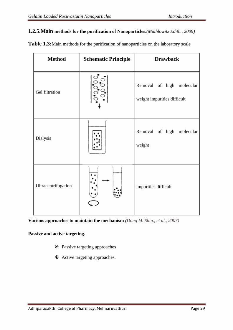
1.2.5.Main methods for the purification of Nanoparticles.(Mathlowitz Edith., 2009)
Table 1.3:Main methods for the purification of nanoparticles on the laboratory scale
Method Schematic Principle Drawback
Gel filtration
Removal of high molecular
weight impurities difficult
Dialysis
Removal of high molecular
weight
Ultracentrifugation
impurities difficult
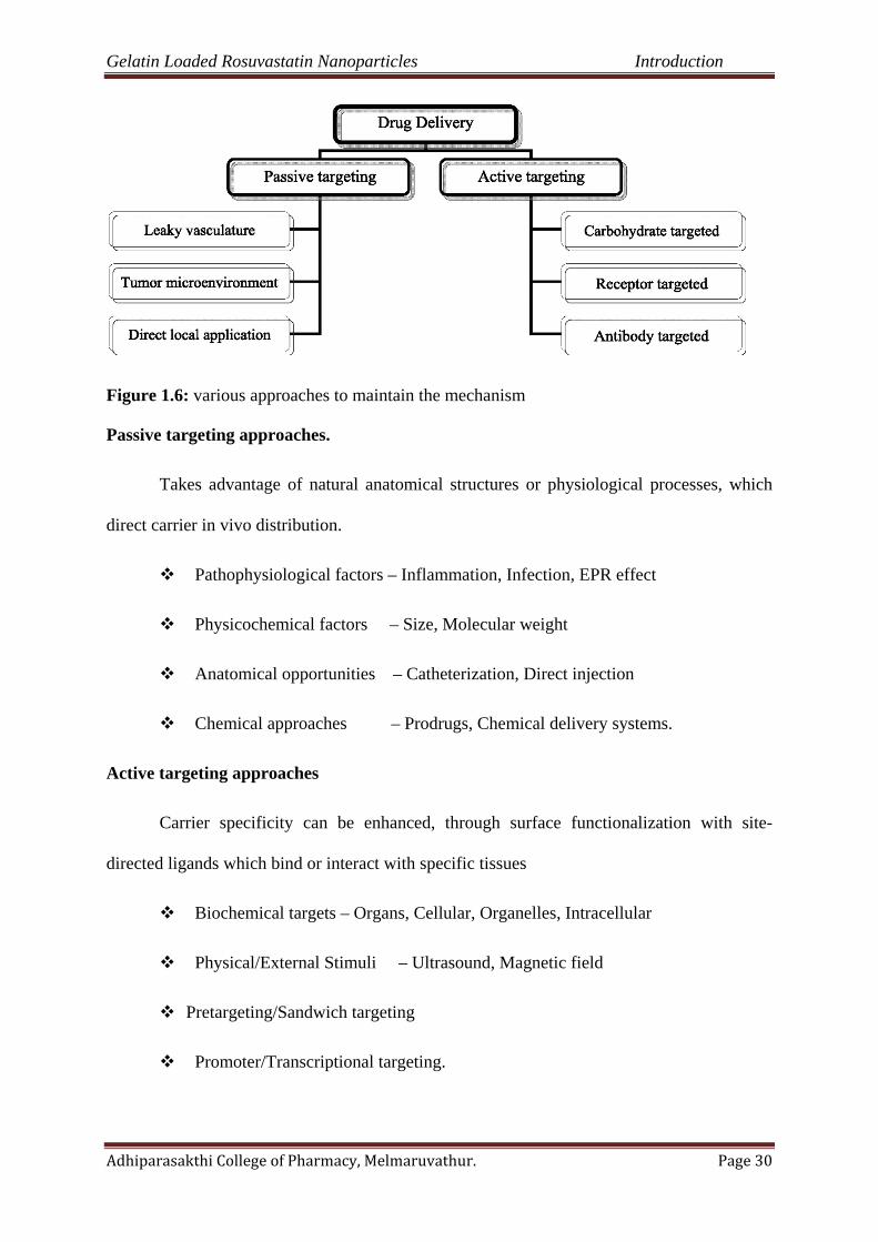
Various approaches to maintain the mechanism (Dong M. Shin., et al., 2007)
Passive and active targeting.
Passive targeting approaches
Active targeting approaches.
Gelatin Loaded Rosuvastatin Nanoparticles Introduction
Adhiparasakthi College of Pharmacy, Melmaruvathur. Page 30
Figure 1.6: various approaches to maintain the mechanism
Passive targeting approaches.
Takes advantage of natural anatomical structures or physiological processes, which
direct carrier in vivo distribution.
Pathophysiological factors – Inflammation, Infection, EPR effect
Physicochemical factors – Size, Molecular weight
Anatomical opportunities – Catheterization, Direct injection
Chemical approaches – Prodrugs, Chemical delivery systems.
Active targeting approaches
Carrier specificity can be enhanced, through surface functionalization with site-
directed ligands which bind or interact with specific tissues
Biochemical targets – Organs, Cellular, Organelles, Intracellular
Physical/External Stimuli – Ultrasound, Magnetic field
Pretargeting/Sandwich targeting
Promoter/Transcriptional targeting.
Gelatin Loaded Rosuvastatin Nanoparticles Introduction
Adhiparasakthi College of Pharmacy, Melmaruvathur. Page 31
Marketed products of novel formulations
Table 1.4:Marketed products of novel formulations
Company Trade name Composition Indication Administration
Enzon Abelect Liposomal
amphotericin B
Fungal
infection
Intravenous
Berna Biotech Epaxal Liposomal IRIV
Vaccine
Hepatitis A Intramuscular
Novavax Estrasorb Micellular
estradiol
Menopausal
therapy
Topical
Nektar,
Hoffmann-
La Roche
Pegasys PEG–a-
interferon 2a
Hepatitis B,
Hepatitis C
Subcutaneous
Genzyme Pegasys Poly(allylamine
hydrochloride)
End-stage
renal disease
Oral
Health implications of Nanoparticles.(Rakesh P. Patel.,et al., 2008)
Nanoparticles can enter the human body in several ways;
(i) The lungs where a rapid translocation through the blood stream to vital organs is possible,
including crossing the BBB,
(ii) The intestinal tract,
(iii) The skin
a) Skin
Particles 500–1000 nm in size, theoretically beyond the realms of nanotechnology,
can penetrate and reach the lower levels of human skin, 128 and smaller particles are
Gelatin Loaded Rosuvastatin Nanoparticles Introduction
Adhiparasakthi College of Pharmacy, Melmaruvathur. Page 32
likely to move deeper into the skin particles are often used in sunscreens to absorb UV
light and therefore to protect skin against sunburn or genetic damage. It has been reported
by Lademann et al in that micrometer-sized particles of get through the human stratum
corneum and even into some hair follicles – including their deeper parts.
b) Intestinal tract
The epithelium of the small and large intestines is in close contact with ingested
material so that nutrients can be utilized. A mixture of disaccharides, peptides, fatty acids,
and monoglycerides generated by digestion in small intestine are further transformed and
taken in the villi.. Charged particles, such as carboxylated polystyrene nanoparticles or
those composed of positively charged polymers exhibit poor oral bioavailability through
electrostatic repulsion and mucus entrapment. The smaller the particle diameter the faster
they could permutate the mucus to reach the colonic enterocytes; 14 nm diameter
permeated within 2 min, 415 nm particles took 30 min, while 1000-nm particles were
unable to translocate this barrier.
c) Lung
Based on three particle-types titanium dioxide , carbon black, and diesel particles,
hazard studies in rats demonstrate that ultrafine or nanoparticles administered to the lung
produce more potent adverse effects in the form of inflammation and subsequent tumors
compared with larger sized particles of identical chemical composition at equivalent mass
concentrations or intratracheally-instilled doses. Surface properties, such as surface
chemistry and area, may play a significant role in nanoparticle particle toxicity.

Gelatin Loaded Rosuvastatin Nanoparticles Introduction
Adhiparasakthi College of Pharmacy, Melmaruvathur. Page 33
1.3.Antilipedimicdrugs
Lipids.(Anne Marie Helmenstine.,2005)
The term 'lipid' was first used by the German biochemist Bloor in 1943 for a major
class of tissue components and foodstuffs.
A lipid is a fat-soluble molecule. To put it another way, lipids are insoluble in water
but soluble in at least one organic solvent. The other major classes of organic compounds
(nucleic acids, proteins, and carbohydrates) are much more soluble in water than in an
organic solvent. Lipids do not share a common molecule structure.
Lipids are biomolecules which are soluble in organic non-polar solvents.
Consequently, fats and lipids are insoluble in water. Glycerides and waxes form a sub-group
of compounds which have an ester as the major functional group and include.waxes,
triglycerides, and phospholipids. Lipids without ester functional groups include.steroids, fatty
acids, soaps, sphingolipids, and prostaglandins.
Figure 1.7: Life cycle of cholesterol

Gelatin Loaded Rosuvastatin Nanoparticles Introduction
Adhiparasakthi College of Pharmacy, Melmaruvathur. Page 34
Definition of lipids(James Richard Fromm.,1977)
A lipid is defined as a water-insoluble biomolecule which has a high solubility in
nonpolar organic solvents such as chloroform. The simplest lipids are the fats, which are
triesters made up of one glycerol and three fatty acids.
The term fats is also used as a general synonym for lipids, so the more precise terms
triacylglycerols or triglycerides are preferable for the simplest lipids. Triacylglycerols are
used primarily for energy storage in animals. More complex lipids, the phospholipids,
glycolipids, and cholesterol, are the major constituents of biological cell membranes.
Figure 1.8: Types of lipids

Gelatin Loaded Rosuvastatin Nanoparticles Introduction
Adhiparasakthi College of Pharmacy, Melmaruvathur. Page 35
Examples of common lipids(Charles E.Ophardt.,2003)
Examples of common lipids include
• Butter
• vegetable oil
• cholesterol and other steroids
• waxes
• phospholipids
• Fat-soluble vitamins.
Classification of lipids.(Charles E.Ophardt.,2003)
Table 1.5:Classification of lipids
Lipid Classification and Examples
Fatty Acids Glycerides
Saturated Fatty Acids Steroids
Unsaturated Fatty Acids Lipoproteins
Soap (salt of fatty acid) Triglycerides
Prostaglandins Phosphoglycerides
Non glyceride Lipids
Waxes -
Sphingolipids -

Gelatin Loaded Rosuvastatin Nanoparticles Introduction
Adhiparasakthi College of Pharmacy, Melmaruvathur. Page 36
1.3.1. Lipid structure.
Although there is no single common structure for lipids, the most commonly
occurring class of lipids are triglycerides, which are fats and oils. Triglycerides have a
glycerol backbone bonded to three fatty acids. If the three fatty acids are identical then the
triglyceride is termed a simple triglyceride. Otherwise, the triglyceride is called a mixed
triglyceride.
The second most abundant class of lipids are the phospholipids, which are found in animal and
plant cell membranes. Phospholipids also contain glycerol and fatty acids, plus the contain
phosphoric acid and a low-molecular-weight alcohol. Common phospholipids include
lecithinsand cephalins.
The most important lipids present in blood plasma include fatty acids, triglycerides,
cholesterol, phospholipids and steroid hormones.
Triglycerides, cholesterol and phospholipids.
Triglycerides are esters of fatty acids (e.g., stearic C-18 or palmitic C-16) and glycerols. Most
of fatty acids are saturated, whereas nonsaturated fatty acids play an important role as
prostaglandin precursors and in the process of cholesterol esterification.
Cholesterol also is a cellular membrane element and precursor of steroid hormones
and biliary acids.
Phospholipids are structurally similar to triglycerides, except that one minor acid is
substituted by a phosphorous group and nitrogen base.
Gelatin Loaded Rosuvastatin Nanoparticles Introduction
Adhiparasakthi College of Pharmacy, Melmaruvathur. Page 37
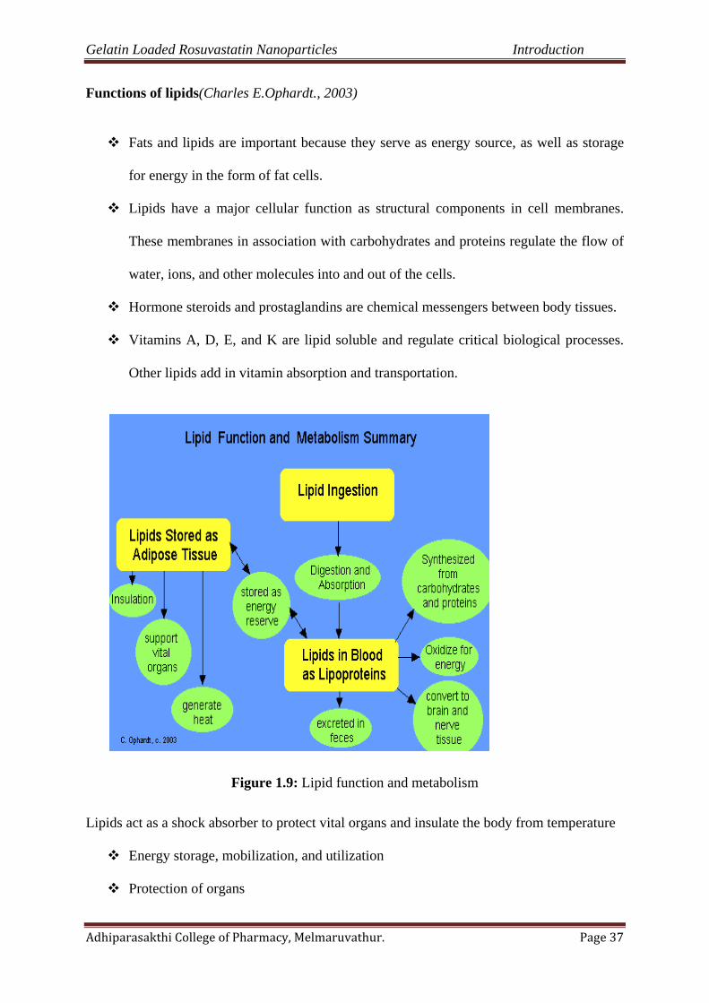
Functions of lipids(Charles E.Ophardt., 2003)
Fats and lipids are important because they serve as energy source, as well as storage
for energy in the form of fat cells.
Lipids have a major cellular function as structural components in cell membranes.
These membranes in association with carbohydrates and proteins regulate the flow of
water, ions, and other molecules into and out of the cells.
Hormone steroids and prostaglandins are chemical messengers between body tissues.
Vitamins A, D, E, and K are lipid soluble and regulate critical biological processes.
Other lipids add in vitamin absorption and transportation.
Figure 1.9: Lipid function and metabolism
Lipids act as a shock absorber to protect vital organs and insulate the body from temperature
Energy storage, mobilization, and utilization
Protection of organs

Gelatin Loaded Rosuvastatin Nanoparticles Introduction
Adhiparasakthi College of Pharmacy, Melmaruvathur. Page 38
Insulation
Storage of vitamins-ADEK
Hormone production.
Lipid metabolism impairments(Charles E.Ophardt., 2003)
Assessment of lipid metabolism impairments is based on plasma concentrations of
cholesterol and triglycerides, and on data obtained by lipoprotein electrophoresis. Therefore,
a fasting period of 14-16 hours is required before blood sampling for analysis. Disorders due
to lipid metabolism impairments are associated with a high risk of atherosclerosis
Figure 1.10: Lipid metabolism impairments
Gelatin Loaded Rosuvastatin Nanoparticles Introduction
Adhiparasakthi College of Pharmacy, Melmaruvathur. Page 39
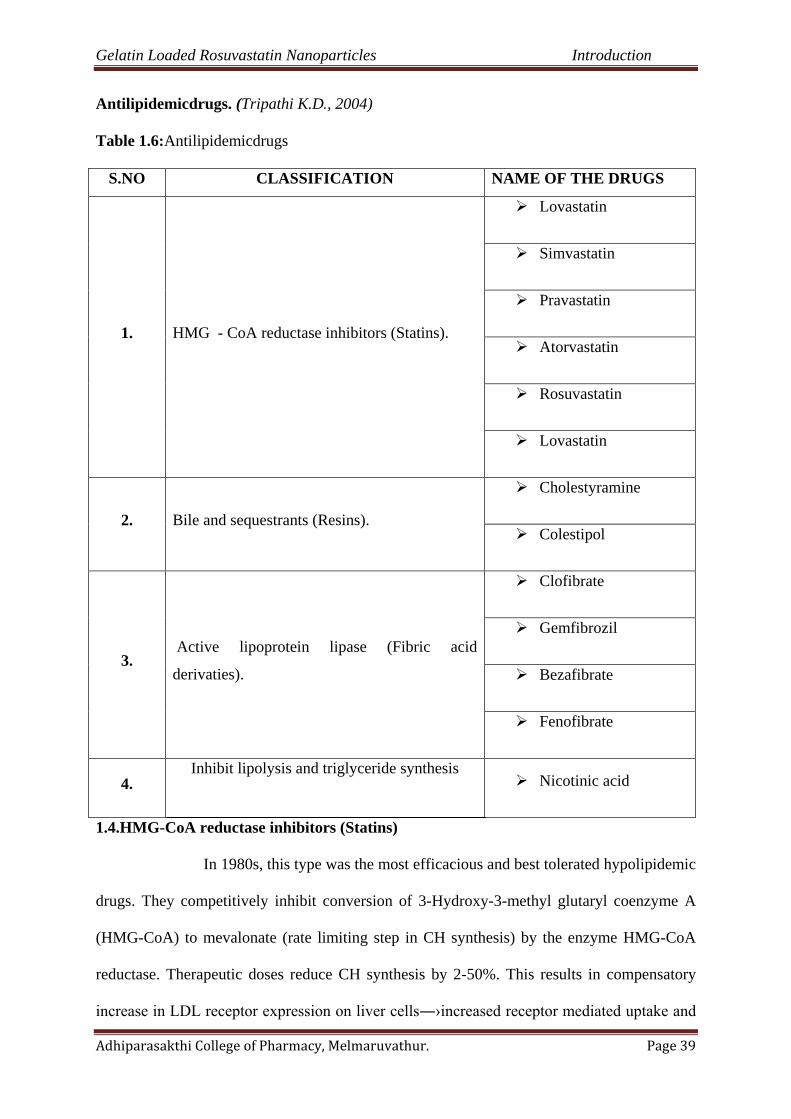
Antilipidemicdrugs. (Tripathi K.D., 2004)
Table 1.6:Antilipidemicdrugs
1.4.HMG-CoA reductase inhibitors (Statins)
In 1980s, this type was the most efficacious and best tolerated hypolipidemic
drugs. They competitively inhibit conversion of 3-Hydroxy-3-methyl glutaryl coenzyme A
(HMG-CoA) to mevalonate (rate limiting step in CH synthesis) by the enzyme HMG-CoA
reductase. Therapeutic doses reduce CH synthesis by 2-50%. This results in compensatory
increase in LDL receptor expression on liver cells―›increased receptor mediated uptake and
S.NO CLASSIFICATION NAME OF THE DRUGS
1. HMG - CoA reductase inhibitors (Statins).
Lovastatin
Simvastatin
Pravastatin
Atorvastatin
Rosuvastatin
Lovastatin
2. Bile and sequestrants (Resins).
Cholestyramine
Colestipol
3. Active lipoprotein lipase (Fibric acid
derivaties).
Clofibrate
Gemfibrozil
Bezafibrate
Fenofibrate
4. Inhibit lipolysis and triglyceride synthesis
Nicotinic acid
Gelatin Loaded Rosuvastatin Nanoparticles Introduction
Adhiparasakthi College of Pharmacy, Melmaruvathur. Page 40
catabolism of IDL and LDL. Over long term, feedback induction of HMG-CoA reductase
tends to increase CH synthesis, but a steady-state is finally attained with a dose-dependent
lowering of LDL-CH levels
The daily dose for lowering LDL-CH by 30-35% is lovastatin 40mg, provastatin 40mg,
simvastatin 20mg, atorvastatin 10mg, rosuvastatin 5mg. Moreover, maximum recommended
doses simvastatin, atorvastatin and rosuvastatin can reduce LDL-CH by upto 45-55%, while
the ceiling effect of lovastatin and pravastatin is 35-40% LDL-CH reduction. All statins
produce peak V lowering after 1-2 weeks therapy.
Gelatin Loaded Rosuvastatin Nanoparticles Introduction
Adhiparasakthi College of Pharmacy, Melmaruvathur. Page 41
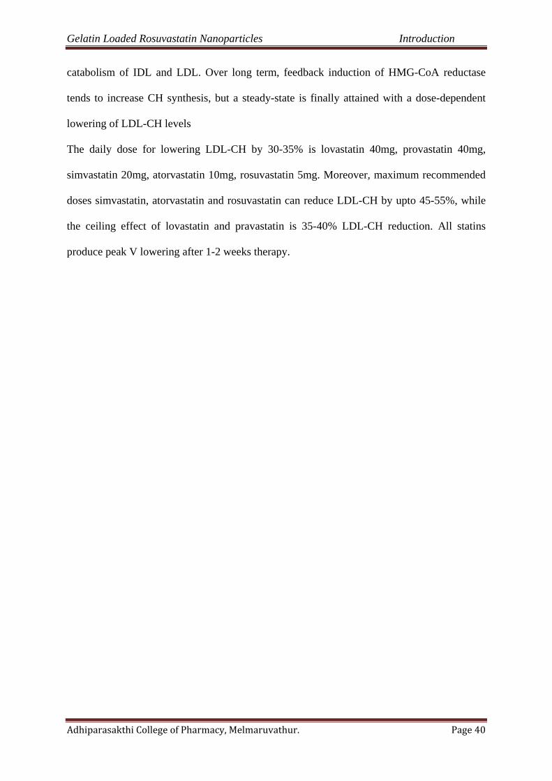
Table 1.7: Mechanism of action and pattern of lipid lowering effect of important hypolipidaemic drug(Michael E. Maragoudakis., 1970)
S.NO CLASSIFICATION NAME OF THE DRUGS
DAILY DOSAGE
MECHANISM OF ACTION EFFECT ON LIPIDS (%)
1.
HMG-CoAreductaseinhibitors (Statins).
Lovastatin (10-80mg)
LDL Decreased 20-55 HDL Increased 5-15 TG Decreased 10-35
Simvastatin (5-40mg)
Pravastatin Decreased CH synthesis by inhibition of rate limiting HMG-CoA reductase
Atorvastatin (10-80mg)
Rosuvastatin (5-20mg)
Lovastatin (10-80mg)
2.
Bile and sequestrants(Resins).
Cholestyramine
(4-16 mg) Decreased bile acid absorption, Increased hepatic conversion of CH to
bile acids Increased LDL receptors on hepatocytes
LDL Decreased 15-30 HDL Increased 3-5 TG not affected, may
increased in some Colestipol (5-30 mg)
3.
Active lipoprotein lipase (Fibric acid derivatives).
Clofibrate
Increased Activity of lipoprotein lipase Decreased release of fatty acids from
adipose tissue
LDL Decreased 5-20 HDL Increased 10-20 TG Increased 20-50
Gemfibrozil (1200 mg)
Bezafibrate (600 mg)
Fenofibrate (200 mg)
Gelatin Loaded Rosuvastatin Nanoparticles Introduction
Adhiparasakthi College of Pharmacy, Melmaruvathur. Page 42
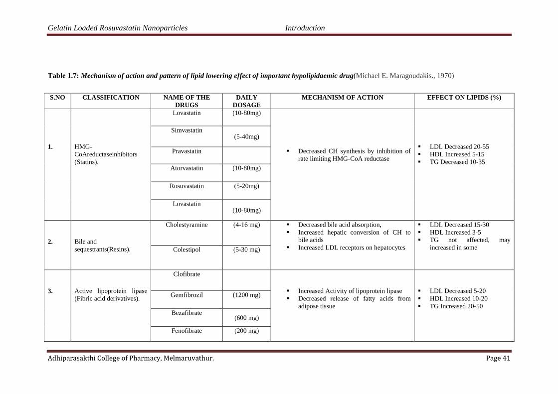
HMG-CoA reductase pathway, which is blocked by statins via inhibiting the rate
limiting enzyme HMG-CoA reductase.
Figure 1.11:HMG-CoA reductase pathway, which is blocked by statins via inhibiting the rate
limiting enzyme HMG-CoA reductase.
The more effacious statins (simvastatin, atorvastatin, rosuvastatin) given at their highe doses
effectively reduce (by 25% to 35%). Because HMG-CoA reductase activity is maximum at
midnight, all statins are administered at bed time to obtain maximum effectiveness. But, this
is not necessary for atorvastatin and rosuvastatin, which have long plasma t1/2
Gelatin Loaded Rosuvastatin Nanoparticles Introduction
Adhiparasakthi College of Pharmacy, Melmaruvathur. Page 43
1.4.1.Other statins drugs. (Richard N. Fogoros, M.D.,2012)
Lovastatin.
It is the first clinically used statin; is lipophilic and given orally in the precursor
lactone form.
Lovastatin 40mg
Systematic (IUPAC) name
1S,3R,7S,8S,8aR)-8-{2-[(2R,4R)-4-hydroxy-6-oxooxan-2-yl]ethyl}-3,7-dimethyl-
1,2,3,7,8,8a-hexahydronaphthalen-1-yl (2S)-2-methylbutanoate
Absorption – incomplete
Metabolism – bile
t1/2- short (1-4 hours)
Dose – 10 to 40 mg
Simvastatin.
It is a hypolipidemic drug used to control elevated cholesterol. Simvastatin is a
member of the statin class of pharmaceuticals, is a synthetic derivate of a fermentation
product of Aspergillusterreus.
Systematic (IUPAC) name
(1S,3R,7S,8S,8aR)-8-{2-[(2R, 4R)-4-hydroxy-6-oxotetrahydro-2H-pyran-2-yl] ethyl}-
3, 7-dimethyl-1, 2,3, 7,8,8a-hexahydronaphthalen-1-yl 2,2-dimethylbutanoate.

Gelatin Loaded Rosuvastatin Nanoparticles Introduction
Adhiparasakthi College of Pharmacy, Melmaruvathur. Page 44
Absorption – incomplete
Metabolism – bile
t1/2- 2-3 hr
Dose – 5 to 20 mg
Pravastatin.
Pravastatin (marketed as Pravachol or Selektine) is a member of the drug class of
statins, used for lowering cholesterol and preventing cardiovascular disease. Initially known
as CS-514, it was originally identified in a bacterium called Nocardiaautotrophica. It is also
hydrophilic and given in the active form.
Pravastatin 10 mg
Systematic (IUPAC) name
(3R, 5R)-3,5-dihydroxy-7-((1R,2S,6S,8R,8aR)-6-hydroxy-2-methyl-8-{[(2S)-2
methylbutanoyl]oxy}-1,2,6,7,8,8a-hexahydronaphthalen-1-yl)-heptanoic acid
Absorption – incomplete
CH lowering effect - less
t1/2- 1-3 hr
Dose – 40 to 80mg/day
Atorvastatin.
This newer statin is more potent and appears to have the highest LDL-CH lowering
efficacy. All statins, including atorvastatin, prevent the production of cholesterol in the liver
by blocking HMG-CoA reductase, an enzyme that makes cholesterol.
Atorvastatin 40 mg
t1/2 - 18 - 24 hr
Dose – 10 to 40 mg

Gelatin Loaded Rosuvastatin Nanoparticles Introduction
Adhiparasakthi College of Pharmacy, Melmaruvathur. Page 45
Rosuvastatin.
This is the latest and the most potent statin (10 mg rosuvastatin ≈ 20 mg atorvastatin),
crestor 10 mg
t1/2 - 18 - 24 hr
LDL-CH reduction – Greater
TG levels – raised
Dose – 5 mg
Equivalentdosages of statin groups.
Table 1.8:Equivalentdosages of statin groups
% LDL
Reduction
(approx.)
Atorvastatin Fluvastatin Lovastatin Pravastatin Rosuvastatin Simvastatin
10-20% -- 20 mg 10 mg 10 mg -- 5 mg
20-30% -- 40 mg 20 mg 20 mg -- 10 mg
30-40% 10 mg 80 mg* 40 mg 40 mg 5 mg 20 mg
40-45% 20 mg -- 80 mg* 80 mg* 5–10 mg 40 mg
46-50% 40 mg -- -- -- 10–20 mg 80 mg*
50-55% 80 mg -- -- -- 20 mg --
56-60% -- -- -- -- 40 mg --
Gelatin Loaded Rosuvastatin Nanoparticles Introduction
Adhiparasakthi College of Pharmacy, Melmaruvathur. Page 46
Adverse effects of statins.(Tripath K.D., 2006)
Headache
Nausea
Bowel upset
Sleep disturbances
Rise in serum transaminase, but liver damage is rare
Muscle tenderness
Myopathy
Uses of statins.(Richard N. Fogoros., 2012)
Statins improve blood cholesterol levels primarily by inhibiting a liver enzyme called
HMG Co-A reductase.
Reducing the size of plaques in the arteries.
Stabilizing plaques, so they are less likely to rupture (and therefore less likely to cause
acute heart attacks).
Reducing inflammation (which is now thought to be an important component of
plaque formation and rupture).
Reducing CRP levels
Decreasing blood clot formation (Blood clot formation at the site of plaque rupture is
the cause of most heart attacks).
Improving overall vascular function.
NEED
AND
OBJECTIVE
Gelatin Loaded Rosuvastatin Nanoparticles Need and Objectives
Adhiparasakthi College of Pharmacy, Melmaruvathur Page 47
2. NEED AND OBJECTIVES
Application of nanotechnology in drug delivery system has opened up new areas of research
in controlled release of drugs. The nanoparticle represents promising drug delivery system of
controlled and targeted drug release which shows and maintains the therapeutic concentration for
long period of time. The reported bio- availability of Rosuvastain is less. Hence the present
study was undertaken to develop the bioavailability of the drug. While forming the nanoparticle
formulation it increased the absorption and bio- availability. The present work of nanoparticles
are focused on Rosuvastatin loaded gelatin nanoparticle by two step desolvation method. Hence
the novel delivery system applied to antilipedimic drugs. Some biodegradable carriers degrade
in the body. This problem is overcome in this nanoparticles carriers. In long term therapy
fluctuations in the plasma concentrations, with high concentration peaks are common for drugs
with rapid absorption and elimination. Such characteristic makes Rosuvastatin is a suitable
candidate for to prepare desired nanoparticulate drug delivery system. Rosuvastatin is the widely
used category of anti lipedimic drug in the treatment of high cholesterol condition.The aim of
present work was to formulate by using polymer like gelatin A and Gelatin B nanoparticles
containing Rosuvastatin in order to provide therapeutic effect.
2.1 Objectives
• Formulation of gelatin loaded Rosuvastatin nanoparticles by two step desolvation
method.
• To characterize the nanoparticles for its physiochemical properties .
• To study the their in-vitro release profile and release kinetics.
• To perform Stability studies as per ICH guidelines.
• To Study the zeta potenial

Plan of work

Gelatin Loaded Rosuvastatin Nanoparticles Plan of Work
Adhiparasakthi College of Pharmacy, Melmaruvathur. Page 48
• LITERATURE REVIEW
• SELECTION OF DRUG, POLYMER
• EXPERIMENTAL WORK
a) PREFORMULATION STUDIES
Identification of drug
By FTIR spectroscopy
By melting point
Physicochemical parameters
Organoleptic properties
Solubility profile
Loss on drying
Analytical methods
Determination of λ max
Development of standard curve of Rosuvastatin
Determination of percentage purity of drug
Determination of compatibility for drug with polymer
By DSC thermal analysis
Formulation of nanoparticles
By two step desolvation method
Determination of drug loading efficiency, drug loading capacity
Characterization of nanoparticles
By scanning electron microscopy
In-vitro dissolution studies
By dialysis bag method
Kinetics of drug release
Stability studies
Zeta potential
3.PLAN OF WORK

Literature review

Gelatin Loaded Rosuvastatin Nanoparticles Literature Review
Adhiparasakthi College of Pharmacy, Melmaruvathur. Page 49
The Literature review Indicating that advancement in nanoparticles drug delivery
system is given below
Adlin jino nesalin J., et al., (2009) were developed and characterized nanoparticles
containing Flutamide .It is used in the treatment of prostate carcinoma having short biological
half life of 5-6 hrs. It is good candidate for the formulation of sustained release dosage form. coat
ratio 1:4 gives better sustained release for about 12 hrs as compared as other formulation.
Vandana singh A., et al., (2010) were developed and characterized Rosiglitazone
loaded gelatin nanoparticles by two step desolvation method. The sustained release of the drug
could maintain the therapeutic concentration for long time. The encapsulation efficiency was
found to be between in the range of 80-90%. The release profile was dependent on korsmeyer-
peppas equation.
Rahul Nair K., et al., (2011) was formulated and evaluated on solid lipid nanoparticles
of water soluble drug Isoniazed. The entrapment efficiency of drug in the SLN has been
improved. The SLNs were prepared by ethanol injection method using tristearin and
phospholipon 80 H using 32 factorial design. The different combination of tween 80
concentrations and varied sonication time were used to prepare SLN. The prepared SLN were
spherical in shape and process mean average size of 164.9 nm. It was for treatment of
tuberculosis.
4. LITERATURE REVIEW

Gelatin Loaded Rosuvastatin Nanoparticles Literature Review
Adhiparasakthi College of Pharmacy, Melmaruvathur. Page 50
Tamizharasi S., et al., (2009) had prepared Lamivudine loaded polymethacrylic acid
nanoparticles. They were specially designed to release the drug in the vignity of target tissue. It
was prepared and evaluated polymethacrylic acid nanoparticles containing lamivudine in
different drug and polymer ratio by nanoprecipitation method. The particle size was found to be
121 to 403 nm.
Shanmughavel R., et al., (2010) In this study preparation and characterization of
biopolymeric gelatin nanoparticles for encapsulating the antimicrobial drug Sulfadiazine and its
vivo drug release in phosphate buffer saline had been investicated. It was prepared by two step
desolvation method. It had been proved to be effective treatment of meningococcal meningitis.
The release was occurred upto 30% in a controlled manner.
DongGon Kim., et al., (2006) had prepared the Retinol encapsulated chitosan
nanoparticles, reconstitute in aqueous solution and application of cosmetic and pharmaceutical
applications. These nanoparticles have a spherical shape and the obtained particle size is the 50
to 200nm. Solubility of Retinol is able to increase by encapsulation into chitosan nanoparticles
more than 1600-fold. X-ray diffractions patterns also showed that crystal peaks of retinol were
disappeared by encapsulation into chitosan nanoparticles.
Joseph Nisha Mary S., et al., (2006) investigated a nanoparticles for the cancer
treatment. Nanoparticles were prepared by the crosslinking method with different concentration
of polymer, surfactant. So these nanoparticles showed a sustain release kinetics and good
encapsulating properties.

Gelatin Loaded Rosuvastatin Nanoparticles Literature Review
Adhiparasakthi College of Pharmacy, Melmaruvathur. Page 51
Kalaria D., et al., (2008) were prepared Adoxorubicin loaded nanoparticles by emulsion
method and stability studies was done in simulated fluids. The pharmacokinetic studies and
toxicity studies were conducted in rats. So the nanoparticles showed good stability in simulated
intestinal fluid and shows a sustain release, enhanced bioavailability and lower toxicity.
Kowsalya R., et al., (2005) was prepared and physicochemical evaluation of Cephalexin
loaded polymethacrylic acid nanoparticles. The association of Cephalexin with nanoparticles was
performed by emulsion polymerization in continuous aqueous phase. SEM indicated that
Cephalexin nanoparticle have a discrete spherical structure without aggregation. The particle
size, drug content and drug recovery of the nanoparticles was gradually increased with increase
in the proportion of polymethacrylic acid polymer. The in-vitro release studies indicated that
Cephalexin nanoparticles provide sustained release over a period of 21hours and follow zero
order kinetics.
Lieven Baert., et al., (2009) developed a long acting formulation containing Rilivirine
nanoparticles for HIV treatment. The Nanosuspensions were prepared by wet milling in aqueous
carrier. The obtained particle size is 200-800nm and stable for six months, stability up to six
months. Following single dose administration 3 months in dogs and 3 weeks in mice shows the
sustain release
Muhammad Rafeeq P., et al., (2010) the objective of our study is to load first line anti
tubercular drug, Isoniazid in chitosan nanoparticles in order to enhance bioavailability and to
reduce dose frequency. Chitosan was dissolved in acetic acid aqueous solution at various
concentrations, drug was dispersed in above chitosan solution kept over magnetic stirrer at room
temperature for a period of 30 minute. The tripolyphosphate aqueous solution with various
concentrations added drop wise to the above sonication and centrifugation with ultracentrifuge.
Gelatin Loaded Rosuvastatin Nanoparticles Literature Review
Adhiparasakthi College of Pharmacy, Melmaruvathur. Page 52
After freeze drying the nanoparticles were collected. SEM studies show that formulation no 2
having optimum nanosized particles. Zeta potential shows good positive potentials. It shows
good encapsulation efficiency. And good release profile follows first order release kinetics
Qiang zhang., et al., (1999) stearic acid nanoparticles were prepared in this study by
melt-homogenization to investigate the possibility of them as a new kind of drug carrier system.
Cyclosporine a as a model drug was then encapsulated into stearic acid nanoparticles. Following
the establishment of assay for cyclosporine a analysis in stearic acid nanoparticles or blood
samples, the encapsulation ratio of cyclosporine a to stearic acid nanoparticles was estimated and
pharmacokinetics as well as bioavailability of cyclosporine a stearic acid nanoparticles after oral
administration to wistar rats were studied. The mean diameter of cyclosporine a stearic acid
nanoparticles was 316.1 nm, while the encapsulation ratio of cyclosporine a to stearic acid
nanoparticles reached to 88.36%. The relative bioavailability of cyclosporine a stearic acid
nanoparticle over reference was nearly 80%, and the time to reach maximum concentration of
Cyclosporine.
YoungWook., et al., (2008) was developed the natural gelatin based nanoparticles by
method of modified desolvation by natural cross linker for protein drug delivery. The obtained
recombinant human gelatin nanoparticles deliver a modified protein in sustained release manner
without initial burst.
Yungchih kuo., et al., (2005) was performing the loading efficiency of Rosuvastatin, a
human immunodeficiency antiretroviral agent, on the external surfaces of
polybutylcyanoacrylate and methylmethacrylatesulphopropylmethacrylate, were prepared. The
experimental results indicate that the larger the polymeric nanoparticles, the smaller loading
efficiency of Rosuvastatin on the two kinds of biomaterials. Freeze-drying of the two

Gelatin Loaded Rosuvastatin Nanoparticles Literature Review
Adhiparasakthi College of Pharmacy, Melmaruvathur. Page 53
nanoparticles, however, yields an increase in particle size and an increase in loading efficiency of
Rosuvastatin, in general. Stability studies at 4◦C over 6 weeks leads to an increase in particle size
and a decrease in loading efficiency of Rosuvastatin. Loading efficiency of Rosuvastatin on both
of the two nanoparticles decreases with a variation in pH value.
Yvette Konan N., et al., (2002) had studied on preparation and characterization of sterile
and freeze dried nanoparticles. By using salting out procedure to produced the sub-200nm
nanoparticles. After freeze drying complete dispersion of all the types of polyester nanoparticles
in presence of lyoprotectants such as saccharides and the sterility testing showed that no
microbial contamination.

Drug
AnD
Polymer Profile

Gelatin loaded Rosuvastatin Nanoparticles Drug and Polymer profile
Adhiparasakthi College of Pharmacy, Melmaruvathur. Page 54
5. DRUG AND POLYMER PROFILE
5.1: Drug profile (McTaggart F., et al., 2001)
CRESTOR (Rosuvastatin calcium) is a synthetic lipid lowering agent for oral administration.
Rosuvastatin is an antilipidemic agent that competitively inhibits hydroxymethylglutaryl-
coenzyme A (HMG-CoA) reductase. HMG-CoA reducuase catalyzes the conversion of HMG-
CoA to mevalonic acid, the rate-limiting step in cholesterol biosynthesis. Rosuvastatin belongs to
a class of medications called statins and is used to reduce plasma cholesterol levels and prevent
cardiovascular disease.
Brand name
Astende (Lazar (Argentina))
Cirantan (AstraZeneca (Netherlands))
Cresadex (Drugtech (Chile))
Crestor (AstraZeneca)
Provisacor (AstraZeneca (Italy, Netherlands) )
Razel (Glenmark (India))
Rosedex (Roux-Ocefa (Argentina))
Rosimol (Sandoz (Argentina))
Rosumed (Labomed (Chile))
Rosustatin (Montpellier (Argentina))
Rosuvas (Ranbaxy (India))
Rosuvast (Bago (Argentina))
Rosvel (Laboratorios Chile (Chile))

Gelatin loaded Rosuvastatin Nanoparticles Drug and Polymer profile
Adhiparasakthi College of Pharmacy, Melmaruvathur. Page 55
Chemical data
Molecular formula : (C22H27FN3O6S)2Ca
Molecular weight : 1001.1 g/mol
Solubility :sparingly soluble in water and methanol and slightly soluble in
ethanol.
Melting point : 155.4°C to 155°C
Colour : Off-white to creamish white crystalline powder.
Plasma concentration : reached in 3–5 hour
Molecular structure:
Category:(McTaggart F., et al., 2001)
Anticholesteremic Agents
HMG-CoA Reductase Inhibitors
Gelatin loaded Rosuvastatin Nanoparticles Drug and Polymer profile
Adhiparasakthi College of Pharmacy, Melmaruvathur. Page 56
Pharmacokinetics data
Bioavailability- 20%
Metabolism- Liver
Half-life- 19 h
Excretion- Urine / Faeces
Volume - 134L
Protein binding- 90%
a) Absorption:
Bioavailability is approximately 20%
b) Distribution:
Rosuvastatin is 88% bound to plasma proteins, mostly albumin. This binding is reversible
and independent of plasma concentrations.
c)Volume of distribution:
134 L/kg
d) Metabolism:
Rosuvastatin is not extensively metabolized; approximately 10% of a radio labeled dose is
recovered as metabolite. The metabolite is N- desmethylRosuvastatin, which is formed
principally by cytochrome P450 2C9, and in vitro studies have demonstrated that N-
desmethylRosuvastatin has approximately one-sixth to one-half the HMG-CoA reductase
inhibitory activity of the parent compound. Overall, greater than 90% of active plasma HMG-
CoA reductase inhibitory activity is accounted for by the parent compound.

Gelatin loaded Rosuvastatin Nanoparticles Drug and Polymer profile
Adhiparasakthi College of Pharmacy, Melmaruvathur. Page 57
e) Excretion:
Following oral administration, Rosuvastatin and its metabolites are primarily excreted in the
feces (90%). After an intravenous dose, approximately 28% of total body clearance was via the
renal route, and 72% by the hepatic route.
5.1.1:Mechanism of action(Tripathy K.D., 2008)
Rosuvastatin is a selective and competitive inhibitor of HMG-CoA reductase, the rate-
limiting enzyme that converts 3-hydroxy-3-methylglutaryl coenzyme A to mevalonate, a
precursor of cholesterol. First, it increases the number of hepatic LDL receptors on the cell
surface to enhance uptake and catabolism of LDL. Second, Rosuvastatin inhibits hepatic
synthesis of VLDL, which reduces the total number of VLDL and LDL particles.
Rosuvastatin pathway
Rosuvastatin inhibits cholesterol synthesis via the mevalonate pathway by inhibiting 3-
hydroxy-3-methylglutaryl-coenzyme A (HMG-CoA) reductase. HMG-CoA reductase is the
enzyme responsible for the conversion of HMG-CoA to mevalonic acid, the rate-limiting step of
cholesterol synthesis by this pathway. The active form of statins bears a chemical resemblance to
the reduced HMG-CoA reaction intermediate that is formed during catalysis. Unlike Lovastatin
and simvastatin, which undergo in vivo hydrolysis to their active form Rosuvastatin is
synthetically produced in active form. Cholesterol biosynthesis accounts for approximately 80%
of cholesterol in the body; thus, inhibiting this process can significantly lower cholesterol levels.
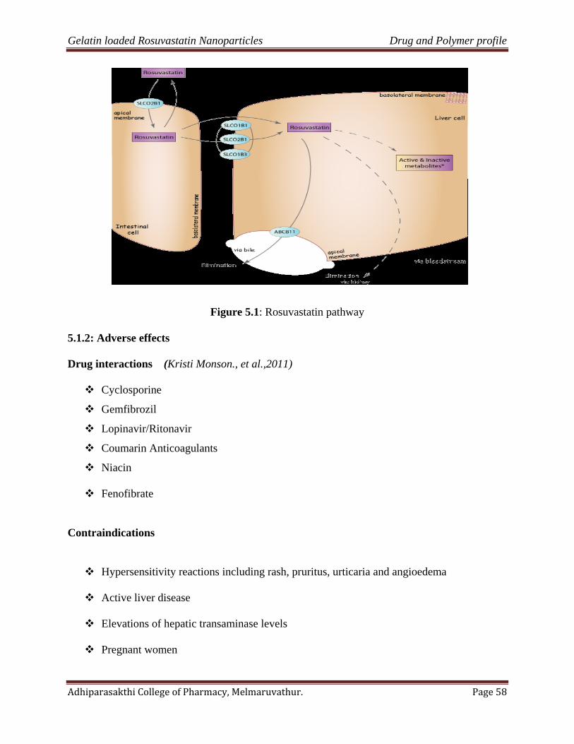
Gelatin loaded Rosuvastatin Nanoparticles Drug and Polymer profile
Adhiparasakthi College of Pharmacy, Melmaruvathur. Page 58
Figure 5.1: Rosuvastatin pathway
5.1.2: Adverse effects
Drug interactions (Kristi Monson., et al.,2011)
Cyclosporine
Gemfibrozil
Lopinavir/Ritonavir
Coumarin Anticoagulants
Niacin
Fenofibrate
Contraindications
Hypersensitivity reactions including rash, pruritus, urticaria and angioedema
Active liver disease
Elevations of hepatic transaminase levels
Pregnant women
Gelatin loaded Rosuvastatin Nanoparticles Drug and Polymer profile
Adhiparasakthi College of Pharmacy, Melmaruvathur. Page 59
Dosage forms
5 mg: Yellow, round, biconvex, coated tablets.
10 mg: Pink, round, biconvex, coated tablets.
20 mg: Pink, round, biconvex, coated tablets.
40 mg: Pink, oval, biconvex, coated tablets.
5.2Polymer profile(Wayne Goates Kansas., et al., 2003)
Gelatin is a natural, biodegradable protein obtained by acid- or base-catalyzed Hydrolysis
of collagen. It is a heterogeneous mixture of single- or multi-stranded Polypeptides composed
predominantly of glycine, proline, and hydroxyproline Residues and is degraded in vivo to
amino acids. Gelatin nanoparticles are prepared by a two-step, de solvation process. The
concentrated gelatin liquid particles are isolated and hardened by chemical cross-linking with
glutaraldehyde. Alternately, these particles can be prepared using a simple o/w emulsion or
w/o/w micro emulsion method. Gelatin nanoparticles have been used to deliver paclitaxel,
methotrexate, doxorubicin, DNA, double-stranded oligonucleotides, and genes. Pegylation of the
particles significantly enhances their circulation time in the blood stream and increases their
uptake into cells by endocytosis. Antibody-modified gelatin nanoparticles have been used for
targeted uptake by lymphocytes.
Description:
Gelatin occurs as a light-amber to faintly yellow-colored, vitreous, brittle solid. It is
practically odorless and tasteless and is available as translucent sheets add granules.
Gelatin contasins:

Gelatin loaded Rosuvastatin Nanoparticles Drug and Polymer profile
Adhiparasakthi College of Pharmacy, Melmaruvathur. Page 60
• 84-90% protein
• 1-2% mineral salts
• 8-15% water
• It is free from additives and preservatives
Types of gelatin:(GMAP Publishers, 2005)
Two main types of gelatin.
Type A, is derived from collagen with exclusively acid pretreatment.
Type B, is the result of an alkaline pretreatment of the collagen.
Typical specifications for Type Aand Type B are:
Table 5.1: Typical specifications for Type Aand Type B
Parameter Type A Type B
pH 3.8 - 5.5 5.0 - 7.5
Isoelectric Point 7.0 - 9.0 4.7 - 6.0
Gel strength (bloom 50 – 300 50 - 300
Viscosity (mps) 15 – 75 20 - 75
Chemical formula :C102H151039N31
Gelatin loaded Rosuvastatin Nanoparticles Drug and Polymer profile
Adhiparasakthi College of Pharmacy, Melmaruvathur. Page 61
5.2.1:Bio physicochemical properties of gelatin
Color: color varying from a very pale yellow to dark amber
Physical appearance: Occurs as flakes.
Viscosity:
4.3-4.7 mPa s (4.3-4.7 cP) for a 6.67% w/v aqueous solution at 60.c
18.5-20.5 mPa s (18.5-20.5 cP) for a 12.5% w/v aqueous solution at 60.c
Molecular weight: 15000 – 250 000
5.2.2:The most favorable operating conditions for the extraction of gelatin are: -
Table 5.2: The extraction of gelatin
STAGES TEMPERATURE TIME PH
First stage 64 oC 5 hr 7
Second stage 74 oC 3.72 hr 7
Third stage 84 oC 3.69 hr 7
Fourth stage 98 oC , 3.83 hr 7
5.2.3: Chemical properties of gelatin:
Together with water, it forms a semi-solid colloidgel.
Gelatin forms a solution of high viscosity in water,
Gelatin solutions show viscoelastic flow and streaming birefringence.
Gelatin is put into contact with cold water, some of the material dissolves.
solubility of the gelatin is determined by the method of manufacture.
Gelatin is also soluble in most polar solvents.
Gelatins are very sensitive to temperature variations.

Gelatin loaded Rosuvastatin Nanoparticles Drug and Polymer profile
Adhiparasakthi College of Pharmacy, Melmaruvathur. Page 62
The viscosity of the gelatin/water mixture increases with concentration and when kept
cool (≈ 4 °C).
There are also vegetable gelatins such as agar.
Safety:
In general, when used in oral formulations gelatin may be regarded as a nontoxic &
nonirritant material. Gelatin capsules adhering to the esophageal lining which may cause local
irritation.
Handling precautions:
Eye protection and gloves are recommended. Gelatin should be handled in a well-
ventilated environment
Uses of gelatin:(Wayne GoatesKansas., et al., 2003)
binders for paper money
cosmetics
bonding for the tip of matches
bakery products
photographic film
whipping agent in dairy products
medicine emulsions
hardening of jams and jellies
treatment of wounds as a sponge
Gelatin loaded Rosuvastatin Nanoparticles Drug and Polymer profile
Adhiparasakthi College of Pharmacy, Melmaruvathur. Page 63
5.2.4:Pharmaceuticalaspects of gelatin: (GMAP Publishers, 2005)
P Gelatin is the most pure and perfect protein
available
H It is absolutely harmless and controlled by high quality standards for such
applications
A Active and readily and rapidly accepted by the
body
R Reliable and safe ingredient in sensitive products
M Significant in medicinal and medical applications
A Contains practically all essential amino acids
required for body cells and tissue nourishment
C Sourced from natural collagens
E Essential to all humans
U A unique and useful product used through the
centuries
T Significant in thermoreversible and tabletting applications
I Truly an international product
C A clear and clean product
A Easily available from several producers who are
members of gmap
L Used in several life supporting applications, such as
plasma extenders.

Gelatin loaded Rosuvastatin Nanoparticles Drug and Polymer profile
Adhiparasakthi College of Pharmacy, Melmaruvathur. Page 64
Pharmaceutical applications: (Sylvie Audoly., et al., 2006)
Hard capsules
Hard capsules are composed mainly of gelatin, and eventually of an opacifier and
coloring agents. They are obtained by dipping metallic formers called pins, into the gelatin
solution, removing and allowing the solution to gel in the air and dry. Pharmaceutical customer
will fill the hard capsules mainlywith powders or solid forms (tablets, …), but with the
improvement of the technology including the sealing of the capsules to avoid leakage, hard
capsules can also be filled with liquids or pastes when it was dedicated to soft capsules few
years ago. The resistance, the elasticity and the solubility of the shell will depend on physical
characteristics of the gelatin.
Soft capsules
Softgels are a one-piece, hermetically sealed capsule with an outer shell made of
gelatin, water, plasticizers (glycerol, sorbitol,…) which gives elasticity and softness to the walls
and sometimes coloring agents plus opacifier. Medium Bloom are preferred for elasticity and
easiness to work on the machine. In this technology, the encapsulation of the drugs (oily
products, non aqueous liquids, suspension and astes…) is made simultaneously with the
formation of the walls. The main process used is the rotary die process.
Tablets
Gelatin is used for its binding properties. Used in direct compression or after granulation
of the powder to be used in compression, gelatin or hydrolyzed collagen allow to give
cohesiveness,resistance, hardness to the tablet.

Gelatin loaded Rosuvastatin Nanoparticles Drug and Polymer profile
Adhiparasakthi College of Pharmacy, Melmaruvathur. Page 65
Bloodplasma
Blood plasma substitute replaces blood when large volumes are lost during anaccident for
example and without knowing the blood grouping. Most of the time in Europe, blood plasma
substitute consists of pyrogene free water, gelatin and salts (chlorides), while in the USA starch
preparation are mainly used.Blood plasma substitute reestablishes the blood pressure with the
presence of relatively low molecular weight molecules. One of the advantages of plasma
substitute based on gelatin is the absence of accumulation effect into the body (as it could
happen with starch) due to a reasonably rapid elimination by the metabolism or excretion.
Cholostomy bags
Gelatin is used in association with other polymers as CMCor pectines and anadhesive to
obtain a sticking ring allowingto fix cholostomy bags and to absorb themoisture ofthe skin.
Sticking, coating and water binding properties ofgelatin are especially interesting in that case.
Materials
and
equipMent
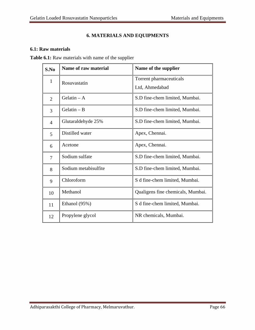
Gelatin Loaded Rosuvastatin Nanoparticles Materials and Equipments
Adhiparasakthi College of Pharmacy, Melmaruvathur. Page 66
6. MATERIALS AND EQUIPMENTS
6.1: Raw materials
Table 6.1: Raw materials with name of the supplier
S.No Name of raw material Name of the supplier
1 Rosuvastatin Torrent pharmaceuticals
Ltd, Ahmedabad
2 Gelatin – A S.D fine-chem limited, Mumbai.
3 Gelatin – B S.D fine-chem limited, Mumbai.
4 Glutaraldehyde 25% S.D fine-chem limited, Mumbai.
5 Distilled water Apex, Chennai.
6 Acetone Apex, Chennai.
7 Sodium sulfate S.D fine-chem limited, Mumbai.
8 Sodium metabisulfite S.D fine-chem limited, Mumbai.
9 Chloroform S d fine-chem limited, Mumbai.
10 Methanol Qualigens fine chemicals, Mumbai.
11 Ethanol (95%) S d fine-chem limited, Mumbai.
12 Propylene glycol NR chemicals, Mumbai.
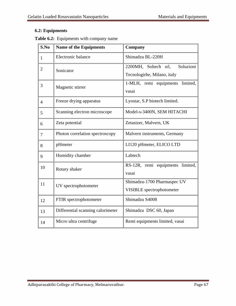
Gelatin Loaded Rosuvastatin Nanoparticles Materials and Equipments
Adhiparasakthi College of Pharmacy, Melmaruvathur. Page 67
6.2: Equipments
Table 6.2: Equipments with company name
S.No Name of the Equipments Company
1 Electronic balance Shimadzu BL-220H
2 Sonicator 2200MH, Soltech srl, Soluzioni
Tecnologirhe, Milano, italy
3 Magnetic stirrer 1-MLH, remi equipments limited,
vasai
4 Freeze drying apparatus Lyostar, S.P biotech limited.
5 Scanning electron microscope Model-s-3400N, SEM HITACHI
6 Zeta potential Zetasizer, Malvern, UK
7 Photon correlation spectroscopy Malvern instruments, Germany
8 pHmeter LI120 pHmeter, ELICO LTD
9 Humidity chamber Labtech
10 Rotary shaker RS-12R, remi equipments limited,
vasai
11 UV spectrophotometer Shimadzu-1700 Pharmaspec UV
VISIBLE spectrophotometer
12 FTIR spectrophotometer Shimadzu S4008
13 Differential scanning calorimeter Shimadzu DSC 60, Japan
14 Micro ultra centrifuge Remi equipments limited, vasai
Gelatin Loaded Rosuvastatin Nanoparticle Experimental work
Adhiparasakthi College of Pharmacy, Melmaruvathur. Page 68
7.1. Preformulation studies
Preformulation testing was an investigation of physical and chemical properties of a drug
substance alone. It was the first step in rational development of dosage form.
7.1.1. Identification of drug
7.1.1.1. Identification by FTIR spectroscopy(SkoogD.A.,et al.,1996;IP, 2007)
Rosuvastatin discs were prepared by pressing the Rosuvastatin with potassium bromide
and the spectra between 4000-1 to 500-1 cm was obtained under the operational conditions. The
absorption maximums in spectrum obtained with the substance being examined correspond in
position and relative intensity to those in the reference spectrum in Table 8.1 and Figure 8.1
7.1.1.2. Identification by melting point (IP, 2007)
Melting point of the drug was determined by capillary tube method.
7.1.2. Physicochemical parameters
7.1.2.1. Organoleptic properties(Lachman L., et al., 1991; Bankar G.S. and Rhodes C.T., 1996)
The color, odor and taste of the drug were recorded using descriptive terminology.
7.1.2.2. Solubility study (Merck Index, IP, 2007)
It is important to know about solubility characteristic of a drug in aqueous system, since
they must possess some limited aqueous solubility to elicit a therapeutic response. The solubility
of drug was recorded by using various descriptive terminology specified in Indian
pharmacopoeia, 2007.The solubility study was shown in Table 8.2.
7. EXPERIMENTAL WORK
Gelatin Loaded Rosuvastatin Nanoparticle Experimental work
Adhiparasakthi College of Pharmacy, Melmaruvathur. Page 69
7.1.2.3. Loss on drying(IP, 2007)
Loss on drying is the loss of weight expressed as percentage w/w resulting from water and
volatile matter of any kind that can be driven off under specified condition. The accurately
weighed 1gm of sample was transferred instoppered-glass shallow weighing bottle and
accurately weighed the bottle. The bottle was transferred in oven and substance was dried at
105°C for 3 hours. The bottle was removed from oven and reweighed; loss on drying was
calculated by following equation,.It was shown in Table 8.3
𝐋𝐋𝐋𝐋𝐋𝐋 = 𝐈𝐈𝐈𝐈𝐈𝐈𝐈𝐈𝐈𝐈𝐈𝐈𝐈𝐈𝐈𝐈𝐈𝐈𝐈𝐈𝐈𝐈𝐈𝐈𝐈𝐈𝐈𝐈𝐈𝐈𝐈𝐈𝐈𝐈𝐈𝐈𝐈𝐈𝐈𝐈𝐈𝐈𝐈𝐈𝐈𝐈𝐈𝐈 − 𝐅𝐅𝐈𝐈𝐈𝐈𝐈𝐈𝐈𝐈𝐈𝐈𝐈𝐈𝐈𝐈𝐈𝐈𝐈𝐈𝐈𝐈𝐈𝐈𝐈𝐈𝐈𝐈𝐈𝐈𝐈𝐈𝐈𝐈𝐈𝐈𝐈𝐈𝐈𝐈𝐈𝐈𝐈𝐈
𝐈𝐈𝐈𝐈𝐈𝐈𝐈𝐈𝐈𝐈𝐈𝐈𝐈𝐈𝐈𝐈𝐈𝐈𝐈𝐈𝐈𝐈𝐈𝐈𝐈𝐈𝐈𝐈𝐈𝐈𝐈𝐈𝐈𝐈𝐈𝐈𝐈𝐈𝐈𝐈𝐈𝐈𝐈𝐈𝐈𝐈𝐈𝐈× 𝟏𝟏𝟏𝟏𝟏𝟏
7.1.3. Analytical methods
7.1.3.1. Determination of λ max (Alka Gupta., et al., 2005)
The absorption maximum of the standard solution was scanned between 200-400 nm regions on
UV-VISIBLE spectrophotometer. The absorption maximum obtained with the substance being
examined corresponds in position and relative intensity to those in the reference spectrum
Development of standard curve of Rosuvastatin in methanol: (Alka Gupta., et al., 2005)
25 mg of Rosuvastatin Calcium was weighed accurately and transferred into 50 ml
volumetric flask and dissolved in Methanol, after dissolution the volume was made up to the
mark with Methanol (500µg/ ml). Further dilution was made by pipetting 1 ml of mother liquor
into 50 ml s to acquire 10 µg/ ml solution made up with methanol. The absorbance
measurements of these solutions were carried out against methanol as blank at 244 nm. A
calibration curve of Rosuvastatin was plotted.
Gelatin Loaded Rosuvastatin Nanoparticle Experimental work
Adhiparasakthi College of Pharmacy, Melmaruvathur. Page 70
Preparation of methanol solution(IP, 2007)
Methanol was prepared according to I.P. 2007.
Preparation of stock solution of Rosuvastatin in methanol:
Accurately weighed 100 mg of Rosuvastatin was dissolved in little quantity of 100ml of
methanol and 1ml of solution was taken & make up to 10ml (100µg/ml) volume was the same to
prepared standard solution having concentration of 100 µg/ ml.
Procedure:
From the stock solution, aliquots of 1, 2, 3, 4, 5,6 and 7 ml were transferred to 10 ml
volumetric flasks and final volume was made to 100ml with methanol. Absorbance values of
these solutions were measured against blank (methanol) at 244 nm using uv-visible
spectrophotometer
Development of standard curve of Rosuvastatin in 6.8 phosphate buffer:(IP, 2007)
Preparation of 6.8 phosphate buffer:
Place the 50 ml of 0.2M potassium dihydrogen phosphate in a 200 ml of volumetric
flask.And add specified volume of 0.2 M sodium hydroxide then add water to volume to
required.
Preparation of 0.2 M sodium hydroxide:
Dissolve the sodium hydroxide in water to produce 40 to 60 %w/v solution and allow to
stand and finally add 8 gm of sodium hydroxide in 1000 ml.
Preparation of 0.2 M potassium dihydrogen phosphate:
Dissolve the 27.218 gm of potassium dihydrogen phosphate in water to dilute with water
1000ml.

Gelatin Loaded Rosuvastatin Nanoparticle Experimental work
Adhiparasakthi College of Pharmacy, Melmaruvathur. Page 71
Preparation of stock solution of Rosuvastatin in 6.8 phosphate buffer:
Accurately weighed 100 mg of Rosuvastatin was dissolved in little quantity of 100ml of
6.8 phosphate buffer and 1ml of solution was taken & make up to 10ml (100µg/ml) volume was
the same to prepared standard solution having concentration of 100 µg/ ml.
Procedure:
From the stock solution, aliquots of 1, 2, 3, 4, 5,6 and 7 ml were transferred to 10 ml
volumetric flasks and final volume was made to 100ml with methanol. Absorbance values of
these solutions were measured against blank (6.8 phosphate buffer) at 240 nm using uv-visible
spectrophotometer
Determination of percentage purity of drug (Dhirendra K., et al., 2010; IP, 2007)
Standard solution Preparation:
Standard solution of Rosuvastatin calcium was prepared by dissolving 10mg of
Rosuvastatin calcium in 100ml of methanol. To obtained the concentration 100µg/ml. Further
diluted 5-50ml by same solvent to obtained a solution containing 10µg/ml.
Sample solution preparation:
Pure raw materials of Rosuvastatin calcium were weighed and powdered. Amount of
powdered equivalent to 20 mg taken into a 200ml volumetric flask, 140 ml methanol added to
dissolve the drug, cooled the flask up to the room temperature and methanol was added to make
up the volume up to the mark,Centrifuged at 4000 rpm for 10mins, further diluted 5ml to 50ml
with same solvent to obtain a solution containing 10 μg/ml. Same procedure was used for two
other market samples of Rosuvastatin calcium.
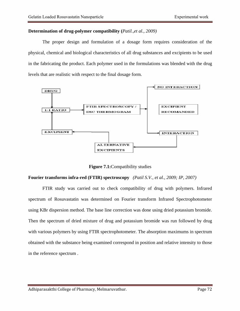
Gelatin Loaded Rosuvastatin Nanoparticle Experimental work
Adhiparasakthi College of Pharmacy, Melmaruvathur. Page 72
Determination of drug-polymer compatibility (Patil.,et al., 2009)
The proper design and formulation of a dosage form requires consideration of the
physical, chemical and biological characteristics of all drug substances and excipients to be used
in the fabricating the product. Each polymer used in the formulations was blended with the drug
levels that are realistic with respect to the final dosage form.
Figure 7.1:Compatibility studies
Fourier transforms infra-red (FTIR) spectroscopy (Patil S.V., et al., 2009; IP, 2007)
FTIR study was carried out to check compatibility of drug with polymers. Infrared
spectrum of Rosuvastatin was determined on Fourier transform Infrared Spectrophotometer
using KBr dispersion method. The base line correction was done using dried potassium bromide.
Then the spectrum of dried mixture of drug and potassium bromide was run followed by drug
with various polymers by using FTIR spectrophotometer. The absorption maximums in spectrum
obtained with the substance being examined correspond in position and relative intensity to those
in the reference spectrum .
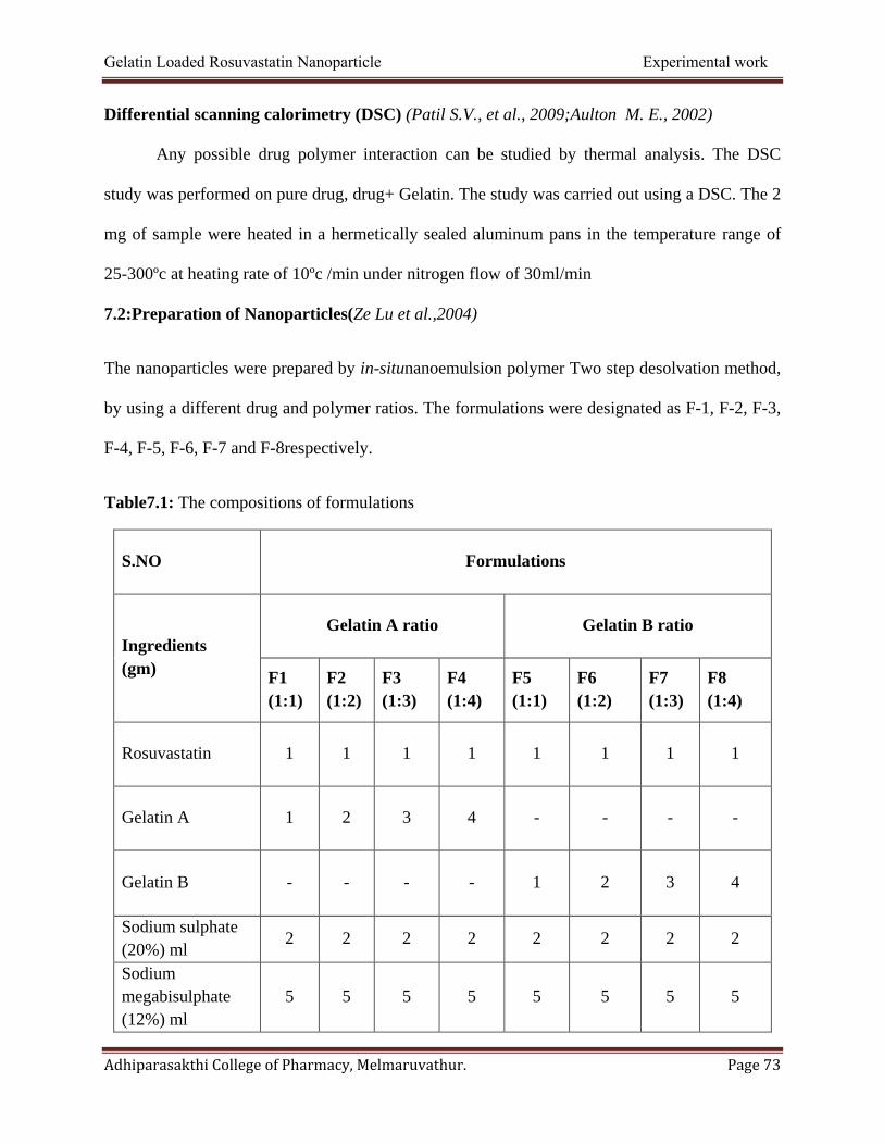
Gelatin Loaded Rosuvastatin Nanoparticle Experimental work
Adhiparasakthi College of Pharmacy, Melmaruvathur. Page 73
Differential scanning calorimetry (DSC) (Patil S.V., et al., 2009;Aulton M. E., 2002)
Any possible drug polymer interaction can be studied by thermal analysis. The DSC
study was performed on pure drug, drug+ Gelatin. The study was carried out using a DSC. The 2
mg of sample were heated in a hermetically sealed aluminum pans in the temperature range of
25-300ºc at heating rate of 10ºc /min under nitrogen flow of 30ml/min
7.2:Preparation of Nanoparticles(Ze Lu et al.,2004)
The nanoparticles were prepared by in-situnanoemulsion polymer Two step desolvation method,
by using a different drug and polymer ratios. The formulations were designated as F-1, F-2, F-3,
F-4, F-5, F-6, F-7 and F-8respectively.
Table7.1: The compositions of formulations
S.NO Formulations
Ingredients (gm)
Gelatin A ratio Gelatin B ratio
F1 (1:1)
F2 (1:2)
F3 (1:3)
F4 (1:4)
F5 (1:1)
F6 (1:2)
F7 (1:3)
F8 (1:4)
Rosuvastatin 1 1 1 1 1 1 1 1
Gelatin A 1 2 3 4 - - - -
Gelatin B - - - - 1 2 3 4
Sodium sulphate (20%) ml
2 2 2 2 2 2 2 2
Sodium megabisulphate (12%) ml
5 5 5 5 5 5 5 5

Gelatin Loaded Rosuvastatin Nanoparticle Experimental work
Adhiparasakthi College of Pharmacy, Melmaruvathur. Page 74
Two step desolvationmethod:(Ze Lu et al.,2004)
Gelatin (200 mg) was dissolved in 10 ml of water containing of 2% Tween 20. The
solution was heated at 40°C with constant stirring at 300 rpm. To this solution, 2 ml of a 20%
aqueous solution of sodium sulfate was added slowly, followed by 1 of isopropanolcontaining2
mg of Rosuvastatin. A remaining of sodium sulfate solution (5.5–6 ml) was added
untilthesolution turned turbid, (which indicated the formation of gelatin aggregates.)
Approximately 1 ml of distilled water was then added until the solution turned clear. An aqueous
solution of glutaraldehyde[25%, (0.4 ml)] was added to cross-link with the gelatin. Sodium
metabisulfitesolution [12%, (5 ml)] was addedafter 5 minutes to stop the cross-linking process.
After 1 hour, the crude product was purified on a Sephadex G-50 column. The nanoparticle
containing fraction was lyophilized in a freeze drier over a 48 hour period.
7.3. Characterization of Nanoparticles
7.3.1. Characterization of Rosuvastatinloaded gelatin Nanoparticles
Encapsulation efficiency(vandanasingh ., et al., 2010)
The determination of drug entrapment, the amount of drug present in the clear
supernatant after centrifugation was determined(w) by spectrophotometry at 240 nm. A standard
calibration curve of drug was plotted. The amount of drug in the supernatant was then subtracted
from the total amount of drug added during the preparation (W). Effectively (W-w) will give the
amount of drug entrapped in the pellet.

Gelatin Loaded Rosuvastatin Nanoparticle Experimental work
Adhiparasakthi College of Pharmacy, Melmaruvathur. Page 75
Amount of drug bound by total amount of nanoparticles Drug encapsulation efficiency = x100
Amount of drug taken.
Particle size distribution (MaincentPandKimy., 1997)
Every sample was diluted with phosphate buffered saline pH 6.8.The surface charge (Zeta
potential) was determined by measuring the electrophoretic mobility of nanoparticles using a
Malvern zeta sizer. Samples were prepared by dilution with phosphate buffer saline pH 6.8. The
zeta potential value was calculated by the software using Smoluchowski’sequation.It was shown
in Figure 8.15
Scanning electron microscopy (SEM)
The surface morphology (roundness, smoothness and formation of aggregate and the size
of nanoparticles formulation were studied by SEM. The solid sample of freeze dried nanoparticle
were dispersed in water for SEM analysis was coated with a thin layer of platinum or gold using
the PVD process at a 30MA current from the distance of 50nm during
Fourier transform infra-red (FTIR) spectroscopy (Patil S.V., et al., 2009; IP, 2007)
FTIR study was carried out to check compatibility of drug with polymers. Infrared
spectrum of Stavudine was determined on Fourier transform Infrared Spectrophotometer using
KBr dispersion method. The base line correction was done using dried potassium bromide. Then
the spectrum of dried mixture of drug and potassium bromide was run followed by drug with
various polymers by using FTIR spectrophotometer. The absorption maximums in spectrum
obtained with the substance being examined correspond in position and relative intensity to those
in the reference spectrum represented in Table 8.1 and also shown in Figure 8.1

Gelatin Loaded Rosuvastatin Nanoparticle Experimental work
Adhiparasakthi College of Pharmacy, Melmaruvathur. Page 76
Differential scanning calorimetry (DSC)(Patil S.V., et al., 2009)
Any possible drug polymer interaction can be studied by thermal analysis. The DSC study was
performed on pure drug, drug+ Gelatin. The study was carried out using a DSC. The 2 mg of
sample were heated in a hermetically sealed aluminum pans in the temperature range of 25-300ºc
at heating rate of 10ºc /min under nitrogen flow of 30ml/min.
Study of in-vitro releases(vandanasingh .,et al.,2010)
In vitro release study across cellophane membrane (cut off 3500 Da) precluding gelatin
was performed in a specially designed diffusion chamber consisting of two compartments
separated by the membrane. Drug loaded nanoparticles (5 ml equivalent to 8 mg of the drug)
were placed in the donor compartment and the receptor compartment was filled with 50 ml
phosphate buffer (pH 6.8). To determine the amount of Rosuvastatin diffused through the
cellophane membrane, sample (1 ml) was withdrawn from the receiver compartment at the
prefixed time intervals and the drug concentration was measured spectrophotometrically at 240
nm. After each withdrawal, same amount of phosphate buffer was replaced in the receiver
chamberindex.
7.4. Release kinetics data (BenoyBrataBhowmik., et al., 2006)
To study the released kinetics of In-vitro drug release, data was applied to kinetic models
such as zero order, first order, Higuchi and Korsmeyer- Peppas.
Zero order
Where K0 - is the zero-order rate constant expressed in units of concentration/time
t -is the time in hrs.
C = K0t
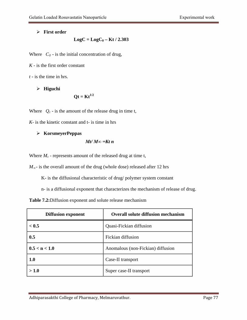
Gelatin Loaded Rosuvastatin Nanoparticle Experimental work
Adhiparasakthi College of Pharmacy, Melmaruvathur. Page 77
First order
Where C0 - is the initial concentration of drug,
K - is the first order constant
t - is the time in hrs.
Higuchi
Where Qt - is the amount of the release drug in time t,
K- is the kinetic constant and t- is time in hrs
KorsmeyerPeppas
Where Mt - represents amount of the released drug at time t,
M∞- is the overall amount of the drug (whole dose) released after 12 hrs
K- is the diffusional characteristic of drug/ polymer system constant
n- is a diffusional exponent that characterizes the mechanism of release of drug.
Table 7.2:Diffusion exponent and solute release mechanism
Diffusion exponent Overall solute diffusion mechanism
< 0.5 Quasi-Fickian diffusion
0.5 Fickian diffusion
0.5 < n < 1.0 Anomalous (non-Fickian) diffusion
1.0 Case-II transport
> 1.0 Super case-II transport
LogC = LogC0 – Kt / 2.303
Qt = Kt1/2
Mt/ M∞ =Kt n
Gelatin Loaded Rosuvastatin Nanoparticle Experimental work
Adhiparasakthi College of Pharmacy, Melmaruvathur. Page 78
7.5: Stability studies (Manavalan R. and Ramasamy S., 2004)
The International Conference on Harmonization (ICH) Guidelines titled “Stability testing
of New Drug Substances and Products (QIA) describes the stability test requirements for drug
registration application in the European Union, Japan and the States of America
ICH specifies the length of study and storage conditions
Long-Term Testing: 250 C ±2
0 C at 60% RH ±5% for 12 Months
Accelerated Testing: 400 C ±2
0 C at 75% RH ±5% for 6 Months
In present study the selected formulation F1 was exposed up to 3 months of stability
studies at room temperature, accelerated condition and refrigerator temperature (250± 20C at 60%
RH±5%RH, 400C ±20C at 750% RH ±5% RH and 50C ±3
0C ) to find out the effect of aging on
drug loading efficiency.
Procedure: (KalariaD.R.,et al., 2008:Jospech Nisha Carry., et al., 2006)
Stability studies were carried out by freeze dried nanoparticles as per ICH titled QIA
guide lines meant for refrigerated product over a period of 90 days. Nanoparticles were kept in
stability chamber with temperature at 250± 20C at 60% RH±5%RH, 400C ±20C at 75% RH
±5%RH and 50C ±30C. Sucrose (5%) was used as Cryoprotectant during the freeze drying
process. Samples were withdrawn predetermined time intervals and analyzed the drug loading
efficiency.

Gelatin Loaded Rosuvastatin Nanoparticle Experimental work
Adhiparasakthi College of Pharmacy, Melmaruvathur. Page 79
7.6. Zeta potential:(Maincent P., et al 1997)
The zeta potential value of the sample was measured by zetasizer.A nanoparticle
formulation which gives best results between particle size, drug loading efficiency, in-vitro drug
release was determined for zeta potential. Zeta potential it can help to understand the
characteristics of a suspension by understanding how individual colloids interact with one
another. Each colloid carries aelectrical charge is high enough, the colloids will remain discrete,
disperse and in suspension. The report was showed in Figure 8.33.

RESULT
and
dISCUSSIOn
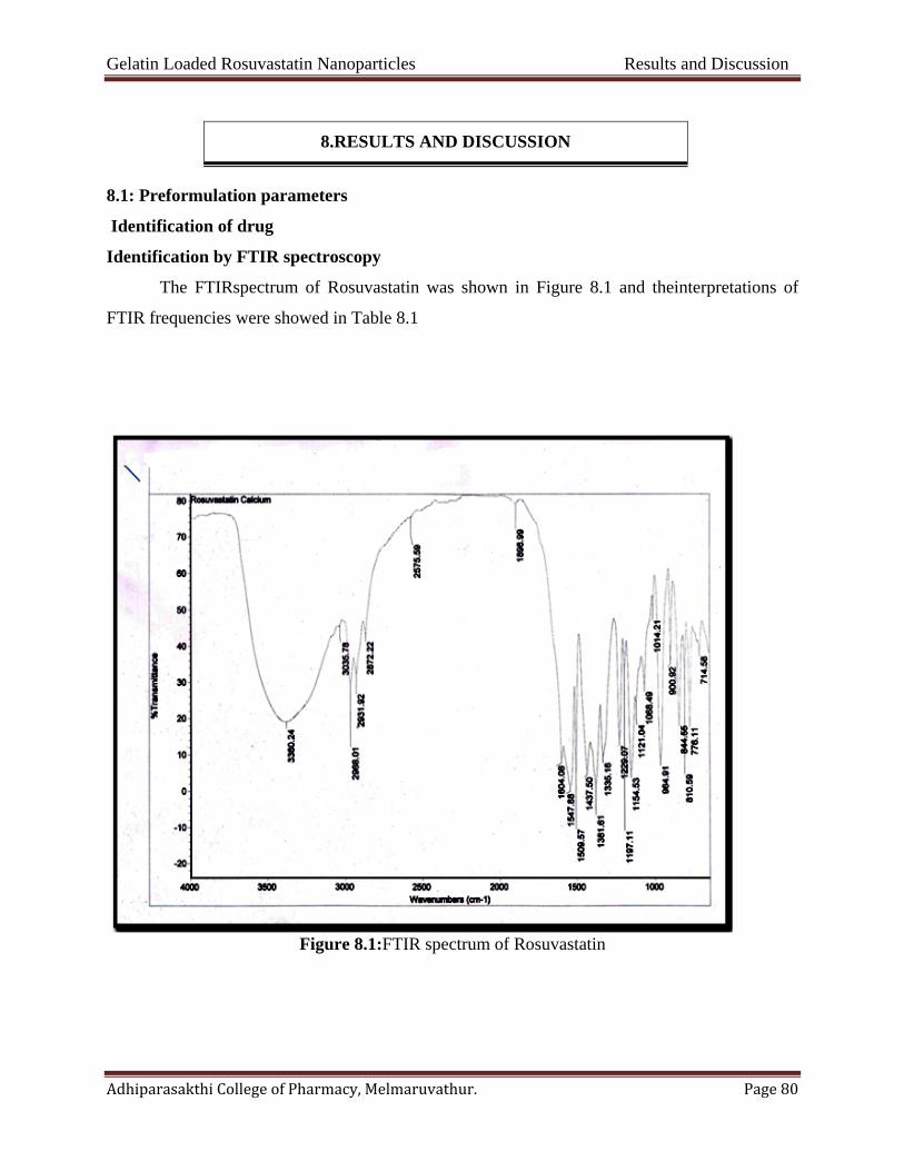
Gelatin Loaded Rosuvastatin Nanoparticles Results and Discussion
Adhiparasakthi College of Pharmacy, Melmaruvathur. Page 80
8.RESULTS AND DISCUSSION
8.1: Preformulation parameters
Identification of drug
Identification by FTIR spectroscopy
The FTIRspectrum of Rosuvastatin was shown in Figure 8.1 and theinterpretations of
FTIR frequencies were showed in Table 8.1
Figure 8.1:FTIR spectrum of Rosuvastatin
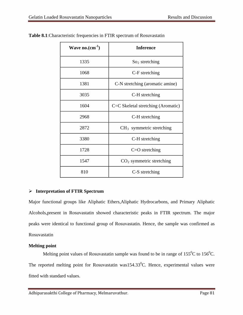
Gelatin Loaded Rosuvastatin Nanoparticles Results and Discussion
Adhiparasakthi College of Pharmacy, Melmaruvathur. Page 81
Table 8.1:Characteristic frequencies in FTIR spectrum of Rosuvastatin
Wave no.(cm-1) Inference
1335 So2 stretching
1068 C-F stretching
1381 C-N stretching (aromatic amine)
3035 C-H stretching
1604 C=C Skeletal stretching (Aromatic)
2968 C-H stretching
2872 CH3 symmetric stretching
3380 C-H stretching
1728 C=O stretching
1547 CO2 symmetric stretching
810 C-S stretching
Interpretation of FTIR Spectrum
Major functional groups like Aliphatic Ethers,Aliphatic Hydrocarbons, and Primary Aliphatic
Alcohols,present in Rosuvastatin showed characteristic peaks in FTIR spectrum. The major
peaks were identical to functional group of Rosuvastatin. Hence, the sample was confirmed as
Rosuvastatin
Melting point
Melting point values of Rosuvastatin sample was found to be in range of 1550C to 1560C.
The reported melting point for Rosuvastatin was154.330C. Hence, experimental values were
fitted with standard values.

Gelatin Loaded Rosuvastatin Nanoparticles Results and Discussion
Adhiparasakthi College of Pharmacy, Melmaruvathur. Page 82
Solubility study
Table 8.2:Solubilityprofile of Rosuvastatin calcium in polar and non-polar solvents
From the above data the solubility of Rosuvastatin was confirmed in various polar and
non polar solvents.
S. No SOLVENTS EXTENT OF SOLUBILITY CATEGORY
1. Distilled water 10 mg of substance was not soluble upto 100 ml of solvent
Practically insoluble
2. Methanol 10 mg in 0.31 ml Sparingly soluble
3. Ethanol 10 mg in 0.36 ml Sparingly soluble
4. Acetonitrile 10 mg in 0.05 ml Freely soluble
5. Di-methylFormamide 10 mg in 0.04ml Freely soluble
6. Acetone 10 mg in 0.02 ml Freely soluble
7. Glacial acetic acid 10 mg in 0.04 ml Freely Soluble
8. Cyclohexane 10 mg in 0.3 ml Soluble
9. Chloroform 10 mg in 0.01 ml Freely soluble
10. Petroleum ether 10 mg in 0.45 ml Sparingly soluble
11. n- Butanol 10 mg in 0.07 ml Freely soluble
12 Carbon tetra chloride 10 mg in 0.04 ml Freely soluble
13 Benzene 10 mg in 0.09 ml Freely insoluble
15 Iso propyl alcohol 10 mg in 0.55 ml Sparingly soluble
16 Hexane 10 mg in 0.29 ml Soluble
17 Diethyl ether 10 mg in 0.09 ml Freely soluble
18 Dichloro methane 10 mg in 0.02 ml Freely soluble

Gelatin Loaded Rosuvastatin Nanoparticles Results and Discussion
Adhiparasakthi College of Pharmacy, Melmaruvathur. Page 83
Physicochemical parameters of drug
Organoleptic properties
Odour:Odourless
Colour: White or almost white
Nature: Crystalline powder.
Loss on drying
The percentage loss on drying after 3 hours was found to be as follows
Table 8.3: Percentage loss on drying for Rosuvastatin
The sample passes test for loss on drying as per the limit specified in IP, 2007 (N.M.T. 1%).It
was shown in Table 8.3
S. No Percentage LOD Avg. percentage LOD
1 0.7
0.5666±0.25
2 0.4
3 0.8
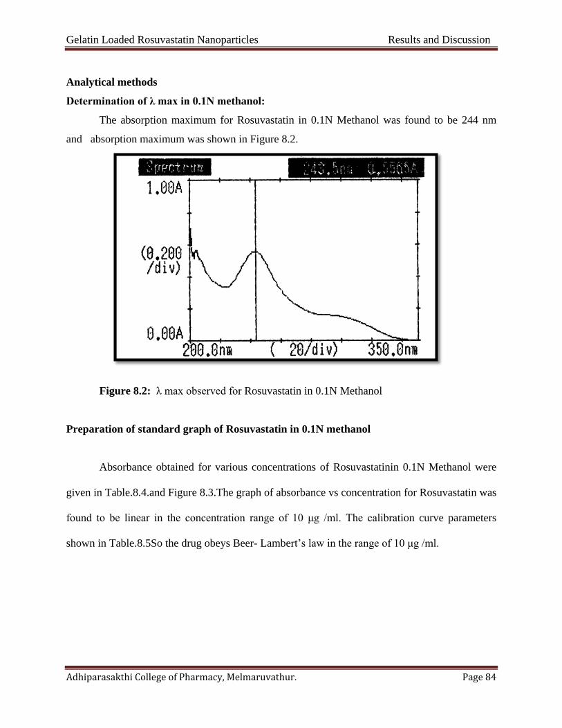
Gelatin Loaded Rosuvastatin Nanoparticles Results and Discussion
Adhiparasakthi College of Pharmacy, Melmaruvathur. Page 84
Analytical methods
Determination of λ max in 0.1N methanol:
The absorption maximum for Rosuvastatin in 0.1N Methanol was found to be 244 nm
and absorption maximum was shown in Figure 8.2.
Figure 8.2: λ max observed for Rosuvastatin in 0.1N Methanol
Preparation of standard graph of Rosuvastatin in 0.1N methanol
Absorbance obtained for various concentrations of Rosuvastatinin 0.1N Methanol were
given in Table.8.4.and Figure 8.3.The graph of absorbance vs concentration for Rosuvastatin was
found to be linear in the concentration range of 10 μg /ml. The calibration curve parameters
shown in Table.8.5So the drug obeys Beer- Lambert’s law in the range of 10 μg /ml.

Gelatin Loaded Rosuvastatin Nanoparticles Results and Discussion
Adhiparasakthi College of Pharmacy, Melmaruvathur. Page 85
Table 8.4:Data of concentration and absorbance for Rosuvastatin in 0.1N Methanol
S.NO Concentration
(µg/ml)
Absorbance
at 244nm
1 0 0
2 1 0.062
3 2 0.122
4 3 0.183
5 4 0.245
6 5 0.308
7 6 0.371
Figure 8.3: Calibration curve of Rosuvastatin calcium in methanol at 244 nm
0
0.05
0.1
0.15
0.2
0.25
0.3
0.35
0.4
0 1 2 3 4 5 6
Abso
rban
ce a
t 244
nm
Concentration (µg/ml)
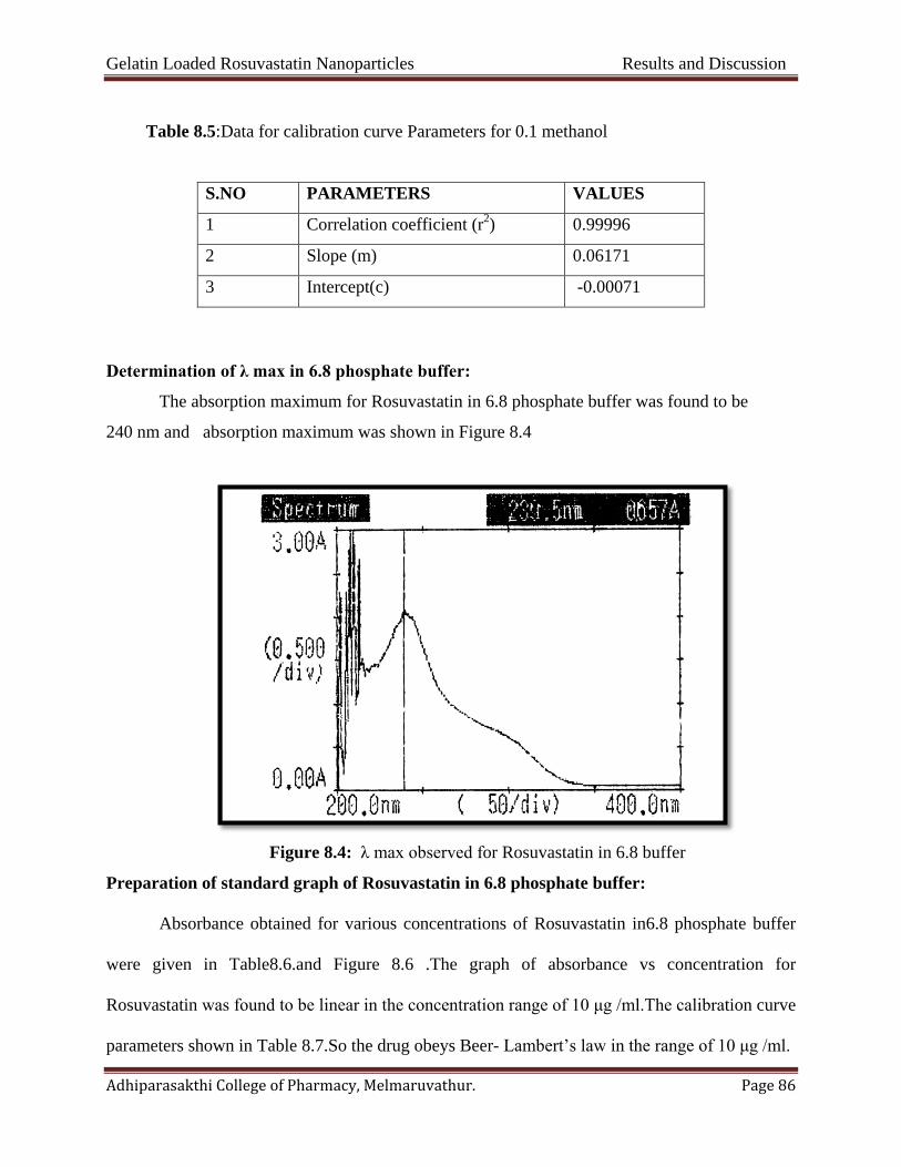
Gelatin Loaded Rosuvastatin Nanoparticles Results and Discussion
Adhiparasakthi College of Pharmacy, Melmaruvathur. Page 86
Table 8.5:Data for calibration curve Parameters for 0.1 methanol
S.NO PARAMETERS VALUES
1 Correlation coefficient (r2) 0.99996
2 Slope (m) 0.06171
3 Intercept(c) -0.00071
Determination of λ max in 6.8 phosphate buffer:
The absorption maximum for Rosuvastatin in 6.8 phosphate buffer was found to be
240 nm and absorption maximum was shown in Figure 8.4
Figure 8.4: λ max observed for Rosuvastatin in 6.8 buffer
Preparation of standard graph of Rosuvastatin in 6.8 phosphate buffer:
Absorbance obtained for various concentrations of Rosuvastatin in6.8 phosphate buffer
were given in Table8.6.and Figure 8.6 .The graph of absorbance vs concentration for
Rosuvastatin was found to be linear in the concentration range of 10 μg /ml.The calibration curve
parameters shown in Table 8.7.So the drug obeys Beer- Lambert’s law in the range of 10 μg /ml.

Gelatin Loaded Rosuvastatin Nanoparticles Results and Discussion
Adhiparasakthi College of Pharmacy, Melmaruvathur. Page 87
Table 8.6: Calibration curve of Rosuvastatin calcium in 6.8 phosphate buffer at 240 nm
S.NO Concentration (µg/ml)
Absorbance
at 240nm
1 0 0
2 1 0.071
3 2 0.142
4 3 0.211
5 4 0.278
6 5 0.349
7 6 0.423
Figure 8.5:Calibration curveofRosuvastatin calcium in 6.8 buffer at 240 nm
0
0.05
0.1
0.15
0.2
0.25
0.3
0.35
0.4
0.45
0 1 2 3 4 5 6
Abso
rban
ce a
t 240
nm
Concentration (µg/ml)
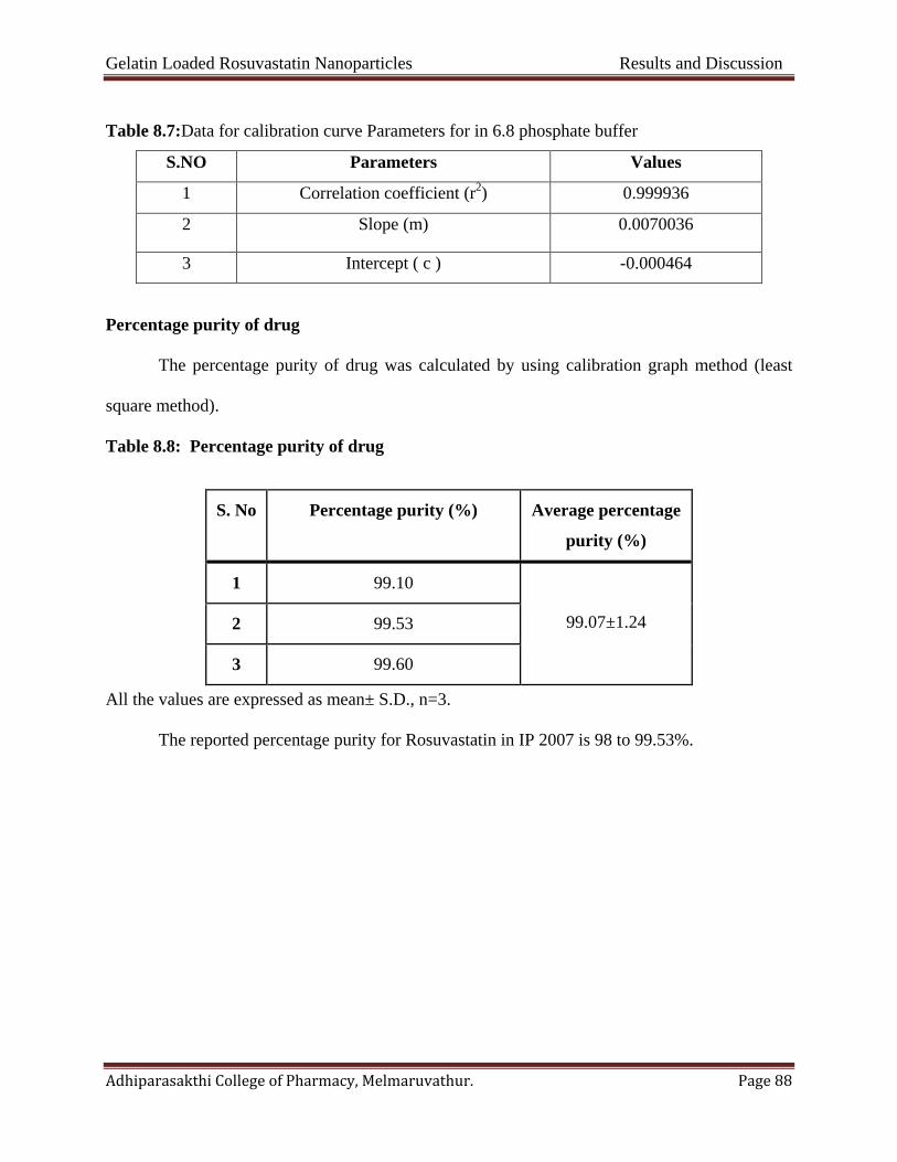
Gelatin Loaded Rosuvastatin Nanoparticles Results and Discussion
Adhiparasakthi College of Pharmacy, Melmaruvathur. Page 88
Table 8.7:Data for calibration curve Parameters for in 6.8 phosphate buffer
S.NO Parameters Values
1 Correlation coefficient (r2) 0.999936
2 Slope (m) 0.0070036
3 Intercept ( c ) -0.000464
Percentage purity of drug
The percentage purity of drug was calculated by using calibration graph method (least
square method).
Table 8.8: Percentage purity of drug
All the values are expressed as mean± S.D., n=3.
The reported percentage purity for Rosuvastatin in IP 2007 is 98 to 99.53%.
S. No Percentage purity (%) Average percentage
purity (%)
1 99.10
99.07±1.24 2 99.53
3 99.60
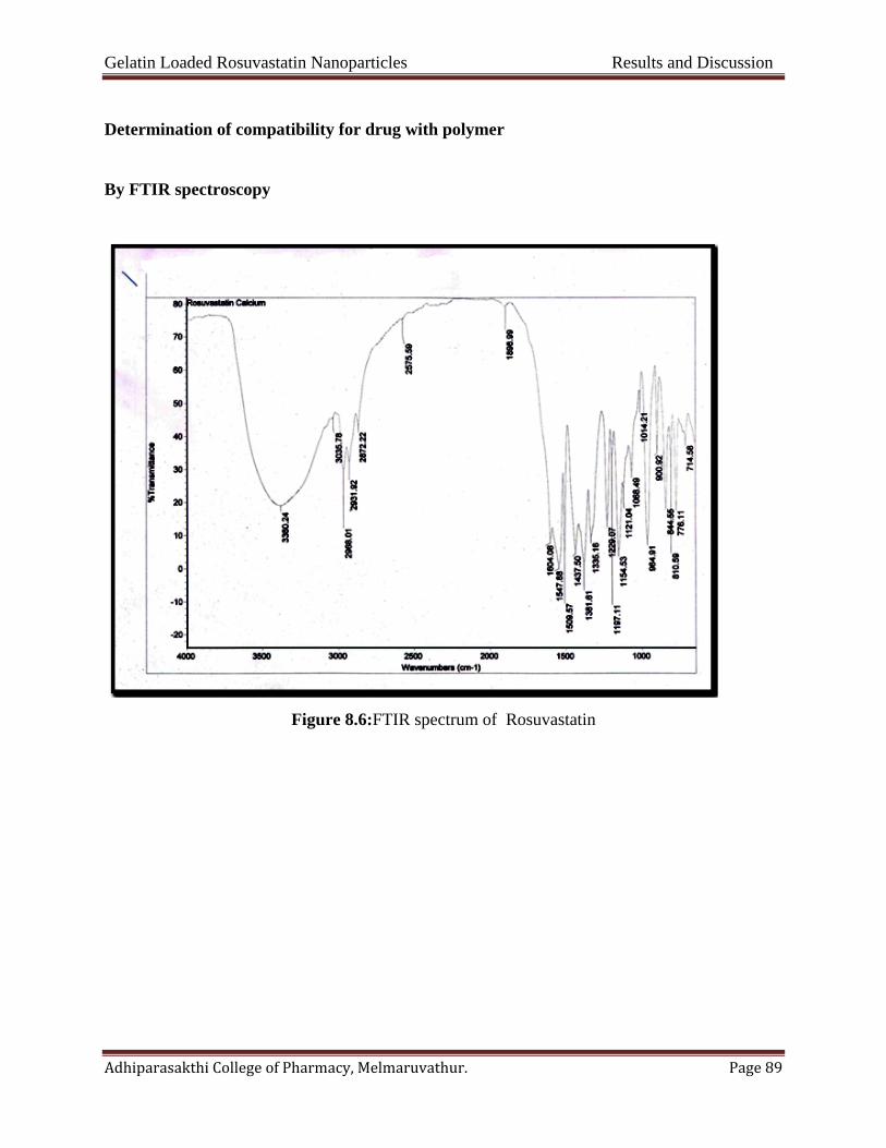
Gelatin Loaded Rosuvastatin Nanoparticles Results and Discussion
Adhiparasakthi College of Pharmacy, Melmaruvathur. Page 89
Determination of compatibility for drug with polymer
By FTIR spectroscopy
Figure 8.6:FTIR spectrum of Rosuvastatin
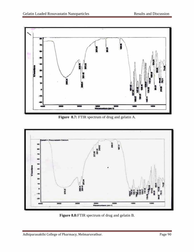
Gelatin Loaded Rosuvastatin Nanoparticles Results and Discussion
Adhiparasakthi College of Pharmacy, Melmaruvathur. Page 90
Figure 8.7: FTIR spectrum of drug and gelatin A.
Figure 8.8:FTIR spectrum of drug and gelatin B.
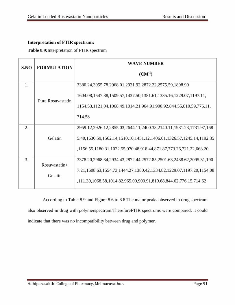
Gelatin Loaded Rosuvastatin Nanoparticles Results and Discussion
Adhiparasakthi College of Pharmacy, Melmaruvathur. Page 91
Interpretation of FTIR spectrum:
Table 8.9:Interpretation of FTIR spectrum
S.NO FORMULATION WAVE NUMBER
(CM-1)
1.
Pure Rosuvastatin
3380.24,3055.78,2968.01,2931.92,2872.22,2575.59,1898.99
1604.08,1547.88,1509.57,1437.50,1381.61,1335.16,1229.07,1197.11,
1154.53,1121.04,1068.49,1014.21,964.91,900.92,844.55,810.59,776.11,
714.58
2.
Gelatin
2959.12,2926.12,2855.03,2644.11,2400.33,2140.11,1981.23,1731.97,168
5.40,1630.59,1562.14,1510.10,1451.12,1406.01,1326.57,1245.14,1192.35
,1156.55,1180.31,1022.55,970.48,918.44,871.87,773.26,721.22,668.20
3. Rosuvastatin+
Gelatin
3378.20,2968.34,2934.43,2872.44,2572.85,2501.63,2438.62,2095.31,190
7.21,1608.63,1554.73,1444.27,1380.42,1334.82,1229.07,1197.20,1154.08
,111.30,1068.58,1014.82,965.00,900.91,810.68,844.62,776.15,714.62
According to Table 8.9 and Figure 8.6 to 8.8.The major peaks observed in drug spectrum
also observed in drug with polymerspectrum.ThereforeFTIR spectrums were compared; it could
indicate that there was no incompatibility between drug and polymer.

Gelatin Loaded Rosuvastatin Nanoparticles Results and Discussion
Adhiparasakthi College of Pharmacy, Melmaruvathur. Page 92
By DSC thermal analysis
The compatibility and interactions between drugs and polymer were analysed
byDSCthermogram and results were obtained represented and shown in Figure 8.9 to 8.11.
Figure 8.9: DSC thermo grams of Rosuvastatin
Figure 8.10:DSC thermo grams of Rosuvastatin+ gelatin A

Gelatin Loaded Rosuvastatin Nanoparticles Results and Discussion
Adhiparasakthi College of Pharmacy, Melmaruvathur. Page 93
Figure 8.11:DSC thermo grams of Rosuvastatin+ gelatin B
The possible drug and polymer interaction can be studied by using DSC. The Rosuvastatin
exhibits a sharp endometric peak 155.8 which is corresponding with its melting point at figure
8.10 and the Rosuvastatin + gelatin A and B exhibits the sharp endometric peak 155.5 at figure
8.9. Therefore when compared with pure drug thermo gram. No interaction was found between
drug and polymers.

Gelatin Loaded Rosuvastatin Nanoparticles Results and Discussion
Adhiparasakthi College of Pharmacy, Melmaruvathur. Page 94
8.2. Characterization of nanoparticles
Drug loading efficiency (%le) and drug loading capacity (%LC):
The results were shown in Table 8.10 and Figure.8.12 to 8.13. It is revealed that the highest,
%LE and %LC in the nanoparticle preparation were obtained.
Table 8.10: % LE and % LC of Rosuvastatin nanoparticles with gelatin A and gelatin B
S.NO
Parameter
Formulation (Gelatin A) Formulation (Gelatin B)
F1 F2 F3 F4 F5 F6 F7 F8
1 Drug entrapment
efficiency (LE %) 91.73 72.93 76.50 86.27 69.85 87.74 88.68 68.72
2 Drug loading
capacity (Lc %) 66.21 55.62 49.41 39.54 65.22 54.14 61.33 47.54
Figure 8.12:% LE and % LC of Rosuvastatin nanoparticles with gelatin A
0
20
40
60
80
100
F1 F2 F3 F4
perc
enta
ge e
ntra
pmen
t eff
icie
ncy
Formulation F1-F4
Drug entrapment efficiency (LE %)
Drug loading capacity (LC %)

Gelatin Loaded Rosuvastatin Nanoparticles Results and Discussion
Adhiparasakthi College of Pharmacy, Melmaruvathur. Page 95
Figure 8.13:% LE and % LC of Rosuvastatin nanoparticles with gelatin B
Morphological characterization of F1 to F4 (gelatin A)
Rosuvastatin loaded polymeric nanoparticles were fabricated usingin-situ two step
desolvationmethodusing polymer Gelatin A.
The choice of an in-situ two step desolvation method of encapsulation was usually
determined by the %yield, LE and %LC of the drug and sonication process which allowed
reducing considerably the mean particle size and simultaneously to narrow the width of the size
distribution, i.e. reduces the polydispersity index. The effectiveness of in-situ two step
desolvation method and polymer on nanoparticle preparation was confirmed by their size, in-
vitro release characteristics.
0
20
40
60
80
100
F5 F6 F7 F8
perc
enta
geen
trap
men
t eff
icie
ncy
Formulations F5-F9
Drug entrapment efficiency (LE %)
Drug loading capacity (Lc %)
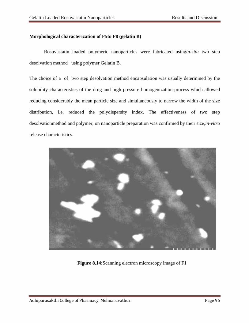
Gelatin Loaded Rosuvastatin Nanoparticles Results and Discussion
Adhiparasakthi College of Pharmacy, Melmaruvathur. Page 96
Morphological characterization of F5to F8 (gelatin B)
Rosuvastatin loaded polymeric nanoparticles were fabricated usingin-situ two step
desolvation method using polymer Gelatin B.
The choice of a of two step desolvation method encapsulation was usually determined by the
solubility characteristics of the drug and high pressure homogenization process which allowed
reducing considerably the mean particle size and simultaneously to narrow the width of the size
distribution, i.e. reduced the polydispersity index. The effectiveness of two step
desolvationmethod and polymer, on nanoparticle preparation was confirmed by their size,in-vitro
release characteristics.
Figure 8.14:Scanning electron microscopy image of F1

Gelatin Loaded Rosuvastatin Nanoparticles Results and Discussion
Adhiparasakthi College of Pharmacy, Melmaruvathur. Page 97
Figure 8.15: Particle size distribution F1
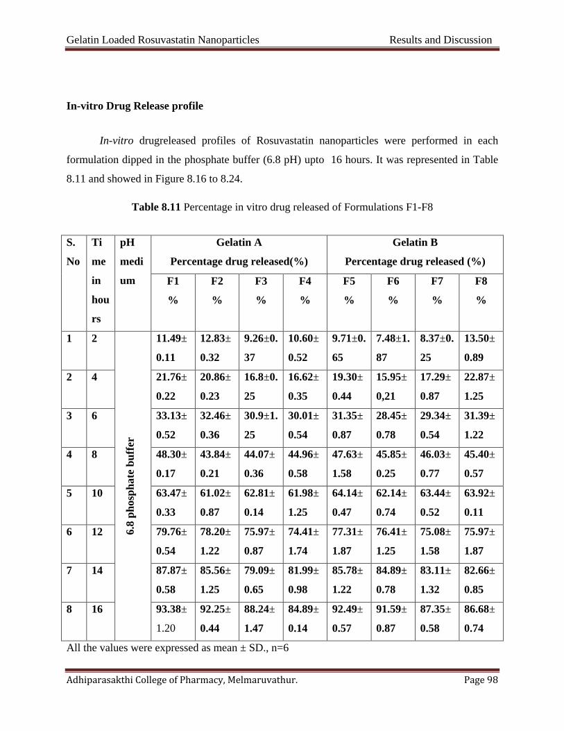
Gelatin Loaded Rosuvastatin Nanoparticles Results and Discussion
Adhiparasakthi College of Pharmacy, Melmaruvathur. Page 98
In-vitro Drug Release profile
In-vitro drugreleased profiles of Rosuvastatin nanoparticles were performed in each
formulation dipped in the phosphate buffer (6.8 pH) upto 16 hours. It was represented in Table
8.11 and showed in Figure 8.16 to 8.24.
Table 8.11 Percentage in vitro drug released of Formulations F1-F8
S.
No
Ti
me
in
hou
rs
pH
medi
um
Gelatin A
Percentage drug released(%)
Gelatin B
Percentage drug released (%)
F1
%
F2
%
F3
%
F4
%
F5
%
F6
%
F7
%
F8
%
1 2
6.8
phos
phat
e bu
ffer
11.49±
0.11
12.83±
0.32
9.26±0.
37
10.60±
0.52
9.71±0.
65
7.48±1.
87
8.37±0.
25
13.50±
0.89
2 4 21.76±
0.22
20.86±
0.23
16.8±0.
25
16.62±
0.35
19.30±
0.44
15.95±
0,21
17.29±
0.87
22.87±
1.25
3 6 33.13±
0.52
32.46±
0.36
30.9±1.
25
30.01±
0.54
31.35±
0.87
28.45±
0.78
29.34±
0.54
31.39±
1.22
4 8 48.30±
0.17
43.84±
0.21
44.07±
0.36
44.96±
0.58
47.63±
1.58
45.85±
0.25
46.03±
0.77
45.40±
0.57
5 10 63.47±
0.33
61.02±
0.87
62.81±
0.14
61.98±
1.25
64.14±
0.47
62.14±
0.74
63.44±
0.52
63.92±
0.11
6 12 79.76±
0.54
78.20±
1.22
75.97±
0.87
74.41±
1.74
77.31±
1.87
76.41±
1.25
75.08±
1.58
75.97±
1.87
7 14 87.87±
0.58
85.56±
1.25
79.09±
0.65
81.99±
0.98
85.78±
1.22
84.89±
0.78
83.11±
1.32
82.66±
0.85
8 16 93.38±
1.20
92.25±
0.44
88.24±
1.47
84.89±
0.14
92.49±
0.57
91.59±
0.87
87.35±
0.58
86.68±
0.74
All the values were expressed as mean ± SD., n=6

Gelatin Loaded Rosuvastatin Nanoparticles Results and Discussion
Adhiparasakthi College of Pharmacy, Melmaruvathur. Page 99
Figure 8.16:Graphical representation of invitro percentage drug released for formulation F1
Figure 8.17:Graphical representation of invitro percentage drug released for formulation F2
/
0
20
40
60
80
100
0 2 4 6 8 10 12 14 16 18
% D
rug
rele
ased
Time in hours
0
20
40
60
80
100
0 2 4 6 8 10 12 14 16 18
% D
rug
rele
ased
Time in hours

Gelatin Loaded Rosuvastatin Nanoparticles Results and Discussion
Adhiparasakthi College of Pharmacy, Melmaruvathur. Page 100
Figure8.18: Graphical representation of invitro percentage drug released for formulation F3
Figure8.19:Graphical representation of invitro percentage drug released forformulation F4
0
20
40
60
80
100
0 2 4 6 8 10 12 14 16 18
% D
rug
rele
ased
Time in hours
0
20
40
60
80
100
0 2 4 6 8 10 12 14 16 18
% D
rug
rele
ased
Time in hours

Gelatin Loaded Rosuvastatin Nanoparticles Results and Discussion
Adhiparasakthi College of Pharmacy, Melmaruvathur. Page 101
Figure 8.20:Graphical representation of invitro percentage drug released for formulation F5
Figure 8.21:Graphical representation of invitro percentage drug released for formulation F6
0
20
40
60
80
100
0 2 4 6 8 10 12 14 16 18
% D
rug
rele
ased
Time in hours
0
20
40
60
80
100
0 2 4 6 8 10 12 14 16 18
% D
rug
rele
ased
Time in hours

Gelatin Loaded Rosuvastatin Nanoparticles Results and Discussion
Adhiparasakthi College of Pharmacy, Melmaruvathur. Page 102
Figure 8.22: Graphical representation of invitro percentage drug released for formulation F7
Figure 8.23:Graphical representation of invitro percentage drug released for formulation F8
0
20
40
60
80
100
0 2 4 6 8 10 12 14 16 18
% D
rug
rele
ased
Time in hours
0
20
40
60
80
100
0 2 4 6 8 10 12 14 16 18
% D
rug
rele
ased
Time in hours

Gelatin Loaded Rosuvastatin Nanoparticles Results and Discussion
Adhiparasakthi College of Pharmacy, Melmaruvathur. Page 103
Figure 8.24: Graphical representation of comprehensive invitro percentage drug released for
formulations F1-F8
Release Kinetics of In-vitro Drug Release
The kinetics of in-vitro drug released was determined by applying the drug release data
to various kinetic models such as zero order first order, Higuchi and Korsmeyer- Peppas. The
results obtained were represented Table 8.12 and Figure 8.25 to 8.32.
0
20
40
60
80
100
120
0 2 4 6 8 10 12 14 16 18
perc
enta
ge d
rug
rele
ased
Time in hours
F1
F2
F3
F4
F5
F6
F7
F8

Gelatin Loaded Rosuvastatin Nanoparticles Results and Discussion
Adhiparasakthi College of Pharmacy, Melmaruvathur. Page 104
Table 8.12: Release kinetics of In-vitro drugrelease gelatin A and gelatin B
Formulations Zero order
R2
First order
R2
Higuch
R2
Peppas
R2
Best Fit
Model
F1 0.991 0.845 0.978 0.837 Zero
F2 0.986 0.873 0.974 0.734 Zero
F3 0.983 0.914 0.975 0.924 Zero
F4 0.981 0.923 0.968 0.864 Zero
F5 0.992 0.838 0.976 0.877 Zero
F6 0.989 0.870 0.964 0.962 Zero
F7 0.983 0.907 0.973 0.821 Zero
F8 0.984 0.929 0.968 0.912 Zero
Figure 8.25:Best fit model (zero order) of formulation F1
R² = 0.991
-20
0
20
40
60
80
100
120
0 2 4 6 8 10 12 14 16 18
% D
rug
rele
ased
Time in hours

Gelatin Loaded Rosuvastatin Nanoparticles Results and Discussion
Adhiparasakthi College of Pharmacy, Melmaruvathur. Page 105
Figure 8.26:Best fit model (zero order) of formulation F2
Figure 8.27Best fit model (zero order) of formulation F3
R² = 0.986
-200
20406080
100120
0 2 4 6 8 10 12 14 16 18
% D
rug
rele
ased
Time in hours
R² = 0.983
-200
20406080
100
0 2 4 6 8 10 12 14 16 18% D
rug
rele
ased
Time in hours
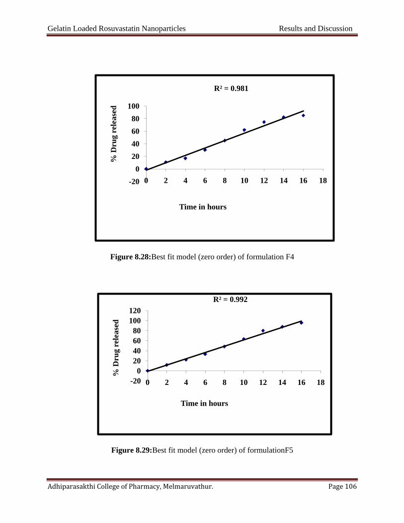
Gelatin Loaded Rosuvastatin Nanoparticles Results and Discussion
Adhiparasakthi College of Pharmacy, Melmaruvathur. Page 106
Figure 8.28:Best fit model (zero order) of formulation F4
Figure 8.29:Best fit model (zero order) of formulationF5
R² = 0.981
-200
20406080
100
0 2 4 6 8 10 12 14 16 18
% D
rug
rele
ased
Time in hours
R² = 0.992
-200
20406080
100120
0 2 4 6 8 10 12 14 16 18
% D
rug
rele
ased
Time in hours
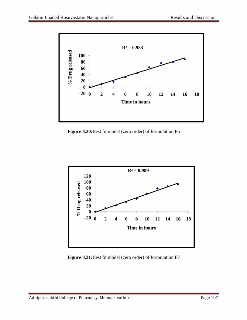
Gelatin Loaded Rosuvastatin Nanoparticles Results and Discussion
Adhiparasakthi College of Pharmacy, Melmaruvathur. Page 107
Figure 8.30:Best fit model (zero order) of formulation F6
Figure 8.31:Best fit model (zero order) of formulation F7
R² = 0.983
-200
20406080
100
0 2 4 6 8 10 12 14 16 18
% D
rug
rele
ased
Time in hours
R² = 0.989
-200
20406080
100120
0 2 4 6 8 10 12 14 16 18
% D
rug
rele
ased
Time in hours

Gelatin Loaded Rosuvastatin Nanoparticles Results and Discussion
Adhiparasakthi College of Pharmacy, Melmaruvathur. Page 108
Figure 8.32:Best fit model (zero order) of formulation F8
From the data and graphical representations, the Rosuvastatin nanoparticles formulations
were showed well fitted zero order kinetics and formulation F1 was showed best among the
formulations were prepared based on morphology, %drug loading efficiency, in-vitro drug
released profiles and also well fitted the zero order kinetics.
8.3 Stability studies
FormulationF1was kept in room temperature, accelerated condition and refrigerator
temperature. After exposed to specified periods of time the samples were analyzed for %LE.
Results were represented in Table 8.13
R² = 0.984
0
20
40
60
80
100
0 2 4 6 8 10 12 14 16 18
% D
rug
rele
ased
Time in hours

Gelatin Loaded Rosuvastatin Nanoparticles Results and Discussion
Adhiparasakthi College of Pharmacy, Melmaruvathur. Page 109
Table 8.13:Stability studies of optimized formulation (F1).
S.No
Temperature(00c)
Drug loading efficiency(%LE)
1st month 2nd month 3rd month
1 250C±20C at RH 60%±5% 98.45 98.98 97.93
2 400C±20C at RH 75%±5% 98.55 96.80 96.92
3 50C±30C 98.29 98.14 97.35
No major difference was found between %LE before and after stability studies.The
formulation F1 was showed satisfactory physical stability at 250C ±20C at RH 60%±5%, 400C
±20C at RH 75%±5% and 50C±30C.

Gelatin Loaded Rosuvastatin Nanoparticles Results and Discussion
Adhiparasakthi College of Pharmacy, Melmaruvathur. Page 110
8.4. Zeta potential
The zeta potential report of the formulation F1 showed in Figure 8.33. The obtained value
was the positive value.
Figure 8.33: Zeta potential of F1 formulation

SUMMARY
AND
CONCLUSION
Gelatin loaded rosuvastatin nano particles Summary and Conclusion
Adhiparasakthi College of Pharmacy, Melmaruvathur. Page 111
The Rosuvastatin was widely used in the antilipedimic drug.This research work
mainly focused on therapeutic effect of the drug to increased Bioavailability. The reported
Bioavailability of Rosuvastatin was only 20%. While forming the novel drug delivery system in
the form of nanoparticle to increased the bioavalability.
Eight formulations were prepared by using drug with gelatin A and gelatin B in
different ratio of 1:1,1:2,1:3 and1:4 each polymer respectively(F1 to F8). Among the eight
formulations F1 formulation was best because of these formulation shows the optimized result of
colloidal drug delivery systems of nanoparticles have emerged as an efficient means enhancing
the bioavailability with lowest possible dose.
The present work was proposed to prepared nanoparticles loaded with Rosuvastatin to
achieved better bioavailability with low dose of the drug at the site, decreased the risk of adverse
side effects. Rosuvastatin nanoparticles were prepared by in-situ two step desolvation method.
The loading efficiency(F1) and Entrapment efficiency(F1) was 66.21 and 91.73
respectively.
The shape of nanoparticle was found to be spherical by SEM analysis. Formulation with
high polymer content was observed to be fairly spherical. Compatability of drug and polymer
mixture was done by performing FTIR and DSC study. It was concluded that there was no
interaction between drug and polymer.
9. SUMMARY AND CONCLUSION

Gelatin loaded rosuvastatin nano particles Summary and Conclusion
Adhiparasakthi College of Pharmacy, Melmaruvathur. Page 112
The in-vitro released of Rosuvastatin was evaluated in phosphate buffer saline (pH6.8)
up to 16hrs. The formulation F1 was released the drug 93.38% up to16 hours. and chosen the
best among the formulations were prepared.
Zeta potential was determined for the formulation F1 and it was found to be +ve value
of 1.57mv.
The in-vitro drug released data was applied to various kinetic models like zero order
kinetics, Higuchi plot, first order kinetics, and Peppas plot by predict the drug release kinetics
mechanism. The formulation F1 was best fitted the zero order kinetics.
In short term stability studies the formulation F1 was showed that there was no
remarkable changes in the % drug loading efficiency.
Based on the % drug loading efficiency, zeta potential, in-vitro drug release profile,
in-vitro drug release kinetics and stability studies the formulation F1 was found to be best one
among the formulations (F1 to F8) were prepared.
These nanoparticles can be promising agents for rational drug delivery in lipidemic
condition.
Future prospects

Gelatin Loaded Rosuvastatin NanoParticles Future Prospects
Adhiparasakthi College of Pharmacy, Melmaruvathur. Page 113
1. Nano-drug delivery systems that deliver large but highly localized quantities of drugs to
specific areas to be released in controlled ways.
2. Controllable release profiles, especially for sensitive drugs.
3. Materials for nanoparticles that are biocompatible and biodegradable.
4. Architectures / structures, such as biomimetic polymers, nanotubes.
5. Technologies for self-assembly.
6. Advanced polymeric carriers for the delivery of therapeutic peptide/proteins
(biopharmaceutics).
10.FUTURE PROSPECTS

BiBliography
Gelatin Loaded Rosuvastatin Nanoparticles Bibliography
Adhiparasakthi College of Pharmacy, Melmaruvathur. Page 114
1. AdlinJinoNesalin., Gowthamaraja., and Somashekhara. Development and
characterisation of nanoparticle containing flutamide, Journal of pharmtech Research,
2009, 3 (1), 411-415.
2. Alka Gupta., Mishra P., and Shan K. Determination of Rosuvastatin in pure form by UV
visible spectrophotometer method, Journal Of Chemistry, 2009, 6(1),89-92.
3. Anne Marie Helmenstine. Textbook of Lipids, 1st Edition, Expert zoom
publishers , Delhi, 2002,115.
4. Annonyms: DrugBank: Rosuvastatin.
5. Anonymous, http://www.drugbank.ca/drugs/DB00649.
6. Anonymous, The Indian Pharmacopoeia. Vol-1, 2, 3, The Controller of publication, New
Delhi, 2007, 134 ,135 159 ,511,1676 .
7. Anonymous, The Merck Index. An encyclopedia of chemicals, drugs &biologicals.
14thedn., New Jersy, Merck & Co. Inc, 1997. 1428.
8. Aulton M.E. Eds. Pharmaceutics, The science of dosage form design, 2ndEdition,
Churchill Livingstone, New York, 2002, 211-230.
9. Aurelien roux., Roux A Koster., Lenz M., Sorre B., Manneville J. B., Nassoy P and
Bassereau. Textbook of Membrane curvature controls polymerization, 2nd Edition, Goel
Publishers, 2010,1-3.
10. Bankar G.S. and Rhodes C.T. Eds. Modern Pharmaceutics, 3rdEdition, Marcel Dekker,
Inc, New York, 1996, 611-624.
11.BIBLIOGRAPHY
Gelatin Loaded Rosuvastatin Nanoparticles Bibliography
Adhiparasakthi College of Pharmacy, Melmaruvathur. Page 115
11. BenoyBrataBhowmik., Bishwanth S.A., and Arup Mukherjee. Preparation of in vitro
characterization of slow release Testosterone nanoacapsules in alginates, Acta Pharm,
2006, 56, 417-429.
12. Bshsagar.,Gelpernia.,Kisich K., Iseman M.D and Heifits L. Textbook of Nanoparticles,
Novel drug delivery system, AMJ Respair Critcare Med, New York, 2005, 1487-1490.
13. Charles E, and Ophardt. Text book of Lipids, 6th Edition, Virtual Chembook publications,
New York, 2003,126-129.
14. ChayaVenkat., Kuo G.M., Boumpas D.T., Illei G.G., Yarboro C., Pucino F. Text book of
Methods of drug delivery, 2nd Edition, Informa healthcare, New York, 2003, 528-533.
15. Costas Kaparissides., Sofia Alexandridou., KaterinaKotti, and SotiraChaitidou. Text book
of Recent Advances in Novel Drug Delivery Systems, Journal Of Nanotechnology,
2005,115-23.
16. Deepak Thassu., Michel Deleers, and YashwantPathak. Textbook of Nanoparticulate
Drug Delivery Systems, Informa healthcare, USA Newyork, 2007, Vol-166, 52-54.
17. Dhirendra K., Vivek D., Shaila L., Brajesh P., Kavita R.G, and Sarvesh P. Design and
evaluation of sustained release matrix once daily formulation of Stavudine, International
Journal Of Drug Delivery, 2010, 2, 125-134.
18. Dong Gon Kim., YoungJeong., Chanayong Choi., Sung-HeeRoh., Seong-Koo Kang.,
MiKyeong Jang, and Jae Woon Nah. Retinol encapsulated low molecular water soluble
chitosan nanoparticles, International Journal Of Pharmaceutics, 2006, 130-138.
Gelatin Loaded Rosuvastatin Nanoparticles Bibliography
Adhiparasakthi College of Pharmacy, Melmaruvathur. Page 116
19. Dong M Shin., RajniSinha., Gloria J Kim, and ShumingNie. Nanotechnology in cancer
therapeutics Bioconjugated nanoparticles for drug delivery, AACR Publications,
2007,3-8.
20. Galindo-Rodriguez S., Allémann E., Fessi H., and Doelker E. Physicochemical
parameters associated with nanoparticle formation in the salting-out, emulsification-
diffusion, and nanoprecipitation methods, Pharm Res, 2004, 428-1439.
21. Illinois. Text book of Nanoparticles for drug delivery, 2nd Edition, Stanford Publishing
House, Singapore, 2004,68-74.
22. James P., McCulley M.D., and Ward Shine. Textbook of a compositional based model
for the tear film lipid layer, The University of Texas Southwestern Medical Center at
Dallas, 1997,129.
23. James R Fromm, Textbook of Introduction of Lipids, 1st Edition, Med Publishers, Delhi,
1997,320-323.
24. Joseph Nisha Mary., Palani S., Sharma P.K, and Gupta M.K. Development and
Evaluation of Nanoparticles of Mitomycinc, Journal Of Pharmaceutical Research, 2006,
53-56.
25. Kalaria D.R., Sharma G., Beniwal V and Ravi Kumar N.V. Design of Biodegradable
Nanoparticles for oral Delivery of DoxorubicinIn-vivo Pharmacokinetics and Toxicity
Studies in Rats, Pharmaceutical Research, 2009, 26(3), 492-501.
26. Kowsalya R. Formulation and invitro Evaluation of Cephalexin Loaded Polymethacrylic
Acid Nanoparticles, Indian Drugs, 2005, 296-299.
Gelatin Loaded Rosuvastatin Nanoparticles Bibliography
Adhiparasakthi College of Pharmacy, Melmaruvathur. Page 117
27. Kristi Monson., Arthur Schoenstadt. Textbook of Drug Interactions With Rosuvastatin,
6th Edition , Med Publishers, Delhi, 2011, 21-26.
28. Kyung Hyun Min., Kyeongsoon Park., Yoo-Shih Kim., Sang MunBae., Seulki Lee.,
HyungGon Jo., Rang-Woon Park., In-san Kim., Seo Young jeong., Kwangmeyung Kim,
and Ick Chan Kwon. Hydrophobically modified glycol chitosan nanoparticles-
encapsulated campothecin enhance the drug stability and tumor targeting in cancer
therapy, Journal Of Controlled Release, 2008, 208-218.
29. Lachman L., Lieberman H.A, and Kanig J.L. The theory and practice of industrial
pharmacy, 3rd Edition, Varghese Publishing House, Mumbai, 1991, 183-184.
30. Lamprecht Alf. Textbook of NanoTherapeutics Drug Delivery Concepts in Nanoscience,
Pan Stanford Publishing Pvt. Ltd., Jermany, 2009, 8-13.
31. Langmuir., Angela Chemelli., Otto Glatter., Heinrichstr., Sarah Rogers., Richard K
Heenan, and Isabelle Grillo. Textbook of Separation and Purification of Nanoparticles in
a Single Step, ACS Publications, Delhi, 2010, 26 (10),6989–6994.
32. Leroueil-Le Verger M., Fluckiger L., Kim YI., Hoffman M, and Maincent P. Preparation
and characterization of nanoparticles containing an antihypertensive agent, European
Journal of Pharmaceutics And Biophrmaceutics, 1998, 137-143.
33. LievenBaert., GerbenKlooster., Willy Dreies., Marc Francoils., AlfonsWouters., Esther
Basstanie., KoenIterbeke., Fred Stappers., and Jan Rosier. Development of a long-acting
injectable formulation with nanoparticles of Ripivirine (TMC278) for treatment,
European Journal Of Pharmaceutics And Biophrmaceutics, 2009, 549-556.
Gelatin Loaded Rosuvastatin Nanoparticles Bibliography
Adhiparasakthi College of Pharmacy, Melmaruvathur. Page 118
34. Mahua S., Sateesh K, and Ramesh P. Development and validation of RP-HPLC and
ultravioletspectrophotometric methods of analysis for the quantitative estimation of
antiretroviral drugs in pharmaceutical dosage forms, Journal Of Chromatography, 2006,
349–354.
35. Maincent P andKimy. The anti hypertensive effect of orally administration of Nifidifine
loaded nanoparticles, British Journal Of Pharmacology, 1997, 399-404.
36. Manavalan R and Ramasamy S. Physical Pharmaceutics, Accelerated Stability Testing,
1st Edition, Vignesh Publisher, Chennai, 1995, 288-299.
37. Mathlowitz Edith, Encyclopedia of Controlled drug delivery, vol-2, Delhi press,
Newdelhi, 2009, 641-654.
38. McTaggart F., Buckett L., Davidson R., Holdgate G., McCormick A., Schneck D., Smith
G, and Warwick M. Preclinical and clinical pharmacology of Rosuvastatin, Journal Of
Cardiol Publisher, 2001, 87(5A), 28-32.
39. Michael E., Maragoudakis. Text book of Biochemistry(including biophysical chemistry
& molecular biology), ACS Publications, Delhi, 1970, 9 (2), 413–417.
40. Michael Faraday. Textbook of Nanoparticle background, 2nd Edition, Olonano Publishers,
Delhi, 1857, 258-32.
41. Mohan Raj V.J, and Chen Y. Text book of Nanoparticles A Review, Tropical Journal Of
Pharmaceutical Research, 2006, 5(1), 561-573.
42. Muhammed Rafeeq., Junise., Saraswathi Krishnan and Dilip. Devepment and
characterization of chitosan with Isoniazid for the treatment of Tuberculosis, Research
Journal Of Pharmaceutical, Biological And Chemical Sciences, 2010, 4(2), 383-389.
Gelatin Loaded Rosuvastatin Nanoparticles Bibliography
Adhiparasakthi College of Pharmacy, Melmaruvathur. Page 119
43. Krishnadasan., Yashina S, and deMello A.S. Text book of Microfluidic Reactors for
Nanomaterial Synthesi,. Advances in Chemical Engineering, Academic Press, 2010,
Volume 38, 2004, 195-231.
44. Norwegian., Darren McKellep, and Carol B Eckmann. Text book of Nanoparticles can
hinder intracellular transport, Published by Claude Olsen,Newyork ,2011,218-26.
45. PatilS.V., Kuchekar B.S., Janugade B.U and Lade P.D. In-Vitro Studies of Stavudine
Sustained Release from Hydrophilic Matrices, Journal Of Pharmacy Research, 2009,
2(12), 1855-1856.
46. Qiang Zhang., GuoqingYie., Yie Li., Qingsong Yang and Nagai T. Studies on the
cyclosporin A loaded stearic acid nanoparticles, International Journal Of Pharmaceutics,
2000, 153-159.
47. Rahul Nair.K., Vishnu priya, and K.S. ArunKumar, Formulation and evaluation of solid
lipid nanoparticles of water soluble drugs- isoniazid, Journal Of pharmaceutical Science
And Research, 3, 2011, 1256-1264.
48. Rakesh P Patel., Mukeshgohel., Sanjaymodi., Ganeshnirma and Vijayaraghavan. Text
book of Nanoparticles and its applications in field of pharmacy, 2008, 208.
49. Randy P. Carney., Gretchen A,DeVries Cedric Dubois., Hyewon Kim., Jin Young Kim.,
Chetana Singh., Pradip K Ghorai., Joseph B Tracy., Rebecca L Stiles, and Royce W. Text
book of Size Limitations for the Formation of Ordered Striped Nanoparticles, Jouranl Of
The American Society, Department of Materials Science & Engineering, 2008, 798–799.
50. Richard N Fogoros. Textbook of The statin drugs, 2nd Edition, Published by Claude
Olsen, Delhi, 2010,341-398.

Gelatin Loaded Rosuvastatin Nanoparticles Bibliography
Adhiparasakthi College of Pharmacy, Melmaruvathur. Page 120
51. Shanmughavel., Radhika Ramachandran., Piramanayagam. Preparation and
characterization of biopolymeric nanoparticles used in drug delivery, Indian Journal Of
Biochemistry And Biophysics, 2010,56-59.
52. Shoba rani R Hiremath. Text book of Industrial pharmacy, 1stEdition, Black Swan
Pulishers Ltd, Chennai, 2008, 129-137.
53. Skoog D.A., Holler F. and Nieman S. Principles of instrumental analysis, 5thEdition,
Thomson Asia Pvt. Limited, 1996, 410-413.
54. Soppimath K.S., Aminabhavi T.M., Kulkarni A.R, and Rudzinski W.E. Biodegradable
polymeric nanoparticles as drug delivery, Journal Of Controlled Release, 2001,1-20.
55. Steffen Petersen, Text book of Order Milestone for Nanoparticle Characterisation Tool,
1st Edition, Expert Zoom Publishers, 2008, 624-628.
56. Sylvie Audoly, and Rousselot S.A. Textbook of Gelatin in pharmaceutical applications,
Rousselot Expert Zoom publishers, 2006, 17-20.
57. Tamizhrasi., Shukla., Shivkumar., Rathi. Formulation and Evaluation of Lamivudine
loaded polymethacrylicacid nanoparticles, International Journal Of Pharmtech Research,
2009, 3 (1), 411-415
58. Tripathi K.D. Essentials of medical pharmacology, JaypeePuplication, 5th edition., New
Delhi, 2004, 522-530.
59. Ui Yang., Xu Han., Kai Shi Gang Cheng., Chang-Koo Shim, and Fude Cui.
Silver nanoparticles using an emulsion-solvent diffusion method, American Journal
Pharmaceutical Sciences, 2009, 4(2),89-95.

Gelatin Loaded Rosuvastatin Nanoparticles Bibliography
Adhiparasakthi College of Pharmacy, Melmaruvathur. Page 121
60. Vandana Singh., Amrendra Kumar and Chaudhary. Rosuglitazoneloaded gelatin
nanoparticles by two step desolvation Method, International Journal Of Pharmaceutical
Science Review And Reseach, 2010, 7(5), 100-103.
61. Vyas SP, and Khar R.K. Textbook of Targeted and Controlled drug delivery, Novel
carrier systems, 1stEdtion., CBS publishers, Newdelhi, 2002, 331.
62. Wayne Goates., Mary Harris., Gina Watkiss, and Patricia Virginia Smith. Textbook of
Gelatin a natural polymer substance, 1st Edition,Vignesh Publishers, Delhi, 2003,
243-256.
63. WitiakD.T, New man, and Feller. Textbook of Antilipidemic Drugs Medicinal, Chemical
and Biochemical Aspects, Elsevier Science Ltd, 1991,618.
64. Young-ChihKuo, and Fu-Lung Su. Delaviridne and saquinavir across the blood-brain
barrier by polybutylcyanoacrylate, methylmethacrylate sulfopropylmethacrylate, and
solid lipid nanoparticles, International Journal Of Pharmaceutics, 2007, 340, 143-152.
65. Young-Wook Won, and Yong-Hee Kim. Recombinant human gelatin nanoparticles as a
protein drug carrier, Journal Of Controlled Release, 2008, 127, 154-161.
66. Yvette N Konan., Robert Gurny, and Eric Allemann. Preparation and characterization of
Sterile and freeze-dried Sub-200 Nanoparticles, International Journal Pharmaceutics,
2002, 239-252.
67. Zelu., Tengkuang., Max Tsai., Jessie L.S., and Guill Wientjes.M. Paclitaxel loaded
gelatin nanoparticles for intravesical bladder cancer therapy, Journal Of Clinical Cancer
Research, 2004,7577-7684.
68. Zelu., Teng-KuangYeh., Max Tsai. Nanoparticles for Intravesical Bladder Cancer
Therapy, Journal Of Clinical Cancer, 2004, 7677-7679.