Embed Size (px)
Citation preview
1521-009X/43/3/309–316$25.00 http://dx.doi.org/10.1124/dmd.114.061689DRUG METABOLISM AND DISPOSITION Drug Metab Dispos 43:309–316, March 2015Copyright ª 2015 by The American Society for Pharmacology and Experimental Therapeutics
Formation of the Accumulative Human Metabolite andHuman-Specific Glutathione Conjugate of Diclofenac in TK-NOG
Chimeric Mice with Humanized Livers
Hidetaka Kamimura, Satoshi Ito, Kohei Nozawa, Shota Nakamura, Hiroyuki Chijiwa,Shin-ichiro Nagatsuka, Miyuki Kuronuma, Yasuyuki Ohnishi, Hiroshi Suemizu, and Shin-ichi Ninomiya
ADME & Tox. Research Institute, Sekisui Medical Co., Ltd., Tokyo, Japan (H.K., S.I., K.N., S.Nak., H.C., S.Nag., S.Ni); and CentralInstitute for Experimental Animals, Kawasaki, Kanagawa, Japan (M.K., Y.O., H.S.)
Received October 20, 2014; accepted December 10, 2014
ABSTRACT
39-Hydroxy-49-methoxydiclofenac (VI) is a human-specific metaboliteknown to accumulate in the plasma of patients after repeated admin-istration of diclofenac sodium. Diclofenac also produces glutathione-conjugated metabolites, some of which are human-specific. In thepresent study, we investigated whether these metabolites could begenerated in humanized chimeric mice produced from TK-NOGmice. After a single oral administration of diclofenac to humanizedmice, the unchanged drug in plasma peaked at 0.25 hour and thendeclined with a half-life (t1/2) of 2.4 hours. 49-Hydroxydiclofenac (II)and 39-hydroxydiclofenac also peaked at 0.25 hour and were unde-tectable within 24 hours. However, VI peaked at 8 hours and declinedwith a t1/2 of 13 hours. When diclofenac was given once per day, peakand trough levels of VI reached plateau within 3 days. Studies withadministration of II suggested VI was generated via II as an inter-mediate. Among six reported glutathione-conjugated metabolites of
diclofenac, M1 (5-hydroxy-4-(glutathion-S-yl)diclofenac) to M6 (29-(glutathion-S-yl)monoclofenac), we found three dichlorinated conju-gates [M1, M2 (49-hydroxy-39-(glutathion-S-yl)diclofenac), and M3(5-hydroxy-6-(glutathion-S-yl)diclofenac)], and a single mono-chlorinated conjugate [M4 (29-hydroxy-39-(glutathion-S-yl)mono-clofenac) or M5 (49-hydroxy-29-(glutathion-S-yl)monoclofenac)], inthe bile of humanized chimeric mice. M4 and M5 are positionalisomers and have been previously reported as human-specific invitro metabolites likely generated via arene oxide and quinoneimine–type intermediates, respectively. The biliary monochlori-nated metabolite exhibited the same mass spectrum as those ofM4 and M5, and we discuss whether this conjugate correspondedto M4 or M5. Overall, humanized TK-NOG chimeric mice wereconsidered to be a functional tool for the study of drug metabolismof diclofenac in humans.
Introduction
Diclofenac sodium was the first approved nonsteroidal anti-inflammatory drug, and has been widely used to treat pain or inflammationcaused by arthritis or ankylosing spondylitis (Small, 1989). Metabolismstudies in animals and humans revealed that this drug undergoes almostcomplete biotransformation by direct conjugation or oxidation of thearomatic rings followed by conjugation (Stierlin et al., 1979; Stierlin andFaigle, 1979). In humans, 49-hydroxydiclofenac (II), 5-hydroxydiclofenac,39-hydroxydiclofenac (IV), and 49,5-dihydroxydiclofenac are excreted intourine as their free (minor) or conjugated forms. While analyses on theconcentrations of these metabolites in plasma samples from study subjectswere performed, a new metabolite, 39-hydroxy-49-methoxydiclofenac(VI), was found. This metabolite showed distinct pharmacokineticbehavior with a time to maximum plasma concentration (Tmax) and half-life (t1/2) of 12 and 80 hours, respectively, after single oral admin-istration with potential accumulation after repeated dosing of diclofenac(Degen et al., 1988; Faigle et al., 1988). According to Faigle et al.
(1988), unpublished data showed that VI was not found in mouse, rat,or marmoset plasma, but baboon plasma only. The cumulative urinaryexcretion (0–96 hours) of free and conjugated VI in humans was verysmall (ca. 1% of dose), which might explain why VI had not been foundin the urine sample in the previous studies.Formation of a reactive 1-O-acyl glucuronide by direct glucuronidation
of diclofenac is considered to be responsible for the major modification ofproteins (Kretz-Rommel and Boelsterli, 1993). Evidence of this includesthe detection of diclofenac-S-acyl-glutathione in rat bile treated with highdoses of diclofenac (Grillo et al., 2003). The formation of reactivemetabolites of diclofenac by various cytochrome P450s (P450s), followedby their glutathione-dependent inactivation, has been previously studied(Boelsterli, 2003; Tang, 2003). Tang et al. (1999) identified threeglutathione conjugates—namely, 5-hydroxy-4-(glutathion-S-yl)diclofenac(M1), 49-hydroxy-39-(glutathion-S-yl)diclofenac (M2), and 5-hydroxy-6-(glutathion-S-yl)diclofenac (M3)—in rat bile and with in vitro incubationsin rat liver microsomes or human hepatocytes. This suggested the for-mation of quinone imine intermediates during diclofenac metabolism. Onthe other hand, a novel glutathione conjugate, 29-hydroxy-39-(glutathion-S-yl)monoclofenac (M4), was reported to be generated by incubation ofdx.doi.org/10.1124/dmd.114.061689.
ABBREVIATIONS: II, 49-hydroxydiclofenac; IV, 39-hydroxydiclofenac; VI, 39-hydroxy-49-methoxydiclofenac; ALT, alanine aminotransferase; CID,collision-induced dissociation; GCV, ganciclovir; HPLC, high-performance liquid chromatography; LC-MS/MS, liquid chromatography–tandemmass spectrometry; M1, 5-hydroxy-4-(glutathion-S-yl)diclofenac; M2, 49-hydroxy-39-(glutathion-S-yl)diclofenac; M3, 5-hydroxy-6-(glutathion-S-yl)diclofenac; M4, 29-hydroxy-39-(glutathion-S-yl)monoclofenac; M5, 49-hydroxy-29-(glutathion-S-yl)monoclofenac; M6, 29-(glutathion-S-yl)mono-clofenac; P450, cytochrome P450; t1/2, half-life; uPA, urokinase-type plasminogen activator.
309
at ASPE
T Journals on M
ay 26, 2018dm
d.aspetjournals.orgD
ownloaded from
diclofenac with human liver microsomes, suggesting the existence of anarene oxide intermediate as another route of bioactivation (Yan et al.,2005). Experiments using recombinant enzymes and specific inhibitorsdemonstrated that CYP2C9 was involved in the formation of M4, andthis metabolite was not detected in incubations with either rat or monkeyliver microsomes. Around the same time, Yu et al. (2005) reported an-other monochlorinated glutathione conjugate with the same molecularweight, 49-hydroxy-29-(glutathion-S-yl)monoclofenac (M5), followed bythe discovery of the latest glutathione conjugate, 29-(glutathion-S-yl)monoclofenac (M6) (Wen et al., 2008; Boerma et al., 2012). AlthoughM4, M5, and M6 are generated in in vitro studies using recombinantenzymes or liver microsomes fortified with glutathione, in vivo forma-tion of these metabolites has not been previously reported.Chimeric mice with humanized livers hold considerable advantages
for more accurate prediction of human metabolism over conventional invivo animal or in vitro human systems (Yoshizato et al., 2012; Grompeand Strom, 2013; Peltz, 2013). The ability to predict circulating humanmetabolites is one of the characteristic features of this model whichcould satisfy regulatory requirements for the safety testing of drugmetabolites (Kamimura et al., 2010). Histologic examination showingthe connection of human bile canaliculi to a mouse interlobular bileduct suggested easy access to human bile samples, unlike in clinicalexperiments (Kamimura et al., 2010). Following the development ofimmunodeficient humanized mice with a urokinase-type plasminogenactivator (uPA) transgene (Mercer et al., 2001) or a disrupted geneencoding murine fumarylacetoacetate hydrolase (Azuma et al., 2007),NOD/Shi-scid IL2 receptor gamma–null mice expressing a herpessimplex virus type-1 thymidine kinase transgene (TK-NOG mice) withhumanized liver were generated (Hasegawa et al., 2011). Liver-specificexpression of thymidine kinase by the albumin enhancer/promoterresults in damage to murine hepatocytes following administrations of anontoxic dose of ganciclovir (GCV). This new humanized mouse modelhas demonstrated formation of the human metabolites of debrisoquine(Hasegawa et al., 2011), thalidomide including a glutathione conjugateof its 5-hydroxy form in plasma (Yamazaki et al., 2012), and clemizole(Nishimura et al., 2013). These results, the application of this model inpharmacokinetic modeling (Tsukada et al., 2013; Suemizu et al., 2014;Yamashita et al., 2014), and the evaluation of drug-drug interactions(Nishimura et al., 2013; Yamazaki et al., 2013) prompted us to conductdiclofenac metabolism studies using humanized TK-NOG mice. In thisreport, the formation of the human-specific metabolites IV, VI, andglutathione conjugate metabolites is presented. Additionally, adminis-tration of a synthetic version of metabolite II was conducted to elucidatethe pathways of diclofenac metabolism in humans.
Materials and Methods
Diclofenac sodium [2-(2-(2,6-dichlorophenylamino)phenyl)acetic acid so-dium salt] was purchased from UPS (Rockville, MD). 49-Hydroxydiclofenac(synthetic version of metabolite II) was purchased from Becton, Dickinson andCompany (Franklin Lakes, NJ). [13C6]49-Hydroxydiclofenac, used as aninternal standard, was obtained from Toronto Research Chemicals (Ontario,Canada). Other chemicals and solvents were of analytical grade. Hepatocytesfrom a 4-year-old female donor (lot 7F3063; LONZA, Japan, Tokyo) or thosefrom a 9-year old female donor (lot HC2-19; XenoTech, Lenexa, KS) wereused for the transplantation. Metabolic activities of CYP2C9 in the respectivehepatocytes were 1.28 (49-hydroxylation of tolbutamide at 400 mM) or 467(49-hydroxylation of diclofenac at 100 mM) pmol/min per 106 viable cells.
Reference incubation mixture containing M6 was prepared at VU UniversityAmsterdam (Amsterdam, The Netherlands) as described by Boerma et al.(2014). In brief, a final concentration of 500 mM diclofenac was incubated with100 nM horseradish peroxidase, 100 mM glutathione, and 8 mM humanglutathione-S-transferase P1-1 for 1 hour at 37�C. The reaction was terminatedby the addition of methanol.
Animals, Dosing, and Sample Collection. The humanized TK-NOG chimericmice were generated using the method of Hasegawa et al. (2011), with somemodifications. For the plasma concentration studies, 11- or 12-week-old maleTK-NOG mice were given intraperitoneal injections of 6 mg/kg GCV, and then 6and 50 mg/kg GCV was given 2 and 10 days later, respectively. Eighteen daysafter the first GCV dose, mice with elevated serum alanine aminotransferase(ALT) levels of more than 200 IU/l received transplants of 1.1 or 1.5 � 106 ofhuman hepatocytes by intrasplenic injection. For the biliary excretion studies,8- or 10-week-old male TK-NOG mice were given intraperitoneal injectionsof 10 mg/kg GCV, and an additional 50 mg/kg GCV was given to a subset ofthese mice 8 days later. Eight to 17 days after the first dose, mice with serumALT levels of more than 240 IU/l received transplants of 0.6 or 1.0 � 106 ofhuman hepatocytes by intrasplenic injection. Mouse serum levels of humanalbumin were measured and used as a marker for the hepatic replacement ratioby human hepatocytes (Hasegawa et al., 2011). The hepatic replacementratios were calculated with respect to an in-house calibration curve, and thosemice with a ratio greater than 77% were used. TK-NOG mice whose serumALT levels had not been elevated sufficiently by the GCV treatments wereused as control TK-NOG mice. Administration of drugs to and sample col-lections from the mice were performed in compliance with protocols ap-proved by the Sekisui Medical Animal Care and Use Committee.
Control TK-NOG and humanized chimeric mice received a single oral ad-ministration of diclofenac sodium (10 mg/kg as free form) or synthetic metaboliteII (10 mg/kg), and blood samples were collected for analyses at 15 and 30 minutesand 1, 4, 8, 24, and 48 hours. Three weeks later, a group of the same humanizedchimeric mice were administered the same dose of diclofenac sodium orally onceper day for 14 days, and blood samples were taken at 8 and 24 hours after eachdose. Food and water were given ad libitum during the drug treatment. For thebiliary excretion study, a control TK-NOG mouse and a humanized chimericmouse, in which a polyethylene tube was cannulated into the bile duct, receiveddiclofenac sodium (10 mg/kg as free form) or synthetic metabolite II (10 mg/kg),and bile samples were collected for 0–24 hours on dry ice.
Analyses of Plasma Samples. Diclofenac, II, IV, and VI were quantified byliquid chromatography–tandem mass spectrometry (LC-MS/MS). To a 15- or30-ml aliquot of control plasma, the same volume of a standard workingsolution of diclofenac sodium or synthetic metabolite II was added to preparethe standard sample. To a 15- or 30-ml aliquot of the sample plasma, the samevolume of 50% (v/v) methanol in water was added to prepare the analyticalsample. To the analytical and standard samples, 100 ml of internal standardsolution (25 nM) was added. The tube was closed and mixed vigorously for ca.10 seconds using vortex mixing and subsequently centrifuged (15,000g, 4�C,10 minutes). The supernatant was pipetted into an autosampler vial, and50 ml of the sample was injected into the column. The LC-MS/MS analysiswas performed using the LC20AD high-performance liquid chromatography(HPLC) system (Shimadzu, Kyoto, Japan) coupled with an API-4000 tandemmass spectrometer (AB Sciex, Foster City, CA). The column (CAPCELL PAKMG C18, 250 � 4.6 mm i.d., 5 mm; Shiseido, Tokyo, Japan) was maintained at40�C, and diclofenac and its metabolites were eluted with a linear gradient(flow rate of 1.0 ml/min) that escalated from 15% B to 35% B by 15 minutes,was maintained at 35% B for 15 minutes, then escalated again to 90% B over5 minutes, and was maintained at 90% B for 5 minutes [10 mM ammoniumformate in distilled water (pH 5.0) (A) or acetonitrile (B)]. Analytes weredetected in the positive ion mode with electrospray ionization (collision energyof 50, 30, 30, and 45 eV for diclofenac, II, IV, and VI, respectively) using se-lected reaction monitoring: diclofenac, [M+H]+ m/z 296→ 214; II, [M+H]+ m/z312 → 231; IV, [M+H]+ m/z 312 → 231; VI, [M+H]+ m/z 342 → 260. Theretention times of diclofenac, II, IV, VI, and 49-hydroxydiclofenac-13C6 were36, 25, 23, 25, and 25 minutes, respectively. The limits of quantification were1 and 3 ng/ml in plasma, and determination coefficients (R) were .0.993 and.983 for diclofenac and II, respectively. Plasma concentrations of IV and VIwere expressed as peak response since their standard reference compoundswere not available.
Analyses of Bile Samples. Diclofenac glutathione conjugates were ana-lyzed by a liquid chromatography-multiple-stage mass spectrometer using theLC20AD HPLC system coupled with an LTQ Orbitrap XL mass spectrometer(Thermo Fisher Scientific, Waltham, MA). A 10- to 25-ml aliquot of bilesample was injected into the column (CAPCELL Core ADME, 150 � 2.1 mmi.d., 2.7 mm; Shiseido) without any pretreatment. The column was maintained
310 Kamimura et al.
at ASPE
T Journals on M
ay 26, 2018dm
d.aspetjournals.orgD
ownloaded from
at 40�C, and chromatography was performed with a linear gradient from 10%B to 30% B by 30 minutes at a flow rate of 0.2 ml/min [100 mM ammoniumformate in distilled water (pH 5.0) (A) or acetonitrile (B)]. After the analysis,the column was washed by increasing the composition from 30% B to 100%B, and it was maintained at 100% B for 10 minutes and returned to 10% B.The electrospray ionization ion source was operated in positive ion mode, andinstrumental parameters were set as follows: spray voltage 4.5 kV, capillarytemperature 330�C, capillary voltage 3.0 V. Accurate mass chromatogramswere obtained with a resolution of 30,000 by monitoring: M1, M2, and M3,[M+H]+ m/z 617.08394–617.09012; M4 or M5, [M+H]+ m/z 583.12308–583.12892; M6, [M+H]+ m/z 567.12825–567.13393. The calculated high-resolution mass values as protonated ions for C24H27Cl2N4O9S
+ (M1, M2,and M3), C24H28ClN4O9S
+ (M4 or M5), and C24H28ClN4O8S+ (M6) were m/z
617.08703, m/z 583.12600, and m/z 567.13109, respectively.Pharmacokinetic Data Analysis for VI. Metabolite VI was generated from
diclofenac via more than two sequential metabolic reactions, and was consideredto exhibit complicated pharmacokinetics. However, for a simplified simulation inthe multiple-dose study, a one-compartment open model was arbitrarily appliedto the plasma concentration of VI after a single oral administration of diclofenac.Assuming that both formation and elimination rates of VI follow first-orderpharmacokinetics, pharmacokinetic parameters A, kel (elimination rate constant),and kform (formation rate constant), defined by the following equation, wereobtained:
CðtÞ ¼ A� �expð2kel X tÞ2 exp
�2kform X t
��
where C(t) is plasma concentration of VI at time t.
Results
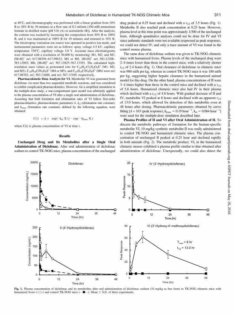
Unchanged Drug and Its Metabolites after a Single OralAdministration of Diclofenac. After oral administration of diclofenacsodium to control TK-NOGmice, plasma concentration of the unchanged
drug peaked at 0.25 hour and declined with a t1/2 of 3.5 hours (Fig. 1).Metabolite II also reached peak concentration at 0.25 hour. However,plasma level at this time point was approximately 1/300 of the unchangedform. Although quantitative analyses could not be done for IV and VIsince authentic standards were not available (expressed as peak response),we could not detect IV, and only a trace amount of VI was found in thecontrol mouse plasma.The same dose of diclofenac sodium was given to TK-NOG chimeric
mice with humanized livers. Plasma levels of the unchanged drug were2–4 times lower than those in the control mice, with a relatively shortert1/2 of 2.4 hours (Fig. 1). Oral clearance of diclofenac in chimeric micewas 900 ml/h per kg, whereas in control TK-NOG mice it was 180 ml/hper kg, suggesting higher hepatic clearance in the humanized animalmodel for this drug. On the other hand, plasma concentrations of II were3–4 times higher than those in the control mice and declined with a t1/2of 5.6 hours. Humanized chimeric mice also had IV in their plasmawhich declined with a t1/2 of 4.8 hours. With gradual decrease of II andIV, metabolite VI peaked at 8 hours and declined with an apparent t1/2of 13.0 hours, which allowed for detection of this metabolite even at48 hours after dosing. Pharmacokinetic parameters obtained by curvefitting [A = 163 (peak response), kform 5 0.19 hour21, kel 5 0.064 hour21]were used for the multiple-dose simulation described later.Plasma Profiles of II and VI after Oral Administration of II. To
discern the metabolic pathways of formation for the human-specificmetabolite VI, 10 mg/kg synthetic metabolite II was orally administeredto control TK-NOG and humanized chimeric mice. The plasma con-centration of unchanged II peaked at 0.25 hour and declined rapidlyin both animals (Fig. 2). The metabolic product, VI, in the humanizedchimeric mouse exhibited a plasma profile similar to that obtained afteradministration of diclofenac. Unexpectedly, we could also detect the
Fig. 1. Plasma concentration of diclofenac and its metabolites after oral administration of diclofenac sodium (10 mg/kg as free form) to TK-NOG chimeric mice withhumanized livers (―u―) and control TK-NOG mice (- -j- -). Mean 6 S.D. of three experiments.
Metabolism of Diclofenac in Humanized TK-NOG Chimeric Mice 311
at ASPE
T Journals on M
ay 26, 2018dm
d.aspetjournals.orgD
ownloaded from
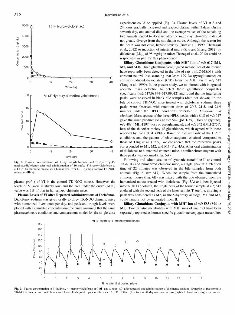
plasma profile of VI in the control TK-NOG mouse. However, thelevels of VI were relatively low, and the area under the curve (AUC)value was 7% of that in humanized chimeric mice.Plasma Levels of VI after Repeated Administrations of Diclofenac.
Diclofenac sodium was given orally to three TK-NOG chimeric micewith humanized livers once per day, and peak and trough levels wereplotted with a simulated concentration-time curve assuming that the samepharmacokinetic conditions and compartment model for the single-dose
experiment could be applied (Fig. 3). Plasma levels of VI at 8 and24 hours gradually increased and reached plateau within 3 days. On theseventh day, one animal died and the average values of the remainingtwo animals tended to decrease after the ninth day. However, data didnot greatly diverge from the simulation curve. Although the reason forthe death was not clear, hepatic toxicity (Bort et al., 1999; Thanagariet al., 2012) or induction of intestinal injury (Zhu and Zhang, 2012) bydiclofenac (LD50 of 95 mg/kg in mice; Thanagari et al., 2012) could beresponsible in part for this phenomenon.Biliary Glutathione Conjugates with MH+ Ion of m/z 617 (M1,
M2, and M3). Three glutathione-conjugated metabolites of diclofenachave reportedly been detected in the bile of rats by LC-MS/MS withconstant neutral loss scanning that loses 129 Da (pyroglutamate) oncollision-induced dissociation (CID) from the MH+ ion of m/z 617(Tang et al., 1999). In the present study, we monitored with integratedaccurate mass detection to detect these glutathione conjugatesspecifically (m/z 617.08394–617.09012) and found that no interferingpeaks were observed in blank bile samples (data not shown). In thebile of control TK-NOG mice treated with diclofenac sodium, threepeaks were observed with retention times of 20.7, 21.5, and 24.9minutes under the HPLC conditions described in Materials andMethods. Mass spectra of the three HPLC peaks with a CID of m/z 617gave the same product ions at m/z 542 ([MH-75]+, loss of glycine),m/z 488 ([MH-129]+, loss of pyroglutamate), and m/z 342 ([MH-275]+,loss of the thioether moiety of glutathione), which agreed with thosereported by Tang et al. (1999). Based on the similarity of the HPLCconditions and the pattern of chromatograms obtained compared tothose of Tang et al. (1999), we considered that the respective peakscorresponded to M1, M2, and M3 (Fig. 4A). After oral administrationof diclofenac to humanized chimeric mice, a similar chromatogram withthree peaks was obtained (Fig. 5A).Following oral administration of synthetic metabolite II to control
TK-NOG and humanized chimeric mice, a single peak at a retentiontime of 22 minutes was observed in the bile samples from bothanimals (Fig. 6, m/z 617). When the sample from the humanizedchimeric mouse (Fig. 6B) was mixed with the bile obtained from thehumanized mouse treated with diclofenac (Fig. 5A) and then injectedinto the HPLC column, the single peak of the former sample at m/z 617coeluted with the second peak of the latter sample. Therefore, this singlepeak was considered as M2, as the 5-hydroxy analogs, M1 and M3,could simply not be generated from II.Biliary Glutathione Conjugate with MH+ Ion of m/z 583 (M4 or
M5). Two in vitro metabolites with MH+ ions of m/z 583 have beenseparately reported as human-specific glutathione conjugate metabolites
Fig. 2. Plasma concentration of 49-hydroxydiclofenac and 39-hydroxy-49-methoxydiclofenac after oral administration of 10 mg/kg 49-hydroxydiclofenac toa TK-NOG chimeric mouse with humanized liver (―u―) and a control TK-NOGmouse (- -j- -).
Fig. 3. Plasma concentration of 39-hydroxy-49-methoxydiclofenac at 0 (d) and 8 hours (s) after repeated oral administration of diclofenac sodium (10 mg/kg as free form) toTK-NOG chimeric mice with humanized livers. Each point represents the mean 6 S.D. of three (first to seventh day) or mean of two (eighth to fourteenth day) experiments.
312 Kamimura et al.
at ASPE
T Journals on M
ay 26, 2018dm
d.aspetjournals.orgD
ownloaded from
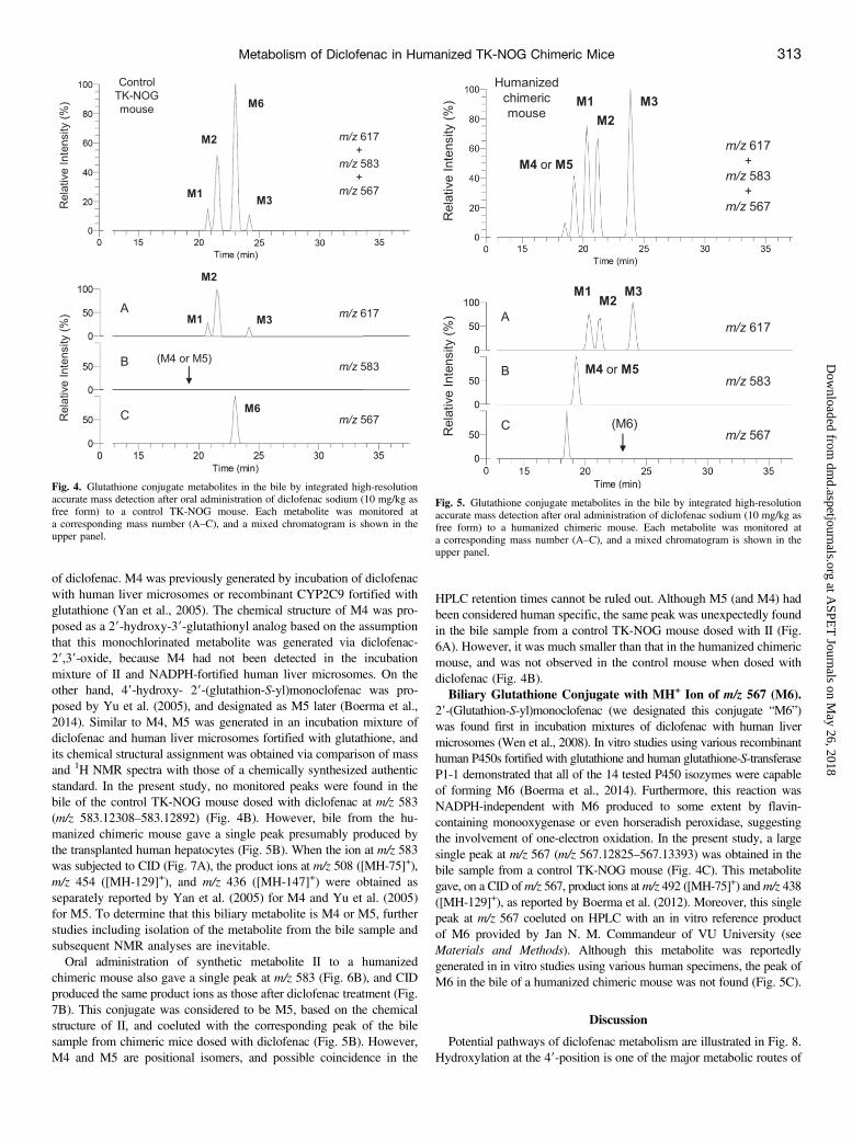
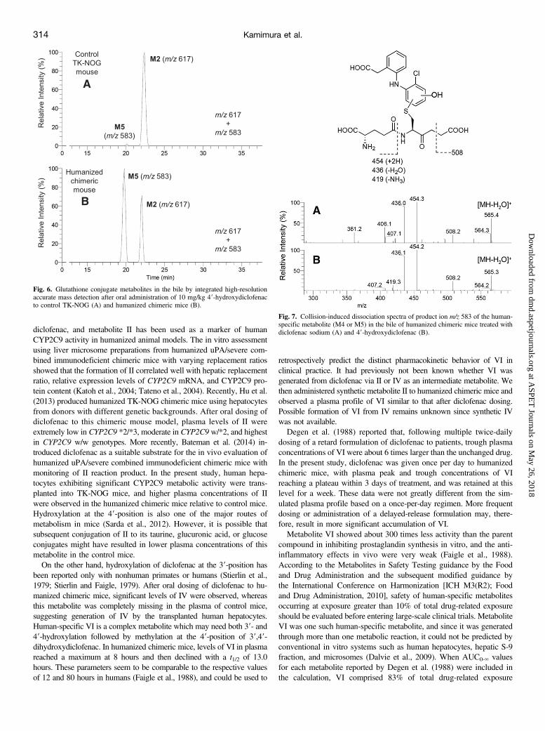
of diclofenac. M4 was previously generated by incubation of diclofenacwith human liver microsomes or recombinant CYP2C9 fortified withglutathione (Yan et al., 2005). The chemical structure of M4 was pro-posed as a 29-hydroxy-39-glutathionyl analog based on the assumptionthat this monochlorinated metabolite was generated via diclofenac-29,39-oxide, because M4 had not been detected in the incubationmixture of II and NADPH-fortified human liver microsomes. On theother hand, 49-hydroxy- 29-(glutathion-S-yl)monoclofenac was pro-posed by Yu et al. (2005), and designated as M5 later (Boerma et al.,2014). Similar to M4, M5 was generated in an incubation mixture ofdiclofenac and human liver microsomes fortified with glutathione, andits chemical structural assignment was obtained via comparison of massand 1H NMR spectra with those of a chemically synthesized authenticstandard. In the present study, no monitored peaks were found in thebile of the control TK-NOG mouse dosed with diclofenac at m/z 583(m/z 583.12308–583.12892) (Fig. 4B). However, bile from the hu-manized chimeric mouse gave a single peak presumably produced bythe transplanted human hepatocytes (Fig. 5B). When the ion at m/z 583was subjected to CID (Fig. 7A), the product ions at m/z 508 ([MH-75]+),m/z 454 ([MH-129]+), and m/z 436 ([MH-147]+) were obtained asseparately reported by Yan et al. (2005) for M4 and Yu et al. (2005)for M5. To determine that this biliary metabolite is M4 or M5, furtherstudies including isolation of the metabolite from the bile sample andsubsequent NMR analyses are inevitable.Oral administration of synthetic metabolite II to a humanized
chimeric mouse also gave a single peak at m/z 583 (Fig. 6B), and CIDproduced the same product ions as those after diclofenac treatment (Fig.7B). This conjugate was considered to be M5, based on the chemicalstructure of II, and coeluted with the corresponding peak of the bilesample from chimeric mice dosed with diclofenac (Fig. 5B). However,M4 and M5 are positional isomers, and possible coincidence in the
HPLC retention times cannot be ruled out. Although M5 (and M4) hadbeen considered human specific, the same peak was unexpectedly foundin the bile sample from a control TK-NOG mouse dosed with II (Fig.6A). However, it was much smaller than that in the humanized chimericmouse, and was not observed in the control mouse when dosed withdiclofenac (Fig. 4B).Biliary Glutathione Conjugate with MH+ Ion of m/z 567 (M6).
29-(Glutathion-S-yl)monoclofenac (we designated this conjugate “M6”)was found first in incubation mixtures of diclofenac with human livermicrosomes (Wen et al., 2008). In vitro studies using various recombinanthuman P450s fortified with glutathione and human glutathione-S-transferaseP1-1 demonstrated that all of the 14 tested P450 isozymes were capableof forming M6 (Boerma et al., 2014). Furthermore, this reaction wasNADPH-independent with M6 produced to some extent by flavin-containing monooxygenase or even horseradish peroxidase, suggestingthe involvement of one-electron oxidation. In the present study, a largesingle peak at m/z 567 (m/z 567.12825–567.13393) was obtained in thebile sample from a control TK-NOG mouse (Fig. 4C). This metabolitegave, on a CID ofm/z 567, product ions atm/z 492 ([MH-75]+) andm/z 438([MH-129]+), as reported by Boerma et al. (2012). Moreover, this singlepeak at m/z 567 coeluted on HPLC with an in vitro reference productof M6 provided by Jan N. M. Commandeur of VU University (seeMaterials and Methods). Although this metabolite was reportedlygenerated in in vitro studies using various human specimens, the peak ofM6 in the bile of a humanized chimeric mouse was not found (Fig. 5C).
Discussion
Potential pathways of diclofenac metabolism are illustrated in Fig. 8.Hydroxylation at the 49-position is one of the major metabolic routes of
Fig. 4. Glutathione conjugate metabolites in the bile by integrated high-resolutionaccurate mass detection after oral administration of diclofenac sodium (10 mg/kg asfree form) to a control TK-NOG mouse. Each metabolite was monitored ata corresponding mass number (A–C), and a mixed chromatogram is shown in theupper panel.
Fig. 5. Glutathione conjugate metabolites in the bile by integrated high-resolutionaccurate mass detection after oral administration of diclofenac sodium (10 mg/kg asfree form) to a humanized chimeric mouse. Each metabolite was monitored ata corresponding mass number (A–C), and a mixed chromatogram is shown in theupper panel.
Metabolism of Diclofenac in Humanized TK-NOG Chimeric Mice 313
at ASPE
T Journals on M
ay 26, 2018dm
d.aspetjournals.orgD
ownloaded from
diclofenac, and metabolite II has been used as a marker of humanCYP2C9 activity in humanized animal models. The in vitro assessmentusing liver microsome preparations from humanized uPA/severe com-bined immunodeficient chimeric mice with varying replacement ratiosshowed that the formation of II correlated well with hepatic replacementratio, relative expression levels of CYP2C9 mRNA, and CYP2C9 pro-tein content (Katoh et al., 2004; Tateno et al., 2004). Recently, Hu et al.(2013) produced humanized TK-NOG chimeric mice using hepatocytesfrom donors with different genetic backgrounds. After oral dosing ofdiclofenac to this chimeric mouse model, plasma levels of II wereextremely low in CYP2C9 *2/*3, moderate in CYP2C9 w/*2, and highestin CYP2C9 w/w genotypes. More recently, Bateman et al. (2014) in-troduced diclofenac as a suitable substrate for the in vivo evaluation ofhumanized uPA/severe combined immunodeficient chimeric mice withmonitoring of II reaction product. In the present study, human hepa-tocytes exhibiting significant CYP2C9 metabolic activity were trans-planted into TK-NOG mice, and higher plasma concentrations of IIwere observed in the humanized chimeric mice relative to control mice.Hydroxylation at the 49-position is also one of the major routes ofmetabolism in mice (Sarda et al., 2012). However, it is possible thatsubsequent conjugation of II to its taurine, glucuronic acid, or glucoseconjugates might have resulted in lower plasma concentrations of thismetabolite in the control mice.On the other hand, hydroxylation of diclofenac at the 39-position has
been reported only with nonhuman primates or humans (Stierlin et al.,1979; Stierlin and Faigle, 1979). After oral dosing of diclofenac to hu-manized chimeric mice, significant levels of IV were observed, whereasthis metabolite was completely missing in the plasma of control mice,suggesting generation of IV by the transplanted human hepatocytes.Human-specific VI is a complex metabolite which may need both 39- and49-hydroxylation followed by methylation at the 49-position of 39,49-dihydroxydiclofenac. In humanized chimeric mice, levels of VI in plasmareached a maximum at 8 hours and then declined with a t1/2 of 13.0hours. These parameters seem to be comparable to the respective valuesof 12 and 80 hours in humans (Faigle et al., 1988), and could be used to
retrospectively predict the distinct pharmacokinetic behavior of VI inclinical practice. It had previously not been known whether VI wasgenerated from diclofenac via II or IV as an intermediate metabolite. Wethen administered synthetic metabolite II to humanized chimeric mice andobserved a plasma profile of VI similar to that after diclofenac dosing.Possible formation of VI from IV remains unknown since synthetic IVwas not available.Degen et al. (1988) reported that, following multiple twice-daily
dosing of a retard formulation of diclofenac to patients, trough plasmaconcentrations of VI were about 6 times larger than the unchanged drug.In the present study, diclofenac was given once per day to humanizedchimeric mice, with plasma peak and trough concentrations of VIreaching a plateau within 3 days of treatment, and was retained at thislevel for a week. These data were not greatly different from the sim-ulated plasma profile based on a once-per-day regimen. More frequentdosing or administration of a delayed-release formulation may, there-fore, result in more significant accumulation of VI.Metabolite VI showed about 300 times less activity than the parent
compound in inhibiting prostaglandin synthesis in vitro, and the anti-inflammatory effects in vivo were very weak (Faigle et al., 1988).According to the Metabolites in Safety Testing guidance by the Foodand Drug Administration and the subsequent modified guidance bythe International Conference on Harmonization [ICH M3(R2); Foodand Drug Administration, 2010], safety of human-specific metabolitesoccurring at exposure greater than 10% of total drug-related exposureshould be evaluated before entering large-scale clinical trials. MetaboliteVI was one such human-specific metabolite, and since it was generatedthrough more than one metabolic reaction, it could not be predicted byconventional in vitro systems such as human hepatocytes, hepatic S-9fraction, and microsomes (Dalvie et al., 2009). When AUC0-‘ valuesfor each metabolite reported by Degen et al. (1988) were included inthe calculation, VI comprised 83% of total drug-related exposure
Fig. 6. Glutathione conjugate metabolites in the bile by integrated high-resolutionaccurate mass detection after oral administration of 10 mg/kg 49-hydroxydiclofenacto control TK-NOG (A) and humanized chimeric mice (B).
Fig. 7. Collision-induced dissociation spectra of product ion m/z 583 of the human-specific metabolite (M4 or M5) in the bile of humanized chimeric mice treated withdiclofenac sodium (A) and 49-hydroxydiclofenac (B).
314 Kamimura et al.
at ASPE
T Journals on M
ay 26, 2018dm
d.aspetjournals.orgD
ownloaded from
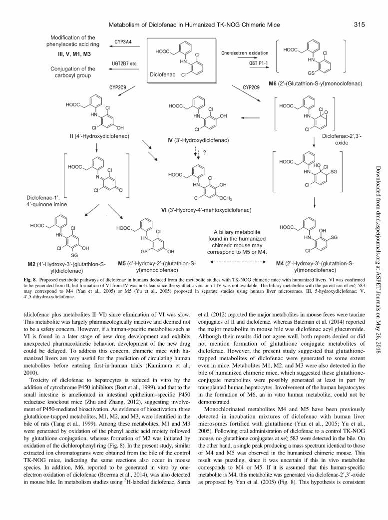
(diclofenac plus metabolites II–VI) since elimination of VI was slow.This metabolite was largely pharmacologically inactive and deemed notto be a safety concern. However, if a human-specific metabolite such asVI is found in a later stage of new drug development and exhibitsunexpected pharmacokinetic behavior, development of the new drugcould be delayed. To address this concern, chimeric mice with hu-manized livers are very useful for the prediction of circulating humanmetabolites before entering first-in-human trials (Kamimura et al.,2010).Toxicity of diclofenac to hepatocytes is reduced in vitro by the
addition of cytochrome P450 inhibitors (Bort et al., 1999), and that to thesmall intestine is ameliorated in intestinal epithelium–specific P450reductase knockout mice (Zhu and Zhang, 2012), suggesting involve-ment of P450-mediated bioactivation. As evidence of bioactivation, threeglutathione-trapped metabolites, M1, M2, and M3, were identified in thebile of rats (Tang et al., 1999). Among these metabolites, M1 and M3were generated by oxidation of the phenyl acetic acid moiety followedby glutathione conjugation, whereas formation of M2 was initiated byoxidation of the dichlorophenyl ring (Fig. 8). In the present study, similarextracted ion chromatograms were obtained from the bile of the controlTK-NOG mice, indicating the same reactions also occur in mousespecies. In addition, M6, reported to be generated in vitro by one-electron oxidation of diclofenac (Boerma et al., 2014), was also detectedin mouse bile. In metabolism studies using 3H-labeled diclofenac, Sarda
et al. (2012) reported the major metabolites in mouse feces were taurineconjugates of II and diclofenac, whereas Bateman et al. (2014) reportedthe major metabolite in mouse bile was diclofenac acyl glucuronide.Although their results did not agree well, both reports denied or didnot mention formation of glutathione conjugate metabolites ofdiclofenac. However, the present study suggested that glutathione-trapped metabolites of diclofenac were generated to some extenteven in mice. Metabolites M1, M2, and M3 were also detected in thebile of humanized chimeric mice, which suggested these glutathione-conjugate metabolites were possibly generated at least in part bytransplanted human hepatocytes. Involvement of the human hepatocytesin the formation of M6, an in vitro human metabolite, could not bedemonstrated.Monochlorinated metabolites M4 and M5 have been previously
detected in incubation mixtures of diclofenac with human livermicrosomes fortified with glutathione (Yan et al., 2005; Yu et al.,2005). Following oral administration of diclofenac to a control TK-NOGmouse, no glutathione conjugates at m/z 583 were detected in the bile. Onthe other hand, a single peak producing a mass spectrum identical to thoseof M4 and M5 was observed in the humanized chimeric mouse. Thisresult was puzzling, since it was uncertain if this in vivo metabolitecorresponds to M4 or M5. If it is assumed that this human-specificmetabolite is M4, this metabolite was generated via diclofenac-29,39-oxideas proposed by Yan et al. (2005) (Fig. 8). This hypothesis is consistent
Fig. 8. Proposed metabolic pathways of diclofenac in humans deduced from the metabolic studies with TK-NOG chimeric mice with humanized livers. VI was confirmedto be generated from II, but formation of VI from IV was not clear since the synthetic version of IV was not available. The biliary metabolite with the parent ion of m/z 583may correspond to M4 (Yan et al., 2005) or M5 (Yu et al., 2005) proposed in separate studies using human liver microsomes. III, 5-hydroxydiclofenac; V,49,5-dihydroxydiclofenac.
Metabolism of Diclofenac in Humanized TK-NOG Chimeric Mice 315
at ASPE
T Journals on M
ay 26, 2018dm
d.aspetjournals.orgD
ownloaded from

with our finding that another possible product from the arene oxide, IV,was completely missing in the plasma of control mice and considered tobe human-specific. Bioactivation of diclofenac to diclofenac-29,39-oxidewas mediated by human CYP2C9 (Yan et al., 2005), and mice may lackthis metabolic pathway. As supporting evidence in the report by Yan et al.(2005), incubation of II with human liver microsomes did not produceM4. However, this result was inconsistent with our finding that a peak atm/z 583 was also detected after administration of II to the humanizedchimeric mouse. If it is assumed that the human-specific metabolite is M5,detection of the peak in the humanized chimeric mouse treated with IIprovides a rational basis for the proposed diclofenac-19,49-quinone imineintermediate for the formation of this metabolite (Yu et al., 2005) (Fig. 8).In this scenario, formation of M5 from the quinone imine intermediate waseither exclusively or predominantly a human metabolic reaction. Evena control TK-NOG mouse dosed with 10 mg/kg II gave a relatively smallpeak at m/z 583, probably because exposure of the mouse to II was muchhigher than that after administration of the same dose of diclofenac.However, further studies including NMR analysis of the biliary metaboliteto distinguish the potential positional isomers are necessary.In conclusion, we could mimic the distinct plasma concentration pro-
file of the human-specific metabolite of diclofenac (VI) by TK-NOGmice with humanized livers. Glutathione conjugate metabolites werealso detected in the bile samples, providing a new means of studyingglutathione-dependent inactivation of reactive human metabolites invivo. As with the conventional humanized animal models, humanizedTK-NOG chimeric mice were considered to be a useful tool for metab-olism studies in new drug development.
Acknowledgments
The authors thank Jan N. M. Commandeur (VU University Amsterdam) forproviding the in vitro reference material for M6; Daisuke Morita (SeikagakuCorporation, Tokyo, Japan) for providing HPLC conditions for the separationof diclofenac and its metabolites II, IV, and VI; and Faraz Kazmi (XenoTech,)for correcting the English manuscript.
Authorship ContributionsParticipated in research design: Kamimura.Conducted experiments: Ito, Nozawa, Nakamura, Chijiwa.Contributed new reagents or analytic tools: Kuronuma, Ohnishi, Suemizu.Performed data analysis: Nagatsuka.Wrote or contributed to the writing of the manuscript: Kamimura, Ito,
Ninomiya.
References
Azuma H, Paulk N, Ranade A, Dorrell C, Al-Dhalimy M, Ellis E, Strom S, Kay MA, Finegold M,and Grompe M (2007) Robust expansion of human hepatocytes in Fah-/-/Rag2-/-/Il2rg-/- mice.Nat Biotechnol 25:903–910.
Bateman TJ, Reddy VG, Kakuni M, Morikawa Y, and Kumar S (2014) Application of chimericmice with humanized liver for study of human-specific drug metabolism. Drug Metab Dispos42:1055–1065.
Boelsterli UA (2003) Diclofenac-induced liver injury: a paradigm of idiosyncratic drug toxicity.Toxicol Appl Pharmacol 192:307–322.
Boerma JS, Dragovic S, Vermeulen NP, and Commandeur JN (2012) Mass spectrometric char-acterization of protein adducts of multiple P450-dependent reactive intermediates of diclofenacto human glutathione-S-transferase P1-1. Chem Res Toxicol 25:2532–2541.
Boerma JS, Vermeulen NP, and Commandeur JN (2014) One-electron oxidation of diclofenac byhuman cytochrome P450s as a potential bioactivation mechanism for formation of 29-(glutathion-S-yl)-deschloro-diclofenac. Chem Biol Interact 207:32–40.
Bort R, Ponsoda X, Jover R, Gómez-Lechón MJ, and Castell JV (1999) Diclofenac toxicityto hepatocytes: a role for drug metabolism in cell toxicity. J Pharmacol Exp Ther 288:65–72.
Dalvie D, Obach RS, Kang P, Prakash C, Loi CM, Hurst S, Nedderman A, Goulet L, Smith E,and Bu HZ, et al. (2009) Assessment of three human in vitro systems in the generation of majorhuman excretory and circulating metabolites. Chem Res Toxicol 22:357–368.
Degen PH, Dieterle W, Schneider W, Theobald W, and Sinterhauf U (1988) Pharmacokinetics ofdiclofenac and five metabolites after single doses in healthy volunteers and after repeated dosesin patients. Xenobiotica 18:1449–1455.
Faigle JW, Böttcher I, Godbillon J, Kriemler HP, Schlumpf E, Schneider W, Schweizer A,Stierlin H, and Winkler T (1988) A new metabolite of diclofenac sodium in human plasma.Xenobiotica 18:1191–1197.
Food and Drug Administration (2010) Guidance for Industry: M3(R2) Nonclinical Safety Studiesfor the Conduct of Human Clinical Trials and Marketing Authorization for Pharmaceuticals,FDA, Rockville, MD.
Grillo MP, Knutson CG, Sanders PE, Waldon DJ, Hua F, and Ware JA (2003) Studies on thechemical reactivity of diclofenac acyl glucuronide with glutathione: identification ofdiclofenac-S-acyl-glutathione in rat bile. Drug Metab Dispos 31:1327–1336.
Grompe M and Strom S (2013) Mice with human livers. Gastroenterology 145:1209–1214.Hasegawa M, Kawai K, Mitsui T, Taniguchi K, Monnai M, Wakui M, Ito M, Suematsu M, PeltzG, and Nakamura M, et al. (2011) The reconstituted ‘humanized liver’ in TK-NOG mice ismature and functional. Biochem Biophys Res Commun 405:405–410.
Hu Y, Wu M, Nishimura T, Zheng M, and Peltz G (2013) Human pharmacogenetic analysis inchimeric mice with ‘humanized livers’. Pharmacogenet Genomics 23:78–83.
Kamimura H, Nakada N, Suzuki K, Mera A, Souda K, Murakami Y, Tanaka K, Iwatsubo T,Kawamura A, and Usui T (2010) Assessment of chimeric mice with humanized liver as a toolfor predicting circulating human metabolites. Drug Metab Pharmacokinet 25:223–235.
Katoh M, Matsui T, Nakajima M, Tateno C, Kataoka M, Soeno Y, Horie T, Iwasaki K, YoshizatoK, and Yokoi T (2004) Expression of human cytochromes P450 in chimeric mice with hu-manized liver. Drug Metab Dispos 32:1402–1410.
Kretz-Rommel A and Boelsterli UA (1993) Diclofenac covalent protein binding is dependent onacyl glucuronide formation and is inversely related to P450-mediated acute cell injury incultured rat hepatocytes. Toxicol Appl Pharmacol 120:155–161.
Mercer DF, Schiller DE, Elliott JF, Douglas DN, Hao C, Rinfret A, Addison WR, Fischer KP,Churchill TA, and Lakey JR, et al. (2001) Hepatitis C virus replication in mice with chimerichuman livers. Nat Med 7:927–933.
Nishimura T, Hu Y, Wu M, Pham E, Suemizu H, Elazar M, Liu M, Idilman R, Yurdaydin C,and Angus P, et al. (2013) Using chimeric mice with humanized livers to predict human drugmetabolism and a drug-drug interaction. J Pharmacol Exp Ther 344:388–396.
Peltz G (2013) Can ‘humanized’ mice improve drug development in the 21st century? TrendsPharmacol Sci 34:255–260.
Sarda S, Page C, Pickup K, Schulz-Utermoehl T, and Wilson I (2012) Diclofenac metabolism inthe mouse: novel in vivo metabolites identified by high performance liquid chromatographycoupled to linear ion trap mass spectrometry. Xenobiotica 42:179–194.
Small RE (1989) Diclofenac sodium. Clin Pharm 8:545–558.Stierlin H and Faigle JW (1979) Biotransformation of diclofenac sodium (Voltaren) in animalsand in man. II. Quantitative determination of the unchanged drug and principal phenolicmetabolites, in urine and bile. Xenobiotica 9:611–621.
Stierlin H, Faigle JW, Sallmann A, Küng W, Richter WJ, Kriemler HP, Alt KO, and Winkler T(1979) Biotransformation of diclofenac sodium (Voltaren) in animals and in man. I. Isolationand identification of principal metabolites. Xenobiotica 9:601–610.
Suemizu H, Sota S, Kuronuma M, Shimizu M, and Yamazaki H (2014) Pharmacokinetics and effectson serum cholinesterase activities of organophosphorus pesticides acephate and chlorpyrifos inchimeric mice transplanted with human hepatocytes. Regul Toxicol Pharmacol 70:468–473.
Tang W (2003) The metabolism of diclofenac—enzymology and toxicology perspectives. CurrDrug Metab 4:319–329.
Tang W, Stearns RA, Bandiera SM, Zhang Y, Raab C, Braun MP, Dean DC, Pang J, Leung KH,and Doss GA, et al. (1999) Studies on cytochrome P-450-mediated bioactivation of diclofenacin rats and in human hepatocytes: identification of glutathione conjugated metabolites. DrugMetab Dispos 27:365–372.
Tateno C, Yoshizane Y, Saito N, Kataoka M, Utoh R, Yamasaki C, Tachibana A, Soeno Y,Asahina K, and Hino H, et al. (2004) Near completely humanized liver in mice shows human-type metabolic responses to drugs. Am J Pathol 165:901–912.
Thanagari BS, Fefar DT, Prajapati KS, Jivani BM, Thakor KB, Patel JH, Ghodasara DJ, Joshi BP,and Undhad VV (2012) Haemato-biochemical alterations induced by diclofenac sodium tox-icity in Swiss albino mice. Vet World 5:417–419.
Tsukada A, Suemizu H, Murayama N, Takano R, Shimizu M, Nakamura M, and Yamazaki H(2013) Plasma concentrations of melengestrol acetate in humans extrapolated from the pharma-cokinetics established in in vivo experiments with rats and chimeric mice with humanized liverand physiologically based pharmacokinetic modeling. Regul Toxicol Pharmacol 65:316–324.
Wen B, Ma L, Nelson SD, and Zhu M (2008) High-throughput screening and characterization ofreactive metabolites using polarity switching of hybrid triple quadrupole linear ion trap massspectrometry. Anal Chem 80:1788–1799.
Yamashita M, Suemizu H, Murayama N, Nishiyama S, Shimizu M, and Yamazaki H (2014)Human plasma concentrations of herbicidal carbamate molinate extrapolated from the phar-macokinetics established in in vivo experiments with chimeric mice with humanized liver andphysiologically based pharmacokinetic modeling. Regul Toxicol Pharmacol 70:214–221.
Yamazaki H, Suemizu H, Murayama N, Utoh M, Shibata N, Nakamura M, and Guengerich FP(2013) In vivo drug interactions of the teratogen thalidomide with midazolam: heterotropiccooperativity of human cytochrome P450 in humanized TK-NOG mice. Chem Res Toxicol 26:486–489.
Yamazaki H, Suemizu H, Shimizu M, Igaya S, Shibata N, Nakamura M, Chowdhury G,and Guengerich FP (2012) In vivo formation of dihydroxylated and glutathione conjugatemetabolites derived from thalidomide and 5-Hydroxythalidomide in humanized TK-NOGmice. Chem Res Toxicol 25:274–276.
Yan Z, Li J, Huebert N, Caldwell GW, Du Y, and Zhong H (2005) Detection of a novel reactivemetabolite of diclofenac: evidence for CYP2C9-mediated bioactivation via arene oxides. DrugMetab Dispos 33:706–713.
Yoshizato K, Tateno C, and Utoh R (2012) Mice with liver composed of human hepatocytes as ananimal model for drug testing. Curr Drug Discov Technol 9:63–76.
Yu LJ, Chen Y, Deninno MP, O’Connell TN, and Hop CE (2005) Identification of a novelglutathione adduct of diclofenac, 49-hydroxy-29-glutathion-deschloro-diclofenac, upon in-cubation with human liver microsomes. Drug Metab Dispos 33:484–488.
Zhu Y and Zhang QY (2012) Role of intestinal cytochrome p450 enzymes in diclofenac-inducedtoxicity in the small intestine. J Pharmacol Exp Ther 343:362–370.
Address correspondence to: Dr. Hidetaka Kamimura, ADME & Tox. ResearchInstitute, Sekisui Medical Co. Ltd., 13-5, Nihonbashi 3-chome, Chuo-ku, Tokyo103-0027 Japan. E-mail: [email protected]
316 Kamimura et al.
at ASPE
T Journals on M
ay 26, 2018dm
d.aspetjournals.orgD
ownloaded from