Embed Size (px)
Citation preview
Rom J Leg Med (3) 173 - 186 (2009) © 2009 Romanian Society of Legal Medicine
________________________ *) Corresponding author: Prof.Dr. MD, PhD, Member of Academy of Medical Science, Academic Hospital no. 2, Craiova, Romania, e-mail: [email protected]
173
Forensic and anatomic signification of phenotype transformations inside fetus membranes Part One: Amnion and lamina chorionica Gheorghe S. Dragoi*, Ileana Dinca, Petru Razvan Melinte, Carmen Diaconu, Georgiana Silca
Received: 14.04.2009/ Accepted in revised form: 26.08.2009 _____________________________________________________________________ Abstract: The authors analyzed macro and micro anatomically the variability of the relationships between fetus membranes – amnion and lamina chorionica – and also the space distribution of under chorial blood vessels depending on the growing of fibrinoid deposits. In order to identify the phenotype transformations inside fetus membranes, a particular attention was directed to the biodynamic and functional structures in funiculus umbilicalis. Key words: amnion, chorion, fetal membranes, funiculus umbilicalis
etus membranes are structures belonging to fetus and placenta system and have a great proof value in forensic expertise. Its genesis and time and space evolution
are dominated by the phenotype transformations of its structural elements. The knowledge of the signification of these transformations has a great importance for understanding the ontogenesis and pathomorphenesis mechanism of those structures. Tropholast, epiblast and extra embryo mesenchym ensure the formation and phenotype transformation of fetus membranes by complex processes of reciprocal induction, genesis and remodeling.
The purpose of this paper is the evaluation of the phenotype changes of the primordial structures for the knowledge of the location and relations between the ancestral elements derivates inside fetus membranes: amnion, lamina chorionica and trophoblastus.
We proposed ourselves as a primary objective the macro and micro anatomic analysis of the relations variability between fetus membranes on one side, and between those last ones and the umbilical and/or chorial blood vessels, on the other side. Materials and methods
We studied a number of 24 fetus and placenta systems whose gestation age was between 10 weeks and 32 weeks; a number of 60 placentas harvested after eutocic birth (48), C section (8) and hysterectomy (4). The macroscopic examination was achieved after fixation in neutral formaldehyde 10% (ph=7.4). The harvested fragments we addressed by usual micro anatomic techniques.
F
Dragoi Gh. et al Forensic and anatomic signification of phenotype transformations inside fetus membranes (I)
174
Results A. The analysis of phenotype transformations inside fetus membranes during ortomorphgenesis 1. Macro anatomic evaluation
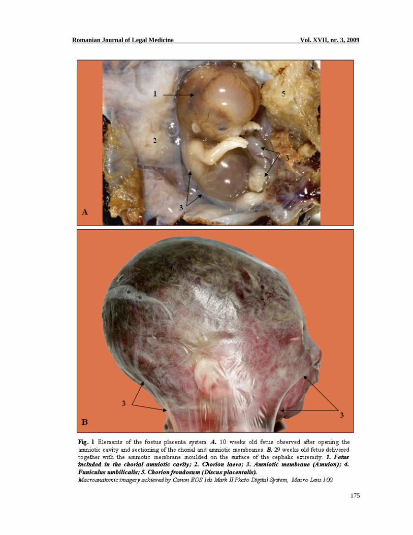
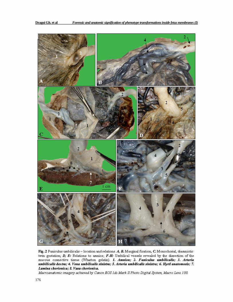
Macroscopic dissection and micro anatomic manipulation of fetus membranes – amnion and lamina chorionica, allowed us to evaluate the dual time and space relations between these structures. The analysis of the location and relations between funiculus umbilicalis and fetus membranes showed the variability of these structures stereo distribution. In our studied cases, funiculus umbilicalis has central fixation (12%), semi central (78%) or marginal (10%). In twin gestation there is the same frequency regarding location (Fig 2, A-C). The umbilical blood vessels were visualized with great difficulty after dissecting the mucous connective tissue (Wharton) because of the absence of some cleavage spaces (Fig 2, F-H). Amnion has continuity relations with funiculus umbilicale (Fig 2, C-E). The trajectory and relations of chorial blood vessels were examined after decollating the amnion (Fig 7A). The ripping of chorial lamina together with the chorial vessels was achieved with great difficulty because of the existence of a large under chorial per vascular stroma (Fig 7, D, E). 2. Micro anatomic evaluation
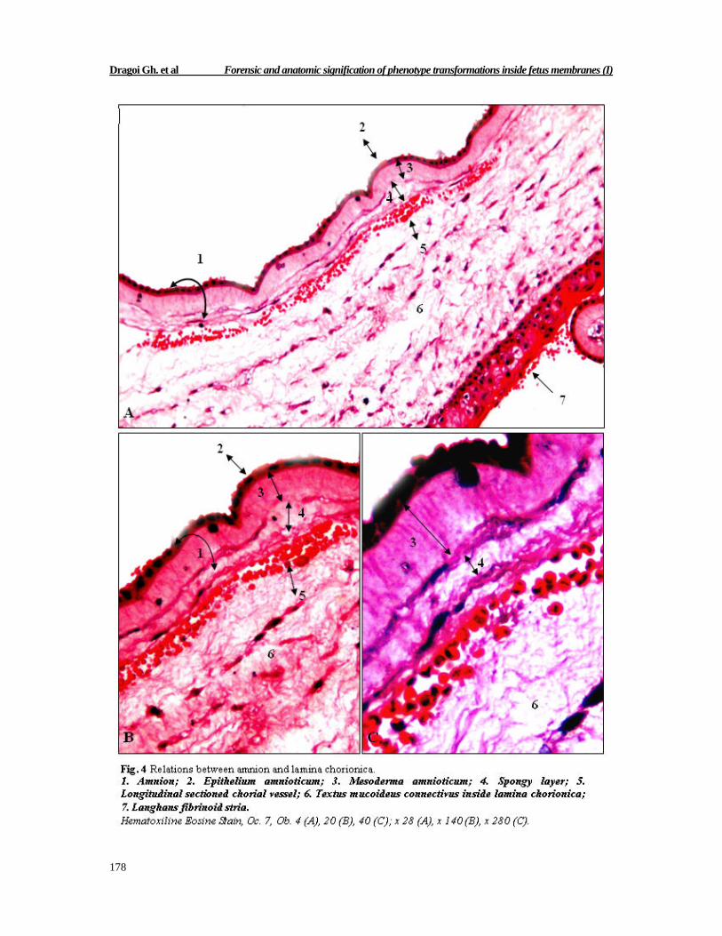
Analyzing the serried sections through the placenta fragments harvested from the secant, transfixing sections, we identified tissue sectors of placenta disc belonging to the two sides of placenta: fetalis and maternalis. Examining with the 4x objective we easily noticed the existence of a non structured space between amnios and lamina chorionica (Fig 4 A).
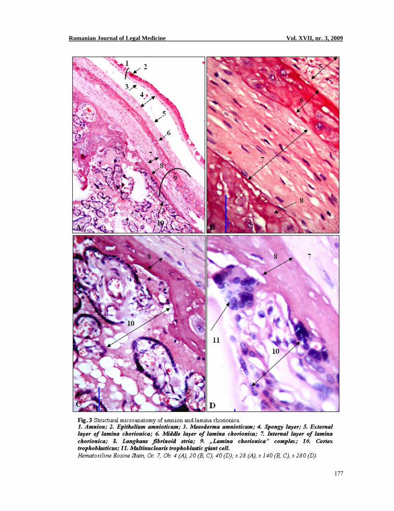
Examining with the 10x and 20x objectives the fetus membrane allowed us to visualize two structural levels: epithelium amnioticum and mezoderma amnioticum (Fig 3A and Fig 4, B, C). Lamina chorionica examined with the 4x objective, appears as a conjunctive structure – mesoderma chorionicum, in which we differentiated three circumferential fascicles: external layer of lamina chorionica, middle layer of lamina chorionica and internal layer of lamina chorionica (Fig 3 A). Frequently we observed the presence of fibrinoid mass inside the middle layer. Langhans fibrionoid stria represents the boundary between the internal layer of lamina chorionica and cortex trophoblasticus (Fig 3 A). Examining with the 20x objective the internal layer of lamina chorionica we noticed the presence of a mucous conjunctive tissue (Fig 3 B) similar to that inside mesoderma amnioticum (Fig 4). In the territory of “cortex trophoblasticus” we easily noticed the presence of numerous multinuclear trophoblast giant cells (Fig 3 D).
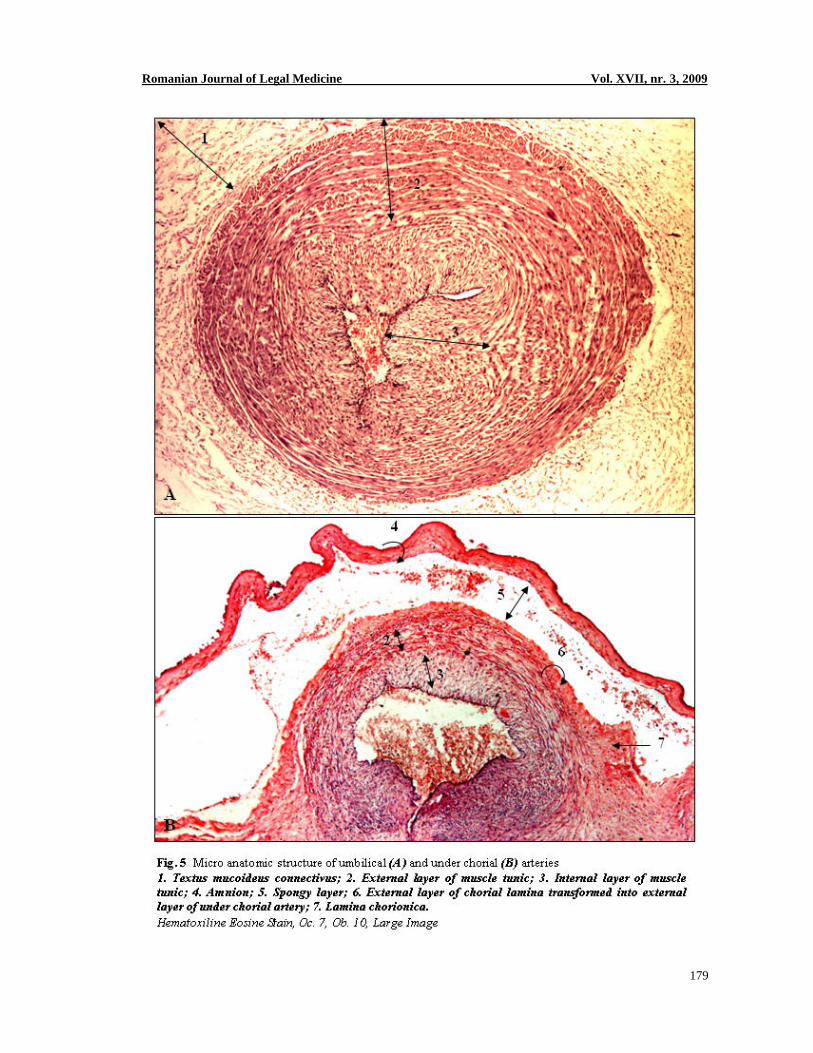
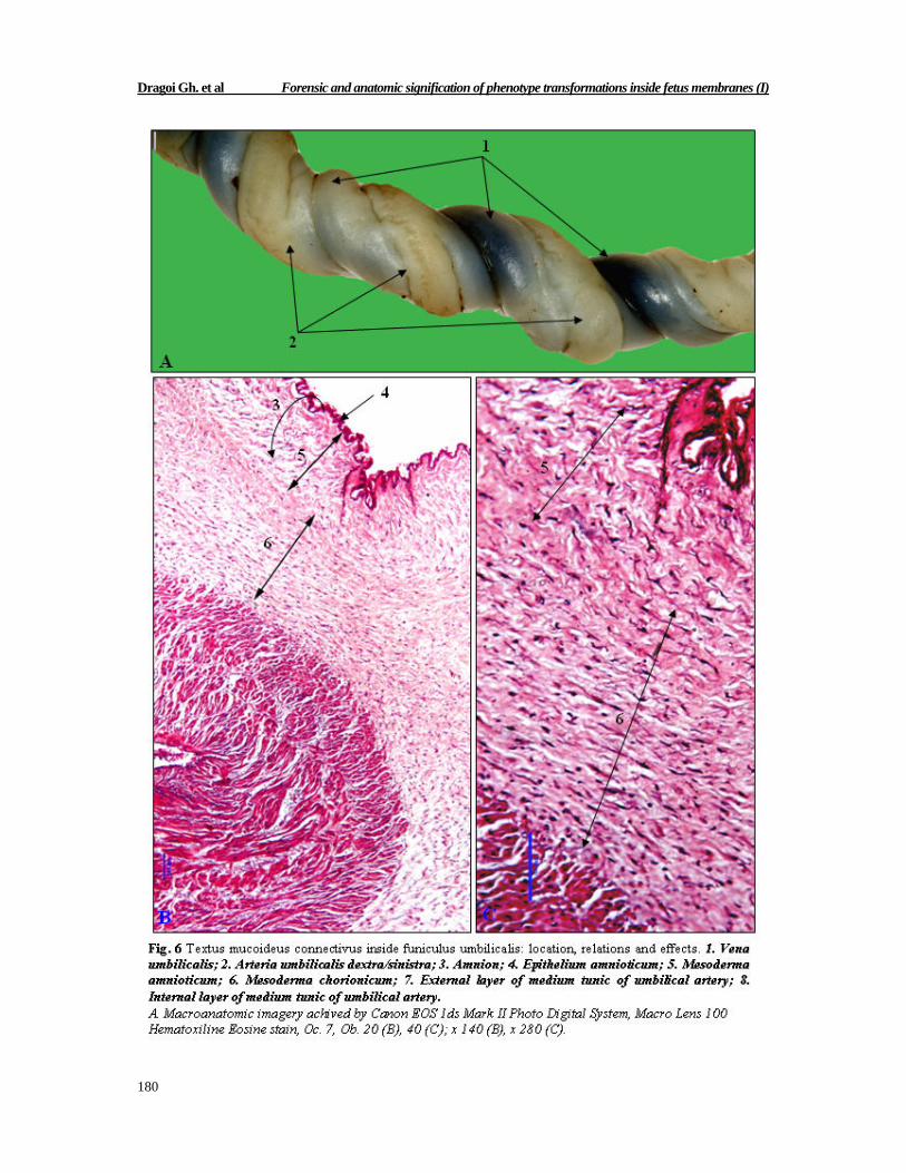
The umbilical and under chorial blood vessels have tight relations to the mucous conjunctive tissue inside mesoderma amnioticum and mesoderma chorionicum that was easily identified per vascular inside umbilical cord (Fig 5 and 6). The muscle cells fascicles inside middle tunic of the umbilical artery and its under chorial branches are grouped in two layers: an external and an internal one (Fig 5 A, B).
In the external layer the fascicles of muscle cells are grouped in lamellas distributed in concentric planes and have helical trajectories directed longitudinally clock wise and anti clock wise (Fig 5 A, B). In the internal layer, the fascicles of muscle cells form concentric lamellas that contract and achieve a kind of “forceps” by folding (Fig 5 A, B). The mucous tissue (Wharton) has continuity relations to the muscle tunic of umbilical artery forming an enormous external tunic (adventitia) to this blood vessel (Fig 6 B, C). In the structure of under chorial vessels we noticed the contribution of chorionic mesoderm to the formation of the external tunic (Fig 5 B).
Romanian Journal of Legal Medicine Vol. XVII, nr. 3, 2009
175
Dragoi Gh. et al Forensic and anatomic signification of phenotype transformations inside fetus membranes (I)
176
Romanian Journal of Legal Medicine Vol. XVII, nr. 3, 2009
177
Dragoi Gh. et al Forensic and anatomic signification of phenotype transformations inside fetus membranes (I)
178
Romanian Journal of Legal Medicine Vol. XVII, nr. 3, 2009
179
Dragoi Gh. et al Forensic and anatomic signification of phenotype transformations inside fetus membranes (I)
180
Romanian Journal of Legal Medicine Vol. XVII, nr. 3, 2009
181
Examined with the 20x and 40x objectives, the mucoid connective tissue (sin. Gelatinous connective tissue) inside umbilical cord wall, is structured on two levels: an external one that belong to mesoderma amnioticum and an internal one derived from mesoderma chorionicum (Fig 6 B, C). The contractile elements organized in lamellas grouped in fascicles oriented variably in space, determine two phenotype transformations inside umbilical cord: the diminishing of the arterial orifice diameter, on one side and the spiral folding of umbilical cord, considered as a whole, on the other side (Fig 5 A and Fig 6 A).
The chorial blood vessels (Vasa chorionica) have contiguity relations to mesoderma chorionicum; this phenomenon is visible on the secant, serried sections through placenta disc (Fig 7, B, C) and after ripping the lamina chorionica (Fig 7, D, E). Under this structure there is a 3D conjunctive network in which vascular branches protrude (Fig 7 E).
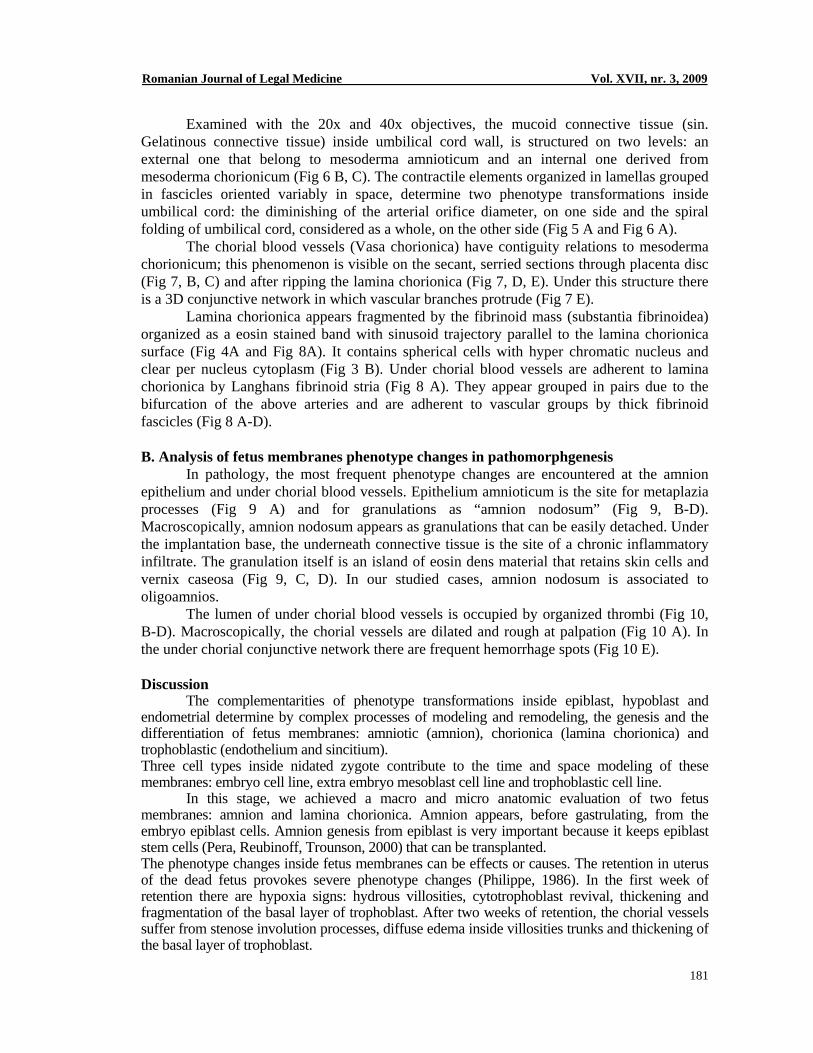
Lamina chorionica appears fragmented by the fibrinoid mass (substantia fibrinoidea) organized as a eosin stained band with sinusoid trajectory parallel to the lamina chorionica surface (Fig 4A and Fig 8A). It contains spherical cells with hyper chromatic nucleus and clear per nucleus cytoplasm (Fig 3 B). Under chorial blood vessels are adherent to lamina chorionica by Langhans fibrinoid stria (Fig 8 A). They appear grouped in pairs due to the bifurcation of the above arteries and are adherent to vascular groups by thick fibrinoid fascicles (Fig 8 A-D). B. Analysis of fetus membranes phenotype changes in pathomorphgenesis
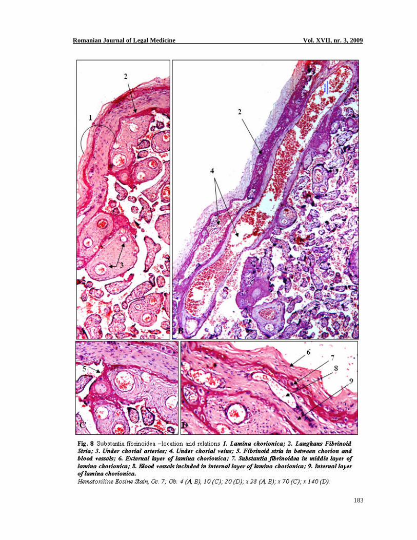
In pathology, the most frequent phenotype changes are encountered at the amnion epithelium and under chorial blood vessels. Epithelium amnioticum is the site for metaplazia processes (Fig 9 A) and for granulations as “amnion nodosum” (Fig 9, B-D). Macroscopically, amnion nodosum appears as granulations that can be easily detached. Under the implantation base, the underneath connective tissue is the site of a chronic inflammatory infiltrate. The granulation itself is an island of eosin dens material that retains skin cells and vernix caseosa (Fig 9, C, D). In our studied cases, amnion nodosum is associated to oligoamnios.
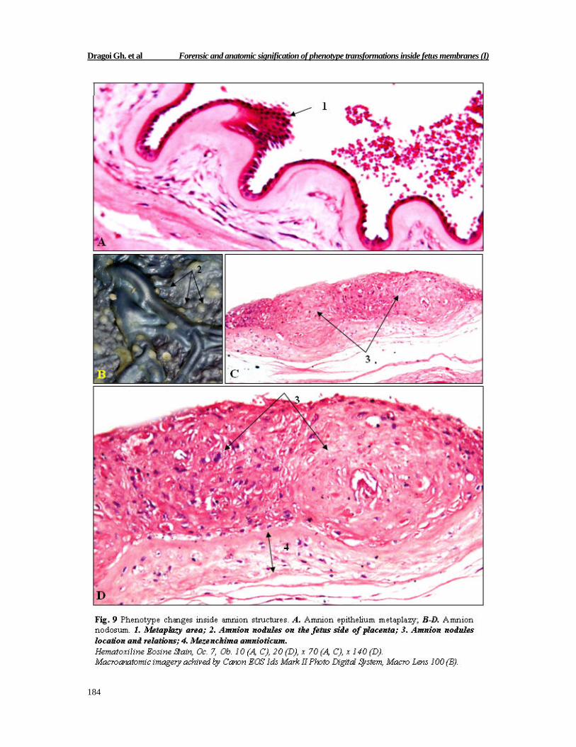
The lumen of under chorial blood vessels is occupied by organized thrombi (Fig 10, B-D). Macroscopically, the chorial vessels are dilated and rough at palpation (Fig 10 A). In the under chorial conjunctive network there are frequent hemorrhage spots (Fig 10 E). Discussion
The complementarities of phenotype transformations inside epiblast, hypoblast and endometrial determine by complex processes of modeling and remodeling, the genesis and the differentiation of fetus membranes: amniotic (amnion), chorionica (lamina chorionica) and trophoblastic (endothelium and sincitium). Three cell types inside nidated zygote contribute to the time and space modeling of these membranes: embryo cell line, extra embryo mesoblast cell line and trophoblastic cell line.
In this stage, we achieved a macro and micro anatomic evaluation of two fetus membranes: amnion and lamina chorionica. Amnion appears, before gastrulating, from the embryo epiblast cells. Amnion genesis from epiblast is very important because it keeps epiblast stem cells (Pera, Reubinoff, Trounson, 2000) that can be transplanted. The phenotype changes inside fetus membranes can be effects or causes. The retention in uterus of the dead fetus provokes severe phenotype changes (Philippe, 1986). In the first week of retention there are hypoxia signs: hydrous villosities, cytotrophoblast revival, thickening and fragmentation of the basal layer of trophoblast. After two weeks of retention, the chorial vessels suffer from stenose involution processes, diffuse edema inside villosities trunks and thickening of the basal layer of trophoblast.

Dragoi Gh. et al Forensic and anatomic signification of phenotype transformations inside fetus membranes (I)
182
Romanian Journal of Legal Medicine Vol. XVII, nr. 3, 2009
183
Dragoi Gh. et al Forensic and anatomic signification of phenotype transformations inside fetus membranes (I)
184
Romanian Journal of Legal Medicine Vol. XVII, nr. 3, 2009
185
Dragoi Gh. et al Forensic and anatomic signification of phenotype transformations inside fetus membranes (I)
186
At three weeks after retention of the dead fetus, the chorial vessels are obliterated completely, the villosity stroma is dense, there are leukocyte infiltrates inside under chorial stroma. After four weeks of retention the chorial villosities are fibrotic, the chorial and trunk blood vessels have no lumen and the trophoblast is replaced with fibrionoid deposits. We noticed the amnion and mesoderm contribution to the external layer of umbilical vessels inside funiculus umbilicalis. In this way, the mucous conjunctive tissue (Wharton) becomes an enormous adventitia to the umbilical vessels and equally ensures the umbilical funicle plasticity and its phenotype variability directed related to the relaxation or contraction of the muscle cells fascicles inside the medium layer of umbilical arteries.
Anatomically speaking, the fetus membranes appear as highly complex structures by implementing the primordial cell lines. In medico legal expertise of abortion, the fetus membranes stand proof during pathomorphgenesis for fetus and placenta hypoxia, gestation age estimation when the data for the last menstruation is unknown, infectious status evaluation, Rh immunization toxemy and last but not least, diabetes; all these are possible because of their phenotype changes. Conclusions
1. The fetus membranes structure is determined by the time and space evolution of epiblast, trophoblast and extra embryo mesenchym derivates.
2. Epithelium amnioticum has its origin in epiblast and keeps the phenotype transformations pattern of its derivates – metaplazy and differentiation.
3. Mesoderma amnioticum and chorionicum contribute to the mucous conjunctive tissue structure inside umbilical funicle.
4. The external layer of under chorial blood vessels is structured by the bounding of chorial mesoderm to the muscle tunic.
5. The mucous conjunctive tissue inside umbilical funicle achieves an enormous adventitia to the umbilical blood vessels.
6. The biodynamic and biokinetics of umbilical arteries are depending on two anatomic factors: muscle layers topography and the relations and integrity of per vascular mucous conjunctive tissue.
Selected References 1. A. Le Moigne, Biologie du développement, 3e Edition, Ed. Masson, Paris, 1955 2. Baergen N. Rebeca, Manual of Benirschke and Kaufman`s Pathology of the Placenta, Ed. Springer, New
York, 2005. 3. Benirschke K., Examination of the placenta, Obstet Gynecol 1991; 18: 309-333. 4. Philippe E., Pathologie faeto-placentarire, 2e Edition, Ed. Masson, Paris, 1986. 5. King B.F., Developmental changes in the fine structure of rhesus monkey amnion, Am. J. Anat. 157: 285-
307, 1980. 6. Bourne G., The microscopic anatomy of the human amnion and chorion, Am J Obstet Gynecol. 1960; 79:
1070-1073. 7. Nanaev A.K., Kohnen G., Milonov A.P. et al., Stromal differentiation and arhitecture of the human
umbilical cord, Placenta 1997; 18: 53-64. 8. Boyd J.D., Hamilton W.J., The human placenta, Cambridge, Heffer, 1970. 9. Gruenwald P., Examination of the placenta by the pathologist, Arch. Pathol. 1964; 77: 41-46. 10. Kaufman P., Huppertz B., Frank H.G., The fibrinoid of the human placenta: origin, composition and
functional relevance, Ann Anat 1996; 178: 485-501. 11. Wigglesworth J.S., Vascular anatomy of the human placenta and its significance for placental pathology, J
Obstet Gynaecol Br Common, 1969; 76: 979