Embed Size (px)
Citation preview
Forebrain connections of the hamsterintergeniculate leaflet:Comparison with those of ventrallateral geniculate nucleus and retina
L.P. MORIN1,2 and J.H. BLANCHARD1
1Department of Psychiatry, Stony Brook University, Stony Brook2Program in Neurobiology and Behavior, Stony Brook University, Stony Brook
(Received December 29, 1998;Accepted June 11, 1999)
Abstract
The hamster intergeniculate leaflet (IGL), part of the circadian rhythm regulatory system, has very extensiveinterconnections with subcortical visual nuclei. The present investigation describes IGL connections with thehamster diencephalon and telencephalon and compares them with ventral lateral geniculate nucleus (VLG)connections and retinal projections. Connections of the geniculate nuclei were evaluated using anterograde transportof iontophoretically injectedPhaseolus vulgarisleucoagglutinin and by retrograde transport of cholera toxinbfragment. The cholera fragment was also injected intraocularly to trace retinal efferents. The IGL has ipsilateral andcontralateral projections to the anterior and posterior hypothalamic nuclei, the ventral preoptic, lateral and dorsalhypothalamic areas, but not to the core ventromedial nucleus and very sparsely to the paraventricular nucleus. Thereare also IGL projections to the medial and lateral zona incerta, anteroventral, anterodorsal, reuniens, parataenial,paraventricular, centrolateral, central medial, and laterodorsal thalamic nuclei. IGL projections to the telencephalonare found in the horizontal limb of the diagonal band, olfactory tubercle, nucleus of the lateral olfactory tract,posterior bed nucleus of the stria terminalis, ventral pallidum, and in nuclei of the medial amygdala. The onlysubstantial VLG projections are to bed nucleus of the stria terminalis, IGL, medial zona incerta, central medial andlaterodorsal thalamic nuclei. Several of the IGL targets, the bed nucleus of the stria terminalis and zona incerta inparticular, send projections back to the IGL and VLG. In addition, cells are present in the caudal cingulate cortexthat project to both nuclei. Retinal projections are found in many of the regions receiving IGL innervation, includingnuclei of the medial basal telencephalon, the posteromedial bed nucleus of the stria terminalis, and nuclei of thehypothalamus. A retinal projection is also visible in the lateral olfactory tract from which it extends rostrally, thenmedially along the base of the rhinal fissure. Fibers also extend caudally, in a superficial location, to perirhinalcortex. The results further demonstrate the widespread connections of the IGL and support the idea that the IGLmodulates olfactory, photic, and circadian rhythm regulation of regulatory physiology and behavior.
Keywords: Circadian rhythm, Visual system, Anatomy, Hypothalamus, Olfactory system, Suprachiasmatic nucleus,Thalamus, Telencephalon, Light, Circadian system
Introduction
The intergeniculate leaflet (IGL) of the lateral geniculate complexwas first described as a thin, retinorecipient nucleus intercalatedbetween the dorsal lateral nucleus and the ventral lateral nucleus(VLG) in the rat (Hickey & Spear, 1976). The IGL has been shownto be a functional constituent of the circadian rhythm system [see(Morin, 1994)]. It receives bilateral, direct retinal innervation (Morinet al., 1992) and its neurons respond to photic input (Zhang &Rusak, 1989). The most prominent efferent projection of the IGLis the geniculohypothalamic tract (GHT) (Harrington et al., 1985;
Card & Moore, 1989; Morin et al., 1992; Morin & Blanchard,1995) projecting to the suprachiasmatic nucleus (SCN), site of thehypothalamic circadian clock mechanism (Klein et al., 1991).
Many of the neurons giving rise to the GHT contain neuropep-tide Y (NPY). Cells containing this neuromodulator provide agood index of IGL boundaries in rats and hamsters. The IGL isnow well characterized with respect to neuropeptide content ofconstituent neurons or afferent terminals (Harrington et al., 1985;Morin et al., 1992; Moore & Speh, 1993; Moore & Card, 1994;Morin & Blanchard, 1995), as well as the migration, development,and location of intrinsic neurons and astrocytes (Morin, 1994).Together, the various criteria identify the IGL as being quite long(about 2 mm in the hamster) with a substantial volume.
Anatomical description of the IGL has focused on its partici-pation as part of the circadian rhythm system. Indeed, a second
Address correspondence and reprint requests to: Lawrence P. Morin,Department of Psychiatry, Health Science Center, SUNY, Stony Brook, NY11794, USA. E-mail [email protected]
Visual Neuroscience(1999),16, 1037–1054. Printed in the USA.Copyright © 1999 Cambridge University Press 0952-5238099 $12.50
1037

large IGL-efferent projection is to the contralateral IGL (Morin &Blanchard, 1995). Despite the indication that the IGL reciprocallyconnects with the superior colliculus (Taylor et al., 1986; Morin &Blanchard, 1998), there has been little effort to discern the fullextent of its ascending or descending projections. In the gerbil,
anterograde tracer injections into the IGL0VLG region revealed awidespread projection pattern to hypothalamic nuclei and zonaincerta (Mikkelsen, 1990). This general pattern has recently beenaffirmed in the rat (Horvath, 1996; Moore et al., 1996; Horvath,1998), with the added observation that the terminal regions of IGL
Abbreviations
AA Anterior amygdalaac Anterior commissureAco Anterior cortical amygdaloid NAD Anterodorsal thalamic NAH Anterior hypothalamusAM Anteromedial thalamic NAOD Anterior olfactory N, dorsalAPT Anterior pretectal NArc Arcuate NAV Anteroventral thalamic NAVPO Anteroventral preoptic areaBST Bed N of the stria terminalisBSTav Bed N of the stria terminalis, anteroventralBSTpi Bed N of the stria terminalis, posterointermediateBSTpl Bed N of the stria terminalis, posterolateralBSTpm Bed N of the stria terminalis, posteromedialBSTs Bed N of the stria terminalis, suprascapularcc Corpus callosumCeL Centralamygdaloid N, lateralCg1 Cingulate cortex, area 1Cg2 Cingulate cortex, area 2CL Central lateral thalamic NCM Central medial thalamic Ncp Cerebral peduncleCPT Commissural pretectal areaDA Dorsal hypothalamic areaDLG Dorsal lateral geniculate NDM Dorsomedial hypothalamic NEP Entopeduncular Nf Fornixfi Fimbriafr Fasciculus retroflexusGHT Geniculohypothalamic tractGP Globus pallidusHb HabenulaHDB Diagonal band of Broca, horizontal limbIAM Interanteromedial thalamic Nic Internal capsuleIGL Intergeniculate leaflet3 Third ventricleIMA Intramedullary thalmic areaIMD Intermediodorsal thalamic NLA Lateroanterior hypothalamic NLD Laterodorsal thalamic NLDVL Lateral dorsal thalamic N, ventrolateralLH Lateral hypothalamic areaLHb Lateral habenulaLO Lateral orbital cortexLOT N lateral olfactory tractLP Lateral posterior thalamic NLPO Lateral preoptic areaLSI Lateral septal N, intermediateLV Lateral ventricleMCPO Magnocellular preoptic NMCPC Magnocellular N of the posterior commissure
MD Mediodorsal thalamic NMDM Mediodorsal thalamic N, medialMe Medial amygdalaMeA Medial amygdaloid N, anteriorMHb Medial habenulaMO Medial orbital cortexMPA Medial preoptic areaMPN Medial preoptic Nmt Mammillothalamic tractMTu Medial tuberal NNPY Neuropeptide Yoc Optic chiasmOPT Olivary pretectal Not Optic tractPa Paraventricular hypothalamuspc Posterior commissurePC Paracentral thalamic NPir Piriform cortexPH Posterior hypothalamic areaPHAL Phaseolus vulgarisleucoagglutinPLi Posterior limitans NPMV Premammillary N, ventralPo Posterior thalamic groupPoMn Posteromedian thalamic NPrC Precommissural NPRh Perirhinal cortexPT Parataenial thalamic NPVA Paraventricular thalamic N, anteriorPVP Paraventricular thalamic N, posteriorRe Reuniens thalamic NRCh Retrochiasmic areaRF Rhinal fissureRh Rhomboid thalamic NRt Reticular thalamic NSCN Suprachiasmatic NSG Suprageniculate thalamic NSHy Striohypothalamic NSI Substantia innominatasm Stria medullarisSM N stria medullarisSO Supraoptic NSON Supraoptic NsPVz Subparaventricular zonest Stria terminalisSubG Subgeniculate NTC Tuber cinereumTu Olfactory tubercleVL Ventrolateral thalamic NVLG Ventral lateral geniculate NVLO Ventrolateral orbital cortexVM Ventral medial thalamic NVMH Ventromedial hypothalamic NVP Ventral posterior thalamic NZIl Zona incerta, lateralZIm Zona incerta, medial
1038 L.P. Morin and J.H. Blanchard

efferents innervate the hypothalamus in a pattern similar to thatdescribed for rat SCN efferents (Watts et al., 1987; Horvath, 1997).
The present study is part of a general effort to provide a thor-ough description of the hamster IGL connections. Initial consid-eration of this topic has demonstrated extensive, often reciprocal,connections between the IGL and tectal or pretectal nuclei (Morin& Blanchard, 1998). In addition, such connections are frequentlybilateral. The VLG connects with many of the same midbrainvisual nuclei as the IGL, often reciprocally and bilaterally as well.However, the two nuclei can be distinguished by criteria of celltype [e.g., location of NPY-immunoreactive (IR) neurons], as wellas of connectivity. One such criterion is that the VLG does notproject to the contralateral IGL. Therefore, the present investiga-tion was conducted to elucidate and compare projections from theIGL and VLG to the hamster forebrain. ThePhaseolus vulgarisleucoagglutinin (PHAL) anterograde tracing method confirmed thepresence of widespread forebrain projections from the IGL, but notfrom the VLG. Retrograde tracing methods using cholera toxinbfragment (CT-b) were used to affirm the PHAL data and the dis-tinctions between the IGL and VLG efferent projection patterns.Also analyzed were retinal projections to rostral diencephalon andtelencephalon which were compared to the patterns of IGL andVLG efferents.
Methods
Adult, outbred male golden hamsters (Charles River, Wilmington,MA) were maintained with free access to food and water under a14-h light010-h dark photoperiod in individual, polypropylene cages.All surgery and perfusions occurred during the light phase of thephotoperiod in animals deeply anesthetized with sodium pentobar-bital (Anpro Pharmaceutical, Arcadia, CA; 100 mg0kg body weight).
Surgery
Animals received injections of CT-b (1%; product #104, List Bio-logical Labs., Inc., Campbell, CA) orPhaseolus vulgarisleucoag-glutinin (PHAL; 2.5%) administered iontophoretically (2mA forCT-b and 5–7mA for PHAL pulsed 7 s ON, 7 s OFF forabout4 min). In some cases, CT-b and PHAL were injected jointly byiontophoresis according to the method of Coolen and Wood (Coolen& Wood, 1998). In these animals, the concentration of each tracerwas reduced to 50% of the concentration when used alone and thestimulus parameters were 6mA for 6–10 min. Retrograde transportof CT-b or anterograde transport of PHAL was permitted for3–5 days. At the end of the transport period, each animal wasdeeply anesthetized with pentobarbital and perfused transcardiallywith physiological saline followed by 4% paraformaldehyde in0.1 M phosphate buffer with sodium m-periodate and lysine added(McLean & Nakane, 1974). The five brains injected with PHALinto the IGL and two of the three that received the same treatmentinto the VLG for which the efferent anatomy is described wereoriginally part of a previous study of connections with midbrainnuclei (Morin & Blanchard, 1998). The third VLG-injected brain(Case 98-17) was obtained specifically for this investigation. Areasidentified, using one of the tracers, as connected to the VLG orIGL were injected in additional animals with the other tracer inorder to verify the existence of such connections.
Histological procedures
Each brain was removed, postfixed for 1 h, cryoprotected in aseries of sucrose solutions to 30% sucrose in phosphate buffer,
then frozen and serially sectioned (30mm) in the coronal plane.Sections were collected in 0.01 M phosphate-buffered saline with0.05% sodium azide. All immunohistochemical reactions wereperformed using free-floating sections. Immunoperoxidase reac-tions were done using the ABC technique (Hsu et al., 1981)(Vector kit) and diaminobenzidine as the chromogen. Antisera toCT-b (goat; List Biological Labs., Inc., Campbell, CA) and PHAL(goat; Vector, Burlingame, CA or rabbit; DAKO, Carpinteria,CA) were used.
Microscopy
Brain sections from PHAL-injected animals were evaluated with aNikon Optiphot microscope using bright- and dark-field tech-niques. All material reacted for PHAL-IR was specifically exam-ined for the presence of terminal boutons among fibers of passage.The results of such inspection were utilized to decide the locationsin which the CT-b retrograde label should be injected. PHALinjections label a cluster of neurons around the injection site. Thevolume described by the cluster was considered the region ofeffective label uptake (Morin & Blanchard, 1998). The center ofeach CT-b injection has a slightly more brilliant appearance whenviewed using dark-field microscopy. It is surrounded by a region ofreaction product that is distinctly denser than slightly more distalareas. As with our previous work on this topic, the volume withinthe boundary of this denser region is considered the zone of ef-fective CT-b uptake. Technical issues concerning the difficulty ofidentifying the precise area within which the applied tracer isactually incorporated for transport by neurons have been discussedpreviously (Morin & Blanchard, 1998). Locations of retrogradelylabeled neurons were viewed in the coronal plane and drawn onpaper using a camera lucida. Coronal drawings of PHAL-IR fiberswere made using a combination of the camera lucida and an in-verted video monitor and associated computer. Drawings weremade directly onto the screen of the video monitor using a lightpen (FTG Data Systems, Stanton, CA) and CorelDraw software.Digitized photomicrographic images of tissue sections were ob-tained using a Sony color video camera and a Snappy image cap-ture device. Composite images were created from the originalsusing Adobe Photoshop.
Results
All animals receiving IGL injections of PHAL (Fig. 1) had patternsof labeled fibers and terminals in the forebrain that were generallysimilar to each other, and those receiving VLG injections hadpatterns of PHAL-IR that were alike. However, there were differ-ences between individuals injected in a particular nucleus withrespect to the apparent density of labeled projections. For example,the density illustrated for the SCN innervation in Case 94-43(Fig. 2E) was much less than that seen in Case 94-33 (Fig. 3B).Innervation differences between individuals may have been relatedto the specific injection site within the overall length of the nucleusor it may have been related to the fact that there were fewer IGLneurons filled with PHAL-IR as in Case 94-43. Table 1 summa-rizes the investigators’ visual estimations of the density of projec-tions to or from the IGL and VLG, and from the retina as determinedfrom all the cases examined. Although the data are suggestive,there were insufficient cases to allow unequivocal conclusionsregarding topographical organization of projections from IGL orVLG to the diencephalon.
IGL, VLG, and retinal projections 1039

Anterograde analysis of IGL-efferent projections
At the level of the anterior commissure (Fig. 2A), fibers and ter-minals are common in the horizontal limb of the diagonal band ofBroca. They are scattered laterally in the olfactory tubercle, nu-cleus of the lateral olfactory tract, and medial nuclei of the amyg-dala (Figs. 2A–2I and 3A–3C).
A few fibers and terminals are also found in the ventral medialand intermediate septum and anterior divisions of the bed nucleusof the stria terminalis (Fig. 2A). Fibers and terminals are dense inthe posteromedial division of the bed nucleus of the stria terminalis(Figs. 2C and 3A). A cascade of fibers spreads ventrolaterallythrough the ventral pallidum into anterior and medial amygdaloidareas. Fibers extend ventrally from the posterior bed nucleus withmoderate density into the lateral preoptic area and lateral hypo-thalamus (Fig. 3A). Numerous fibers with terminals are foundventromedially in the subparaventricular zone of the anterior hy-pothalamus (Figs. 2C–F) and in the SCN (Figs. 2D and 2E). Notethat all other cases which sustained injections further caudally thanCase 94-43 (illustrated in Fig. 2) had much denser innervation tothe SCN similar to that shown for Case 94-33 (Fig. 3B).
Labeled fibers with terminals are present throughout most ofthe hypothalamus, but are not found in the ventromedial nucleusand are sparse in the paraventricular nucleus. Terminals and fibersare also sparse throughout the ventral hypothalamus caudal tothe SCN (Figs. 2G–2L and 3C). The dorsal hypothalamic area ismodestly innervated (Figs. 2H and 3C) and a particularly denseterminal field is evident in the posterior hypothalamic nucleus(Figs. 2K and 2L). Modest innervation is also present in the supra-mammillary region (Fig. 2M).
Abundant fibers enter the dorsolateral thalamus through theposteromedial division of the bed nucleus of the stria terminalis. Inrostral thalamus, there are moderate projections to the parataenialand anterior paraventricular nuclei (Figs. 2B–2D and 3A). There isinnervation of the posterior paraventricular thalamus as well(Figs. 2G–2M and 3C). A particularly robust terminal plexus ispresent in the lateral nucleus reuniens and medial zona incerta(Figs. 2B–2G, 3B, and 3C). Innervation appears to arrive in thezona incerta through the external medullary lamina and across theventral posterolateral thalamic nucleus (Figs. 2F–2M and 3C). Asthese fibers near the midline, some turn dorsally to the nucleusreuniens, but much of the reuniens innervation appears to be de-rived from fibers extending ventromedially between the ventralposteromedial and ventral posterolateral thalamic nuclei (Figs. 2Fand 2G). The rostral appearance of such fibers is visible in Fig. 3C.Fibers and terminals form a modestly dense, continuous, bilateralU-shaped plexus in the centrolateral and paracentral thalamic nu-clei (Figs. 2E, 2F, and 3B). Fibers without terminals are commonlyseen traversing the laterodorsal thalamic nucleus and rostral dor-solateral geniculate nucleus toward midline pretectal structures(Figs. 2F–2H).
Contralateral IGL projections androutes of decussation
As a rule, IGL projections to ipsilateral nuclei are mimicked bysimilar, but much less dense, projections to the contralateral side(Fig. 2). If a terminal field is fairly sparse, as in the region of theolfactory tubercle or amygdala, the visible contralateral projectionstend to be limited to a few fiber segments (Figs. 2D–2G). Midlinestructures such as the SCN, the thalamic central medial nucleus,and the nucleus reuniens receive innervation which is nearly bi-laterally symmetrical (Fig. 2E).
Decussating pathways are evident in the supraoptic decussa-tions, the nucleus reuniens, dorsal hypothalamic area, and centralmedial thalamic nucleus. An additional decussation of IGL fibersexists in the posterior commissure and has been described else-where (Morin & Blanchard, 1998). A clear fiber path extends fromthe ipsilateral dorsolateral bed nucleus of the stria terminalis, pos-terior division, to the same location contralaterallyvia the nucleusreuniens. It is probable that innervation of the contralateral dorsaland lateral hypothalamus arrivesvia this decussation (Fig. 2E).However, fibers in the optic chiasm also appear to exit into adja-cent hypothalamus on both sides of the midline. The SCN is denselyinnervated bilaterally. On the ipsilateral side, numerous adjacentfibers lying in a dorsolateral-ventromedial plane are found dorso-lateral and caudal to the nucleus, suggesting that much of the SCNinnervation might arrive by traversing the hypothalamus from adorsolateral point of entry. Contralaterally, the picture is much thesame in and around the SCN. Although some fibers clearly entereach SCN from the supraoptic decussation, there is no certaintyregarding the percentage of SCN innervation contributed from thisdecussation.
The dorsal hypothalamic area and posterior hypothalamic nu-cleus are regions of immense complexity because of the density ofinnervation and the confusion of efferents or afferents. Fig. 2Lshows, for example, a dense terminal plexus in the posterior nucleusseeming to originate from fibers extending medially in the externalmedullary lamina and medial lemniscus. However, Fig. 2M indi-cates that many fibers are found medially, oriented vertically in theperiventricular fiber system. It is not clear whether these fibers are
Fig. 1. Sites of PHAL iontophoretic injection into the IGL or VLG. Thenumber of each site identifies the experimental case. As described in theMethods section, the shaded area at each site is the region of uptake byneurons determined from the locations of labeled cells.
1040 L.P. Morin and J.H. Blanchard

Fig. 2. Fibers and terminals in the diencephalon and basal telencephalon following iontophoretic injection of PHAL in the IGL(Case 94-43). The injection site (*) was quite well limited to the IGL. Other areas of nonspecific tracer spread (shading) are shownin levels I–M. (Figure continues on next two pages.)
IGL, VLG, and retinal projections 1041
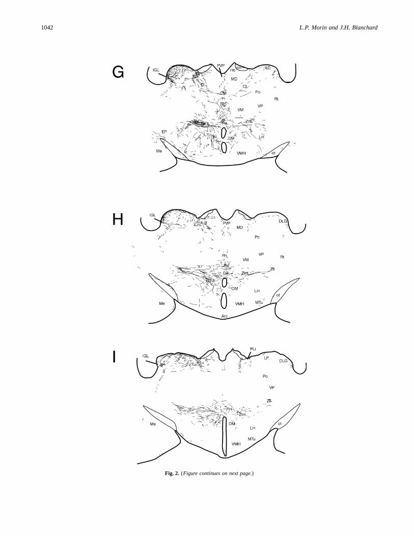
Fig. 2. (Figure continues on next page.)
1042 L.P. Morin and J.H. Blanchard

Fig. 2.
IGL, VLG, and retinal projections 1043

arriving in the posterior hypothalamus from a dorsal route or areprojecting dorsally to pretectal areas from a ventral route.
Anterograde analysis of VLG-efferent projections
The densest innervation of a diencephalic area by the VLG is inthe intramedullary area of the lateral posterior thalamic nucleus(Figs. 4E and 4F). An extensive, moderately dense terminal fieldis also present in the medial and lateral zona incerta (Figs. 4C–4E). Rostrally, sparse innervation of the lateral hypothalamus andnucleus reuniens arrivesvia the posteromedial division of the bednucleus of the stria terminalis (Figs. 4A and 4B). A few fibers areseen medially ventral to the stria medullaris with innervation evidentin the central medial and centrolateral thalamic nuclei (Figs. 4A–4C). There is also sparse innervation of the posterior hypothalamicregion (Figs. 4D–4F). As with the projections of the IGL, it is notpossible to determine whether they arrive from a lateral locationvia the zona incerta or from a dorsal routevia the periventricularfiber system.
Retrograde analysis of IGL- and VLG-efferent projections
Every PHAL injection into the IGL resulted in a small number ofneurons outside the IGL taking up the tracer. For this reason, theretrograde label, CT-b, was applied to sites (Fig. 5) that wereapparent targets of IGL or VLG neurons as determined from thePHAL-IR material. Injection of CT-b into the medial zona incertaresulted in both labeling of neurons in dorsal and medial IGL andin the VLG (Table 1; Fig. 6A). This was the only CT-b injectionsite that resulted in neurons being labeled ipsilaterally and contra-laterally in both brain nuclei. Injection into the posterior bed nu-cleus of the stria terminalis (Fig. 6B) and central medial thalamicnucleus also labeled moderate numbers of neurons in both the IGLand VLG, but were bilateral only in the IGL. In contrast, injectionsinto the anterior amygdala, septum, or parataenial nuclei labeledneurons only in the IGL, primarily on the ipsilateral side but witha few labeled contralaterally. Injections into the thalamic nucleus
reuniens and several hypothalamic locations (anterior, lateral, andposterior nuclei) labeled neurons in the IGL, but not the VLG(cf. anterograde tracing data above). An injection into a very me-dial subparaventricular hypothalamic region (Case 98-5) labeled afew cells bilaterally in the IGL. Injections into the anterior hypo-thalamic nucleus, the lateral hypothalamus (Fig. 6C), and the pos-terior hypothalamus also labeled neurons bilaterally in the IGL. Aninjection of CT-b into the intramedullary area of the lateral pos-terior thalamic nucleus labeled numerous cells in the IGL andVLG.
IGL-afferent projections
After an injection of CT-b into the IGL, labeled neurons are evi-dent in relatively few forebrain structures. In the telencephalon, asmall but consistent collection of cells is found in the caudal cin-gulate cortex (Fig. 7A). PHAL injection into this region labeledfibers and terminals in the IGL. An occasional cell is evident in themedial amygdala, medial septum, and region of the olfactory tu-bercle. A slender group of neurons of modest number is distributedin the posterointermediate division of the bed nucleus of the striaterminalis (Fig. 7B). This set of cells extends caudomedially, be-coming contiguous with the medial zona incerta where there is alsoa modest collection of labeled neurons (Fig. 7C). Caudally, thisgroup is reduced in number and cells are scattered in the medialzona incerta0posterior hypothalamic area. At this level, a secondgroup consisting of a moderately large number of cells is evidentin the lateral zona incerta. Ipsilaterally, this group merges withnumerous labeled neurons in the subgeniculate nucleus furtherlaterally (Fig. 7D).
Small numbers of CT-b labeled cells are also present, as pre-viously reported (Morin et al., 1992), around the SCN, in theanterior hypothalamus, and in the retrochiasmatic region, with anoccasional cell seen in the lateral tuberal hypothalamus. Occa-sional neurons are also found contralaterally in the same areas.Dorsally, a few cells are present in the ventral habenula. Manymore labeled neurons are found dorsally and caudally in mesen-cephalic nuclei of the pretectum and (Morin & Blanchard, 1998).
Fig. 3.Dark-field photomicrographs at (A) rostral, (B) middle, and (C) caudal hypothalamic levels showing PHAL-IR fibers followingan injection of tracer into the IGL (Case 94-33). Scale bar5 500 mm.
1044 L.P. Morin and J.H. Blanchard

Table 1. Connections of the hamster forebrain with the IGL, VLG, and retinain comparison with targets of SCN projectionsa
FromIGL
FromVLG
Fromretina
ToIGL
ToVLG
FromSCNb
Anterior hypothalamus 11 111 102 1Anterior ventral thalamic N 102 2Anterior amygdaloid N, ventral 1 1Anterodorsal thalamic N 1Anteroventral preoptic N 11 1 1c
Arcuate N 1BST, perifornical 111 1 1 1 1BST, posterointermediate 111 1 111 11 1 1BST, posterolateral 1BST, anteromedial 1 1BST, posteromedial 1 1 1 1BST, anteroventral 1Central medial thalamic N 11 1 1Centrolateral thalamic N 11Cingulate cortex 11 1Diagonal band of Broca, horiz. limb 1Dorsal hypothalamic area 1 1 1Dorsomedial hypothalamic N 1 1 1Habenula 1 1 1Intergeniculate leaflet 1111d 1d 111 111 e
Intramedullary thalamic area 1 111 11Lateral olfactory tract N 1 1Lateral hypothalamus 11 11 102Lateroanterior hypothalamic N 11 11 1 102 1Laterodorsal thalamic N 11 11Medial amygdaloid N 1 1 102 1Medial preoptic area 102 1Median preoptic N 1 1Mediodorsal thalamic N 1Olfactory tubercle 1 1 102Parataenial thalamic N 11 1Paraventricular hypothalamic N 102 102 1Paraventricular thalamic N, post. 1 1Paraventricular thalamic N, ant. 1 1Perirhinal cortex 1Piriform cortex 1 1Posterior hypothalamic N 111 1 1Premammillary N 1Retrochiasmatic area 102 1 102 1Reuniens thalamic N 111 102 1Septum, lateral intermediate 102Septum, lateral ventral 1Septum, medial 102Subgeniculate thalamic N 11 1 111 111Subparaventricular zone 11 11 1Suprachiasmatic N 1111 11111 1e
Supramammillary N 1 1Supraoptic N 1Tuberal hypothalamus 1 1 102 102 1Ventrolateral preoptic N 11 11 1Ventromedial hypoth. N, coreVentromedial hypoth. N, shell 11 1Zona incerta, lateral 11 11 1 111 1Zona incerta, medial 111 11 11 1
aEmpty cell: no evident connection;102: very sparse;1: sparse;11: modest;111: moderate;1111: dense; and11111:extremely dense.bData from Morin et al., 1994, except for anteroventral preoptic nucleus.cHamster: Morin and Blanchard (unpublished data); rat: Watts et al., 1987.dContralateral to injection site.eInnervation arises from cells around SCN.
IGL, VLG, and retinal projections 1045
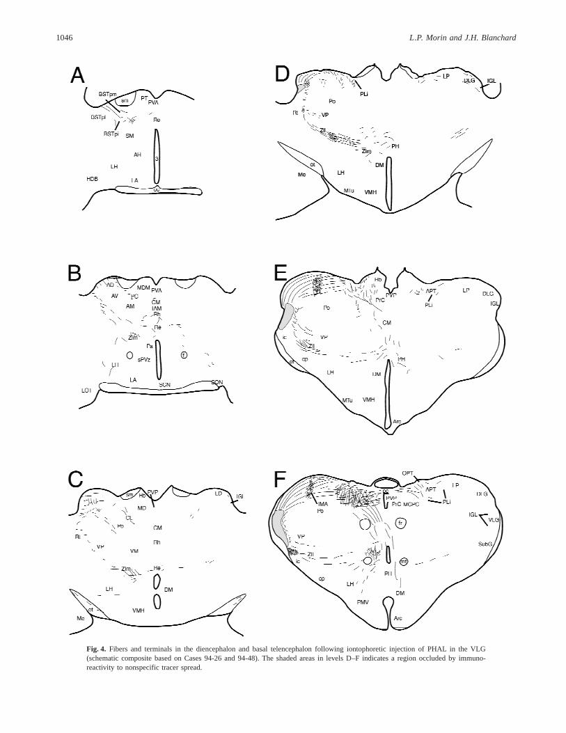
Fig. 4. Fibers and terminals in the diencephalon and basal telencephalon following iontophoretic injection of PHAL in the VLG(schematic composite based on Cases 94-26 and 94-48). The shaded areas in levels D–F indicates a region occluded by immuno-reactivity to nonspecific tracer spread.
1046 L.P. Morin and J.H. Blanchard
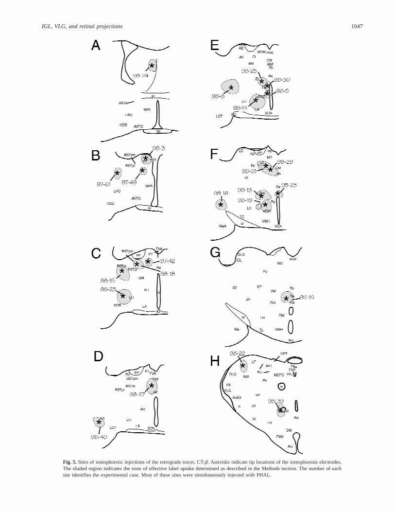
Fig. 5. Sites of iontophoretic injections of the retrograde tracer, CT-b. Asterisks indicate tip locations of the iontophoresis electrodes.The shaded region indicates the zone of effective label uptake determined as described in the Methods section. The number of eachsite identifies the experimental case. Most of these sites were simultaneously injected with PHAL.
IGL, VLG, and retinal projections 1047

VLG-afferent projections
There are few forebrain sites with neurons projecting to the VLG.The largest collections of such cells are found bilaterally in themedial and lateral zona incerta and in the subgeniculate nucleus.
Neurons are also consistently seen in the posterointermediate bednucleus of the stria terminalis and caudal cingulate cortex. Injec-tion of PHAL into the latter area labeled fibers with terminals inthe VLG. Scattered cells are also sparsely distributed in lateroan-terior, retrochiasmatic, and tuberal hypothalamus.
Fig. 6.CT-b-labeled neurons in the IGL following tracer injection in the (A) medial zona incerta (Case 98-13), (B) posterointermediatedivision of the bed nucleus of the stria terminalis (Case 98-18), and (C) far lateral hypothalamus (Case 98-16). The injection sites forthese cases are shown in Figs. 4F, 4C and 4F, respectively. Scale bar5 100 mm.
Fig. 7.Photomicrographs of retrogradely labeled neurons in (A) cingulate cortex, (B) bed nucleus of the stria terminalis, posteromedialdivision, (C) lateral zona incerta, and (D) medial zona incerta after a CT-b injection into the IGL. Scale bar5 200 mm.
1048 L.P. Morin and J.H. Blanchard

Forebrain retinal projections
Asmall, superficial pathway passes contralaterally through the piri-form cortex to a location near the rhinal fissure. It persists in closeproximity to the rhinal fissure for a substantial distance caudally intoperirhinal cortex (Fig. 8A). A pathway adjacent to the rhinal fissure(Figs. 8B and 8C) also extends rostrally, eventually extending alongthe base of the fissure medially between olfactory bulb and orbitalcortex.The rostral and caudal projections, as typically observed, weresubstantially less robust than illustrated by this particular case.
Retinal fibers with terminals are visible in rostral hypothala-mus. There are numerous bilateral projections to the rostroventralpreoptic region (Fig. 9A), particularly in the anteroventrolateralpreoptic nucleus, extending laterally into the olfactory tubercle andpiriform cortex. Numerous retinal fibers innervate the basal por-
tions of several amygdaloid nuclei (Fig. 9B). Fibers do not appearto reach these areas ipsilateral to the injected eye.
A small, bilateral fiber bundle departs from the dorsal optictract adjacent to the dorsal lateral geniculate nucleus (DLG) andextends rostrally as a small profile, but dense, superficial terminalplexus (Fig. 10). The final terminal location is ventrolateral to thestria medullaris in the bed nucleus of the stria terminalis, postero-medial division (Fig. 9A). Caudal to the anteroventrolateral pre-optic nucleus, innervation is evident bilaterally above the chiasmand is moderately dense in the contralateral ventral medial preopticarea and anteroventrolateral preoptic nucleus. Extremely denseinnervation fills the SCN and extends lateral, dorsal, and caudal tothe nucleus (Fig. 9B). A fairly substantial bilateral, retinorecipientzone lateral to the SCN includes the adjacent anterior hypothala-mus, and extends into the ventral perifornical region and lateralhypothalamus. At the caudal end of this region, a small but distinctset of retinal projections can be seen terminating in the basal lateralhypothalamus contiguous with the optic tract and the plexus con-tinues caudally into tuberal hypothalamus. Also at the caudal endof this region, modest numbers of retinal fibers can be seen inner-vating the subgeniculate nucleus and dorsolateral zona incerta (notshown). The peri-SCN innervation extends caudally in a dwindlingterminal plexus that fills the subparaventricular zone. The caudalsubparaventricular retinorecipient plexus merges with a less denseterminal zone in the periventricular and dorsomedial hypothalamicnuclei (Fig. 9C). At this level, a modest terminal plexus is presentin the medial zona incerta. A very few visual fibers are found in theparaventricular hypothalamus and dorsally in the thalamic nucleusreuniens. None are visible in other midline thalamic nuclei or inthe anterior or posterior paraventricular thalamic nuclei. Fibersalso specifically avoid the core region of the ventromedial hypo-thalamic nucleus.
Discussion
The IGL is a major constituent of the subcortical visual system(Morin & Blanchard, 1998). As part of that system, the IGL re-ceives abundant direct retinal projections and has both efferent andafferent connections with virtually all pretectal nuclei and the su-perior colliculus. The present data demonstrate similar widespreadconnections between the IGL and certain diencephalic nuclei, par-ticularly within the hypothalamus, and with nuclei in the ventraltelencephalon. These are matched by direct retinal projections tothe majority of the same regions. As is the case with mesencephalicnuclei of the subcortical visual shell (Morin & Blanchard, 1997),forebrain nuclei with IGL connections seem to have both directand indirect access to visual signals.
IGL projections to the forebrain
Autoradiographic investigation of rat ventral lateral geniculate pro-jections revealed ample innervation of the hypothalamus (Swansonet al., 1974; Ribak & Peters, 1975), but did not distinguish IGLfrom VLG as the nucleus of origin. The present extensive IGLprojections to hypothalamus in the hamster are similar to what hasbeen described in gerbil (Mikkelsen, 1990) and rat (Moore et al.,1996; Horvath, 1998), also derived from studies using PHAL asthe anterograde tracer. In all three species, a major target is an areaextending from the lateral optic chiasm medially through the lat-eroanterior hypothalamus to the midline, including the SCN, anddorsally to include the subparaventricular zone and adjacent peri-fornical anterior hypothalamus. As in the rat (Horvath, 1998), the
Fig. 8. Dark-field photomicrographs of CT-b-IR retinal projectionsshowing (A) fibers and terminals caudally in superficial perirhinal cortex,(B) fiber segments (arrowheads) rostrally in the lateral olfactory tract, and(C) fibers with terminals (arrowheads) spreading medially along the baseof the rhinal fissure between lateral orbital cortex and olfactory bulb. Scalebar5 10 mm for (A) and 20mm for (B) and (C).
IGL, VLG, and retinal projections 1049

pattern of hypothalamic innervation by the IGL of the hamster isgenerally similar to that of SCN efferent projections (Morin et al.,1994) (Table 1).
Projections to the SCN from IGL neurons are dense, as ex-pected (Card & Moore, 1989; Morin & Blanchard, 1995). As sug-gested previously (Card & Moore, 1989), the SCN may receiveinnervation from the IGLvia two or more routes. There is a thin,dense laminar tract on the medial surface of the optic tract thatcould provide innervation entering ventrally from the supraopticdecussation. A second route to the SCN appears to bevia projec-tions extending ventromedially across the ventral thalamic nucleusand anterior hypothalamus.
Thalamic projections of the IGL have not been fully character-ized, although a dense projection to the contralateral IGL is welldocumented in rat (Card & Moore, 1989) and hamster (Morin &Blanchard, 1995). The IGL also projects bilaterally to the VLG,but does not innervate the major thalamic visual relay nuclei.However, anterograde tracing methods supported the presence of aprojection from the IGL to the intramedullary area of the thalamiclateral posterior nucleus (Morin & Blanchard, 1998). The presentstudy provides retrograde data to affirm this the presence of sucha pathway. The intramedullary area is an exception to the rule thatthalamic structures projecting to cortex are not innervated by theIGL. As such, it probably should be considered as fully distinctfrom the lateral posterior nucleus, rather than a subdivision of it.
Several thalamic nuclei located rostromedially, the parataenial,anterior paraventricular, and paracentral, are innervated by IGLprojections. These, plus the posterointermediate and posteromedialdivisions of the bed nucleus of the stria terminalis, provide avenuesfor IGL fibers to reach midline thalamic nuclei (central medial,reuniens) where there is a substantial degree of decussation con-tributing to innervation of contralateral nuclei. IGL projections tothe habenula, paraventricular thalamic nucleus (anterior and pos-terior), and the parataenial nucleus shown here for the hamsterhave not been previously reported. The thalamic subgeniculatenucleus and zona incerta are also innervated by the IGL with themedial zona incerta, in particular, receiving moderately dense pro-jections in hamster, rat, and gerbil (present data and (Mikkelsen,1990; Horvath, 1998)).
IGL projections have been noted as far rostral as the medialseptum and vertical limb of the diagonal band of Broca in the rat(Horvath, 1998). Innervation of these two regions was sparse inthe hamster. Fibers with terminals from the hamster IGL werefound in the intermediate lateral septum, anterior divisions of thebed nucleus of the stria terminalis, and the horizontal limb of thediagonal band of Broca. This pattern is consistent with the addi-tional observations that IGL innervation extends rostrolaterallyinto basal telencephalic regions, including anterior and medialamygdala.
Retinal projections to forebrain
We have previously described four distinct routes of rostral di-encephalic retinal innervation in the hamster: (1) the largest, toSCN and the anterior hypothalamic area laterally and caudally; (2)to the ventral lateral hypothalamic area; (3) a rostral projection toventral preoptic area; and (4) a site in the putative anterodorsalthalamic (Johnson et al., 1988a) or bed nucleus of the stria termi-nalis (Cooper et al., 1994). The present data, obtained with themore sensitive CT-b immunohistochemical method, largely sub-stantiate the previous report, but also add several new observationsand reinterpretations.
With respect to the first two pathways, the major differenceconcerns the density of visual projections to non-SCN hypothala-mus. The present data indicate substantially greater innervationthan previously reported, particularly in the lateroanterior nucleus,subparaventricular zone, and ventral lateral hypothalamus. Retinalinnervation of the paraventricular hypothalamus is very sparse[present data and (Johnson et al., 1988a); but see (Cooper et al.,1994)]. The present data also show modestly dense medial hypo-thalamic innervation extending to the dorsomedial nucleus, a featurenot previously reported. It should be emphasized that terminatingvisual projections are not present in the core of the ventromedialhypothalamic nucleus, but are found dorsally, medially, and ven-trally in the “shell” region of the ventromedial hypothalamic nucleus.
The present data also demonstrate retinal innervation of theventral preoptic area comparable to, although somewhat denserthan, previous reports (Johnson et al., 1988a). Visual projections
Fig. 9. Dark-field photomicrographs showing retinal ganglion cell projections to hypothalamus and basal telencephalon at (A) rostral,(B) middle, and (C) caudal hypothalamic levels of section. Scale bar5 500 mm.
1050 L.P. Morin and J.H. Blanchard

through this region extend further laterally into telencephalon. Ven-tral telencephalic retinal projections have been identified across 13mammalian orders in 50 species (Cooper et al., 1989, 1994). Thepresent data reveal sparse innervation in anterior and medial amyg-daloid nuclei, with most fibers and terminals located superficially,passing ventral to the nucleus of the lateral olfactory tract into
layer 1 of the piriform cortex. This is consistent with the originaldescription in the hamster (Pickard & Silverman, 1981), as well asa more recent report (Cooper et al., 1994), although some inves-tigators have not seen this projection in either rat or hamster (John-son et al., 1988a).
An unexpected component of the retinal projection pattern isthe presence of a small, but consistent, lateral pathway to thevicinity of the rhinal fissure. The innervation persists caudally,adjacent to the fissure, to the perirhinal cortex. The data also showa similarly small rostral projection into the lateral olfactory tract.The projection extends rostrally in the lateral olfactory tract, even-tually curving medially along the ventral surface of the rhinalfissure, beneath overlying frontal cortex.
The fourth retinal pathway has been previously identified asterminating in the anterodorsal thalamic nuclei (Johnson et al.,1988a) or “encapsulated” part of the bed nucleus of the striaterminalis (Cooper et al., 1994). Analysis of horizontal sectionsshows that this retinal projection actually provides a terminal plexusextending a fairly long distance superficially along the lateral sur-face of the laterodorsal thalamic nucleus. We concur with the viewof Cooper and colleagues that the site of final termination of thispathway is in the bed nucleus of the stria terminalis, posterolateraldivision.
The major thalamic nuclei receiving retinal projections are theVLG, IGL, dorsal lateral geniculate, and lateral posterior nuclei.The present data show sparse innervation of the nucleus reuniensas well. No other thalamic nuclei were consistently identified asreceiving retinal projections [present data and (Johnson et al.,1988a)], although such projections have been reported in othermidline thalamic nuclei, including the anterior paraventricular, andin the anteroventral and anterodorsal nuclei (Cooper et al., 1994).
Forebrain projections to the IGL or VLG
Forebrain afferents of the IGL and VLG have not been widelystudied. The present data show that few structures contain neuronsprojecting to the IGL. The most robust collections of IGL-afferentneurons are found in the posteromedial division of the bed nucleusof the stria terminalis, zona incerta, and subgeniculate nucleus.Smaller numbers are found in the peri-SCN and retrochiasmaticregions [present data and (Morin et al., 1992)]. In the rat, IGL-afferent neurons are found directly in the SCN (Card & Moore,1989; Vrang & Mikkelsen, 1996). The zona incerta and retrochi-asmatic projections to IGL have also been noted in the rat, alongwith innervation from the ventromedial hypothalamic nucleus (Vrang& Mikkelsen, 1996). The latter is not evident in the hamster (presentdata). Many more labeled neurons projecting to the IGL or VLGare found dorsally and caudally in mesencephalic nuclei of thepretectum and tectum (Morin & Blanchard, 1998).
The IGL and VLG receive innervation from the posterior cin-gulate cortex in both rat (Vrang & Mikkelsen, 1996) and hamster(present data). Although both IGL and VLG project to the intra-medullary area of the lateral posterior thalamic nucleus [presentdata and (Morin & Blanchard, 1998)], this area does not recipro-cally project to either.
Connections of the VLG
Initial evaluations of VLG projections to the rat hypothalamuswere made in the absence of knowledge about the IGL (Ribak &Peters, 1975). The present study employed both anterograde andretrograde methods to determine the extent of VLG projections to
Fig. 10. Dark-field photomicrographs of horizontal sections showing (A)the emergence of a retinal projection rostrally (arrowheads) from the optictract adjacent to the dorsal lateral geniculate nucleus, and (B–D) extendingprogressively further rostral, with terminals along the way, finally termi-nating in a narrow part of the bed nucleus of the stria terminalis, postero-medial division. Scale bar5 500 mm.
IGL, VLG, and retinal projections 1051

rostral thalamus, hypothalamus, and telencephalon. The PHAL an-terograde tracing method indicated that a limited number of nucleimight receive projections from the VLG. CT-b retrograde labelconfirmed the presence of innervation from the VLG in most of thenuclei containing at least modestly robust projections. If thePHAL-IR material indicated sparse innervation to a particular lo-cation (e.g. lateral hypothalamus), then a CT-b injection at that sitefailed to retrogradely label VLG neurons. The nuclei identifiedfrom the PHAL-IR material as receiving modestly robust projec-tions include the bed nucleus of the stria terminalis, central medialthalamic nucleus, and the medial zona incerta. Retrograde analysisalso shows a VLG projection to the intramedullary area of thethalamic lateral posterior nucleus in accordance with previous an-terograde data (Morin & Blanchard, 1998). All diencephalic nucleireceiving VLG projections are fairly dorsal and none is hypotha-lamic, a feature consistent with previous reports in rat (Horvath,1998) and gerbil (Mikkelsen, 1990). Thus, it is likely that thelimited immunoreactive fibers and terminals seen in the basal hy-pothalamus after a VLG injection of PHAL are consequent to labelleakage into adjacent IGL. The VLG and IGL do not differ greatlywith respect to the specific nuclei of the subcortical visual shell towhich they are connected. However, one clear trait of the IGL isthe presence of bilaterally efferent projections (although it is notknown whether individual neurons project bilaterally). This is ev-ident both with respect to projections to nuclei of the subcorticalvisual shell (Morin & Blanchard, 1998) and with respect to morerostral projections (present data). We previously demonstrated thatIGL, but not VLG, neurons project to SCN (Morin & Blanchard,1995). The present data generalize this observation by showingthat lateral geniculate neurons projecting to hypothalamus are foundonly in the IGL.
Functional considerations
The visual system has typically been partitioned into two majordivisions, one concerned with sophisticated image analysis andanother concerned with oculomotor control. The circadian visualsystem has been described as a third division of the overall visualplan (Morin, 1994). The two major nuclei of this system are theSCN and IGL. Briefly, the SCN receives a direct retinal projectionthat transmits photic information necessary for entrainment to thecircadian clock in that nucleus (Johnson et al., 1988a,b; Dinget al., 1994). The IGL also receives a direct retinal projection andcan provide indirect photic information to the SCN (Zhang &Rusak, 1989). The IGL is not necessary for rhythm generation orphotic entrainment, but can modulate the effects of light on rhyth-micity (Harrington & Rusak, 1986; Pickard et al., 1987; Johnsonet al., 1989). The IGL is necessary for circadian rhythm phasecontrol by several nonphotic stimuli (Johnson et al., 1988c; Bielloet al., 1991; Wickland & Turek, 1994; Janik & Mrosovsky, 1994).
As a rule, the IGL does not connect with thalamic relay nucleiinvolved with complex visual image analysis (Morin & Blanchard,1998). The intramedullary area of the lateral posterior nucleus,situated ventrolaterally adjacent to the DLG, is a noteworthy ex-ception to this rule. Its neurons project to secondary visual cortexarea 18a (Takahashi, 1985; Morin & Blanchard, 1998) which maybe involved in pattern discrimination (Dean, 1981). The IGL hasextensive interconnections with all subcortical visual nuclei par-ticipating in various forms of oculomotor control (see Morin &Blanchard, 1998 for a discussion). Thus, the circadian visual sys-tem is not fully separable from the two major divisions of thevisual system. At the moment, it is not possible to state the func-
tion of the rather extensive relationship between cortical or sub-cortical visual nuclei and the IGL.
The IGL appears to have similarly extensive connections withforebrain nuclei not customarily thought to have visual function.There is no direct information from the hamster concerning func-tion of second-order visual information conveyed to non-SCN,non-geniculate forebrain structuresvia the IGL. However, datafrom the rat indicate that retinal fibers terminate on IGL neuronsprojecting to non-SCN hypothalamus and that tonic photic stimu-lation may act through the IGL to regulate certain endocrine func-tions in rats (Horvath, 1998). Putative neuroendocrine targets ofIGL projections are dopamine neurons in the rat ventral medialhypothalamus. The present data show a direct retinal projection tothe same region, suggesting redundancy between direct and indi-rect (i.e. from IGL) retinal projections. A similar phenomenon hasbeen demonstrated in the mesencephalic nuclei of the subcorticalvisual shell to the extent that all receive both direct retinal projec-tions and projections from the IGL (Morin & Blanchard, 1997,1998).
Horvath (1998) has emphasized another form of apparent vi-sual projection redundancy in his description of similar hypotha-lamic innervation patterns independently arising from IGL andSCN neurons. Similarity of innervation is a theme also discussedby Cooper and colleagues (Cooper et al., 1994) who have indicatedthat olfaction-related projections to hypothalamus and direct reti-nal projections often innervate common areas. However, there isvery little evidence to support any function of direct retinal inputrelated to olfaction-mediated physiology or behavior. Neither isthere much data supporting the possibility of physiological or be-havioral regulation by either direct, non-SCN, retinohypothalamicinnervation, or indirect innervation from the IGL. The non-SCN,direct, and indirect visual projections to hypothalamus could beinvolved in circadian rhythm “masking.” Masking effects are seenwhen a stimulus induces increased or decreased amplitude of arhythmic variable (e.g. number of daily wheel revolutions) ratherthan through a change in phase or period of the pacemaker systemregulating the timing of that variable. In the rat, the acoustic startleresponse is amplified by a concomitant bright light (Walker &Davis, 1997). This effect is blocked by lesions of the bed nucleusof the stria terminalis. It is possible that the visual projectiondescribed here as innervating the nearby border of stria medullarisand anterodorsal thalamic nucleus contributes to that response fa-cilitation. However, our previous study of retinal forebrain projec-tions did not identify such a pathway in rat (Johnson et al., 1988b).
The mechanism generating circadian rhythmicity normally pro-vides an oscillatory substrate modulating the likelihood of sleep(Borbely, 1975; Ibuka & Kawamura, 1975). However, light canhave direct effects on this and other behaviors in addition to indi-rect actions through circadian clock regulation. For example, lightsOFF will acutely induce rapid-eye-movement (REM) sleep (Lisk& Sawyer, 1966) and is associated with reduced slow wave sleep(Borbely, 1975). Lights ON will acutely inhibit waking, feeding,drinking, and locomotion (Borbely & Huston, 1974; Borbely et al.,1975). The acute effects of lights off on REM sleep may be me-diated through pretectal nuclei (Miller et al., 1997). Whether theacute effects of light or dark on feeding, drinking, and locomotionare also mediated through pretectal nuclei or are simultaneouslyelicited through direct or indirect photic input to hypothalamushave not yet been studied.
In summary, the present data show widespread projections ofthe IGL to the forebrain, especially the hypothalamus. Both olfac-tory and retinal projections appear to project to many of the same
1052 L.P. Morin and J.H. Blanchard

regions. The pattern of ascending IGL projections is easily distin-guishable from that contributed by VLG neurons. Through its ex-tensive projections to both forebrain and midbrain, the IGL appearsto have the capacity, largely untested at this time, to modulatenumerous aspects of normal physiology and behavior.
Acknowledgments
This research was supported by NIH NINDS Grant NS22168.
References
Biello, S.M., Harrington, M.E. & Mason, R. (1991). Geniculo-hypothalamic tract lesions block chlordiazepoxide-induced phase ad-vances in Syrian hamsters.Brain Research552, 47–52.
Borbely, A.A. (1975). Circadian rhythm of vigilance in rats: Modulationby short light–dark cycles.Neuroscience Letters1, 67–71.
Borbely, A.A. & Huston, J.P. (1974). Effects of two-hour light–darkcycles on feeding, drinking and motor activity of the rat.Physiologyand Behavior13, 795–802.
Borbely, A.A., Huston, J.P. & Waser, P.G. (1975). Control of sleep statesin the rat by short light–dark cycles.Brain Research95, 89–101.
Card, J.P. & Moore, R.Y. (1989). Organization of lateral geniculate-hypothalamic connections in the rat.Journal of Comparative Neurol-ogy 284, 135–147.
Coolen, L.M. & Wood, R.I. (1998). Bidirectional connections of themedial amygdaloid nucleus in the syrian hamster brain: Simultaneousanterograde and retrograde tract tracing.Journal of Comparative Neu-rology 399, 189–209.
Cooper, H.M., Mick, G. & Magnin, M. (1989). Retinal projection tomammalian telencephalon.Brain Research477, 350–357.
Cooper, H.M., Parvopassu, F., Herbin, M. & Magnin, M. (1994). Neuro-anatomical pathways linking vision and olfaction in mammals.Psy-choneuroendocrinology19, 623–639.
Dean, P. (1981). Grating detection and visual acuity after lesions of striatecortex in hooded rats.Experimental Brain Research43, 145–153.
Ding, J.M., Chen, D., Weber, E.T., Faiman, L.E., Rea, M.A. & Gillette,M.U. (1994). Resetting the biological clock: Mediation of nocturnalcircadian shifts by glutamate and NO.Science266, 1713–1717.
Harrington, M.E., Nance, D.M. & Rusak, B. (1985). Neuropeptide Yimmunoreactivity in the hamster geniculo-suprachiasmatic tract.BrainResearch Bulletin15, 465–472.
Harrington, M.E. & Rusak, B. (1986). Lesions of the thalamic inter-geniculate leaflet alter hamster circadian rhythms.Journal of Biologi-cal Rhythms1, 309–325.
Hickey, T.L. & Spear, P.D. (1976). Retinogeniculate projections in hoodedand albino rats: An autoradiographic study.Experimental Brain Re-search24, 523–529.
Horvath, T.L. (1996). Evidence that circadian and visual signals from theSCN and LGN are integrated on neuroendocrine dopamine cells in therat hypothalamus.Society for Neuroscience26, 31.
Horvath, T.L. (1997). Suprachiasmatic efferents avoid phenestrated cap-illaries but innervate neuroendocrine cells, including those producingdopamine.Endocrinology138, 1312–1320.
Horvath, T.L. (1998). An alternate pathway for visual signal integrationinto the hypothalamo-pituitary axis: Retinorecipient intergeniculateneurons project to various regions of the hypothalamus and innervateneuroendocrine cells including those producing dopamine.Journal ofNeuroscience18, 1546–1558.
Hsu, S.-M., Raine, L. & Fanger, H. (1981). Use of avidin-biotin-perioxidase complex (ABC) in immunoperioxidase techniques: A com-parison between ABC and unlabeled antibody (PAP) procedures.Journalof Histochemistry and Cytochemistry29, 577–580.
Ibuka, N. & Kawamura, H. (1975). Loss of circadian rhythm in sleep-wakefulness cycle in the rat by suprachiasmatic nucleus lesions.BrainResearch96, 76–81.
Janik, D. & Mrosovsky, N. (1994). Intergeniculate leaflet lesions andbehaviorally-induced shifts of circadian rhythms.Brain Research651,174–182.
Johnson, R.F., Moore, R.Y. & Morin, L.P. (1988a). Loss of entrainmentand anatomical plasticity after lesions of the hamster retinohypotha-lamic tract.Brain Research460, 297–313.
Johnson, R.F., Morin, L.P. & Moore, R.Y. (1988b). Retinohypothalamicprojections in the hamster and rat demonstrated using cholera toxin.Brain Research462, 301–312.
Johnson, R.F., Smale, L., Moore, R.Y. & Morin, L.P. (1988c). Lateralgeniculate lesions block circadian phase shift responses to a benzodi-azepine.Proceedings of the National Academy of Sciences of the U.S.A.85, 5301–5304.
Johnson, R.F., Moore, R.Y. & Morin, L.P. (1989). Lateral geniculatelesions alter activity rhythms in the hamster.Brain Research Bulletin22, 411–422.
Klein, D.C., Moore, R.Y. & Reppert, S.M. (1991). SuprachiasmaticNucleus: The Mind’s Clock.New York: Oxford University Press.
Lisk, R.D. & Sawyer, C.H. (1966). Induction of paradoxical sleep bylights-off stimulation.Proceedings of the Society for Experimental Bi-ology and Medicine123, 664–667.
McLean, I.W. & Nakane, P.K. (1974). Periodate-lysine-paraformaldehydefixative: A new fixative for immunoelectron microscopy.Journal ofHistochemistry and Cytochemistry22, 1077–1083.
Mikkelsen, J.D. (1990). Projections from the lateral geniculate nucleus tothe hypothalamus of the Mongolian gerbil (Meriones unguiculatus):An anterograde and retrograde tracing study.Journal of ComparativeNeurology299, 493–508.
Miller, A.M., Oblinger, M.M., Behan, M. & Benca, R.M. (1997).Ibotenic acid lesions of the pretectum affect REM triggering, but notredistribution of sleep in short light–dark cycles.Society for Neurosci-ence23, 2132.
Moore, R.Y. & Card, J.P. (1994). Intergeniculate leaflet: An anatomicallyand functionally distinct subdivision of the lateral geniculate complex.Journal of Comparative Neurology344, 403–430.
Moore, R.Y., Moga, M.M. & Weis, R. (1996). Intergeniculate leaflet andventral lateral geniculate projections in the rat.Society for Neurosci-ence26, 31.
Moore, R.Y. & Speh, J.C. (1993). GABA is the principal neurotransmitterof the circadian system.Neuroscience Letters150, 112–116.
Morin, L.P. (1994). The circadian visual system.Brain Research Review67, 102–127.
Morin, L.P. & Blanchard, J.H. (1995). Organization of the hamsterintergeniculate leaflet: NPY and ENK projections to the suprachias-matic nucleus, intergeniculate leaflet and posterior limitans nucleus.Visual Neuroscience12, 57–67.
Morin, L.P. & Blanchard, J.H. (1997). Neuropeptide Y and enkephalinimmunoreactivity in retinorecipient nuclei of the hamster pretectumand thalamus.Visual Neuroscience14, 765–777.
Morin, L.P. & Blanchard, J.H. (1998). Interconnections among nuclei ofthe subcortical visual shell: The intergeniculate leaflet is a major con-stituent of the hamster subcortical visual system.Journal of Compar-ative Neurology396, 288–309.
Morin, L.P., Blanchard, J.H. & Moore, R.Y. (1992). Intergeniculateleaflet and suprachiasmatic nucleus organization and connections in thehamster.Visual Neuroscience8, 219–230.
Morin, L.P., Goodless-Sanchez, N., Smale, L. & Moore, R.Y. (1994).Projections of the suprachiasmatic nuclei, subparaventricular zone andretrochiasmatic area in the golden hamster.Neuroscience61, 391–410.
Pickard, G.E., Ralph, M.R. & Menaker, M. (1987). The intergeniculateleaflet partially mediates effects of light on circadian rhythms.Journalof Biological Rhythms2, 35–56.
Pickard, G.E. & Silverman, A.J. (1981). Direct retinal projections tohypothalamus, piriform cortex and accessory optic nuclei in the goldenhamster as demonstrated by a sensitive anterograde horseradish per-oxidase technique.Journal of Comparative Neurology196, 155–172.
Ribak, C.E. & Peters, A. (1975). An autoradiographic study of the pro-jections from the lateral geniculate body of the rat.Brain Research92,341–368.
Swanson, L.W., Cowan, W.M. & Jones, E.G. (1974). An autoradio-graphic study of the efferent projections of the ventral lateral geniculatenucleus in the albino rat and cat.Journal of Comparative Neurology156, 143–164.
Takahashi, T. (1985). The organization of the lateral thalamus of thehooded rat.Journal of Comparative Neurology231, 281–309.
Taylor, A.M., Jeffery, G. & Lieberman, A.R. (1986). Subcortical affer-ent and efferent connections of the superior colliculus in the rat andcomparisons between albino and pigmented strains.Experimental BrainResearch62, 131–142.
IGL, VLG, and retinal projections 1053

Vrang, N. & Mikkelsen, J.D. (1996). Afferent connections of the inter-geniculate leaflet of the golden hamster: A combined retrograde andanterograde tracing study.Society for Neuroscience26, 232.
Walker, D.L. & Davis, M. (1997). Double dissociation between theinvolvement of the bed nucleus of the stria terminalis and the centralnucleus of the amygdala in startle increases produced by condi-tioned versus unconditioned fear.Journal of Neuroscience17, 9375–9383.
Watts, A.G., Swanson, L.W. & Sanchez-Watts, G. (1987). Efferent
projections of the suprachiasmatic nucleus: I. Studies using anterogradetransport of Phaseolus vulgaris leucoagglutinin in the rat.Journal ofComparative Neurology258, 204–229.
Wickland, C. & Turek, F.W. (1994). Lesions of the thalamic intergenic-ulate leaflet block activity-induced phase shifts in the circadian activityrhythm of the golden hamster.Brain Research660, 293–300.
Zhang, D.X. & Rusak, B. (1989). Photic sensitivity of geniculate neuronsthat project to the suprachiasmatic nuclei or the contralateral genicu-late.Brain Research504, 161–164.
1054 L.P. Morin and J.H. Blanchard


![Hamster[1] (3)rt](https://img.pdfslide.us/doc/110x75/5491fa67b4795943628b46a3/hamster1-3rt.jpg)