Embed Size (px)
Citation preview

Subscriber access provided by BUFFALO STATE
is published by the American Chemical Society. 1155 Sixteenth Street N.W.,Washington, DC 20036Published by American Chemical Society. Copyright © American Chemical Society.However, no copyright claim is made to original U.S. Government works, or worksproduced by employees of any Commonwealth realm Crown government in the courseof their duties.
Article
Zinc-Doped Mesoporous Graphitic Carbon Nitridefor the Colorimetric Detection of Hydrogen Peroxide
Aftab Ahmed, Peter John, Mian Hasnain Nawaz, Akhtar Hayat, and Muhammad NasirACS Appl. Nano Mater., Just Accepted Manuscript • DOI: 10.1021/acsanm.9b01036 • Publication Date (Web): 11 Jul 2019
Downloaded from pubs.acs.org on July 12, 2019
Just Accepted
“Just Accepted” manuscripts have been peer-reviewed and accepted for publication. They are postedonline prior to technical editing, formatting for publication and author proofing. The American ChemicalSociety provides “Just Accepted” as a service to the research community to expedite the disseminationof scientific material as soon as possible after acceptance. “Just Accepted” manuscripts appear infull in PDF format accompanied by an HTML abstract. “Just Accepted” manuscripts have been fullypeer reviewed, but should not be considered the official version of record. They are citable by theDigital Object Identifier (DOI®). “Just Accepted” is an optional service offered to authors. Therefore,the “Just Accepted” Web site may not include all articles that will be published in the journal. Aftera manuscript is technically edited and formatted, it will be removed from the “Just Accepted” Website and published as an ASAP article. Note that technical editing may introduce minor changesto the manuscript text and/or graphics which could affect content, and all legal disclaimers andethical guidelines that apply to the journal pertain. ACS cannot be held responsible for errors orconsequences arising from the use of information contained in these “Just Accepted” manuscripts.

1
Zinc-Doped Mesoporous Graphitic Carbon Nitride for the Colorimetric
Detection of Hydrogen Peroxide
Aftab Ahmed1,2, Peter John2, Mian Hasnain Nawaz1, Akhtar Hayat1*, Muhammad Nasir1*
1 Interdisciplinary Research Centre in Biomedical Materials (IRCBM), COMSATS University
Islamabad, Lahore Campus, 1.5 Km Defence Road, off Raiwind Road, Lahore, Punjab, Pakistan
54000
2 Government College University Lahore, Katchery Road, Anarkali, Lahore, Punjab, Pakistan
54000
Highlights
• Development of a facile, highly sensitive, low cost, colorimetric detection method for
H2O2 determination
• Zn doping was done in graphitic carbon nitride to increase its electrical properties for
better sensitivity
• Structure and activity relationship were studied to better understand the high efficiency of
the prepared catalyst
ABSTRACT: Recently, graphitic carbon nitride (g-C3N4) has been explored as a peroxidase-like
catalyst for the non-enzymatic colorimetric detection of H2O2. In this study, we have developed a
simple, low cost and eco-friendly hydrogen bond assisted soft template method of zinc ions doping
in mesoporous graphitic-carbon-nitride (Zn-mpg-C3N4) thin nanosheets. Morphology and
Page 1 of 45
ACS Paragon Plus Environment
ACS Applied Nano Materials
123456789101112131415161718192021222324252627282930313233343536373839404142434445464748495051525354555657585960

2
composition of prepared samples were determined by different characterization techniques. PEG-
1500 was beneficial to enhance the porosity and surface area of g-C3N4, whereas Zinc loading in
the framework of g-C3N4 was resulted in the increase in electrical properties. The peroxidase-like
catalytic activity of samples was investigated and compared based on the development of blue
colored reaction mixture by the oxidation reaction between 3,3ʹ,5,5ʹ-tetramethylbenzidine and
hydrogen peroxide (H2O2) through colorimetric method. The as-prepared 10% Zn-mpg-C3N4 has
shown higher peroxidase-like activity as compared to natural HRP, g-C3N4 and mpg-C3N4. This
enhanced peroxidase-like activity was attributed to the thin structured nanosheets, higher specific
surface area, outstanding electron transfer ability, increased band gap and increased in charge
separation of the catalyst through the direct zinc ions doping modification. The steady-state
kinetics mechanism was investigated by using Michaelis-Menten Kinetics and found that the
reaction followed Ping-Pong mechanism. This outstanding catalytic activity permitted us to design
a rapid and convenient colorimetric sensing method to detect H2O2. Under the optimized condition,
the developed sensor exhibited a linear range of 10 to 2000 µM (R-square = 0.9981), limit of
detection as 1.4 M and limit of quantification as 3.0 M for H2O2 detection. In view of advantages
compared with previous methods such as simple, facile operation, cost-effective, eco-friendly,
naked eye observation and rapid response, the developed sensor possesses huge potential and a
promising candidate for enzyme mimic sensing of H2O2 in the environmental as well as in the
biological samples.
KEYWORDS: Graphitic Carbon Nitride; Zn doping; Peroxidase-like activity; Hydrogen
Peroxide; Colorimetric detection.
Page 2 of 45
ACS Paragon Plus Environment
ACS Applied Nano Materials
123456789101112131415161718192021222324252627282930313233343536373839404142434445464748495051525354555657585960

3
1. INTRODUCTION
The powerful oxidizing behavior of hydrogen peroxide (H2O2) has made its critical in many
fields such as in chemical, biological and clinical fields. Therefore, it is of vital importance to
monitor its level for various applications.1 Different analytical techniques are used for the detection
of H2O2 such as those based on colorimetric,2-3 electrochemical,4 photoelectrochemical,5
chemiluminescence,6 and fluorescence detection methods. The colorimetric method of detection
is low cost, simple and practical. This method is easy in terms of monitoring the progress of
reaction by naked eye and does not require any expensive instrument or skilled operator. The initial
assay by this method was performed using natural enzymes as catalyst to convert substrate into an
optical colored product. Despite high selectivity and specificity, natural enzymes undergo
drawbacks such as instability and denaturing phenomena, which have made enzymes unsuitable
for sensing applications.7
Nanoparticles have small size, large surface area, and controlled catalytic potential.8 Some
nanoparticles exhibited enzyme like activities and applications of these enzyme-mimic-activities
in various fields are remained hot research area.9 Such nanoparticles-based artificial enzymes are
referred as nanozymes.10 They are supposed as alternative to natural enzymes and are exploited to
catalyze H2O2 reactions.11 They have low cost, facile synthesis, high stability and easily to
manipulate in the field of optical sensing.12 Different kinds of nanozymes such as carbon based,
metallic,13 metal oxides,14-15 and metal oxide nanocomposites16 have been employed to replace
natural enzymes. Porous type nanomaterials are formed by interconnecting of large mesopores
with surface and structural defects has also shown enzyme like activities.17 The quest for abundant,
inexpensive, metal free, fascinated and tunable electronic structured nanomaterial’s having
specificity, chemical and thermal stability are remained focused area in this area of research.18-19
Page 3 of 45
ACS Paragon Plus Environment
ACS Applied Nano Materials
123456789101112131415161718192021222324252627282930313233343536373839404142434445464748495051525354555657585960

4
Carbon-based nanomaterials are low cost, easily available, thermally and chemically
stable, environmentally friendly and easy to large-scale production. In present colorimetric assay,
among carbon-based nanomaterials, graphitic-carbon-nitride (g-C3N4) was used for the
development of carbon-based biomimetic sensor. It is an organic semiconductor with band gap of
2.7 eV. Its structure is analogous to graphitic structure, which imparts special optical features in
it. However, due to some intrinsic defects in g-C3N4 such as high recombination percentage of
photo generated charge carriers20 and low electron transfer ability, imparts special challenges in
its use for sensing applications. To solve these limitations, various approaches such as doping,
nano-structuring using soft / hard templates, surface modifications, construction of
heterostructures or nanocomposites have been proposed.21 These modifications have improved its
properties22 and enhanced its applications in various fields like sensing and biosensing.23 Among
these modifications, the use of additive for preparation of porous ultrathin nanosheets along with
elemental binding / doping for tuning the structure and reactivity24-25 have built up a novel way to
fabricate g-C3N4 base functional nanostructures and are becoming a hot research topic. It is
expected that peroxidase-like activity of sample would be improved greatly by these modifications.
In past, soft templates like ammonium chloride or thermal treatments was used to obtain different
nano-structures of g-C3N4 such as exfoliated nanosheets, wrinkled nanosheets, nano-horned and
worm shaped products with porosity. However, the choice of templates, which are environmentally
friendly and give high yield in a simple and single step to safe time are remained under study.
Polyethylene glycol (PEG) can act as a model structure directing template because of its
low cost, low toxicity, relative stability, environmental friendliness, and uniform and well-ordered
chain structure. Its ether oxygen forms week hydrogen bonds with melamine that could be
exploited to prepare a controlled, well-structured and self-assembled porous thin nanosheets.26
Page 4 of 45
ACS Paragon Plus Environment
ACS Applied Nano Materials
123456789101112131415161718192021222324252627282930313233343536373839404142434445464748495051525354555657585960

5
Thermal condensation of melamine began in the range of 300 - 350oC, whereas PEG oxidized and
decomposed to carbonaceous gases at 480oC. PEG and carbonaceous gases may prevents the
complete staking of g-C3N4 intermediates compounds, which may result in the formation of porous
thin sheets of g-C3N4.
The inclusion of transition metals and some rare earth metals in g-C3N4 was reported earlier
to improve its catalytic properties by improving surface area, electron transferability and
porosity.27 These metals inclusions were considered due to the availability of six electrons of
nitrogen atoms in each triangle of g-C3N4 structure. The incorporated metallic species in the
structure of g-C3N4 semiconductor can dope electrons to such semiconductors.28 Transition metals
like iron, cobalt and copper ions doped g-C3N4 nanostructures has shown better peroxidase-like
catalytic activity. However, alkali-metal or transition metal-doped g-C3N4 cater some
disadvantages, for example, the thermal stability of the doped ions is poor as they run off
particularly easily into the aqueous solution by proton interaction and relatively weak bonding
with the adjacent carbon nitride layers. Furthermore, the newly created energy bands might act as
recombination centers, leading to decreased quantum efficiencies.27 Compared with different
doped metals, Zinc (Zn) incorporation in the graphitic framework of g-C3N4 is easier as a result of
the interaction between positively changed zinc and negatively charged nitrogen and it does not
destroy the basic graphitic framework at lower concentrations.29 Zn doping is also advantageous,
as it creates a tighter covalent interaction with the nitrogen atoms by contributing the 4s electrons.
The incorporated Zn species capture the photo generated electrons from the conduction band and
accelerate the charge transfer and inhibit the recombination of electron-hole pairs. This results in
the enhancement of electron-hole transfer between g-C3N4 and Zn. Due to this unique charge
distribution and extended -conjugated system by Zn dopant, more active sites are created for
Page 5 of 45
ACS Paragon Plus Environment
ACS Applied Nano Materials
123456789101112131415161718192021222324252627282930313233343536373839404142434445464748495051525354555657585960
6
TMB oxidation.30 Therefore, additive hydrogen bonded assisted method and metallic elemental
doping were considered more feasible techniques to design especial porous nano-structures of the
product. It was also supposed that these preparation techniques may alter size, band gap, electrical
properties and photochemical properties of g-C3N4, which may cause enhancement of the
peroxidase-like activity of the catalyst.
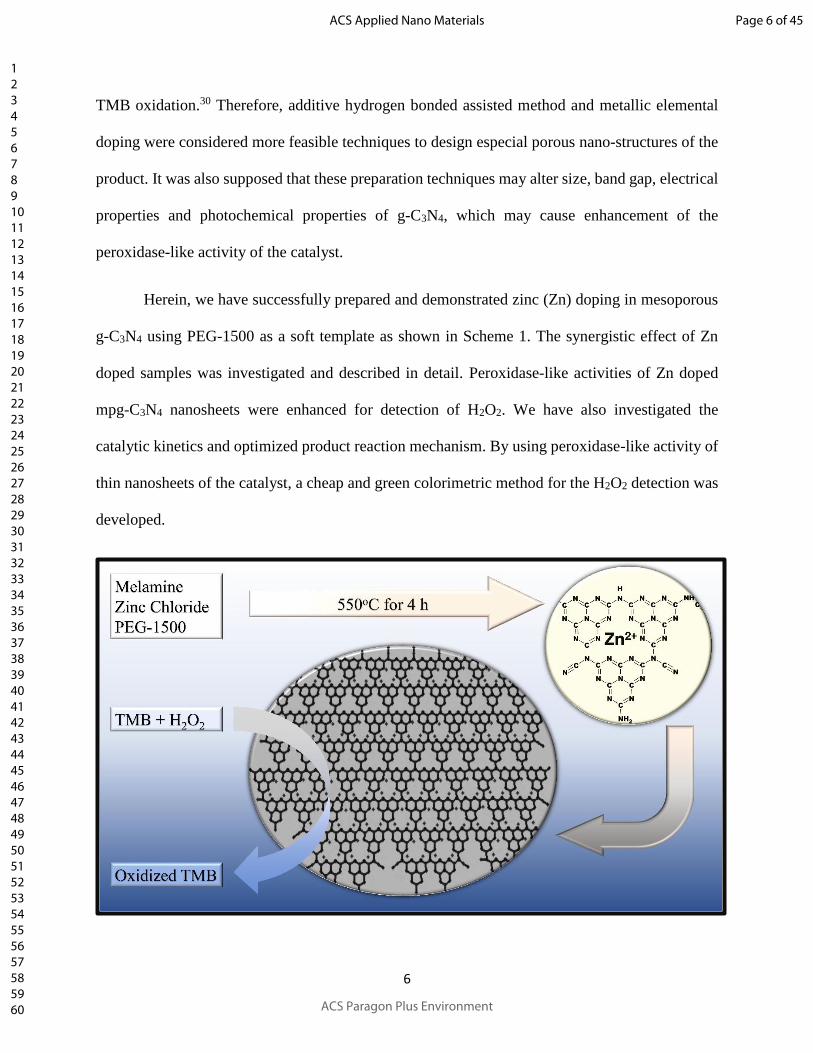
Herein, we have successfully prepared and demonstrated zinc (Zn) doping in mesoporous
g-C3N4 using PEG-1500 as a soft template as shown in Scheme 1. The synergistic effect of Zn
doped samples was investigated and described in detail. Peroxidase-like activities of Zn doped
mpg-C3N4 nanosheets were enhanced for detection of H2O2. We have also investigated the
catalytic kinetics and optimized product reaction mechanism. By using peroxidase-like activity of
thin nanosheets of the catalyst, a cheap and green colorimetric method for the H2O2 detection was
developed.
Page 6 of 45
ACS Paragon Plus Environment
ACS Applied Nano Materials
123456789101112131415161718192021222324252627282930313233343536373839404142434445464748495051525354555657585960
7
Scheme 1. Systematic representation of proposed mechanism for the colorimetric detection of
H2O2 by TMB and Zn doped mpg-C3N4 nanosheets.
2. EXPERIMENTATION
2.1 Reagents and Materials
Melamine was received from Analar. Acetic acid, zinc chloride dihydrated (ZnCl2•2H2O)
and TMB were received from Sigma Aldrich. Sodium acetate anhydrous was obtained from
Daejung. H2O2 (30%) was obtained from Merck KGaA. Polyethylene glycol 1500 (PEG-1500)
was received from Panreac. Dimethyl sulfoxide (DMSO) was received from Lab Scan.
Hydrochloric acid solution was obtained from BDH. Sodium hydroxide pellets was received from
Omicron. All chemicals were of analytical standard, used as received. All solutions were made by
deionized water obtained from an Elga-Pure-LAB@Ultra deionizer.
2.2 Characterizations
X-ray diffraction (XRD) analysis was performed on Rigaku-D/max-2500-PC
diffractometer with a graphite typed monochromator (40kV, 40mA). Nickel filtered Cu-Kα
radiations having wavelength of 1.5418Å was used during the analysis. Surface morphology and
shape were obtained using Vega 3, LMU, Tescan scanning-electron-microscope (SEM). SEM
images were obtained at 10kV. Elemental analysis as well as purity were determined by using
energy-dispersive X-rays spectrometer (EDX) on same SEM device. Double adhesive tape
supported gold coated samples were mounted on aluminum support for analysis. Morphologies
and microstructure of the samples were determined through transmission electron microscopy
(TEM) and measurements were carried out on a transmission electron microscope (JEM-2010F,
JEOL, Japan) with an acceleration voltage of 200 kV. The Brunauer-Emmett-Teller (BET) specific
Page 7 of 45
ACS Paragon Plus Environment
ACS Applied Nano Materials
123456789101112131415161718192021222324252627282930313233343536373839404142434445464748495051525354555657585960

8
surface area, pore sizes distribution and pore volume were measured BET method. The BET and
Barrett-Joyner-Halenda (BJH) measurements were carried out by using Micrometrics ASAP 2020
(V 4.00 H) surface area / porosity analyzer. X-ray photoelectron spectroscopy (XPS) was used to
determine the chemical environment and chemical state of the elements present in the samples.
XPS measurements were carried on Axis Ultra spectrometer system with a mono-chromatized
Aluminum-K X-ray radiation source (225 W). The high resolution XPS spectra were recorded
for carbon (C) 1s, nitrogen (N) 1s and Zn 2p regions. For functional groups determination, Fourier-
transform-infrared (FTIR) spectra were scanned in absorption manner in wavenumber range of
670 - 4000 cm-1 using Thermo-Fisher-Scientific (Nicolet, 6700) spectrophotometer, a resolution
of 8 cm-1 in attenuated total reflectance (ATR) mode at room temperature, using one scan per
spectrum. For estimation of band gap, ultra-violet-visible (UV-Vis) diffuse-reflectance-spectra
(DRS) were scanned on Perkin-Elmer-Lambda 35 spectrophotometer. Barium sulphate was
standard reference and samples were pressed into thin tablet under ambient temperature conditions.
Absorption edge was used for band gap calculation. In colorimetric analysis, to find wavelength
of maximum absorption (λmax) and change of absorption peak intensity of reaction system, UV-
Vis absorption spectra of reaction system were noted using Perkin-Elmer-Lambda 25 UV-Vis
double beam spectrophotometer (UV-25, Perkin Singapore) with quartz cuvette capacity of 3 mL
and bandwidth setting (1 nm) at a wavelength scan rate of 960 nm min-1 in wavelength range 400
- 800 nm. Raman and photoluminescence spectra of samples were recorded using InVia-laser-
scanning-confocal-microscope (Reni Shaw, UK) at 457 nm laser excitation wavelength. The laser
exposure time was set at 10s with grating, objective and laser power of 1800 I min-1, 5x, and 0.1%
respectively.
2.3 Synthesis of Zn-mpg-C3N4
Page 8 of 45
ACS Paragon Plus Environment
ACS Applied Nano Materials
123456789101112131415161718192021222324252627282930313233343536373839404142434445464748495051525354555657585960

9
The preparation of Zn doped mesoporous-graphitic-carbon-nitride (Zn-mpg-C3N4) was
done by calcination technique with minor modifications as reported earlier.31 Briefly, 11.1 g of
melamine and 2.3 g of PEG-1500 were mixed in 50 mL of the ethanol containing ZnCl2•2H2O
solution. The resultant suspension was heated at 80°C with continuous stirring for removal of
ethanol to form a white solid. Fine grounded white solid was transferred into ceramic boats, heated
under static air at temperature ramp of 5°C min-1 to achieve final temperature of 550°C and held
at 550oC for 4 h. After completion of reaction, the product was cooled under normal conditions.
As a result, a pale-yellow product was obtained, which was mechanically ground to fine powder
in pestle mortar and used for checking of peroxidase-activity. Various samples were prepared by
varying amount of ZnCl2•2H2O (0, 0.005, 0.012, 0.019, 0.027, 0.0578, 0.111, 0.333 and 0.555 g)
and wt. percentage of Zn with respect to product amount was calculated (0, 1, 2.2, 3.3, 4.7, 10, 15,
38 and 52). The resultant samples were labelled as wt. % of Zn-mpg-C3N4. The g-C3N4 was
synthesized without the addition of PEG-1500 and ZnCl2•2H2O under the same reaction
conditions.
2.4 Peroxidase-like Catalytic Activity and Steady-State Catalytic Kinetics Assay
To examine and compare peroxidase-like activity, two-dimensional nanostructures having
thickness ranging from 1 to 100 nm called as nanosheets were prepared and used for reaction
between TMB and H2O2. Stock suspension (2.7 mg mL-1) of each sample in distilled water was
prepared by ultrasonication with R60% at 60oC for 30 min. This suspension (75 µL) was separately
added into sodium acetate buffer (2820 L, 100 mM, and pH 4) and mixed well. Then, 45 µL of
TMB (stock 20 mM in DMSO) and 60 µL of H2O2 (stock 20 mM in dist. water) were added in
reaction mixture. Final concentration was 68 µg mL-1, 300 µM and 400 µM for nanosheets, TMB
and H2O2 respectively. Reaction mixture turned blue due to the oxidized TMB and a prominent
Page 9 of 45
ACS Paragon Plus Environment
ACS Applied Nano Materials
123456789101112131415161718192021222324252627282930313233343536373839404142434445464748495051525354555657585960

10
absorption peak at 652 nm (A652 nm) in the UV-Vis absorption spectrum was observed. Such
reaction are dependent on the amount of nanosheets, so measurements were carried out by
controlling the amount of nanosheets suspension in the reaction mixture. Peroxidase-like activity
depends upon the pH of buffer, temperature, catalyst concentration and substrate (TMB, H2O2)
concentration, therefore catalytic measurements were carried by varying pH (2.0 - 6.0),
temperature (20 - 40oC), amount of catalyst suspension (0 - 100 µg mL-1), TMB concentration
([TMB], 0 - 620 µM) and H2O2 concentration ([H2O2], 0 - 16 mM) in 100 mM acetate buffer.
Kinetics studies were performed by determining the A652 nm with respect to reaction time.
Biomimetic determinations for mechanism study were performed by varying [H2O2] at fixed
[TMB] or vice versa. Steady-state dynamics kinetics constant parameters (Km and Vmax) were
calculated from the initial linear ranges of the initial reaction rates of kinetics curves by Line-
weaver-Burk plots (1 / velocity against 1 / [substrate]) of the double reciprocal of Michaelis-
Menten-equation, [V = (Vmaximal × [S]) / (Km + [S])]. Where, V and Vmaximal were velocities of the
reaction, Km was Michaelis-Menten constant and [S] was the concentration of the substrate.
3. RESULTS AND DISCUSSION
To find out the structural morphology, SEM images of prepared samples were obtained as
seen in Figure 1. The g-C3N4 was hierarchical platelet type analogous to the graphite, while micron
sized g-C3N4 was stacked. It was obvious that after introduction of PEG-1500 as soft template in
the synthesis process, mpg-C3N4 has displayed stacked typed morphology with several hundred
nanometers sized chips. This revealed that PEG-1500 was beneficial to increase the number of
small pores on the surface. Moreover, after ZnCl2•2H2O addition along with PEG-1500 in
melamine and the resultant mixture calcination (550oC, 4 h), the SEM image of 10% Zn-mpg-
C3N4 has showed that pores were blocked with Zn loading.
Page 10 of 45
ACS Paragon Plus Environment
ACS Applied Nano Materials
123456789101112131415161718192021222324252627282930313233343536373839404142434445464748495051525354555657585960

11
Figure 1. SEM images of prepared g-C3N4 based modified samples.
EDX images of the prepared samples are presented in Figure S1. EDX labelled peaks in g-
C3N4 as well as in mpg-C3N4 were corresponding to C and N atoms, whereas in 10% Zn-mpg-
C3N4 were corresponding to C, N and Zn atoms. Some other peaks were corresponding to elements
Page 11 of 45
ACS Paragon Plus Environment
ACS Applied Nano Materials
123456789101112131415161718192021222324252627282930313233343536373839404142434445464748495051525354555657585960

12
used for sputter coating for EDX analysis. Table 1 represented the comparison of elemental
percentages.
Table 1. Comparison of g-C3N4, mpg-C3N4, and 10% Zn-mpg-C3N4 EDX elemental analysis.
Samples Wt. %Zn Wt. %N Wt. %C Atomic %Zn Atomic %N Atomic %C
g-C3N4 - 49 51 - 46 54
mpg-C3N4 - 58 42 - 54 46
10% Zn-mpg-
C3N4
28 27 45 7 32 62
EDX results supported the presence of N and C atoms in g-C3N4 as well as in mpg-C3N4
whereas Zn, N and C atoms in 10% Zn-mpg-C3N4. However, higher percentage of C atoms was
due to the involvement of C atoms form laser light and from double adhesive tape.
To determine the specific surface areas, porosity and pore-size-distribution of prepared
samples, nitrogen adsorption-desorption isotherms were obtained. Figure 2 showed that all the
samples exhibited type IV adsorption isotherms according to Brunauer-Deming-Deming-Teller
(BDDT) classification system. The amount of nitrogen gas adsorbed for 10% Zn-mpg-C3N4 was
much larger than the amount of nitrogen gas for g-C3N4, which indicated the formation of enlarged
mesopores. According to the International Union of Pure and Applied Chemistry (IUPAC)
guideline, H3 type hysteresis loop was observed in the range from 0.65-1.0 P/Po for 10% Zn-mpg-
C3N4, which also indicated the mesoporous features in the sample. These mesopores was
originated from the stacking of g-C3N4 nanosheets since the PEG-1500 and doped Zn species
strongly effected the interlayers packing of g-C3N4 nanosheets through N-bridge linking. Such
effect was resulted in an increase of space between the interlayers. The BET surface area of 10%
Zn-mpg-C3N4 was determined as 9.8 m2/g, which was about 3.6 times of the g-C3N4 (2.8 m2/g).
Page 12 of 45
ACS Paragon Plus Environment
ACS Applied Nano Materials
123456789101112131415161718192021222324252627282930313233343536373839404142434445464748495051525354555657585960

13
This enhancement in surface area was ascribed to the generation of mesoporous structure,
reduction of thickness and size of the mpg-C3N4 layer. Furthermore, the pore-size-distribution was
also determined from the adsorption branches of the isotherms by using BJH method as shown in
insert of Figure 2. The BJH adsorption pore diameter for g-C3N4 and 10% Zn-mpg-C3N4 was
calculated as 80.4 and 18.8 nm, whereas adsorption cumulative volume of pores was calculated as
0.014 and 0.029 cm3/g respectively. These finding revealed that the pores in 10% Zn-mpg-C3N4
were corresponding to the mesoporous structure. The BET average pore width of 10% Zn-mpg-
C3N4 (12.4 nm) exhibited a quite broad pore-size-distribution (2 - 90 nm) with small mesopores
(2.5 and 4.6 nm) and large mesopores (31.1 nm), which further confirmed the generation of more
mesopores in the 10% Zn-mpg-C3N4. The small mesopores were produced due to the porosity in
nanosheets created by soft template and Zn-N interaction due to the splitting of the large layers
during the synthesis process. The large mesopores were formed by the packing of g-C3N4 layers.
Therefore, the introduction of mesoporosity into g-C3N4 has enlarged the surface area and
decreased the pore volume, which were beneficial in providing more possible redox reaction active
sites. Thus, this work showed that 10% Zn-mpg-C3N4 with enhanced porous surface areas can be
prepared by using soft template like PEG-1500.
Page 13 of 45
ACS Paragon Plus Environment
ACS Applied Nano Materials
123456789101112131415161718192021222324252627282930313233343536373839404142434445464748495051525354555657585960

14
Figure 2. Nitrogen adsorption-desorption isotherms and corresponding BJH pore size distribution
curves (inset) of the samples.
The morphologies and microstructure of g-C3N4 and Zn-doped mpg-C3N4 samples were
investigated via TEM. Figure 3A showed the TEM image of g-C3N4. It can be clearly seen that
aggregate of free-standing nanosheets with edges tended to be ragged to minimize their surface
area. A dense and stacked architecture was obtained. Figure 3B and Figure 3C showed the low-
resolution and high-resolution TEM images of 10% Zn-mpg-C3N4. High-resolution image showed
the mesoporous structure consisting of irregular, wrinkled and small flat sheets. Many pores with
mean diameter of 10 - 45 nm were distributed on the sheets. Furthermore, this pore size was much
greater than the size of Zn2+ (~74 pm). The presence of somewhat transparency indicated that
sample was made of few atomic layers with mesoporous structure, but low-resolution image
Page 14 of 45
ACS Paragon Plus Environment
ACS Applied Nano Materials
123456789101112131415161718192021222324252627282930313233343536373839404142434445464748495051525354555657585960

15
supported the presence of Zn in the porous structure. It is speculated that PEG-1500 proceeded to
the crystal lattice during the formation of mpg-C3N4, which may be changed its morphology.
Considering above observations, it can also be concluded that the 10% Zn-mpg-C3N4 sample was
more fragmental, thin and smaller with mesoporous structures as compared to earlier reported
samples. Meanwhile, Zn element and its oxides forms were not found in TEM image, which
representing that the state of Zn in 10% Zn-mpg-C3N4 sample was ionic. The thin nanosheets with
porous structure resulted in the increase of surface to volume ratio and reactive site numbers, which
can enhance the peroxidase-like catalytic activity of Zn-mpg-C3N4.
Page 15 of 45
ACS Paragon Plus Environment
ACS Applied Nano Materials
123456789101112131415161718192021222324252627282930313233343536373839404142434445464748495051525354555657585960

16
Figure 3. TEM images of g-C3N4 (A) and 10% Zn-mpg-C3N4 (B and C).
As can be seen in Figure 4, XRD patterns confirmed the presence of graphitic like structure
in all the samples. Diffraction peaks in spectra was corresponding to the structure of g-C3N4. A
minor diffraction peak around 15.8o in the spectra of g-C3N4, mpg-C3N4 and 10% Zn-mpg-C3N4
were corresponding to index (100) planes with interplanar distance of 0.55 nm was arising from
the in-planar structural packing motifs. Intensity of this peak was decreased in the case of mpg-
Page 16 of 45
ACS Paragon Plus Environment
ACS Applied Nano Materials
123456789101112131415161718192021222324252627282930313233343536373839404142434445464748495051525354555657585960

17
C3N4 and Zn-mpg-C3N4. Similarly, a major peak in the spectra of g-C3N4 and mpg-C3N4 was
observed around 26.5o corresponding to index (002) planes with interplanar distance of 0.336 nm
arising from the typical interplanar stacking of hexagonal g-C3N4 (JCPDS-PDF 01-087-1526)
(JCPS-PDF 01-087-1522).32 A decrease in intensity of major peak in mpg-C3N4 spectra indicated
the presence of thin and porous structure in mpg-C3N4 nanosheets. However, in the case of 10%
Zn-mpg-C3N4 spectra, this peak was slight shifted and further decrease in peak intensity was
observed. In addition of (100) and (002) indexed planes, a small variation in intensity and small
shift in 2 of indexed planes (001), (201), (102), (111), (300), (220), (302) and (203) in XRD
patterns of 10% Zn-mpg-C3N4 were observed as compared to that of the g-C3N4 and mpg-C3N4
XRD patterns. These variations were due to decrease in crystallinity, generation of edge defects
and zinc ion doping in the thin and porous structure of mpg-C3N4. There was no diffraction peak
observed corresponding to Zn or ZnO phase in the samples, which also indicated that Zn metal
ions were chemically coordinated / embedded in host graphitic structured planes possibly in the
form of Zn-N coordination bonds. Therefore, introduction of PEG-1500 and external Zn ion
doping treatment during calcination process alleviated the crystallinity and generated porosity in
the samples.25, 28
Page 17 of 45
ACS Paragon Plus Environment
ACS Applied Nano Materials
123456789101112131415161718192021222324252627282930313233343536373839404142434445464748495051525354555657585960

18
Figure 4. XRD analysis of g-C3N4, mpg-C3N4 and 10% Zn-mpg-C3N4.
XPS measurements of the samples were carried out to determine the chemical environment
and chemical state of the elements (C, N and Zn). Surface probing technique was used to determine
the local structure of g-C3N4 and metallic nature of Zn. Figure 5 showed the high resolution XPS
spectra of C, N and Zn elements in the prepared samples. High resolution C 1s XPS spectra in
Figure 5A showed the presence of mainly three different Gaussians-Lorentzian peaks upon
deconvolution. The C 1s peak at binding energy of 284.5 eV was ascribed to the graphitic or
amorphous C atoms from instrument and surface adsorbed adventitious hydrocarbons in purely
carbonaceous environment containing C-C coordination and determined as standard C. The C 1s
peaks at 287.6 and 288.3 eV was ascribed to sp2-hybridized C atoms in aromatic ring bonding with
-NH2 group, sp3-hybridized C atoms (C-(N)3) and sp2-hybridized C atoms attached to the N in
Page 18 of 45
ACS Paragon Plus Environment
ACS Applied Nano Materials
123456789101112131415161718192021222324252627282930313233343536373839404142434445464748495051525354555657585960

19
aromatic ring as N-C=N to form a pure graphitic site in a C-N-C coordination. Figure 5B showed
N 1s high resolution XPS spectra. N 1s peak were deconvoluted into four different peaks with
binding energies centered at 398.6, 400.1, 401.2 and 404.5 eV. The main dominant peaks at 398.6
eV and 400.1 eV were attributed to sp2 hybridized N atoms in C-N=C group of triazine ring and to
the tertiary N atoms in N-(C)3 group, which connecting C6N7 structural motif. The peaks at 401.2
eV and 404.5 eV were corresponded to N atoms with ≡N and -N- bonding and charge effect,
respectively. Deconvoluted high resolution XPS spectra of Zn 2p and Zn LMM Auger region of
samples are shown in Figure 5C. A doublet in Zn 2p spectrum was appeared with binding energies
located at 1044.9 and 1021.8 eV, assigned to Zn 2p1/2 and Zn 2p3/2 lines respectively. The
difference of binding energies between these peaks is 23.1 eV, which indicated that Zn ions in the
sample were of a +2 oxidation states. Further, binding energy of Zn 2p3/2 peak was lower than
binding energy of ZnCl2 (1021.9 eV). The presence of peak at 496.5 eV in Zn LMM Auger
spectrum confirmed the existence of Zn-N bonds. The Zn-N bonds helped in charge transfer from
guest Zn to the host g-C3N4, which was important for enhancing peroxidase-like activity of
catalyst. The binding energies of C 1s and N1s signals for g-C3N4 and 10% Zn-mpg-C3N4 samples
was very close to each other, but integral intensity of these signals was decreased with Zn doping.
Compared to g-C3N4, the percentage of C-NH2/C-NH in 10% Zn-mpg-C3N4 was decreased. This
decrease in percentage indicated that thin nanosheets of 10% Zn-mpg-C3N4 was formed during the
heat treatment of mixture of melamine, PEG-1500 and ZnCl2•2H2O. From these observations, we
can conclude that aromatic units of g-C3N4 was somewhat broken by the Zn doping. Based on XPS
and XRD analysis, we believed that the product obtained from the calcination of melamine, PEG-
1500 and ZnCl2•2H2O are basically Zn-doped mpg-C3N4.
Page 19 of 45
ACS Paragon Plus Environment
ACS Applied Nano Materials
123456789101112131415161718192021222324252627282930313233343536373839404142434445464748495051525354555657585960

20
Figure 5. XPS spectra of C1s (A), N1s (B), Zn 2p and Auger Zn LMM (C) peaks of the samples.
In FTIR spectra (Figure 6), all characteristics absorption peaks in mpg-C3N4 and Zn doped
mpg-C3N4 were almost like to that as in the case of g-C3N4. This showed that graphitic network in
Zn doped mpg-C3N4 were similar with the network of g-C3N4. This revealed that Zn loading and
use of PEG-1500 were not able to modify or change the basic structure and central chemical
skeleton in modified g-C3N4 samples. Typical bands around 800 cm-1 in finger print region was
assigned to breathing vibration of tri-s-triazine / s-triazine ring sextant out of the plan bending
system, which was linked by -NH- group. High intensity broad absorbance bands in the range of
1225 - 1572 cm-1 was assigned to aromatic heterocyclic stretching vibration in C=N and C-NH-C
Page 20 of 45
ACS Paragon Plus Environment
ACS Applied Nano Materials
123456789101112131415161718192021222324252627282930313233343536373839404142434445464748495051525354555657585960

21
in finger print region likely derivative of s-triazine. Small absorption peaks around 2300 - 2404
cm-1 was assigned to -C≡N terminal groups and to the cumulated bonds (-N=C=N-) or alike
species. A low intensity and broad set of peaks were found above 3000 cm-1, which were attributed
to the vibrational stretching mode in primary (-NH2) or secondary (-NH) amine. These bands
clearly indicated the presence of dandling hydrogen atoms in -C-N layers in all synthesized
nanomaterials.
Figure 6. FTIR spectra of g-C3N4 based modified samples.
UV-Vis DRS analysis of pure g-C3N4 and Zn modified mpg-C3N4 were done by comparing
spectra. As shown in Figure 7, Zn doped mpg-C3N4 spectrum exhibited strong absorption in the
whole visible region. Absorption below 450 nm to the near-ultraviolet was resulted from the charge
transfer response of g-C3N4 and its functionalized materials from valence-band occupied by N 2p
Page 21 of 45
ACS Paragon Plus Environment
ACS Applied Nano Materials
123456789101112131415161718192021222324252627282930313233343536373839404142434445464748495051525354555657585960

22
orbitals to conduction-band resulted from C 2p orbitals. Absorption peaks in UV region was
attributed to the band gap of polymeric melon units of g-C3N4. Absorption edge of Zn doped mpg-
C3N4 showed a shift in wavelength region as the dopant concentration was varied. It was decreased,
reached its minimum value and then increased with respect to the dopant concentration. Estimated
band gap was calculated using formula (Band gap = 1240 / Absorption edge (λ)) in electron volt
(eV). Insert in Figure 7 showed that g-C3N4 has band gap of 2.74 eV, whereas Zn doped mpg-C3N4
possessed band gap of 2.94, 2.95, 2.96, 2.99, 2.99, 3.0, 2.98, 2.96 and 2.93 eV for different wt.
percentage of Zn content as 0, 1, 2.2, 3.3, 4.7, 10, 15, 38 and 52 in mpg-C3N4 respectively. This
variation in band gap with Zn dopant was originated due to the repulsive interaction of 3d orbitals
of host (Zn) and 2p orbitals of guest (nitrogen) in doped g-C3N4.33 It can also be seen that Zn doped
mpg-C3N4 has higher absorbance intensity in UV region, whereas less absorbance in visible region
than g-C3N4. These variations in spectra indicated that Zn was coordinated with -conjugated
system of g-C3N4, and doping strategy was useful for tuning the band gap and to facilitate the
charge transfer in aromatic CN heterocyclic conjugated system.33
Page 22 of 45
ACS Paragon Plus Environment
ACS Applied Nano Materials
123456789101112131415161718192021222324252627282930313233343536373839404142434445464748495051525354555657585960

23
Figure 7. UV-Visible DRS spectra (insert showed the change in band gap with respect to dopant
amount) of g-C3N4 and its modified forms.
Raman spectra (Figure S2) showed the coordination of Zn species and maintenance of
graphitic structure in the synthesized samples.34 PL spectra (Figure 8) under an excitation
wavelength of 457 nm was scanned and observed a main PL emission around 588 nm in the spectra
of all the samples. The PL intensity of g-C3N4 sample was more than the PL intensity of mpg-
C3N4. PL intensity was depended on the dopant concentration and was quenched with the increase
in Zn loading, reached to minimum value, after that it was began to increase. It can be seen in
Figure 8 that PL intensity of 10% Zn-mpg-C3N4 was lowest due to loading of optimum Zn content.
At optimum Zn loading, recombination of photo generated charge carriers was inhibited and an
Page 23 of 45
ACS Paragon Plus Environment
ACS Applied Nano Materials
123456789101112131415161718192021222324252627282930313233343536373839404142434445464748495051525354555657585960

24
enhancement in electron-hole transfer between Zn and mpg-C3N4 was occurred. This factor
increased the availability of more holes for oxidation of TMB. Therefore, metallic elemental
doping strategy was useful to tune the electronic related properties, photo generated charge
carrier’s recombination and electronic transferability in the samples.35
Figure 8. PL spectra at excitation wavelength of 457 nm of g-C3N4 and Zn modified mpg-C3N4.
3.1 Peroxidase-Activity of Nanosheets
A comparison of peroxidase-like activity of g-C3N4, mpg-C3N4 and 10% Zn-mpg-C3N4 is
shown in term of their UV-Vis absorption spectra in Figure 9A.
Page 24 of 45
ACS Paragon Plus Environment
ACS Applied Nano Materials
123456789101112131415161718192021222324252627282930313233343536373839404142434445464748495051525354555657585960

25
Figure 9. UV-Vis absorption spectra for comparison of peroxidase-like activity after one-hour
incubation of the reaction mixture (A) Effect of dopant concentration (B) pH (C) temperature (D)
Page 25 of 45
ACS Paragon Plus Environment
ACS Applied Nano Materials
123456789101112131415161718192021222324252627282930313233343536373839404142434445464748495051525354555657585960

26
and catalyst concentration (E) on peroxidase-like activity. The reaction mixture containing 68 µg
mL-1 nanosheets, 300 µM TMB, 400 µM of H2O2 in sodium acetate buffer (pH 4.0, 100 mM).
Inserts showed the digital images of color change in the reaction systems.
The A652 nm was linked to the charge-transfer complexes resulting from one-electron
oxidation of TMB and reaction mixtures were turned to a blue color (Insert of Figure 9A). It is
clear from Figure 9A, that A652 nm of 10% Zn-mpg-C3N4 (curve g) reaction mixture was higher
than that of A652 nm of mpg-C3N4 (curve f), g-C3N4 (curve e), and ZnO (curve d) reaction systems.
This indicated that Zn ions coordination in thin mesoporous nanosheets of g-C3N4 has displayed a
major role for enhancing peroxidase-like catalytic activity. In turn, many light-triggered reactive
oxygen species were generated due to the presence of more catalytic active sites and novel
functionalities in Zn-mpg-C3N4. Effect of precursor PEG-1500 in the synthesis of mpg-C3N4 can
be clearly observed, when a comparison of A652 nm of mpg-C3N4 (curve f) was done with the A652
nm of the simple g-C3N4 (curve e) and ZnO (curve d). The A652 nm of mpg-C3N4 reaction system
was higher than the A652 nm of g-C3N4 and ZnO reaction systems. This higher peroxidase-like
catalytic activity of mpg-C3N4 was attributed to its thin and porous morphology as compared to
simple g-C3N4 and ZnO. Further, in the absence of H2O2 the reaction system consisting of g-C3N4
nanosheets and TMB has displayed no obvious change (curve b), which indicated that no redox
reaction occurred. In contrast, in the absence of H2O2 the reaction between 10% Zn-mpg-C3N4
nanosheets and TMB has displayed small change in color (curve c). These observations indicated
the higher electron transferability, higher charge separation efficiency and higher intrinsic
peroxidase-like activity of 10% Zn-mpg-C3N4 nanosheets as compared to that of the pure g-C3N4
nanosheets. Therefore, Zn doping has enabled electron coupling between Zn metals and mpg-C3N4
by shaped unique electronic structures. These unique electronic structures favored the electron
Page 26 of 45
ACS Paragon Plus Environment
ACS Applied Nano Materials
123456789101112131415161718192021222324252627282930313233343536373839404142434445464748495051525354555657585960
27
mobilization along the electronically delocalized planar thin nanosheets by accelerating the
electron transfer process. There was no peak at A652 nm and no blue color in the reaction mixture
was observed, when TMB was mixed with H2O2 (curve a) in the absence of nanosheets. These
observations indicated that the reaction may be occurred only, if catalyst nanosheets were present
in the reaction mixture.
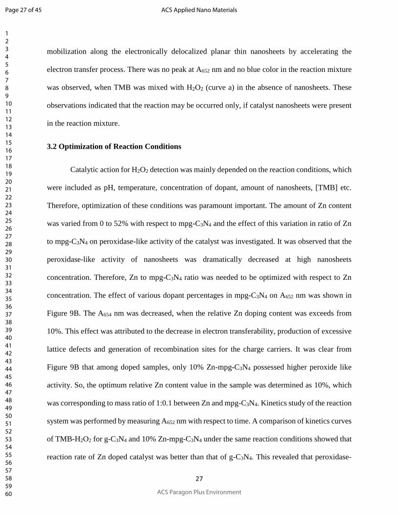
3.2 Optimization of Reaction Conditions
Catalytic action for H2O2 detection was mainly depended on the reaction conditions, which
were included as pH, temperature, concentration of dopant, amount of nanosheets, [TMB] etc.
Therefore, optimization of these conditions was paramount important. The amount of Zn content
was varied from 0 to 52% with respect to mpg-C3N4 and the effect of this variation in ratio of Zn
to mpg-C3N4 on peroxidase-like activity of the catalyst was investigated. It was observed that the
peroxidase-like activity of nanosheets was dramatically decreased at high nanosheets
concentration. Therefore, Zn to mpg-C3N4 ratio was needed to be optimized with respect to Zn
concentration. The effect of various dopant percentages in mpg-C3N4 on A652 nm was shown in
Figure 9B. The A654 nm was decreased, when the relative Zn doping content was exceeds from
10%. This effect was attributed to the decrease in electron transferability, production of excessive
lattice defects and generation of recombination sites for the charge carriers. It was clear from
Figure 9B that among doped samples, only 10% Zn-mpg-C3N4 possessed higher peroxide like
activity. So, the optimum relative Zn content value in the sample was determined as 10%, which
was corresponding to mass ratio of 1:0.1 between Zn and mpg-C3N4. Kinetics study of the reaction
system was performed by measuring A652 nm with respect to time. A comparison of kinetics curves
of TMB-H2O2 for g-C3N4 and 10% Zn-mpg-C3N4 under the same reaction conditions showed that
reaction rate of Zn doped catalyst was better than that of g-C3N4. This revealed that peroxidase-
Page 27 of 45
ACS Paragon Plus Environment
ACS Applied Nano Materials
123456789101112131415161718192021222324252627282930313233343536373839404142434445464748495051525354555657585960

28
like activity as well as kinetics of the reaction was improved by Zn modification in the catalyst.
This improvement was related to the presence of Zn-N active sites in the porous morphology, high
electron transferability, and low recombination rate due to capturing of conduction band electrons
by Zn ions and enlargement in surface area to provide greater catalytic active sites for 10% Zn-
mpg-C3N4 as compared to pure g-C3N4.
The pH of acetate buffer was optimized in range from 2.0 - 6.0 and temperature was
optimized in the range from 20 - 40oC. Effect of change in pH and change in temperature on 10%
Zn-mpg-C3N4 catalytic activity was presented in Figure 9C and Figure 9D respectively. Optimal
pH for the system was calculated as 4.0, which showed that stable catalytic activity of nanosheets
and TMB oxidation befall in weak acidic condition as already been reported for some other
nanomaterials.6, 24, 33, 36-37 Furthermore, the relative activity of reaction system was better at 30oC,
which indicated that our material worked well near room temperature. The effect of amount of
10% Zn-mpg-C3N4 nanosheets on peroxidase-like activity was determined by investigating time
dependent absorbance change against various amount of 10% Zn-mpg-C3N4 from 0 - 100 µg mL-
1. It can be seen form Figure S3 that A652 nm was increased as the concentration of nanosheets
suspension was increased and time was prolonged. The maximum reaction level was observed at
68 µg mL-1. A linear relationship between sample amount and catalytic activity was obtained as
depicted in Figure 9E. However, it can be seen in figure that at higher nanosheets concentration
deviation from linearity was occurred.
3.3 Mechanism and Steady-State Catalytic Kinetics Assay of H2O2 Using 10% Zn-mpg-C3N4
Nanosheets
To explore mechanism of peroxidase-like catalytic activity of 10% Zn-mpg-C3N4, steady-
state kinetics assay was performed by keeping concentration of substrate constant, while varying
Page 28 of 45
ACS Paragon Plus Environment
ACS Applied Nano Materials
123456789101112131415161718192021222324252627282930313233343536373839404142434445464748495051525354555657585960
29
concentration of analyte. Kinetics curves are shown in Figure S4, when [TMB] (300 µM) was kept
fixed and [H2O2] was varied. Similar kinetics curves in Figure S5 are obtained, when [H2O2] (400
µM) was fixed and [TMB] was varied. As excessive [H2O2] or [TMB] inhibited the rate of reaction,
so equilibrium concentrations were required to be determined.
Page 29 of 45
ACS Paragon Plus Environment
ACS Applied Nano Materials
123456789101112131415161718192021222324252627282930313233343536373839404142434445464748495051525354555657585960

30
Page 30 of 45
ACS Paragon Plus Environment
ACS Applied Nano Materials
123456789101112131415161718192021222324252627282930313233343536373839404142434445464748495051525354555657585960

31
Figure 10. Michaelis-Menten curves of 10% Zn-mpg-C3N4 for H2O2 (A) and for TMB (B). Double
reciprocal plot for H2O2 (C) and for TMB (D). Ping-Pong mechanism for H2O2 (E) and TMB (F).
Apparent steady-state reaction rates and initial velocity for both TMB and H2O2 were
determined by calculating the slopes of linear portion of absorbance changes at 652 nm with time
curves. The concentration of oxidized TMB (TMB•+) was determined by applying the Beer-
Lambert law (A = εCl, where A was absorbance, ‘l’ was path length (l = 1 cm), C is concentration
of substrate and ε was extinction coefficient (ε = 39,000 M-1 cm-1) respectively). Typical
Michaelis-Menten curves were drawn for suitable range of H2O2 and TMB as shown in Figure
10A and Figure 10B respectively. The value of Km affects the reaction kinetics and represents the
binding affinities of catalytic nanomaterials to the substrate. Table 2 represents the relationship
between Km, nanomaterials and substrates. The smaller Km represents the stronger affinity in
catalytic nanomaterials and substrates.
Table 2. Comparison of Km and Vmax of horseradish peroxide (HRP), g-C3N4, mpg-C3N4 and 10%
Zn-mpg-C3N4.
Catalyst Substrate Km (mM) Vmax Ref.
10% Zn-mpg-C3N4 TMB
H2O2
0.1
0.311
1.33 (10-8 Ms-1)
1.7 (10-8 Ms-1)
Present work
HRP TMB
H2O2
0.275
0.214
1.24 (10-8 Ms-1)
2.46 (10-8 Ms-1)
38
Co-g-C3N4-2 TMB
H2O2
0.113
318.58
8.46 (10-8 Ms-1)
9.46 (10-8 Ms-1)
33
MnSe-g-C3N4 TMB
H2O2
0.137
0.623
2.40 (10-3 s-1)
2.85 (10-3 s-1)
37
Cu NPs/g-C3N4 TMB
H2O2
0.389
9.27
5.84 (10-7 s-1)
3.84 (10-7 s-1)
36
Page 31 of 45
ACS Paragon Plus Environment
ACS Applied Nano Materials
123456789101112131415161718192021222324252627282930313233343536373839404142434445464748495051525354555657585960

32
g-C3N4 TMB
H2O2
0.031
4.565
1.35 (104 Ms-1)
1.72 (104 Ms-1)
3
Fe-g-C3N4 TMB
H2O2
0.030
8.956
1.95 (104 Ms-1)
1.46 (104 Ms-1)
3
Se-g-C3N4 TMB
H2O2
0.307
0.298
20.5 (10-4 s-1)
43.3 (10-4 s-1)
24
Apparent Km value of 10% Zn-mpg-C3N4 nanosheets with TMB substrate (0.1 mM) was
three times lower than Km of HRP (0.275 mM) with TMB substrate. This was attributed to high
surface area of several small pores on nanosheets and presence of more catalytic active sites on
the porous material, which resulted in strong adsorption affinity between carbon nitride nanosheets
and TMB as compared to the natural HRP. The reaction rate at saturation point of nanozymes with
substrate was explained by Vmax values. The Vmax of 10% Zn-mpg-C3N4 with TMB substrate
(1.33×10⁻⁸ Ms-1) was 1.1 times higher than that of natural HRP (1.24×10⁻⁸ Ms-1), which represented
a higher reaction rate of 10% Zn-mpg-C3N4 nanosheets. This was attributed to intrinsic electron
transfer capability of graphitic-structure and presence of Zn content in the nanosheets. The
catalytic activity of nanosheets was highly efficient and was like natural enzymes. The Km value
of 10% Zn-mpg-C3N4 for H2O2 substrate (0.311 mM) was 1.5 times higher than Km value (0.214
mM) of natural HRP, which was consistent with the observation that a higher [H2O2] was required
to achieve maximal activity for 10% Zn-mpg-C3N4 nanosheets. Double-reciprocal plots between
initial velocities against various [H2O2] analyte by keeping substrate [TMB] constant and vice
versa are shown in Figure 10C and Figure 10D respectively. Graphs showed that the reaction
catalyzed by 10% Zn-mpg-C3N4 nanosheets followed typical Michaelis-Menten model. A
comparison of double-reciprocal plots between initial velocities with variable [H2O2] against three
Page 32 of 45
ACS Paragon Plus Environment
ACS Applied Nano Materials
123456789101112131415161718192021222324252627282930313233343536373839404142434445464748495051525354555657585960

33
[TMB] (200, 300 and 500 M) substrates and variable [TMB] against three [H2O2] (200, 400 and
800 M) substrates were carried out to investigate Ping-Pong mechanism as depicted in Figure
10E and 10F respectively. Double reciprocal plots were characterized by straight parallel lines and
showed a Ping-Pong mechanism for these nanomaterials as shown by HRP. Ping-Pong mechanism
is a mechanism of a non-sequential double-displacement enzymatic reaction in which enzyme is
changed to an intermediate form, when first reacting substrate is changed to the product. This
intermediate form of enzyme is temporary and changes to standard form by bouncing back and
forth to change the second reacting substrate to product. In this mechanism, enzyme is acted like
a Ping-Pong ball. In steady-state catalytic kinetics assay, a reaction follow Ping-Pong mechanism,
when a plot between 1 / Velocity against 1 / [First substrate] at varying concentrations of second
substrate is a series of parallel lines and vice versa as shown in Figure 10E and 10F. It means that
Zn-mpg-C3N4 nanosheets responded to first reacting substrate to release product of this substrate
before responding with second reacting substrate. Such, catalytic mechanism of reaction was
associated with the −conjugated electronic structures of 10% Zn-mpg-C3N4 nanosheets and its
affinities with substrates like H2O2 and TMB. Catalyst nanosheets were −rich and could adsorbed
TMB via − interaction. Due to proximity, electron transfer was occurred from lone pair of amino
groups of TMB to catalyst nanosheets, which resulted in the oxidation of TMB. Electrons would
transfer from nanosheets to H2O2. Product from this substrate was hydroxyl radicals due to the
decomposition of H2O2 under the catalytic effect of 10% Zn-mpg-C3N4 nanosheets. Hydroxyl
radicals facilitated the oxidation of TMB. Possible reaction mechanism of peroxidase-like catalytic
activity of 10% Zn-mpg-C3N4 nanosheets was shown in Scheme 1. Similarly, other metal
coordinated (Cu2+, Pd NPs, Co3+, MnSe NPs, Fe3+) peroxidase-mimic catalyst of g-C3N4 has been
reported. From rest of these, our synthesized Zn2+ coordinated thin mesoporous nanosheets of g-
Page 33 of 45
ACS Paragon Plus Environment
ACS Applied Nano Materials
123456789101112131415161718192021222324252627282930313233343536373839404142434445464748495051525354555657585960
34
C3N4 has displayed better kinetics and catalytic activity. This indicated that our prepared
nanomaterial has more catalytic active sites and has generated many light-triggered reactive
oxygen species due to presence of novel functionalities in it.
3.4 Sensing of H2O2
Graphitic structure facilitates the electron transfer process and such structure type aromatic
carbon compounds exhibit intrinsic peroxidase-like activity. The reduction of H2O2 takes place via
an electron transfer process by donation of electrons from carbon network on micro level. This
electron transfer process can be enhanced through different techniques such as metallic doping and
nano-structuring. Therefore, for better peroxidase-like activity of nanosheets, it was supposed that
Zn doped mpg-C3N4 would serve as natural enzyme and may be used as catalyst in H2O2 reduction.
Nanosheets activity was determined by the amount of oxidized TMB in H2O2 by UV-Vis
absorption spectra. It was noted that oxidized TMB imparted blue color to the reaction mixture,
which was corresponding to a maximum absorption intensity at 652 nm. Without nanosheets, the
reaction mixture containing H2O2, TMB or both in sodium acetate buffer gave no blue color, so no
oxidation of TMB occurred. However, upon the addition of nanosheets in reaction system,
oxidation of TMB was occurred and a blue color was developed with an absorption maxima at 652
nm. This was the indication of nanosheets catalytic activity.
According to excellent peroxidase-like activity of our synthesized 10% Zn-mpg-C3N4
nanosheets under optimum experimental conditions of pH, temperature, [TMB] and nanosheets
amount, a colorimetric sensor for the detection of H2O2 using TMB substrate was developed. Using
typical [H2O2] and sensor response, UV-Vis absorption curves under optimum experimental
conditions (pH 4.0, 25oC) were obtained and shown in Figure 11A.
Page 34 of 45
ACS Paragon Plus Environment
ACS Applied Nano Materials
123456789101112131415161718192021222324252627282930313233343536373839404142434445464748495051525354555657585960

35
Figure 11. Typical UV-Visible absorption spectra (A), sensor dose response curve (B), and
linear-calibration plot (C).
Graph between absorbance at lambda max (max) of 652 nm versus [H2O2] in the presence
of nanosheets is shown in Figure 11B. Absorption intensity was increased as [H2O2] was varied
between 0 - 2000 µM, while at higher [H2O2] peroxidase-like activity of catalyst was inhibited.
There was a slight increase in absorption intensity beyond 400 µM of [H2O2], which was supposed
as reaction equilibrium [H2O2]. A linear range of the sensor was determined from linear calibration
curve as 10 - 2000 µM using absorbance versus [H2O2] as depicted in Figure 11C. Quantification
limit (LOQ) and detection limit (LOD) of developed H2O2 sensor were found from linear
Page 35 of 45
ACS Paragon Plus Environment
ACS Applied Nano Materials
123456789101112131415161718192021222324252627282930313233343536373839404142434445464748495051525354555657585960

36
regression-equation of H2O2 dose and response curve by using 10σ/s and 3.3σ/s respectively.
Sigma (σ) and (s) are standard error of estimate and slop of linear regression-line respectively.
LOD was obtained as low as 1.4 M and LOQ as 3.0 M. Table 3 represented a comparison of
linear ranges and LODs of different g-C3N4 based colorimetric sensors.
Table 3. Comparison of linear ranges, LODs of different nanomaterials as peroxidase mimetic for
H2O2 assays.
Materials Linear Range (µM) LOD (µM) Reference
10% Zn-mpg-C3N4 10 - 2000 (R2 = 0.9981) 1.4 Present Work
g-C3N4 1 - 100 (R2 = 0.994) 0.4 39
Utg-C3N4 NSs 10 - 100 (R2 = 0.9902) 5.6 40
g-C3N4 5 - 30 (R2 = 0.9866) 0.9 2
Se-g-C3N4 16 – 4000 1.6 24
MeSe-g-C3N4 18 - 1800 (R2 = 0.9986) 1.8 37
4. CONCLUSION
In summary, we have used calcination method to synthesize g-C3N4 and thin porous Zn
doped mpg-C3N4 by using mixture of melamine, PEG-1500 and zinc chloride. Introduction of
PEG-1500 as soft template in reaction mixture was beneficial for the enhancement in porosity and
to increase in amount of Zn loading in the graphitic framework of g-C3N4. Results indicated that
Zn-mpg-C3N4 possessed enhanced intrinsic peroxidase-like activity. The synergistic effects of
high conductivity, electron transfer capability of mpg-C3N4 support and intrinsic peroxidase-like
activity of Zn doping, facilitated in enhancement of peroxidase-like catalysis. Steady-state kinetics
analysis specified that catalysis followed Ping-Pong mechanism. In the presence of 10% Zn-mpg-
C3N4 nanosheets, TMB and H2O2 were reacted and reaction mixture color was turned to blue,
Page 36 of 45
ACS Paragon Plus Environment
ACS Applied Nano Materials
123456789101112131415161718192021222324252627282930313233343536373839404142434445464748495051525354555657585960

37
which was the basis for the development of a colorimetric assay for H2O2. Peroxidase-mimics
activity of 10% Zn-mpg-C3N4 has presented several advantages over natural enzymes such as
facile preparation, low cost, fast response and high stability, which might be allowed this
nanomaterial to be used as an enzyme free peroxidase-mimics for potential applications in
biotechnology and medical diagnostic areas.
ASSOCIATED CONTENT
Supporting Information
The supporting information (PDF) includes Figure S1 to S5 for EDX images and analysis, Raman
spectra, kinetics study of different amount of catalyst, steady state kinetics assay at fix [TMB] and
variable [H2O2] and vice versa. The supporting information is available free of charges on the ACS
Publications website at http://pubs.acs.org.
AUTHOR INFORMATION
Corresponding author
*Muhammad Nasir: [email protected]
*Akhtar Hayat: [email protected]
ORCID iD
Muhammad Nasir: 0000-0002-6742-2721
Author Contributions
All authors have contributed and approved the final version of the manuscript.
Funding Sources
Page 37 of 45
ACS Paragon Plus Environment
ACS Applied Nano Materials
123456789101112131415161718192021222324252627282930313233343536373839404142434445464748495051525354555657585960

38
This work was supported by Higher Education Commission Pakistan through its NRPU grant 20-
4993/R&D/HEC/14 and Pakistan Science Foundation and National Natural Science Foundation
China through its PSF-NSFC funded project (Project No. PSF/NSFC-II/Eng/P-COMSATS-Lhr
(07).
Notes
The authors declare no competing financial interest.
ACKNOWLEDGMENT
Aftab Ahmed thanks Interdisciplinary Research Centre in Biomedical Materials (IRCBM),
COMSATS University Islamabad, Lahore Campus to allow him to use its facility for the execution
of his research work and acknowledges the efforts his seniors and fellow colleagues in guiding
him to complete it.
Page 38 of 45
ACS Paragon Plus Environment
ACS Applied Nano Materials
123456789101112131415161718192021222324252627282930313233343536373839404142434445464748495051525354555657585960

39
TOC Graphic
Page 39 of 45
ACS Paragon Plus Environment
ACS Applied Nano Materials
123456789101112131415161718192021222324252627282930313233343536373839404142434445464748495051525354555657585960

40
REFERENCES
(1) Salimi, A.; Hallaj, R.; Soltanian, S.; Mamkhezri, H. Nanomolar detection of hydrogen peroxide
on glassy carbon electrode modified with electrodeposited cobalt oxide nanoparticles. Anal Chim
Acta 2007, 594, 24-31.
(2) Lin, T.; Zhong, L.; Wang, J.; Guo, L.; Wu, H.; Guo, Q.; Fu, F.; Chen, G. Graphite-like carbon
nitrides as peroxidase mimetics and their applications to glucose detection. Biosensors and
Bioelectronics 2014, 59, 89-93.
(3) Tian, J.; Liu, Q.; Asiri, A. M.; Qusti, A. H.; Al-Youbi, A. O.; Sun, X. Ultrathin graphitic carbon
nitride nanosheets: a novel peroxidase mimetic, Fe doping-mediated catalytic performance
enhancement and application to rapid, highly sensitive optical detection of glucose. Nanoscale
2013, 5, 11604-9.
(4) Tian, H.; Fan, H.; Ma, J.; Ma, L.; Dong, G. Noble metal-free modified electrode of exfoliated
graphitic carbon nitride/ZnO nanosheets for highly efficient hydrogen peroxide sensing.
Electrochimica Acta 2017, 247, 787-794.
(5) Xi, X.; Li, J.; Wang, H.; Zhao, Q.; Li, H. Non-enzymatic photoelectrochemical sensing of
hydrogen peroxide using hierarchically structured zinc oxide hybridized with graphite-like carbon
nitride. Microchimica Acta 2015, 182, 1273-1279.
(6) Vazquez-Gonzalez, M.; Liao, W. C.; Cazelles, R.; Wang, S.; Yu, X.; Gutkin, V.; Willner, I.
Mimicking Horseradish Peroxidase Functions Using Cu(2+)-Modified Carbon Nitride
Nanoparticles or Cu(2+)-Modified Carbon Dots as Heterogeneous Catalysts. ACS Nano 2017, 11,
3247-3253.
Page 40 of 45
ACS Paragon Plus Environment
ACS Applied Nano Materials
123456789101112131415161718192021222324252627282930313233343536373839404142434445464748495051525354555657585960

41
(7) Breslow, R. Biomimetic Chemistry and Artificial Enzymes: Catalysis by Design. Accounts of
Chemical Research 2002, 28, 146-153.
(8) Hu, P.; Han, L.; Zhu, C.; Dong, S. J. Nanoreactors: a novel biosensing platform for protein
assay. Chemical Communications 2013, 49, 1705-1707.
(9) Wu, J.; Wang, X.; Wang, Q.; Lou, Z.; Li, S.; Zhu, Y.; Qin, L.; Wei, H. Nanomaterials with
enzyme-like characteristics (nanozymes): next-generation artificial enzymes (II). Chem Soc Rev
2019, 48, 1004-1076.
(10) Wei, H.; Wang, E. Nanomaterials with enzyme-like characteristics (nanozymes): next-
generation artificial enzymes. Chem Soc Rev 2013, 42, 6060-93.
(11) Lin, Y.; Ren, J.; Qu, X. Catalytically active nanomaterials: a promising candidate for artificial
enzymes. Acc Chem Res 2014, 47, 1097-105.
(12) He, L.; Su, Y.; Lanhong, J.; Shi, S. Recent advances of cerium oxide nanoparticles in
synthesis, luminescence and biomedical studies: a review. Journal of Rare Earths 2015, 33, 791-
799.
(13) Challier, L.; Gal, F.; Carrot, G.; Perez, H.; Noel, V. Hybrid platinum nanoparticle ensemble
for the electrocatalytic oxidation of H2O2: Toward nanostructured biosensor design.
Electrochemistry Communications 2013, 28, 118-121.
(14) Nasir, M.; Rauf, S.; Muhammad, N.; Hasnain Nawaz, M.; Anwar Chaudhry, A.; Hamza
Malik, M.; Ahmad Shahid, S.; Hayat, A. Biomimetic nitrogen doped titania nanoparticles as a
colorimetric platform for hydrogen peroxide detection. J Colloid Interface Sci 2017, 505, 1147-
1157.
Page 41 of 45
ACS Paragon Plus Environment
ACS Applied Nano Materials
123456789101112131415161718192021222324252627282930313233343536373839404142434445464748495051525354555657585960
42
(15) Gao, L.; Zhuang, J.; Nie, L.; Zhang, J.; Zhang, Y.; Gu, N.; Wang, T.; Feng, J.; Yang, D.;
Perrett, S.; Yan, X. Intrinsic peroxidase-like activity of ferromagnetic nanoparticles. Nat
Nanotechnol 2007, 2, 577-83.
(16) Cheng, X.; Huang, L.; Yang, X.; Elzatahry, A. A.; Alghamdi, A.; Deng, Y. Rational design
of a stable peroxidase mimic for colorimetric detection of H2O2 and glucose: A synergistic
CeO2/Zeolite Y nanocomposite. Journal of Colloid and Interface Science 2019, 535, 425-435.
(17) Ge, J.; Yang, X.; Luo, J.; Ma, J.; Zou, Y.; Li, J.; Luo, W.; Cheng, X.; Deng, Y. Ordered
mesoporous CoO/CeO2 heterostructures with highly crystallized walls and enhanced peroxidase-
like bioactivity. Applied Materials Today 2019, 15, 482-493.
(18) Wang, H.; Wan, K.; Shi, X. Recent Advances in Nanozyme Research. Adv Mater 2018,
e1805368.
(19) Zhou, Y.; Liu, B.; Yang, R.; Liu, J. Filling in the Gaps between Nanozymes and Enzymes:
Challenges and Opportunities. Bioconjug Chem 2017, 28, 2903-2909.
(20) Wang, X.; Maeda, K.; Thomas, A.; Takanabe, K.; Xin, G.; Carlsson, J. M.; Domen, K.;
Antonietti, M. A metal-free polymeric photocatalyst for hydrogen production from water under
visible light. Nat Mater 2009, 8, 76-80.
(21) Zhang, J.; Zhang, G.; Chen, X.; Lin, S.; Mohlmann, L.; Dolega, G.; Lipner, G.; Antonietti,
M.; Blechert, S.; Wang, X. Co-monomer control of carbon nitride semiconductors to optimize
hydrogen evolution with visible light. Angew Chem Int Ed Engl 2012, 51, 3183-7.
(22) Li, X.; Zhu, L.; Zhou, Y.; Yin, H.; Ai, S. Enhanced Photoelectrochemical Method for
Sensitive Detection of Protein Kinase A Activity Using TiO2/g-C3N4, PAMAM Dendrimer, and
Alkaline Phosphatase. Anal Chem 2017, 89, 2369-2376.
Page 42 of 45
ACS Paragon Plus Environment
ACS Applied Nano Materials
123456789101112131415161718192021222324252627282930313233343536373839404142434445464748495051525354555657585960
43
(23) Wang, H.; Qi, C.; He, W.; Wang, M.; Jiang, W.; Yin, H.; Ai, S. A sensitive
photoelectrochemical immunoassay of N6-methyladenosine based on dual-signal amplification
strategy: Ru doped in SiO2 nanosphere and carboxylated g-C3N4. Biosensors and Bioelectronics
2018, 99, 281-288.
(24) Qiao, F.; Wang, J.; Ai, S.; Li, L. As a new peroxidase mimetics: The synthesis of selenium
doped graphitic carbon nitride nanosheets and applications on colorimetric detection of H2O2 and
xanthine. Sensors and Actuators B: Chemical 2015, 216, 418-427.
(25) Wang, Y.; Wang, Y.; Li, Y.; Shi, H.; Xu, Y.; Qin, H.; Li, X.; Zuo, Y.; Kang, S.; Cui, L. Simple
synthesis of Zr-doped graphitic carbon nitride towards enhanced photocatalytic performance under
simulated solar light irradiation. Catalysis Communications 2015, 72, 24-28.
(26) Xing, W.; Chen, G.; Li, C.; Sun, J.; Han, Z.; Zhou, Y.; Hu, Y.; Meng, Q. Construction of
Large-Scale Ultrathin Graphitic Carbon Nitride Nanosheets by a Hydrogen-Bond-Assisted
Strategy for Improved Photocatalytic Hydrogen Production and Ciprofloxacin Degradation
Activity. ChemCatChem 2016, 8, 2838-2845.
(27) Jiang, L.; Yuan, X.; Pan, Y.; Liang, J.; Zeng, G.; Wu, Z.; Wang, H. Doping of graphitic carbon
nitride for photocatalysis: A reveiw. Applied Catalysis B: Environmental 2017, 217, 388-406.
(28) Yue, B.; Li, Q.; Iwai, H.; Kako, T.; Ye, J. Hydrogen production using zinc-doped carbon
nitride catalyst irradiated with visible light. Sci Technol Adv Mater 2011, 12, 034401.
(29) Yue, B.; Li, Q.; Iwai, H.; Kako, T.; Ye, J. Hydrogen production using zinc-doped carbon
nitride catalyst irradiated with visible light. Science and Technology of Advanced Materials 2011,
12, 034401.
Page 43 of 45
ACS Paragon Plus Environment
ACS Applied Nano Materials
123456789101112131415161718192021222324252627282930313233343536373839404142434445464748495051525354555657585960

44
(30) Wang, Y.; Zhou, X.; Xu, W.; Sun, Y.; Wang, T.; Zhang, Y.; Dong, J.; Hou, W.; Wu, N.; Wu,
L.; Zhou, B.; Wu, Y.; Du, Y.; Zhong, W. Zn-doped tri-s-triazine crystalline carbon nitrides for
efficient hydrogen evolution photocatalysis. Applied Catalysis A: General 2019, 582, 117118.
(31) Xu, J.; Long, K.-Z.; Wang, Y.; Xue, B.; Li, Y.-X. Fast and facile preparation of metal-doped
g-C3N4 composites for catalytic synthesis of dimethyl carbonate. Applied Catalysis A: General
2015, 496, 1-8.
(32) Lee, S. C.; Lintang, H. O.; Yuliati, L. A urea precursor to synthesize carbon nitride with
mesoporosity for enhanced activity in the photocatalytic removal of phenol. Chem Asian J 2012,
7, 2139-44.
(33) Mu, J.; Li, J.; Zhao, X.; Yang, E.-C.; Zhao, X.-J. Cobalt-doped graphitic carbon nitride with
enhanced peroxidase-like activity for wastewater treatment. RSC Advances 2016, 6, 35568-35576.
(34) Sadhukhan, M.; Barman, S. Bottom-up fabrication of two-dimensional carbon nitride and
highly sensitive electrochemical sensors for mercuric ions. Journal of Materials Chemistry A 2013,
1, 2752.
(35) Le, S.; Jiang, T.; Zhao, Q.; Liu, X.; Li, Y.; Fang, B.; Gong, M. Cu-doped mesoporous graphitic
carbon nitride for enhanced visible-light driven photocatalysis. RSC Advances 2016, 6, 38811-
38819.
(36) Wang, N.; Han, Z.; Fan, H.; Ai, S. Copper nanoparticles modified graphitic carbon nitride
nanosheets as a peroxidase mimetic for glucose detection. RSC Advances 2015, 5, 91302-91307.
(37) Qiao, F.; Qi, Q.; Wang, Z.; Xu, K.; Ai, S. MnSe-loaded g-C 3 N 4 nanocomposite with
synergistic peroxidase-like catalysis: Synthesis and application toward colorimetric biosensing of
H 2 O 2 and glucose. Sensors and Actuators B: Chemical 2016, 229, 379-386.
Page 44 of 45
ACS Paragon Plus Environment
ACS Applied Nano Materials
123456789101112131415161718192021222324252627282930313233343536373839404142434445464748495051525354555657585960

45
(38) Song, Y.; Qu, K.; Zhao, C.; Ren, J.; Qu, X. Graphene oxide: intrinsic peroxidase catalytic
activity and its application to glucose detection. Adv Mater 2010, 22, 2206-10.
(39) Liu, S.; Tian, J.; Wang, L.; Luo, Y.; Sun, X. A general strategy for the production of
photoluminescent carbon nitride dots from organic amines and their application as novel
peroxidase-like catalysts for colorimetric detection of H2O2and glucose. RSC Adv. 2012, 2, 411-
413.
(40) Lu, Q.; Deng, J.; Hou, Y.; Wang, H.; Li, H.; Zhang, Y. One-step electrochemical synthesis of
ultrathin graphitic carbon nitride nanosheets and their application to the detection of uric acid.
Chem Commun (Camb) 2015, 51, 12251-3.
Page 45 of 45
ACS Paragon Plus Environment
ACS Applied Nano Materials
123456789101112131415161718192021222324252627282930313233343536373839404142434445464748495051525354555657585960