Embed Size (px)
Citation preview

For Review O
nly
Early Cystic Fibrosis Lung Disease Detected by
Bronchoalveolar Lavage and Lung Clearance Index
Journal: American Journal of Respiratory and Critical Care Medicine
Manuscript ID: Blue-201109-1631OC.R2
Manuscript Type: OC - Original Contribution
Date Submitted by the Author:
08-Jan-2012
Complete List of Authors: Belessis, Yvonne; Sydney Children's Hospital, Randwick, Department of Respiratory Medicine Dixon, Barbara; Sydney Children's Hospital, Randwick, Department of Respiratory Medicine Hawkins, Glenn; Prince of Wales Hospital, Randwick, Department of
Microbiology, South Eastern Area Laboratory Service Pereira, John; Prince of Wales Hospital, Randwick, Department of Radiology Peat, Jenny; Sydney Children's Hospital, Randwick, Clinical Trials Unit McDonald, Rebecca; Sydney Children's Hospital, Randwick, Department of Respiratory Medicine Field, Penny; Sydney Children's Hospital, Randwick, Department of Respiratory Medicine Numa, Andrew Morton, John Lui, Kei Jaffe, Adam
Keywords: lung clearance index, cystic fibrosis, bronchoalveolar lavage, Pseudomonas aeruginosa

For Review O
nly
1 | P a g e
Early Cystic Fibrosis Lung Disease detected by Bronchoalveolar Lavage and Lung 1
Clearance Index 2
Yvonne Belessis1,2, Barbara Dixon1, Glenn Hawkins3, John Pereira4, Jenny Peat5, Rebecca 3
MacDonald1, Penny Field1, Andrew Numa1,2, John Morton1,2, Kei Lui2,6 and Adam Jaffe1,2 4
1Department of Respiratory Medicine, Sydney Children’s Hospital, Randwick, New South 5
Wales, 2School of Women’s and Children’s Health, Faculty of Medicine, University of New 6
South Wales, 3Department of Microbiology, South Eastern Area Laboratory Service, 7
Randwick, 4Department of Radiology, Prince of Wales Hospital, Randwick, New South 8
Wales, 5Clinical Trials Unit, Sydney Children’s Hospital, Randwick, 6Department of 9
Neonatology, Royal Hospital for Women 10
Please address correspondence to: 11
Dr. Yvonne Belessis, MBBS (Hons), FRACP, MPH, PhD 12
Department of Respiratory Medicine 13
Sydney Children’s Hospital Network, Randwick Campus 14
61 High Street 15
Randwick, NSW Australia 2031 16
Tel: 612-9382-1111 17
Fax: 612-9382-1580 18
Email: [email protected] 19
20
Funding: 21
Nil external 22
Running Title: 23
LCI detects early CF inflammation and Pseudomonas 24
Page 1 of 56

For Review O
nly
2 | P a g e
Descriptor No: 9.17 1
Author contribution: 2
Conception and Design: YB, JM, KL, AN, 3
Performance of Bronchoscopies: YB 4
Performance of Multiple Breath Washout Testing: YB, BD, RMcD, AN 5
Data Collection and Interpretation: YB, BD, RMcD, GH, PF JP, JP, AN AJ, KL, 6
Drafting the Manuscript: YB 7
Contribution to Final Draft and Important Intellectual Content—YB, AJ, PF, KL, 8
Word Count: 5433 9
10
11
12
13
14
15
16
17
18
19
20
Page 2 of 56

For Review O
nly
3 | P a g e
At a Glance Commentary 1
Scientific Knowledge on the Subject 2
Airway infection, inflammation and structural change such as bronchiectasis are present 3
within the first few months of life even in newborn-screened infants with cystic fibrosis (CF). 4
A decline in lung function has also been reported particularly in infants infected with 5
Pseudomonas aeruginosa. As these early pathologic events occur mostly in the absence of 6
symptoms, the role of bronchoalveolar lavage (BAL), infant lung function testing (ILFT) and 7
high resolution computed tomography (HRCT) for surveillance of early CF lung disease are 8
under evaluation. The lung clearance index (LCI), a simple measure of ventilation 9
inhomogeneity reflecting small airways disease, has been shown to sensitively identify 10
abnormal lung function in preschool and older children with cystic fibrosis. However its role 11
as a non-invasive marker of early small airway disease in infants and young children with CF 12
and its variability in this younger age group has not been determined. 13
What this Study Adds to the Field 14
The LCI is a repeatable measure of small airway function in healthy infants and young 15
children and similarly aged children with CF. The LCI is elevated in stable, well nourished 16
newborn-screened infants and young children with CF compared to their healthy peers. An 17
abnormal LCI is associated with airway inflammation and Pseudomonas aeruginosa. Our 18
findings suggest that the LCI may be a useful marker of early CF lung disease. Furthermore 19
our LCI repeatability data highlight the potential role of the LCI as an objective outcome 20
measure for future intervention trials involving young children with CF. 21
22
23
Page 3 of 56

For Review O
nly
4 | P a g e
ABSTRACT 1
Rationale: Unrecognised airway infection and inflammation in young children with cystic 2
fibrosis (CF) may lead to irreversible lung disease therefore early detection and treatment is 3
highly desirable. 4
Objectives: To determine whether the lung clearance index (LCI) is a sensitive and repeatable 5
non-invasive measure of airway infection and inflammation in newborn-screened children 6
with CF. 7
Methods: Forty-seven well children with CF (mean age, 1.55 years) and 25 healthy children 8
(mean age, 1.26 years) underwent multiple-breath washout testing. LCI within and between-9
test variability was assessed. Children with CF also had surveillance bronchoalveolar lavage 10
(BAL) performed. 11
Measurements and Main Results: Mean (SD) LCI in healthy children was 6.45 (0.49). LCI 12
was higher in children with CF, 7.21 (0.81), P < 0.001. The upper limit of normal for LCI 13
was 7.41. Fifteen (32%) children with CF had an elevated LCI. LCI measurements were 14
repeatable and reproducible. Airway infection was present in 17 (36%) children with CF 15
including 7 (15%) with Pseudomonas aeruginosa. Polymicrobial growth was associated with 16
worse inflammation. LCI was higher in children with Pseudomonas, 7.92 (1.16), than in 17
children without Pseudomonas, 7.02 (0.56), P = 0.038. LCI correlated with BAL interleukin-18
8, R2 =0.20, P =0.004 and neutrophil count R2 =0.21, P =0.001. A LCI below the upper limit 19
of normality had a high negative predictive value (93%) in excluding Pseudomonas. 20
Conclusions: LCI is elevated early in CF especially in the presence of Pseudomonas and 21
airway inflammation. LCI is a feasible, repeatable and sensitive non-invasive marker of lung 22
disease in young children with CF. 23
Page 4 of 56
For Review O
nly
5 | P a g e
Word count: 250 1
Key words: lung clearance index, bronchoalveolar lavage, cystic fibrosis, Pseudomonas 2
aeruginosa 3
4
5
6
7
8
9
10
11
12
13
14
15
16
17
18
19
Page 5 of 56

For Review O
nly
6 | P a g e
INTRODUCTION 1
Cystic fibrosis lung disease is characterised by unremitting cycles of airway infection and 2
inflammation which begin early in life and are often clinically inapparent.1 During infancy 3
airway inflammation may be associated with significant structural change including 4
bronchiectasis.2 As early childhood is a crucial period of rapid lung development, 5
unrecognised airway disease may have irrevocable consequences for later respiratory health.3 6
Therefore investigations which can sensitively identify early disease and which are also non-7
invasive, repeatable and simple to apply clinically may have the greatest potential to improve 8
outcomes.4 9
Oropharyngeal cultures,5;6 chest x-rays7 and serum antibodies8 are insensitive to detect early 10
CF lung disease. Therefore the indications for bronchoalveolar lavage (BAL), high resolution 11
computed tomography (HRCT) and infant lung function testing (ILFT) are currently being 12
determined. Additionally clinically relevant outcome measures are needed to evaluate new 13
therapies in trials involving younger children with CF.9-11 Bronchoalveolar lavage is the 14
present standard for diagnosing lower airway infection and inflammation in young 15
children.12;13 However BAL remains invasive and limited sampling may underestimate 16
infection or inflammation due to regional variability.14;15 Few centres perform surveillance 17
BAL in young children with CF.16;17 18
Given the inherent invasiveness of BAL, the optimum interval between bronchoscopic 19
sampling in order to monitor early disease is unknown. Similarly HRCT, which has 20
sensitively revealed structural abnormalities in infants as young as 3 months,2 has cumulative 21
radiation exposure concerns which may hamper its role as a serial outcome measure of early 22
CF lung isease.18 23
Page 6 of 56

For Review O
nly
7 | P a g e
In contrast, ILFT has demonstrated both an ability to non-invasively identify early airway 1
disease in cross-sectional studies19-21 and to define its evolution over time.16;22 Both the raised 2
volume rapid thoraco-abdominal compression technique21 and the multiple-breath washout 3
(MBW) method20 have detected abnormal lung function in infants with CF. However a recent 4
multicentre study using the raised volume rapid thoraco-abdominal compression technique 5
identified “poor feasibility, low repeatability and the need for large sample sizes to detect 6
reasonable treatment effects” as important constraints with this technique in clinical trials 7
involving infants with CF.23 8
The lung clearance index (LCI), a commonly reported MBW outcome measure, may more 9
sensitively detect early functional pulmonary impairment, as it reflects ventilation 10
inhomogeneity due to small airway pathology, the hallmark of early CF lung disease. The 11
LCI has identified early disease more sensitively than spirometry in preschool children with 12
CF.24 However the ability of the LCI to detect presymptomatic disease in newborn-screened 13
infants and young children with CF, as well as its variability in this age group, remains 14
unknown. 15
The aims of the current research therefore, were to evaluate the feasibility and repeatability of 16
the LCI in infants and young children with and without CF and to determine the association 17
between the LCI and airway inflammation and infection. Hence our objective was to 18
determine the utility and sensitivity of the LCI as a non-invasive measure of early lung 19
disease in infants and young children with CF. We hypothesised that the LCI would be 20
elevated in presymptomatic/minimally symptomatic newborn-screened infants and young 21
children with CF when compared to their healthy peers reflecting early subclinical lung 22
disease. Some of the results of this study have been previously reported in the form of 23
abstracts.25;26 24
Page 7 of 56

For Review O
nly
8 | P a g e
METHODS 1
Subjects 2
Children with CF less than 3 years of age, admitted for an annual BAL to The Sydney 3
Children’s Hospital, Randwick, between June 2004 and August 2009 as part of an early 4
disease surveillance programme, were recruited for MBW testing. Infants were identified 5
through newborn screening or by meconium ileus presentation and the diagnosis confirmed 6
by sweat chloride concentration > 60 mmol/mL or by CF genetic mutation analysis. 7
Exclusion criteria were (1) respiratory infection within 3 weeks and (2) co-existing cardiac, 8
renal, neuromuscular conditions or lung disease of prematurity. Parents completed a detailed 9
symptom and history questionnaire, (E1 in the online supplement). 10
Control subjects were infants and young children attending The Sydney Children’s Hospital, 11
Randwick, between April 2005 and April 2009, either for a sedated echocardiograph, where 12
normal cardiac structure and function were found, or a dimercaptosuccinic acid scan for 13
previous urinary tract infection. Parental consent was obtained for MBW testing as an add-on 14
procedure. Exclusion criteria were (1) presence of cardiac, respiratory, or neuromuscular 15
disease (2) prematurity (3) respiratory hospitalisation (4) history of asthma, wheezing, 16
breathlessness, chronic cough or use of anti-asthma medication and (5) respiratory infection 17
within 3 weeks. 18
Multiple-breath washout test 19
Multiple breath washout testing was performed at the bedside in the paediatric medical 20
procedures unit (Medical Day Unit) using a commercially available mainstream ultrasonic 21
flowmeter (Exhalyzer ® D, Eco Medics AG, Duernten, Switzerland) with sulphur 22
hexafluoride as the tracer gas. The LCI was determined by dedicated data acquisition and 23
Page 8 of 56

For Review O
nly
9 | P a g e
analysis software, (WBeath, version 3.19.8.0, ndd Medical Technologies, Switzerland). 1
Children were examined, weighed and measured and then sedated with oral chloral hydrate as 2
per guidelines.27;28 3
All children, (CF and non-CF) were tested using the same equipment and technique. 4
However the dose of chloral hydrate in the non-CF children scheduled for a 5
dimercaptosuccinic acid scan was lower (30-50 mg/kg compared to 50-80 mg/kg) according 6
to the hospital’s sedation protocol for that procedure. The MBW equipment was leak tested 7
and calibrated prior to each patient assessment. 8
MBW was performed with the child in the supine position during quiet sleep after regular 9
tidal breathing was established (usually 1-2 minutes). The wash-in was initiated if there was 10
no evidence of mask leak or an unstable end-expiratory level. A minimum of two (ideally 3-11
5) complete wash-in/wash-out curves were obtained for each child without adjusting the 12
mask or body position. This formed the first set of measurements and was usually complete 13
within 10-15 minutes. After a 5-10 minute interval and mask repositioning a second set of 14
curves was obtained to allow assessment of between-test LCI reproducibility. 15
All wash-in/wash-out curves were saved but only recordings which met acceptability criteria 16
were used to derive the LCI.29 Hence the LCI was determined from wash-out curves in which 17
there was no evidence of leak, sighs, hiccoughing, swallowing or arousal and in which the 18
functional residual capacity measurements differed less than 10% in relation to the lower 19
value of the other curves within the set.29 The mean LCI was determined from 3 (minimum 2) 20
acceptable wash-out curves within each set.29 21
A chest x-ray was performed after MBW testing once the child was awake. Children 22
remained overnight (for 24-hour pH monitoring) and underwent BAL on the following day or 23
Page 9 of 56

For Review O
nly
10 | P a g e
were readmitted 48 hours later to the Day Surgery Unit for this. They were subsequently 1
discharged home after a 4-hour period of observation post-bronchoscopy. 2
CF specific chest x-ray scores 3
Two CF-specific scores, the Brasfield Score (BS)30 and the Modified Crispin Norman Score 4
(MCNS)31 were used to assess structural lung disease and were scored by a single paediatric 5
radiologist blinded to the child’s clinical status. Severity cut-off values were used to assess 6
“irreversible” lung disease.32;33 For the BS this was < 21 and for the MCNS this was > 5. 7
Bronchoalveolar lavage in children with CF 8
Bronchoalveolar lavage was performed under general anaesthesia within 72 hours of MBW 9
testing. Suctioning through the bronchoscope (Olympus models BF-3C40, BF-3C160, and 10
BF-XP16F, Olympus Corporation of America, New York, USA) was avoided until the tip 11
had passed beyond the carina. 12
The bronchoscope was sequentially wedged into the right upper lobe (RUL), right middle 13
lobe (RML) and lingula. A single aliquot (1 mL/kg, minimum 10 mL, maximum 20 mL) of 14
warmed non-bacteriostatic sterile saline was instilled into each lobe and BAL fluid 15
immediately aspirated. Three-lobe lavage was performed to optimise detection of airway 16
infection14 and inflammation.34 Similarly topical anaesthesia was applied only after BAL 17
samples were collected to prevent bacterial growth inhibition.35;36 18
Pooled BAL fluid samples were processed for cell count and differential, interleukin-8 (IL-19
8)37 and the complex of neutrophil elastase with alpha1-protease inhibitor (NE/α1-PI 20
complex). Additionally quantitative bacterial microbiology and viral immunofluorescence 21
and culture were performed.37Airway infection was defined as pathogen growth > 105 22
Page 10 of 56
For Review O
nly
11 | P a g e
colony-forming units per milliliter (cfu/mL) of BAL fluid, or a positive viral 1
immunofluorescence or culture.1 2
Statistical analysis 3
Statistical analyses were performed using SPSS version 15.0 (SPSS Inc., Chicago, IL). The 4
LCI was the main outcome measure. The mean LCI value from set 1 in each child was used 5
for all analyses. Based on available LCI data in 43 children with CF and 28 healthy controls, 6
we estimated that 25 children per group (healthy and CF) were required to detect a 1 SD 7
difference in LCI with 80% power at the 5% significance level.38 In addition, an interim 8
analysis of our own data demonstrated that a sample size of 16 per group would be sufficient 9
to detect a 1 SD difference in LCI between infected and non-infected children with CF with 10
80% power and a 5% significance level. 11
Lung clearance index within-test repeatability was assessed by the coefficient of variation 12
(CV) of acceptable curves within set 1. The between-test LCI reproducibility was determined 13
by Bland-Altman analysis39 using the mean LCI values from sets 1 and 2. 14
A LCI greater than the upper limit of normality (ULN), defined as the mean LCI + 1.96 SD 15
value in healthy children, was classified as abnormal. Categorical data were compared with 16
chi square or Fisher’s exact tests and continuous data with t tests or Mann-Whitney U tests as 17
appropriate. Associations between continuous data such as age, inflammatory markers, chest 18
x-ray scores, pathogen density and LCI were assessed by scatter plot and linear regression. 19
Variables were log transformed if required. Negative BAL cultures were assigned a value of 20
1 (100) cfu/mL to allow log transformation of pathogen density and because the lower level 21
of sensitivity of quantitative culture was 101 cfu/mL. Each child contributed only one set of 22
data. Receiver operating characteristic (ROC) curve analysis was used to assess the 23
discriminative ability of the LCI to detect infection and inflammation. A P-value of < 0.05 24
Page 11 of 56

For Review O
nly
12 | P a g e
was considered statistically significant. The study was approved by the South Eastern Sydney 1
Area Health Service Human Research Ethics Committee and registered at the Australian and 2
New Zealand Clinical Trial Register (ACTRN: ACTRN12611000945921). 3
4
RESULTS 5
Sedated MBW testing was attempted in 50 out of 55 eligible children with CF. Parents of 5 6
children declined lung function. All 50 children with CF were adequately sedated for the 7
duration of MBW testing. However in 2 children, quality criteria for LCI were not met. In 8
one sedated child MBW testing was not performed as this child developed intermittent 9
oxygen desaturation (88-94%) due to upper airway obstruction and required low flow 10
oxygen. Hence 47 children with CF, mean age (range) 1.55 (0.36-3.10) years, had technically 11
acceptable LCI measurements. The mean age (range) of the 8 eligible children who did not 12
provide technically acceptable data was 1.96 (0.13-2.98) years. 13
Thirty-six healthy non-CF children were given chloral hydrate of which 25, mean age (range) 14
1.26 (0.32-3.24) years were able to be sedated and had technically acceptable LCI data. 15
Eleven non-CF children, mean age 1.62 (0.50-3.50) years did not sedate in time or woke up 16
prematurely before test completion. Therefore overall 72 of 86 (84%) sedated children 17
provided technically acceptable LCI data. 18
No major adverse events, defined as termination of procedure, need for resuscitation, 19
intubation for suspected aspiration, need for supplemental oxygen greater than 1 hour, need 20
for positive expiratory pressure, fever ≥ 38.50 or unexpected admission were related to 21
bronchoscopy/BAL. 22
Page 12 of 56

For Review O
nly
13 | P a g e
All children with CF had a normal clinical examination and 45 (96%) were receiving anti-1
staphylococcal prophylaxis. There was no difference in age, gender, nutritional status, 2
tobacco smoke exposure, family history of asthma or breastfeeding duration between healthy 3
children and children with CF (Table 1). 4
LCI variability 5
Within-test repeatability 6
The mean (SD) CVLCI was almost identical for children with CF and healthy children at 3.9% 7
(2.4), range 0-9.9% and 3.8% (2.5), range 0.5-11.2% respectively. There was no relationship 8
between CVLCI and age in either group, P = 0.16 and P = 0.83 respectively. 9
Within-test reproducibility 10
The LCI between-occasion reproducibility (between the first and second set of wash-in/wash-11
out curves for each child measured 5-10 minutes apart) is shown in the mean-versus-12
difference (Bland–Altman) plot of Figure 1. The mean (SD) difference in LCI between the 13
two sets in healthy children was -0.07 (0.31) and the 95% limits of agreement were -0.69; 14
0.54. In children with CF this was 0.11 (0.30) and -0.48; 0.70 respectively demonstrating 15
good and comparable reproducibility with healthy controls. 16
LCI in children with CF and controls 17
The mean (SD) LCI in the 25 healthy children was 6.45 (0.49), range 5.42-7.37 with ULN of 18
7.41. In comparison the LCI was higher in the 47 children with CF, 7.21 (0.81), range 6.19-19
11.04, P < 0.001. Importantly 15 (32%) children with CF, age range 0.36-2.67 years, had an 20
elevated LCI, (Figure 2). 21
Page 13 of 56

For Review O
nly
14 | P a g e
A difference in LCI between CF and non-CF children was apparent even in infants less than 1
12 months of age. The mean (SD) LCI in 14 infants with CF was 7.53 (1.10) compared to 2
6.73 (0.37), in healthy infants, P = 0.022. Six out of 14 (43%) infants with CF had an LCI 3
above the ULN. 4
In healthy children the LCI was negatively related to age (P = 0.026) but not gender, weight 5
or height. There was no correlation between LCI and age, gender, height or weight in 6
children with CF. Similarly symptoms such as cough or wheeze or risk factors such as 7
parental smoking were not associated with an abnormal LCI, (Table E1, online supplement). 8
Infection burden in CF 9
Airway infection (> 105 cfu/mL BAL fluid) was present in 17 (36%) children with CF. In 11 10
(23.5%) children a respiratory pathogen was present but in low colony densities (101-105 11
cfu/mL). In 19 (40.5%) children no pathogens were detected, (Table E2). Seven (15%) 12
children had P. aeruginosa infection. A further 3 isolated P. aeruginosa in low colony 13
densities. 14
P. aeruginosa and H. influenzae were the most common organisms detected, (Table E2). 15
Staphylococcus aureus was uncommon (2%). Fifteen children (32%) isolated one organism 16
(of any colony density). Thirteen children (28%) had polymicrobial growth. In 10 children, 2 17
organisms were detected and in 3 children, 3 organisms were detected. There was no 18
relationship between increasing age and number of pathogens, P = 0.190. No viruses were 19
identified by immunofluorescence or culture although 2 children were cytomegalovirus 20
polymerase chain reaction positive. 21
Clinical characteristics including genotype, nutritional status and respiratory symptoms did 22
not distinguish children with or without infection, (Table E3). Similarly previous respiratory 23
Page 14 of 56

For Review O
nly
15 | P a g e
admission or intravenous antibiotic use in the last year were not associated with infection 1
although numbers were small (n = 10 and n = 9 respectively), Table E3. 2
Airway inflammation in CF 3
Inflammatory markers were higher in infected BAL samples, (Table E4). There were 4
significant correlations between inflammatory markers and actual pathogen load. Pathogen 5
density explained 56% of the variability in BAL neutrophil percentage, P < 0.001 and 44% of 6
the variability in IL-8 levels, P < 0.001 (Figures E1 and E2). However there was no 7
association between NE/α1PI complex and infection. 8
Airway inflammation was also related to the number of pathogens in BAL fluid indicating 9
that polymicrobial growth was associated with worse inflammation in children with CF 10
(Table 2). 11
Chest x-ray scores 12
The BS ranged from 19-25 and the MCNS from 0-8 suggesting mild structural lung disease. 13
There was no association between either score and airway infection, P = 0.138 and P = 0.806, 14
BS and MCNS respectively. Furthermore proposed cut-off scores indicating progression to 15
“irreversible” CF lung disease did not relate to airway infection, BS < 21, P = 0.237 and 16
MCNS > 5, P = 0.417. 17
LCI and infection 18
The mean (SD) LCI was not different between children with and without airway infection, 19
LCI = 7.54 (1.10) versus 7.02 (0.51), P = 0.083, (Figure 3). However there was a positive 20
correlation between LCI and pathogen load in children with CF, R2 = 0.101, P = 0.031, see 21
Figure 4. Additionally there was a progressive increase in mean LCI when the 3 groups of 22
children were compared, that is, healthy children (6.45 (0.49)) versus uninfected children 23
Page 15 of 56

For Review O
nly
16 | P a g e
with CF (7.02 (0.51)) versus infected children with CF (7.54 (1.10)) see Figure 5. Compared 1
with healthy controls, LCI was significantly elevated in both infected children with CF (P < 2
0.001) and non-infected children with CF (P < 0.001). 3
LCI and inflammation 4
Figures 6 and 7 demonstrate the significant relationships between LCI, IL-8 and airway 5
neutrophils. Bronchoalveolar lavage IL-8 levels explained 20% of the variability in LCI, P = 6
0.004. The absolute neutrophil count explained 21% of the variability in LCI P = 0.001. 7
P. aeruginosa and LCI 8
Ten children isolated P. aeruginosa in any colony density, of which 7 (15%) had growths > 9
105 cfu/mL. In the 3 children with low P. aeruginosa colony counts (101 – 105 cfu/mL), BAL 10
neutrophil percentage counts were 42%, 43%, and 81%, respectively indicating a vigorous 11
neutrophilic response even with low bacterial loads of this organism. There was no age 12
difference between children with P. aeruginosa, mean (SD) age 1.75 (0.84) years and 13
children without P. aeruginosa, aged 1.50 (0.74) years, P = 0.369. The youngest child with P. 14
aeruginosa was 4 months old and one child had the mucoid phenotype. 15
As we would treat the first or early isolation of any growth of P. aeruginosa to prevent 16
chronic infection, we examined the effect of P. aeruginosa in any colony count on airway 17
inflammation. Children with P. aeruginosa had significantly increased inflammatory markers 18
compared with children with other non-pseudomonal pathogens, (Table 3). 19
Furthermore, in children with CF, the LCI was significantly higher in those with P. 20
aeruginosa than in subjects without this pathogen, 7.92 (1.16) versus 7.02 (0.56), P = 0.038 21
(Figure 8). ROC curve analysis compared the discriminative ability of the LCI and BAL 22
Page 16 of 56

For Review O
nly
17 | P a g e
markers of inflammation to detect any growth of P. aeruginosa (Figure 9). LCI had similar 1
ability to detect P. aeruginosa as increased BAL neutrophils and IL-8 levels. 2
Based on ROC curve analysis, a BAL IL-8 level of 672 pg/mL had the best combination of 3
sensitivity and specificity to detect the presence of any colony count of P. aeruginosa 4
(sensitivity = 100% and specificity = 40%). Similarly the value of 40% neutrophils had the 5
best combination of sensitivity (100%) and specificity (74%) for detecting any growth of P. 6
aeruginosa. A LCI ≥7.41 (the upper limit of normality) had a sensitivity of 67%, specificity 7
of 80% and a positive predictive value of 47% to detect P. aeruginosa whereas an LCI < 7.41 8
had a negative predictive value of 93%. 9
LCI and airway inflammation by pathogen group 10
To test the hypothesis that the ability of the LCI to detect P. aeruginosa was related to this 11
organism’s greater potential to induce inflammation, children with CF were assigned to 3 12
groups based on their culture results (Table 5). Group 1 comprised of children with “nil 13
growth/growth of commensals”, Group 2 consisted of children who isolated a pathogen, 14
multiple or otherwise, in any colony count but excluded P. aeruginosa, and Group 3 15
comprised of children who isolated P. aeruginosa (Table 5). Neutrophil percentage (72% 16
(22) v 42% (29), P < 0.05) and LCI (8.0 (1.1) v 7.0 (0.6), P <0.01) were significantly higher 17
in infants and children with P. aeruginosa compared to infected children without P. 18
aeruginosa, suggesting that isolation of P. aeruginosa is associated with worse inflammation 19
and greater ventilation inhomogeneity. 20
21
DISCUSSION 22
Page 17 of 56

For Review O
nly
18 | P a g e
The results of the present study demonstrate important relationships between airway 1
infection, inflammation and ventilation inhomogeneity assessed by BAL and LCI in young 2
children with CF. The LCI was elevated in presymptomatic/minimally symptomatic 3
newborn-screened infants and young children with CF especially in the presence of airway 4
inflammation and P. aeruginosa. LCI measurements were repeatable both in healthy non-CF 5
infants and young children and in similar-aged children with CF, using a portable MBW 6
system in a clinical setting. This study highlights the feasibility, reproducibility and 7
sensitivity of the LCI as a non-invasive measure of small airway function and marker of early 8
lung disease in children with CF. 9
We established a mean (SD) LCI value of 6.45 (0.49) with an ULN of 7.41 for healthy 10
children aged 0.32 to 3.24 years using a mainstream ultrasonic flowmeter. This is consistent 11
with the literature reporting LCI for healthy preschool children19 and healthy school-aged 12
children assessed by mass spectrometry;40 healthy children assessed by a side-stream 13
ultrasonic flowmeter;41 and healthy children and adults assessed by a photoacoustic 14
analyser,42 see Table 6. As such it validates the use of our non-CF group as healthy controls 15
and confirms the constancy of LCI values across the age spectrum in healthy individuals 16
whether assessed by mass spectrometry, photoacoustic gas analysis or an ultrasonic 17
flowmeter. 18
We found a negative relationship between the LCI and age in healthy, non-CF infants and 19
young children. A longitudinal study assessing ventilation inhomogeneity in preterm and 20
term healthy control infants followed from the newborn period to 15-18 months of age also 21
found a significant decrease in LCI between these two time points.43 During early infancy 22
rapid alveolarisation is associated with dysanaptic growth, that is the growth and 23
development of the alveoli is greater than the growth of the airway, and this physiologic 24
process may contribute to greater ventilation inhomogeneity during this period.43 The lack of 25
Page 18 of 56

For Review O
nly
19 | P a g e
a similar fall in LCI during infancy in children with CF may reflect persisting ventilation 1
inhomogeneity due to evolving early airway disease. Larger studies of ventilation 2
inhomogeneity in healthy infants and young children assessed longitudinally from birth 3
through to 2-3 years may be needed to confirm our observations and precisely define the 4
normal range for LCI in very young subjects thereby clarifying the relationship between LCI 5
and normal postnatal lung growth. 6
This study has also shown that the LCI is a highly repeatable measure of lung function in 7
young children. We demonstrated a within-test CV LCI < 5% in both healthy subjects and in 8
children with CF. This agrees with reports of LCI within-test repeatability in preschool 9
children with CF,19 older children40 and adults with CF42 and studies using different methods 10
of inert gas analysis, see Table E5. For example, the mean (SD) CVLCI, was 7.8% (5.4) in 11
awake children with CF, aged 2-6 years measured by MS,19 6.2% (2.9) in CF children aged 6-12
16 years also measured by MS,40 and 4.4% (2.8) in adult CF subjects, aged 17-49 years, 13
measured by the photoacoustic gas analyser.42 We found no relationship between age and 14
CVLCI, a finding also reported by Aurora et al in school aged children using MS.40 15
The short-term (over 5-10 minutes) reproducibility between mean LCI measurements in set 1 16
and set 2, reported in the current research, has not been previously determined in children 17
with CF of this age group. This study found a 5-10 minute between-test reproducibility of 18
approximately ± 0.60 units in both healthy children and children with CF. The latter is an 19
important finding as the minimal clinically important difference for the LCI is not known for 20
young children with mild disease and therefore our data potentially defines the physiologic 21
variation in LCI for this age group. We did not assess the day-to-day LCI reproducibility as 22
we could not justify repeat sedation for multiple testing occasions over short time periods. 23
Page 19 of 56

For Review O
nly
20 | P a g e
No major adverse events were related to either BAL or MBW testing. Safety of sedation for 1
ILF testing has been long established.44 However one child with unsuspected upper airway 2
obstruction developed intermittent oxygen desaturation requiring low flow oxygen. This child 3
subsequently underwent adenotonsillectomy. Similarly the procedure of bronchoscopy/BAL 4
performed under general anaesthesia is generally safe, well tolerated45 and acceptable to 5
parents. 6
Important strengths of this study were that children with CF were identified by newborn 7
screening and had been segregated from birth into cohort groups according to infection status 8
determined by annual BAL. This is the first study to demonstrate an elevated LCI in 9
clinically well infants and young children with CF diagnosed early though newborn 10
screening. Both BAL and LCI were performed as elective procedures when the child was 11
well with no clinical evidence of a respiratory exacerbation. The LCI was elevated in 32% of 12
children despite early diagnosis (mean age 3.8 weeks), normal nutrition, regular clinical 13
assessment and absence of respiratory symptoms, highlighting the subclinical onset of early 14
CF lung disease. In the only other study which has assessed the LCI in infants with CF, 15
subjects were diagnosed after symptomatic presentation and were lighter and shorter than 16
their healthy peers indicating clinically established disease.20 17
In this study, proportionally fewer children with CF had an abnormal LCI and the difference 18
in LCI between children with CF and healthy controls was modest compared to values 19
reported in successively older CF cohorts, (Table 6).19;38;40;42;46 This suggests that the LCI 20
reflects disease progression as well as sensitively identifying early lung disease although 21
longitudinal studies are required to confirm this. 22
A major strength of this study was the assessment of the LCI in young children with CF in 23
which concurrent lower respiratory infection and inflammation were quantitatively 24
Page 20 of 56

For Review O
nly
21 | P a g e
determined. In addition our three-lobe BAL method which included the RUL and preceded 1
topical anaesthesia, may have optimised the assessment of both airway infection and 2
inflammation. 3
The results of the present study demonstrate a significant infection burden in non/minimally 4
symptomatic, well nourished, screened infants and young children with CF assessed 5
electively. Airway infection was present in 36% of children with CF. P. aeruginosa infection 6
was present in 15% and polymicrobial growth was present in 28%. S. aureus detection was 7
infrequent which, given the high adherence to anti-staphylococcal prophylaxis, supports the 8
preventative role of this strategy for early infection. 9
Clinical parameters such as symptoms of cough and wheeze in the month prior to 10
bronchoscopy or previous admission for a respiratory exacerbation were not associated with 11
infection or an abnormal LCI. Conversely 24% of children with demonstrated airway 12
infection and 60% of children with an abnormal LCI had no cough or wheeze within 12 13
months prior to their BAL underscoring the lack of sensitivity of symptoms in the detection 14
of early CF lung disease. 15
The strong association between airway infection and inflammation has been previously 16
reported1;37 and is confirmed in this study. However a novel finding was that undiagnosed 17
polymicrobial growth, present in almost one-third of children, was associated with 18
significantly greater airway inflammation. 19
In this study the LCI was not higher in children with CF and airway infection. However the 20
LCI was positively correlated with pathogen density suggesting that the LCI does reflect the 21
impact of early infection. 22
Page 21 of 56

For Review O
nly
22 | P a g e
Perhaps the most important finding was the strong association between the LCI and the 1
presence of P. aeruginosa in the lower airways with a mean difference of 1.03 units between 2
young children with and without this organism. Early P. aeruginosa isolation may be 3
asymptomatic yet associated with airway inflammation and structural disease: its initial low 4
colony density and non-mucoid status may present a window of opportunity for eradication. 5
Therefore its isolation in any amount has clinical relevance as an eradication protocol would 6
most likely be initiated.47 Hence we evaluated the results of all 10 children with P. 7
aeruginosa irrespective of pathogen load. 8
Although the sensitivity of the LCI to detect P. aeruginosa was modest, its high negative 9
predictive value (93%) suggests that the LCI has the potential to rule out P. aeruginosa in the 10
majority of well young children with CF. In comparison a recent study using the RVRTC 11
technique was unable to detect lower baseline lung function in the presence of P. aeruginosa 12
despite a more rapid decline in FEV0.5 at follow-up.16 13
We found that 68% of well young children with CF at elective BAL had airway neutrophil 14
levels which exceeded values reported in healthy young children.48;49 Airway neutrophils 15
were significantly higher in infected children, in children with two or more pathogens and in 16
children with P. aeruginosa. The latter finding suggests that this organism has greater 17
pathogenic potential for inducing airway inflammation. 18
This study demonstrated significant relationships between the LCI and airway inflammation. 19
Higher LCI values were associated with increased airway neutrophils and IL-8 levels, a 20
finding which has not previously been reported. There are inconsistent reports in the literature 21
regarding the association between markers of airway inflammation and lung function 22
abnormalities in infants with CF. Brennan et al50
demonstrated significant relationships 23
between parenchymal hysteresivity and tissue damping and neutrophilic inflammation whilst 24
Page 22 of 56

For Review O
nly
23 | P a g e
Dakin et al37 reported a significant association between specific respiratory system 1
compliance and both IL-8 and percentage neutrophils. Recently Pillarisetti et al16 reported an 2
association between neutrophil elastase and FVC and FEV0.5 but not FEF75 and no association 3
between any of these lung function parameters and IL-8, IL-1 or total cell count. Similarly 4
both Nixon et al51 and Linnane et al
52 were unable to demonstrate an association between 5
measures of forced expiration and airway inflammation. 6
Our data suggest that the LCI may be more sensitive than timed expiratory flows to detect 7
early inflammatory disease. It is also possible that our three-lobe lavage technique may have 8
enhanced our ability to detect inflammation. In addition sampling each lobe once and then 9
pooling the resultant lavage fluid is likely to produce a more bronchial sample with higher 10
neutrophil counts.13 This practice is used by some37;51;53 but not all centres16 performing 11
surveillance BAL making comparisons challenging. 12
We recognise that this study has limitations which include the lack of a robust measure of 13
structural lung disease. The two commonly used CF specific chest x-ray scoring systems did 14
not distinguish between children with or without infection. High resolution CT sensitively 15
detects early structural lung change in infants with CF.2;17 Recently it has been suggested that 16
the LCI and HRCT have comparable sensitivity to detect lung disease in older (6-10 years), 17
non-screened children with CF.18 However Hall et al54 reported that the LCI did not relate to 18
the presence of bronchiectasis in infants with CF suggesting that, the LCI may detect early 19
infection and inflammation sensitively but not the onset of structural lung disease. 20
We acknowledge that our cut-off for airway infection (≥105 cfu/mL BAL fluid) is contentious 21
and that other BAL studies have used lower diagnostic thresholds including ≥ 104 cfu/mL,17 22
and ≥ 103 cfu/mL.53 We chose this threshold as it is commonly used,1;37;55 consensus 23
Page 23 of 56

For Review O
nly
24 | P a g e
endorsed12;13 and is based on the marked increase in BAL IL-8 concentrations seen at this 1
pathogen load indicating a significant host response.5;55 2
Furthermore, the concept of lower airway sterility in healthy individuals has recently been 3
questioned with the detection, using sensitive molecular techniques, of a lung microbiome 4
which is indistinguishable from upper respiratory flora except in biomass (lower).56 Whether 5
this is a transient colonisation or a normal microbiological population of the lower airways in 6
unknown. Additionally it is recognised that the procedure of BAL in itself is not free of 7
contamination risk as the bronchoscope passes through the upper airway, with its myriad of 8
organisms, including common CF respiratory pathogens such as H. influenzae and S. aureus. 9
Therefore defining a “threshold” colony count which constitutes lower airway infection or 10
clinically relevant disease in CF remains problematic especially in the current era of early 11
disease surveillance. For this reason we considered the effect of any growth of P. aeruginosa 12
on airway inflammation and ventilation inhomogeneity. 13
The role of BAL in young children with CF unable to expectorate has recently been 14
questioned as result of the findings of a randomised controlled trial of Australian newborn 15
screened infants in which BAL-directed therapy did not improve medium term outcomes.53 In 16
this multicentre study BAL was performed when the child was unwell or had identified P. 17
aeruginosa from oropharyngeal cultures. This design was very different from our own study 18
and the results cannot be extrapolated to surveillance BAL performed electively. We 19
acknowledge that, to date, there are no data to demonstrate that our approach of annual 20
surveillance BAL leads to improved outcomes. However, while surveillance BAL, 21
particularly 3-lobe sampling, has not been shown to alter outcomes, we believe that this 22
technique is valuable for identifying clinically inapparent infection and assessing occult 23
lower airway inflammation. 24
Page 24 of 56

For Review O
nly
25 | P a g e
Conclusions 1
We have provided data demonstrating that the LCI is a feasible, repeatable and sensitive non-2
invasive marker of early lung disease in well newborn-screened young children with CF. 3
Reproducible measurements of the LCI were achievable in sedated infants and young 4
children using a portable MBW system measured at the bedside in a paediatric medical 5
procedures unit. We acknowledge that our results represent a single time point in the clinical 6
course of each child and that a longitudinal assessment of the LCI with respect to changes in 7
airway infection and inflammation is required to confirm the role of LCI as an early marker 8
of CF lung disease. However our study emphasises that subclinical infection, inflammation 9
and functional lung disease can be evaluated through sensitive techniques such as BAL and 10
MBW in infants and young children with CF and our repeatability data highlight the potential 11
role of the LCI as an outcome measure for future intervention trials involving infants and 12
young children with CF. 13
Acknowledgments 14
The authors thank the children and their families for their participation in the study, Dr Linda 15
Xu for cytokine analysis and Ms Rhonda Bell for patient recruitment. We would also like to 16
thank Professor Janet Stocks, Dr Sooky Lum, Dr Jane Pillow, Dr Andreas Schibler and 17
especially A/Professor Graham Hall for their support in establishing ILFT at Sydney 18
Children’s Hospital, Randwick and the Sydney Children’s Hospital Foundation whose 19
generosity made this study possible. 20
21
22
23
Page 25 of 56

For Review O
nly
26 | P a g e
Table 1 Clinical characteristics of the study population 1
Healthy Children
(n=25)
Cystic Fibrosis
(n=47) P
Male, n (%) 10 (40) 22 (47) 0.580
Age (years) Mean (SD)
Range
1.26 (0.69)
0.32 – 3.24
1.55 (0.76)
0.36 – 3.10
0.113
Weight (kg) Mean (SD)
Weight < 10th
centile, n (%)
10.1 (2.4)
3 (12)
10.8 (2.2)
6 (13)
0.264
1.0
Height (cm) Mean (SD)
Height < 10th
centile, n (%)
77.6 (9.5)
0
80.9 (9.1)
4 (9)
0.152
0.2.19
Antenatal smoking, n (%) * 1 (5) 4 (9) 1.0
Household smokers, n (%) * 1 (5) 9 (19) 0.152
Breastfeeding, months Mean (SD)
Range
6.1 (4.8)
0 - 14
4.3 (4.6)
0 - 15
0.158
Asthma/Atopy 1st
degree relatives n % * 13 (59) 31(66) 0.580
Cough** last year, n (%)
Cough last month, n (%)
0
0
17 (36)
23 (49)
<0.001
<0.001
Wheeze last year, n (%)
Wheeze last month, n (%)
0
0
19 (40)
10 (21)
<0.001
<0.001
Respiratory admission, n (%) 0 10 (21) <0.001
IV Antibiotics last year, n (%) 0 9 (20) <0.001
Previous P. aeruginosa (BAL) n % 0 1 (2) <0.001
2
Legend: * Percentages apply to numbers of responses returned. ** Cough lasting > 3 weeks. 3
4
5
6
7
Page 26 of 56

For Review O
nly
27 | P a g e
Figure 1 Bland-Altman analysis of LCI measured 5-10 minutes apart in all children 1
Average of LCI measurements between Set 1 and Set 2
11.0010.009.008.007.006.005.00
LC
I D
iffe
ren
ce b
etw
ee
n S
et
1 a
nd
Set
2
1.50
1.00
0.50
0.00
-0.50
-1.00
Mean difference = 0.05
Cystic Fibrosis
Healthy Children
2
Legend: The heavy broken line is the mean difference in LCI measured 5-10 minutes apart from all children and the lighter 3
broken lines represent the 95% limits of agreement of this difference (mean ± 1.96SD). 4
5
Figure 2 LCI and age in all children 6
Age (years) in all Children
4.003.002.001.000.00
Lu
ng
Cle
ara
nc
e In
de
x
11.00
10.00
9.00
8.00
7.00
6.00
5.00
Upper limit of normality = 7.41
Cystic Fibrosis
Healthy Children
7
Legend: The broken lines are the mean ± 1.96SD (95% limits of normality) for LCI for healthy children. The ULN is 7.41. 8
There was no relationship between age and the LCI, R2 = 0.028 P = 0.164 when all children were combined. 15 children 9
with CF had an abnormal LCI (> 7.41) 10
Page 27 of 56

For Review O
nly
28 | P a g e
Table 2 Association between pathogen number and BAL inflammation in children with CF 1
≤ 1 Pathogen
(n=34)
≥ 2 Pathogens
(n=13)
P value
Neutrophils % Median [IQR] 25 [7-42] 75 [62-91] <0.001
Neutrophil count * Median [IQR] 32 [10-115] 402 [95-2465] 0.001
Total cell count * Median [IQR] 213 [125-391] 682 [162-2716] 0.039
IL-8 pg/mL Median [IQR] 1144 [387-3048] 4085 [2340-6638] 0.003
NE/α1PI complex # Mean (SD) 126 (114) 146 (121) 0.734
2
Legend: * x103 /mL. # Neutrophil elastase/alpha 1-protease inhibitor complex, ng/mL, n=25. 3
4
5
Figure 3 LCI and airway infection in children with CF 6
Lung
Cle
aran
ce In
dex
in C
hild
ren
with
CF
11.00
10.00
9.00
8.00
7.00
6.00
No BAL Infection (n = 30) BAL Infection (n = 17) 7
Legend: There was no association between LCI and airway infection in CF children, P = 0.083. The broken line represents 8
the ULN for healthy controls, LCI=7.41. Bars represent the mean (7.02 non-infected versus 7.54 infected). 9
10
11
12
13
Page 28 of 56

For Review O
nly
29 | P a g e
Figure 4 Association between LCI and BAL pathogen density in children with CF 1
Pathogen Density (log10 cfu/mL BAL)
8.006.004.002.000.00
Lu
ng
Cle
ara
nce
In
de
x
11.00
10.00
9.00
8.00
7.00
6.00
R Sq Linear = 0.099
2
Legend: There was a weak relationship between LCI and pathogen load in young children with CF, R2 = 0.10, P = 0.031. 3
4
Figure 5 LCI in healthy versus non-infected and infected children with CF 5
Healthy (n = 25) CF Non-infected (n = 30) CF Infected (n = 17)
3.002.001.00
Lu
ng
Cle
ara
nce
In
dex
11.00
10.00
9.00
8.00
7.00
6.00
6
Legend: Healthy vs non-infected CF, LCI = 6.45 (0.49) vs 7.02 (0.51), P < 0.001. CF non-infected vs infected, LCI = 7.02 7
(0.51) vs 7.54 (1.10), P = 0.083. Healthy vs infected CF, LCI = 6.45 (0.49) vs 7.54 (1.10), P = 0.001. The broken line 8
represents the ULN for LCI=7.41. Horizontal bars represent the mean. Kruskal-Wallis analysis. 9
P < 0.001 P = 0.083
P < 0.001
Page 29 of 56

For Review O
nly
30 | P a g e
Figure 6 Association between BAL Interleukin-8 and LCI in children with CF 1
BAL Interleukin-8 (pg/mL)
80006000400020000
Lu
ng
Cle
ara
nce
In
de
x
11.00
10.00
9.00
8.00
7.00
6.00
R Sq Linear = 0.203
2
Legend: There was a significant relationship between LCI and IL-8 in young children with CF, R2 = 0.20, P = 0.004. 3
4
5
Figure 7 Association between BAL absolute neutrophil count and LCI in children with CF 6
BAL Absolute Neutrphil Count x 1000 (log10)
4.003.002.001.000.00
Lu
ng
Cle
ara
nc
e In
dex
11.00
10.00
9.00
8.00
7.00
6.00
R Sq Linear = 0.211
7
Legend: There was a significant relationship between LCI and absolute neutrophil count in infants and young children with 8
CF, R2 = 0.21, P = 0.001. 9
10
Page 30 of 56

For Review O
nly
31 | P a g e
Table 3 Airway inflammation and LCI in CF children with or without P. aeruginosa (PsA) 1
No PsA
Isolated
(n=37)
PsA
≥101
cfu/mL
(n=10)
P
Age (years) Mean (SD) 1.50 (0.74) 1.75 (0.84) 0.369
Airway Inflammation (BAL)
Neutrophils % Median [IQR]
Neutrophil count * Median [IQR]
Total cell count * Median [IQR]
IL-8 pg/mL Median [IQR]
NE/α1PI complex # Mean (SD)
25 [8-47]
38 [9-144]
218 [134-490]
1435 [387-3671]
125 (100)
77 [45-91]
375 [45-2160]
470 [92-2366]
3940 [2234-7026]
174 (158)
<0.001
0.009
0.219
0.012
0.368
Lung Clearance Index
Mean (SD) §
Median [IQR]
7.02 (0.56)
6.94 [6.53-7.38]
7.92 (1.16)
7.63 [7.24-7.93]
0.038
0.002
2
Legend: * x103 /mL. # Neutrophil elastase/alpha 1-protease inhibitor complex, ng/mL, n=25. Values were normally 3
distributed. LCI values were non-normally distributed and hence medians and means are expressed. § t-test equal variances 4
not assumed. 5
6
Figure 8 LCI in CF children with and without P. aeruginosa (any 7
growth)
Lu
ng
Cle
aran
ce In
dex
11.00
10.00
9.00
8.00
7.00
6.00
All other isolates (n = 37) Pseudomonas (n = 10) 8
Legend; Children without P. aeruginosa, median LCI = 6.94 [IQR 6.53-7.38] versus children with P. aeruginosa, LCI = 9
7.63 [7.24-7.93], P = 0.002. This relationship remained significant when the outlier was removed, P = 0.006. The broken 10
line represents the ULN for LCI, 7.41. 11
Page 31 of 56

For Review O
nly
32 | P a g e
Figure 9 ROC curves for LCI, BAL neutrophils %, and BAL IL-8 for the detection of P. aeruginosa (any 1
growth) in children with CF 2
1 - Specificity
1.00.80.60.40.20.0
Se
ns
itiv
ity
1.0
0.8
0.6
0.4
0.2
0.0
Reference Line
BAL Interleukin-8
BAL Neutrophils %
LCI
3
Table 4 Area under the curve (AUC) for LCI, BAL neutrophils % and BAL IL-8 for detection of P. 4
aeruginosa in children with CF 5
AUC 95% CI P
BAL Neutrophils % 0.874 0.766-0.982 0.001
LCI 0.819 0.686-0.951 0.004
BAL IL-8 0.774 0.609-0.940 0.014
6
Legend: The LCI had comparable discriminative ability to detect P. aeruginosa ≥101 cfu/mL as BAL interleukin-8 and BAL 7
percentage neutrophils. 8
9
10
11
12
13
14
15
16
17
18
19
Page 32 of 56

For Review O
nly
33 | P a g e
Table 5 BAL inflammatory markers and LCI by infection group in children with CF 1
Group 1
(n=18)
Group 2
(n=17)
Group 3
(n=10)
BAL negative BAL pathogens,
PsA negative BAL PsA Positive
P
IL-8 pg/mL
Median [IQR] 598 [291-1519] 2427 [851-4959]* 3940 [2234-7026]**
, ¢ 0.003
Neutrophils % 14 (12) 42 (29) §
72 (22)§, †
<0.001
ANC x 103/mL
Median [IQR] 13 [6-35] 159 [28-628]
§ 375 [45-2160]
§, ¢
<0.001
TCC x 103/mL
Median [IQR] 0.2 [0.1-0.3] 0.4 [0.2-1.3]
# 0.5 [0.1-2.4]
#, ¢
0.079
NE/α1PI 74 (78) 148 (103) #
174 (158) #, ¢
0.703
LCI 6.9 (0.6) 7.0 (0.6) #
8.0 (1.1)**, ф
0.004
2
Legend: Data are mean (SD) unless otherwise stated. P values are overall Kruskal Wallis testing and a significant value 3
indicates that at least 2 of the groups were significantly different. ANC = absolute neutrophil count, TCC = total cell count 4
*P < 0.05 relative to reference group (Group1). 5
**P < 0.01 relative to reference group. 6
§ P < 0.001 relative to reference group. 7
# Not significant relative to reference group. 8
†P < 0.05 relative to Non-PsA infected group. 9
ф P < 0.01 relative to Non-PsA infected group. 10
¢ Not significant relative to the Non-PsA infected group. 11
¥ P < 0.001 relative to the Non-PsA infected group. 12
13
14
15
16
17
18
19
20
21
22
23
24
25
Page 33 of 56

For Review O
nly
34 | P a g e
Table 6 Comparison of data for LCI in CF from infancy to adulthood 1
Study Subjects Age MBW
Equipment
LCI mean
(SD)
LCI
Mean
difference
(95% CI)
% CF
children
with a high
LCI
Current study
25 healthy
47 CF (screened)
0.32-3.24 yrs
0.36-3.10 yrs
USFM*
mainstream
6.45 (0.49)
7.21 (0.81)
0.76
(0.40 - 1.11)
32%
Lum et al
200720
21 healthy
39 CF (non-screened)
15.3-77.9 wks
7.6-94.1 wks
MS#
7.20 (0.30)
8.40 (1.50)
1.20
(0.70 - 1.70)
56.4%
Aurora et al
200519
30 healthy
30 CF (non-screened)
4.31 (0.84) yrs
4.43 (0.77) yrs
MS
6.89 (0.44)
9.61 (2.19)
2.72
(1.90 - 3.54)
73%
Gustafsson et
al 200338
28 healthy
43 CF (non-screened)
4.5-18.7 yrs
3.0-18.2 yrs
MS
6.33 (0.43)
8.33 (2.48)
2.00
(1.06 - 2.95)
63%
Fuchs et al
200846
22 healthy
26 CF (non-screened)
6.8-18.9 yrs
4.7-17.6 yrs
USFM
sidestream
6.70 (0.50)
10.20 (2.80)
3.5
(2.36 - 4.68)
77%
Aurora et al
200440
33 healthy
22 CF (non-screened)
11.3 (3.1) yrs
11.5 (3.2) yrs
MS
6.45 (0.49)
11.53 (2.86)
5.08
(4.07 - 6.10)
95%
Horsley et al
200842
12 healthy children
48 healthy adults
33 adult CF
6-16 yrs
19-58 yrs
17-49 yrs
P∞
6.30 (0.50)
6.70 (0.40)
13.10 (3.80)
Not stated Not stated
2
Legend: * USFM = ultrasonic flowmeter, # MS = mass spectrometry, P∞= photoacoustic. Ages as ranges or mean (SD), non-3
screened = not diagnosed by newborn screening 4
5
6
7
8
9
10
11
12
13
14
15
Page 34 of 56

For Review O
nly
35 | P a g e
1
Reference List 2
3
1. Armstrong, D. S., K. Grimwood, R. Carzino, J. B. Carlin, A. Olinsky, and P. D. Phelan. 1995. 4
Lower respiratory infection and inflammation in infants with newly diagnosed cystic fibrosis. 5
BMJ 310:1571-1572. 6
2. Stick, S. M., S. Brennan, C. Murray, T. Douglas, B. S. Ungern-Sternberg, L. W. Garratt, C. L. 7
Gangell, N. de Klerk, B. Linnane, S. Ranganathan, P. Robinson, C. Robertson, P. D. Sly, and 8
Australian Respiratory Early Surveillance Team for Cystic Fibrosis (AREST CF). 2009. 9
Bronchiectasis in infants and preschool children diagnosed with cystic fibrosis after newborn 10
screening. Journal of Pediatrics 155:623-628. 11
3. Sly, P. D. and S. Brennan. 2004. Detecting early lung disease in cystic fibrosis: are current 12
techniques sufficient? Thorax 59:1008-1010. 13
4. Stick, S. M. and P. D. Sly. 2011. Exciting new clinical trials in cystic fibrosis: infants need not 14
apply. Am.J.Respir.Crit Care Med. 183:1577-1578. 15
5. Armstrong, D. S., K. Grimwood, J. B. Carlin, R. Carzino, A. Olinsky, and P. D. Phelan. 1996. 16
Bronchoalveolar lavage or oropharyngeal cultures to identify lower respiratory pathogens in 17
infants with cystic fibrosis. Pediatric Pulmonology 21:267-275. 18
6. Rosenfeld, M., J. Emerson, F. Accurso, D. Armstrong, R. Castile, K. Grimwood, P. Hiatt, K. 19
McCoy, S. McNamara, B. Ramsey, and J. Wagener. 1999. Diagnostic accuracy of 20
oropharyngeal cultures in infants and young children with cystic fibrosis. Pediatric 21
Pulmonology 28:321-328. 22
Page 35 of 56

For Review O
nly
36 | P a g e
7. Brody, A. S., P. L. Molina, J. S. Klein, B. S. Rothman, M. Ramagopal, and D. R. Swartz. 1999. 1
High-resolution computed tomography of the chest in children with cystic fibrosis: support 2
for use as an outcome surrogate. Pediatr.Radiol. 29:731-735. 3
8. Douglas, T. A., S. Brennan, L. Berry, K. Winfield, C. E. Wainwright, K. Grimwood, S. M. Stick, P. 4
D. Sly, and on behalf of the members of AREST CF and the ACFBAL Trial. 2010. Value of 5
serology in predicting Pseudomonas aeruginosa infection in young children with cystic 6
fibrosis. Thorax 65:985-990. 7
9. Davis, S. D., A. S. Brody, M. J. Emond, L. C. Brumback, and M. Rosenfeld. 2007. Endpoints for 8
clinical trials in young children with cystic fibrosis. Proc.Am.Thorac.Soc. 4:418-430. 9
10. Hall, G. L. and J. Stocks. 2011. Intervention trials and ventilation distribution in mild cystic 10
fibrosis lung disease: will it all come out in the wash? Eur.Respir.J. 37:757-759. 11
11. Sly, P. D. 2008. Early detection of lung disease in CF: do we have the necessary techniques? 12
Paediatr.Respir.Rev. 9:149-150. 13
12. Brennan, S., C. Gangell, C. Wainwright, and P. D. Sly. 2008. Disease surveillance using 14
bronchoalveolar lavage. [Review] [70 refs]. Paediatric Respiratory Reviews 9:151-159. 15
13. de Blic, J., F. Midulla, A. Barbato, A. Clement, I. Dab, E. Eber, C. Green, J. Grigg, S. Kotecha, G. 16
Kurland, P. Pohunek, F. Ratjen, and G. Rossi. 2000. Bronchoalveolar lavage in children. ERS 17
Task Force on bronchoalveolar lavage in children. European Respiratory Society. [Review] 18
[136 refs]. Eur Respir J 15:217-231. 19
14. Gilchrist, F. J., S. Salamat, S. Clayton, J. Peach, J. Alexander, and W. Lenney. 2011. 20
Bronchoalveolar lavage in children with cystic fibrosis: how many lobes should be sampled? 21
Arch.Dis.Child 96:215-217. 22
Page 36 of 56

For Review O
nly
37 | P a g e
15. Meyer, K. C. and A. Sharma. 1997. Regional Variability of Lung Inflammation in Cystic 1
Fibrosis. Am.J.Respir.Crit.Care Med. 156:1536-1540. 2
16. Pillarisetti, N., E. Williamson, B. Linnane, B. Skoric, C. F. Robertson, P. Robinson, J. Massie, G. 3
L. Hall, P. Sly, S. Stick, and S. Ranganathan. 2011. Infection, Inflammation, and Lung Function 4
Decline in Infants with Cystic Fibrosis. Am.J.Respir.Crit Care Med. 184:75-81. 5
17. Sly, P. D., S. Brennan, C. Gangell, N. de Klerk, C. Murray, L. Mott, S. M. Stick, P. J. Robinson, C. 6
F. Robertson, S. C. Ranganathan, and Australian Respiratory Early Surveillance Team for 7
Cystic Fibrosis (AREST-CF). 2009. Lung disease at diagnosis in infants with cystic fibrosis 8
detected by newborn screening. American Journal of Respiratory & Critical Care Medicine 9
180:146-152. 10
18. Owens, C. M., P. Aurora, S. Stanojevic, A. Bush, A. Wade, C. Oliver, A. Calder, J. Price, S. B. 11
Carr, A. Shankar, and J. Stocks. 2011. Lung Clearance Index and HRCT are complementary 12
markers of lung abnormalities in young children with CF. Thorax 66:481-488. 13
19. Aurora, P., A. Bush, P. Gustafsson, C. Oliver, C. Wallis, J. Price, J. Stroobant, S. Carr, J. Stocks, 14
and F. C. London Cystic. 2005. Multiple-breath washout as a marker of lung disease in 15
preschool children with cystic fibrosis. American Journal of Respiratory & Critical Care 16
Medicine 171:249-256. 17
20. Lum, S., P. Gustafsson, H. Ljungberg, G. Hulskamp, A. Bush, S. B. Carr, R. Castle, A. F. Hoo, J. 18
Price, S. Ranganathan, J. Stroobant, A. Wade, C. Wallis, H. Wyatt, and J. Stocks. 2007. Early 19
detection of cystic fibrosis lung disease: multiple-breath washout versus raised volume tests. 20
Thorax 62:341-347. 21
21. Ranganathan, S. C., C. Dezateux, A. Bush, S. B. Carr, R. A. Castle, S. Madge, J. Price, J. 22
Stroobant, A. Wade, C. Wallis, J. Stocks, and London Collaborative Cystic Fibrosis Group. 23
2001. Airway function in infants newly diagnosed with cystic fibrosis. Lancet 358:1964-1965. 24
Page 37 of 56

For Review O
nly
38 | P a g e
22. Ranganathan, S. C., F. Parsons, C. Gangell, S. Brennan, S. M. Stick, and P. D. Sly. 2011. 1
Evolution of pulmonary inflammation and nutritional status in infants and young children 2
with cystic fibrosis. Thorax 66:408-413. 3
23. Davis, S. D., M. Rosenfeld, G. S. Kerby, L. Brumback, M. H. Kloster, J. D. Acton, A. A. Colin, C. 4
K. Conrad, M. A. Hart, P. W. Hiatt, P. J. Mogayzel, R. C. Johnson, S. L. Wilcox, and R. G. 5
Castile. 2010. Multicenter evaluation of infant lung function tests as cystic fibrosis clinical 6
trial endpoints. Am.J.Respir.Crit Care Med. 182:1387-1397. 7
24. Aurora, P. 2006. Multiple-breath washout in preschool children--FRC and ventilation 8
inhomogeneity. Paediatr.Respir.Rev. 7 Suppl 1:S14-S16. 9
25. Belessis, Y, O'Donovan, B, Numa, A. H., Morton, J. R., Lui, K, and Jaffe, A. The lung clearance 10
index measured by an ultrasonic flowmeter detects early lung disease in infants and young 11
children with cystic fibrosis identified by newborn screening. Pediatr.Pulmonol. Suppl 12
29[A352], 331. 2006. Ref Type: Abstract 13
26. Belessis, Y, O'Donovan, B, Numa, A. H., Hawkins, G, Xu, L, Lui, K, Morton, J. R., and Jaffe, A. 14
Early detection of cystic fibrosis lung disease: Lung clearance index predicts bronchoalveolar 15
lavage evidence of infection and inflammation. Respirology. 14[(Suppl 1)], A15. 2009. Ref 16
Type: Abstract 17
27. 1993. American Academy of Pediatrics Committee on Drugs and Committee on 18
Environmental Health: Use of chloral hydrate for sedation in children. Pediatrics 92:471-473. 19
28. Gaultier, C., M. E. Fletcher, C. Beardsmore, S. England, and E. Motoyama. 1995. Respiratory 20
function measurements in infants: measurement conditions. Working Group of the 21
European Respiratory Society and the American Thoracic Society. Eur.Respir.J. 8:1057-1066. 22
29. Beydon, N., S. D. Davis, E. Lombardi, J. L. Allen, H. G. Arets, P. Aurora, H. Bisgaard, G. M. 23
Davis, F. M. Ducharme, H. Eigen, M. Gappa, C. Gaultier, P. M. Gustafsson, G. L. Hall, Z. 24
Hantos, M. J. Healy, M. H. Jones, B. Klug, K. C. Lodrup Carlsen, S. A. McKenzie, F. Marchal, O. 25
Page 38 of 56

For Review O
nly
39 | P a g e
H. Mayer, P. J. Merkus, M. G. Morris, E. Oostveen, J. J. Pillow, P. C. Seddon, M. Silverman, P. 1
D. Sly, J. Stocks, R. S. Tepper, D. Vilozni, and N. M. Wilson. 2007. An official American 2
Thoracic Society/European Respiratory Society statement: pulmonary function testing in 3
preschool children. Am.J.Respir.Crit Care Med. 175:1304-1345. 4
30. Brasfield, D., G. Hicks, S. Soong, J. Peters, and R. Tiller. 1980. Evaluation of scoring system of 5
the chest radiograph in cystic fibrosis: a collaborative study. AJR Am.J.Roentgenol. 134:1195-6
1198. 7
31. Chrispin, A. R. and A. P. Norman. 1974. The systematic evaluation of the chest radiograph in 8
cystic fibrosis. Pediatr.Radiol. 2:101-105. 9
32. Farrell, P. M., Z. Li, M. R. Kosorok, A. Laxova, C. G. Green, J. Collins, H. C. Lai, L. M. Makholm, 10
M. J. Rock, and M. L. Splaingard. 2003. Longitudinal evaluation of bronchopulmonary disease 11
in children with cystic fibrosis. Pediatr.Pulmonol. 36:230-240. 12
33. Terheggen-Lagro, S. W. J., H. G. M. Arets, J. van der Laag, and C. K. van der Ent. 2007. 13
Radiological and functional changes over 3 years in young children with cystic fibrosis. Eur 14
Respir J 30:279-285. 15
34. Davis, S. D., L. A. Fordham, A. S. Brody, T. L. Noah, G. Z. Retsch-Bogart, B. F. Qaqish, B. C. 16
Yankaskas, R. C. Johnson, and M. W. Leigh. 2007. Computed tomography reflects lower 17
airway inflammation and tracks changes in early cystic fibrosis. American Journal of 18
Respiratory & Critical Care Medicine 175:943-950. 19
35. Chandan, S. S., J. Faoagali, and C. E. Wainwright. 2005. Sensitivity of respiratory bacteria to 20
lignocaine. Pathology 37:305-307. 21
36. Olsen, K. M., T. E. Peddicord, G. D. Campbell, and M. E. Rupp. 2000. Antimicrobial effects of 22
lidocaine in bronchoalveolar lavage fluid. J.Antimicrob.Chemother. 45:217-219. 23
Page 39 of 56

For Review O
nly
40 | P a g e
37. Dakin, C. J., A. H. Numa, H. Wang, J. R. Morton, C. C. Vertzyas, and R. L. Henry. 2002. 1
Inflammation, infection, and pulmonary function in infants and young children with cystic 2
fibrosis. American Journal of Respiratory & Critical Care Medicine 165:904-910. 3
38. Gustafsson, P. M., P. Aurora, and A. Lindblad. 2003. Evaluation of ventilation maldistribution 4
as an early indicator of lung disease in children with cystic fibrosis. Eur Respir J 22:972-979. 5
39. Bland, J. M. and D. G. Altman. 1986. Statistical methods for assessing agreement between 6
two methods of clinical measurement. Lancet 1:307-310. 7
40. Aurora, P., P. Gustafsson, A. Bush, A. Lindblad, C. Oliver, C. E. Wallis, and J. Stocks. 2004. 8
Multiple breath inert gas washout as a measure of ventilation distribution in children with 9
cystic fibrosis. Thorax 59:1068-1073. 10
41. Fuchs, S. I., J. Eder, H. Ellemunter, and M. Gappa. 2009. Lung clearance index: normal values, 11
repeatability, and reproducibility in healthy children and adolescents. Pediatr.Pulmonol. 12
44:1180-1185. 13
42. Horsley, A. R., P. M. Gustafsson, K. A. Macleod, C. Saunders, A. P. Greening, D. J. Porteous, J. 14
C. Davies, S. Cunningham, E. W. Alton, and J. A. Innes. 2008. Lung clearance index is a 15
sensitive, repeatable and practical measure of airways disease in adults with cystic fibrosis. 16
Thorax 63:135-140. 17
43. Schulzke, S. M., G. L. Hall, E. A. Nathan, K. Simmer, G. Nolan, and J. J. Pillow. 2009. Lung 18
Volume and Ventilation Inhomogeneity in Preterm Infants at 15-18 Months Corrected Age. 19
J.Pediatr. 20
44. Stocks, J., P. Sly, RS. Tepper, and W. Morgan. 1996. Infant Respiratory Function Testing. New 21
York: Wiley-Liss, Inc.. 22
45. Wainwright, C. E., K. Grimwood, J. B. Carlin, S. Vidmar, P. J. Cooper, P. W. Francis, C. A. 23
Byrnes, B. F. Whitehead, A. J. Martin, I. F. Robertson, D. M. Cooper, C. J. Dakin, I. B. Masters, 24
R. J. Massie, P. J. Robinson, S. Ranganathan, D. S. Armstrong, L. K. Patterson, and C. F. 25
Page 40 of 56

For Review O
nly
41 | P a g e
Robertson. 2008. Safety of bronchoalveolar lavage in young children with cystic fibrosis. 1
Pediatric Pulmonology 43:965-972. 2
46. Fuchs, S. I., J. Sturz, S. Junge, M. Ballmann, and M. Gappa. 2008. A novel sidestream 3
ultrasonic flow sensor for multiple breath washout in children. Pediatr.Pulmonol. 43:731-4
738. 5
47. Frederiksen, B., C. Koch, and N. Hoiby. 1997. Antibiotic treatment of initial colonization with 6
Pseudomonas aeruginosa postpones chronic infection and prevents deterioration of 7
pulmonary function in cystic fibrosis. Pediatr.Pulmonol. 23:330-335. 8
48. Midulla, F., A. Villani, R. Merolla, L. Bjermer, T. Sandstrom, and R. Ronchetti. 1995. 9
Bronchoalveolar lavage studies in children without parenchymal lung disease: cellular 10
constituents and protein levels. Pediatr.Pulmonol. 20:112-118. 11
49. Ratjen, F., M. Bredendiek, M. Brendel, J. Meltzer, and U. Costabel. 1994. Differential 12
cytology of bronchoalveolar lavage fluid in normal children. Eur.Respir.J. 7:1865-1870. 13
50. Brennan, S., G. L. Hall, F. Horak, A. Moeller, P. M. Pitrez, A. Franzmann, S. Turner, N. de Klerk, 14
P. Franklin, K. R. Winfield, E. Balding, S. M. Stick, and P. D. Sly. 2005. Correlation of forced 15
oscillation technique in preschool children with cystic fibrosis with pulmonary inflammation. 16
Thorax 60:159-163. 17
51. Nixon, G. M., D. S. Armstrong, R. Carzino, J. B. Carlin, A. Olinsky, C. F. Robertson, K. 18
Grimwood, and C. Wainwright. 2002. Early airway infection, inflammation, and lung function 19
in cystic fibrosis. Arch.Dis.Child 87:306-311. 20
52. Linnane, B. M., G. L. Hall, G. Nolan, S. Brennan, S. M. Stick, P. D. Sly, C. F. Robertson, P. J. 21
Robinson, P. J. Franklin, S. W. Turner, S. C. Ranganathan, and C. F. AREST. 2008. Lung 22
function in infants with cystic fibrosis diagnosed by newborn screening. American Journal of 23
Respiratory & Critical Care Medicine 178:1238-1244. 24
Page 41 of 56
For Review O
nly
42 | P a g e
53. Wainwright, C. E., S. Vidmar, D. S. Armstrong, C. A. Byrnes, J. B. Carlin, J. Cheney, P. J. 1
Cooper, K. Grimwood, M. Moodie, C. F. Robertson, and H. A. Tiddens. 2011. Effect of 2
Bronchoalveolar Lavage Directed Therapy on Pseudomonas aeruginosa Infection and 3
Structural Lung Injury in Children With Cystic Fibrosis. JAMA: The Journal of the American 4
Medical Association 306:163-171. 5
54. Hall, G. L., Logie K.M., Parsons F., S. M. Schulzke, G. Nolan, C. Murray, S. C. Ranganathan, P. 6
Robinson, P. Sly, and S. Stick. 2011. Air trapping on chest CT is associated with worse 7
ventilation distribution in infants with cystic fibrosis diagnosed following newborn screening. 8
PLoS.One. 6. 9
55. Khan, T. Z., J. S. Wagener, T. Bost, J. Martinez, F. J. Accurso, and D. W. Riches. 1995. Early 10
pulmonary inflammation in infants with cystic fibrosis. American Journal of Respiratory & 11
Critical Care Medicine 151:1075-1082. 12
56. Charlson, E. S., K. Bittinger, A. R. Haas, A. S. Fitzgerald, I. Frank, A. Yadav, F. D. Bushman, and 13
R. G. Collman. 2011. Topographical Continuity of Bacterial Populations in the Healthy Human 14
Respiratory Tract. Am.J.Respir.Crit.Care Med. 184:957-963. 15
16
17
18
19
20
21
22
23
24
Page 42 of 56

For Review O
nly
1 | P a g e
1
2
3
ONLINE DATA SUPPLEMENT 4
5
Early Cystic Fibrosis Lung Disease detected by Bronchoalveolar Lavage and Lung 6
Clearance Index 7
Yvonne Belessis1,2, Barbara Dixon1, Glenn Hawkins3, John Periera4, Jenny Peat5, Rebecca 8
MacDonald1, Penny Field1, Andrew Numa1,2, John Morton1,2, Kei Lui2,6 and Adam Jaffe1,2 9
10
11
12
13
14
15
16
17
18
19
20
21
22
23
Page 43 of 56

For Review O
nly
2 | P a g e
E1. Cystic Fibrosis Data Collection Questionnaire 1
2
LUNG FUNCTION TEST DATA COLLECTION SHEET (Page 1) 3
DEMOGRAPHIC DATA 4
Date of Study …………………………Study Number ………………………… 5
MRN ………………………… 6
Child’s Family Name ………………………… 7
Child’s Given Name ………………………… 8
Date of Birth ………………………… 9
Gender Male …. Female…….. 10
CF Genotype DF508/DF508 Yes. 11
DF508/Other Yes 12
Other/Other Yes 13
Sweat Chloride …………………………(mmol/L) 14
Ethnicity Caucasian 15
Asian/Subcontinent 16
Indigenous Australian 17
African…………………….…… 18
Mother’s Full Name ……………………….…… 19
Father’s Full Name ……………………………. 20
Home Address ………………………….… 21
Contact numbers Home………………………… Mobile…………………………. 22
CURRENT SYMPTOMS 23
In the last year has your child had a cough lasting more than 3 weeks? Yes…No…. 24
If yes, has this cough occurred 25
Mostly at night? =1 26
Night and day? =2 27
Mostly with physio? =3 28
Mostly with food/meals? =4 29
Mostly with exercise? =5 30
Mostly with signs of a cold? =6 31
Page 44 of 56

For Review O
nly
3 | P a g e
LUNG FUNCTION TEST DATA COLLECTION SHEET (Page 2) 1
In the last 12 months how many times has your child had a cough that lasted more than 3 weeks? 2
0 1 2 3 more than 3 times 3
Has your child had a cough in the last month? Yes…. No…. 4
If yes has this been 5
Mostly at night? =1 6
Night and day? =2 7
Mostly with physio? =3 8
Mostly with food/meals? =4 9
Mostly with exercise? =5 10
Mostly with signs of a cold? =6 11
In the last month is your child’s cough 12
Mostly dry? =1 13
Mostly wet? =2 14
Productive of sputum (mucus)? =3 15
Did your child have any wheezing episodes in their first year? Yes… No… 16
If yes, how many?………………………………………………… 17
In the last year has your child ever wheezed? Yes… No… 18
Less than three episodes in the last year =1 19
More than three episodes in the last year =2 20
Has this wheeze occurred 21
Mostly at night? =1 22
Night and day? =2 23
Mostly with physio? =3 24
Mostly with food/meals? =4 25
Mostly with exercise? =5 26
Mostly with signs of a cold? =6 27
Has your child been wheezing in the last month? Yes…. No…. 28
Mostly at night? =1 29
Night and day? =2 30
Page 45 of 56

For Review O
nly
4 | P a g e
LUNG FUNCTION TEST DATA COLLECTION SHEET (Page 3) 1
Mostly with physio? =3 2
Mostly with food/meals? =4 3
Mostly with exercise? =5 4
Mostly with signs of a cold? =6 5
Has a doctor diagnosed asthma in your child? Yes… No… 6
In the last year has your child vomited after meals? Yes… No… 7
Mostly with a gastro-like illness/infection? =1 8
Mostly when unwell for other reasons? =2 9
Mostly just related to feeds? =3 10
Mostly when they have eaten too much? =4 11
For other reasons? =5 12
Please state…………………………………………. 13
In the last year has your child seemed in pain or uncomfortable after meals? Yes… No… 14
Mostly with a gastro-like illness/infection? =1 15
Mostly when unwell for other reasons? =2 16
Mostly just related to feeds? =3 17
Mostly when enzymes are forgotten? =4 18
For other reasons? =5 19
Please state…………………………………………………… 20
In the last year has your child refused food even when hungry? Yes… No… 21
Mostly with a gastro-like illness/infection? =1 22
Mostly when unwell for other reasons? =2 23
Mostly because they disliked the meal? =3 24
For other reasons =4 25
Please state…………………………………………………… 26
Does your child gag or choke with food? Yes… No… 27
Very rarely? =1 28
Frequently? =2 29
Does your child have trouble gaining enough weight? Yes… No… 30
Page 46 of 56

For Review O
nly
5 | P a g e
LUNG FUNCTION TEST DATA COLLECTION SHEET (Page 4) 1
Has a doctor diagnosed reflux in your child? Yes… No… 2
Has your child been treated for reflux in the last 12 months? Yes… No… 3
Does your child have a hoarse or croaky voice? Yes… No… 4
Does your child snore most nights of the week? Yes… No… 5
Is the snoring associated with long pauses between breaths? Yes… No… 6
Does your child work very hard to breathe when asleep? Yes… No… 7
Does your child have any allergies? Yes… No… 8
Please describe…………………………………………………………………………. 9
Has your child had hay fever in the last 12 months? Yes… No… 10
Has your child had eczema in the last 12 months? Yes… No… 11
PAST HISTORY 12
Gestational age Weeks……………Days…………… 13
Birth weight (g) ………………………… 14
Birth length (cm) ………………………… 15
Did your child require any assistance with their breathing at birth? Yes… No… 16
Resuscitation of baby (tick one or more) 17
None =1 18
Suction =2 19
Oxygen given =3 20
Bag and mask =4 21
Intubation and ventilation =5 22
Cardiac Massage =6 23
Name of hospital where child was ventilated…………………………………………. 24
Did the mother smoke at all during the pregnancy? Yes… No… 25
If yes, how many cigarettes each day on average during the pregnancy? 26
None =1 27
< 10 per day =2 28
> 10 per day =3 29
Not sure =4 30
Duration of breast feeding (months) ……………………………………………….… 31
Page 47 of 56

For Review O
nly
6 | P a g e
LUNG FUNCTION TEST DATA COLLECTION SHEET (Page 5) 1
Date of CF diagnosis ………………………… 2
Mode of Presentation 3
Newborn screen =1 4
Meconium ileus =2 5
Failure to thrive/malabsorption =3 6
Recurrent chest infections =4 7
Recurrent wheezy episodes =5 8
Prolonged jaundice =6 9
Antenatal bowel pathology =7 10
Rectal prolapse =8 11
Family history =9 12
Infection Episodes ever 13
Lower respiratory tract infection � � No. of episodes?.…… No. admitted?......... 14
Bronchiolitis � No. of episodes?.…… No. admitted?…… 15
Pneumonia � No. of episodes?.…… No. admitted? …… 16
Infection Episodes in the last year 17
Upper respiratory infection � No of episodes?.…… No. admitted?........ 18
Lower respiratory infection � No of episodes?.…… No. admitted?…… 19
Bronchiolitis � No of episodes?.…… No. admitted? …… 20
Pneumonia � No of episodes?.…… No. admitted?…… 21
Hospitalisation History: 22
1.Date of admission 23
Reason for admission 24
Days of IV Antibiotics if given 25
2. Date of admission 26
Reason for admission 27
Days of IV Antibiotics if given 28
3. Date of admission 29
Reason for admission 30
Days of IV Antibiotics if given 31
Does your child have any other medical condition/congenital disease? Yes � No � 32
If yes, please describe …………….……………….………….. 33
Page 48 of 56

For Review O
nly
7 | P a g e
LUNG FUNCTION TEST DATA COLLECTION SHEET (Page 6) 1
CURRENT MEDICATIONS/MANAGEMENT 2
Pulmonary 3
Type Name Strength Dose 4
Oral antibiotics i.e. Flopen, Amoxyl, Bactrim 5
Inhaled antibiotics i.e. Tobramycin 6
Steroids i.e. Predmix/Readipred/ Flixotide/Seretide 7
Bronchodilators i.e. Ventolin 8
Nutritional 9
Type Name Strength Dose 10
Pancreatic enzymes i.e. Creon 11
Vitamin supplements i.e. Vitabdeck 12
Antacids i.e. Zantac, Losec 13
Extra courses of oral antibiotics for CF related conditions 14
Last year? ……………………………….. 15
Ever? ……………………………….. 16
Extra courses of steroids for CF related conditions 17
Last year? ……………………………….. 18
Ever? ……………………………….. 19
Number of courses of intravenous antibiotic treatment ever.……………………. 20
Is your child on daily inhaled antibiotic therapy? Yes… No… 21
Name and dose of antibiotic………………………………………………………………… 22
How often is physiotherapy given? 23
Never � Daily � Weekly � 24
…………..times/day 25
…………..tomes/week 26
Does your child receive nutritional supplements? Yes � No � 27
Type and amount per day ……………………… 28
Is your child gastrostomy fed? Yes � No � 29
Type and amount per day ……………………… 30
Page 49 of 56
For Review O
nly
8 | P a g e
LUNG FUNCTION TEST DATA COLLECTION SHEET (Page 7) 1
FAMILY HISTORY 2
Is there anyone else in your extended family that had or has CF? Yes � No � 3
Please describe……………………………………………………………………..…………. 4
Is there a history of wheeze in any family members? Yes � No � 5
Please describe……………………………………………………………………..……………. 6
Is there a history of chest trouble of any kind in the family? Yes � No � 7
Please describe……………………………………………………………………..…………… 8
Is there a history of eczema in any family members? Yes � No � 9
Please describe……………………………………………………………………..…………… 10
Is there a history of hay fever in any family members? Yes � No � 11
Please describe……………………………………………………………………..…………….. 12
Does the mother smoke currently? Yes � No � 13
Number of cigarettes per day………………………………………………………………… 14
Does the father smoke currently? Yes � No � 15
Number of cigarettes per day………………………………………………………………… 16
Number of smokers living at home…………….…………….……………………. 17
Has your child been exposed to cigarette smoke in the last 24 hrs? Yes � No � 18
INVESTIGATIONS 19
Date(s) of previous chest x-rays ………………………………………… 20
Result ………………………………………… 21
Date(s) of previous BAL ………………………………………… 22
Result ………………………………………… 23
Date(s) of pH probe study ………………………………………… 24
Result …………………………………………. 25
Date(s) of previous CT scan (chest) ……………………………… 26
Result …………………………………… 27
Thank you 28
Page 50 of 56

For Review O
nly
9 | P a g e
Table E1 Clinical variables and an abnormal LCI in children with CF 1
CF
Normal LCI (≤ 7.41)
(n=32)
CF
Abnormal LCI (> 7.41)
(n=15)
P
Male n (%) 16 (50) 6 (40) 0.522
Age (years ) (SD) 1.6 (0.8) 1.5 (0.8) 0.937
Weight (kg) Mean (SD) 10.7 (1.9) 10.8 (2.7) 0.857
Height (cm) Mean (SD) 81.0 (8.5) 80.8 (10.5) 0.932
Antenatal smoking, n (%) 3 (9) 1 (7) 1.0
Household smokers, n (%) 7(22) 2 (13) 0.697
Breastfeeding, months Mean (SD) 4.3 (4.7) 4.2 (4.8) 0.905
Asthma / Atopy 1st
degree relatives, n (%) 20 (63) 11 (73) 0.465
Cough* last year, n (%)
Cough last month, n (%)
11 (34)
16 (50)
6 (40)
7 (47)
0.708
0.831
Wheeze last year, n (%)
Wheeze last month, n (%)
13 (41)
9 (28)
6 (40)
1 (7)
0.968
0.135
Anti-staphylococcus prophylaxis, n (%) 30 (94) 15 (100) 1.0
2
Legend: No clinical variables were associated with an elevated LCI in children with CF. * Cough lasting > 3 weeks. 3
4
5
6
7
8
9
10
11
12
13
Page 51 of 56

For Review O
nly
10 | P a g e
Table E2 Type and density of the predominant pathogen in BAL in children with CF 1
Number (%) <105
cfu/mL
(n=30)
≥105
cfu/mL
(n=17)
Highest Colony
Count x 105
cfu/mL
P .aeruginosa* 10 (22) 3* 7 400
H. influenzae 10 (22) 2 8 660
B. catarrhalis 3 (6) 1 2 60
E. coli 2 (4) 2 0 0.06
S. pneumoniae 1 (2) 1 0 0.16
S. aureus 1 (2) 1 0 0.18
S. maltophilia 1 (2) 1 0 0.0044
Commensals 17 (36) 16 1 12
Nil growth 2 (4) 2 N/A N/A
TOTAL 47 (100%) 27 18**
2
Legend: *Included are two Pseudomonas species. Percentages are rounded up / down to total 100%. ** This includes BAL 3
growth of commensals = 12 x 105 cfu/mL in one child. 4
5
6
7
8
9
10
11
12
13
14
15
16
Page 52 of 56

For Review O
nly
11 | P a g e
Table E3 Clinical characteristics of children with CF by infection status 1
No Infection
(n=30)
Infection
(n=17)
P value
Male, n % 14 (47) 8 (47) 0.979
Age (years) Mean (SD)
Range
1.45 (0.76)
0.51 – 3.10
1.73 (0.75)
0.36 – 2.86
0.233
Weight (kg) Mean (SD)
Weight <10th
centile, n, %
10.7 (2.2)
4 (13)
10.9 (2.2)
2 (12)
0.773
1.0
Height (cm) Mean (SD)
Height <10th
centile, n, %
80.3 (8.7)
2 (7)
82.1 (9.8)
2 (12)
0.505
0.613
Homozygous F508del n, % 19 (63) 10 (59) 0.760
Maternal antenatal smoking, n, % 3(10) 1( 6) 1.0
Household smokers, n, % 8 (27) 1 (6) 0.127
Breastfeeding, months Mean (SD)
Range
3.9 (4.4)
0 - 15
5.0 (5.1)
0 - 15
0.454
Asthma/Atopy 1st
degree relatives n, % 20 (67) 11 (65) 0.892
Cough * last year n, %
Cough last month n, %
8 (27)
13 (43)
9 (53)
10 (59)
0.072
0.307
Wheeze last year n, %
Wheeze last month n, %
13 (43)
7 (23)
6 (35)
3 (18)
0.589
0.727
Anti-staphylococcal prophylaxis, n, % 29 (97) 16 (94) 1.0
PPIs / H2 receptor antagonists, n, % 9 (30) 6 (35) 0.708
Respiratory admission ever n, % 9 (30) 1 (6) 0.07
IV Antibiotics last year n, % 7 (23) 2 (12) 0.455
2
Legend: Results are presented as mean (SD) or numbers and percentages. * Cough lasting > 3 weeks. 3
4
5
6
Page 53 of 56
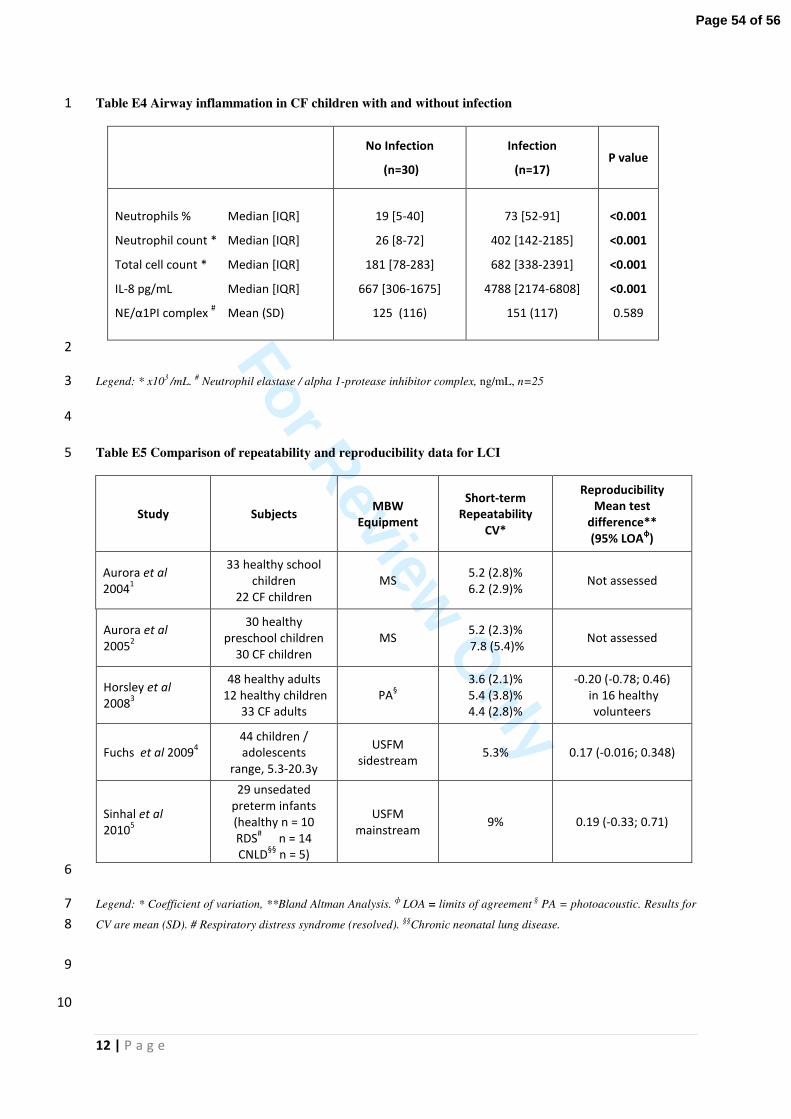
For Review O
nly
12 | P a g e
Table E4 Airway inflammation in CF children with and without infection 1
No Infection
(n=30)
Infection
(n=17) P value
Neutrophils % Median [IQR]
Neutrophil count * Median [IQR]
Total cell count * Median [IQR]
IL-8 pg/mL Median [IQR]
NE/α1PI complex # Mean (SD)
19 [5-40]
26 [8-72]
181 [78-283]
667 [306-1675]
125 (116)
73 [52-91]
402 [142-2185]
682 [338-2391]
4788 [2174-6808]
151 (117)
<0.001
<0.001
<0.001
<0.001
0.589
2
Legend: * x103 /mL. # Neutrophil elastase / alpha 1-protease inhibitor complex, ng/mL, n=25 3
4
Table E5 Comparison of repeatability and reproducibility data for LCI 5
Study Subjects MBW
Equipment
Short-term
Repeatability
CV*
Reproducibility
Mean test
difference**
(95% LOAф)
Aurora et al
20041
33 healthy school
children
22 CF children
MS 5.2 (2.8)%
6.2 (2.9)% Not assessed
Aurora et al
20052
30 healthy
preschool children
30 CF children
MS 5.2 (2.3)%
7.8 (5.4)% Not assessed
Horsley et al
20083
48 healthy adults
12 healthy children
33 CF adults
PA§
3.6 (2.1)%
5.4 (3.8)%
4.4 (2.8)%
-0.20 (-0.78; 0.46)
in 16 healthy
volunteers
Fuchs et al 20094
44 children /
adolescents
range, 5.3-20.3y
USFM
sidestream 5.3% 0.17 (-0.016; 0.348)
Sinhal et al
20105
29 unsedated
preterm infants
(healthy n = 10
RDS#
n = 14
CNLD§§
n = 5)
USFM
mainstream 9% 0.19 (-0.33; 0.71)
6
Legend: * Coefficient of variation, **Bland Altman Analysis. ф LOA = limits of agreement § PA = photoacoustic. Results for 7
CV are mean (SD). # Respiratory distress syndrome (resolved). §§Chronic neonatal lung disease. 8
9
10
Page 54 of 56
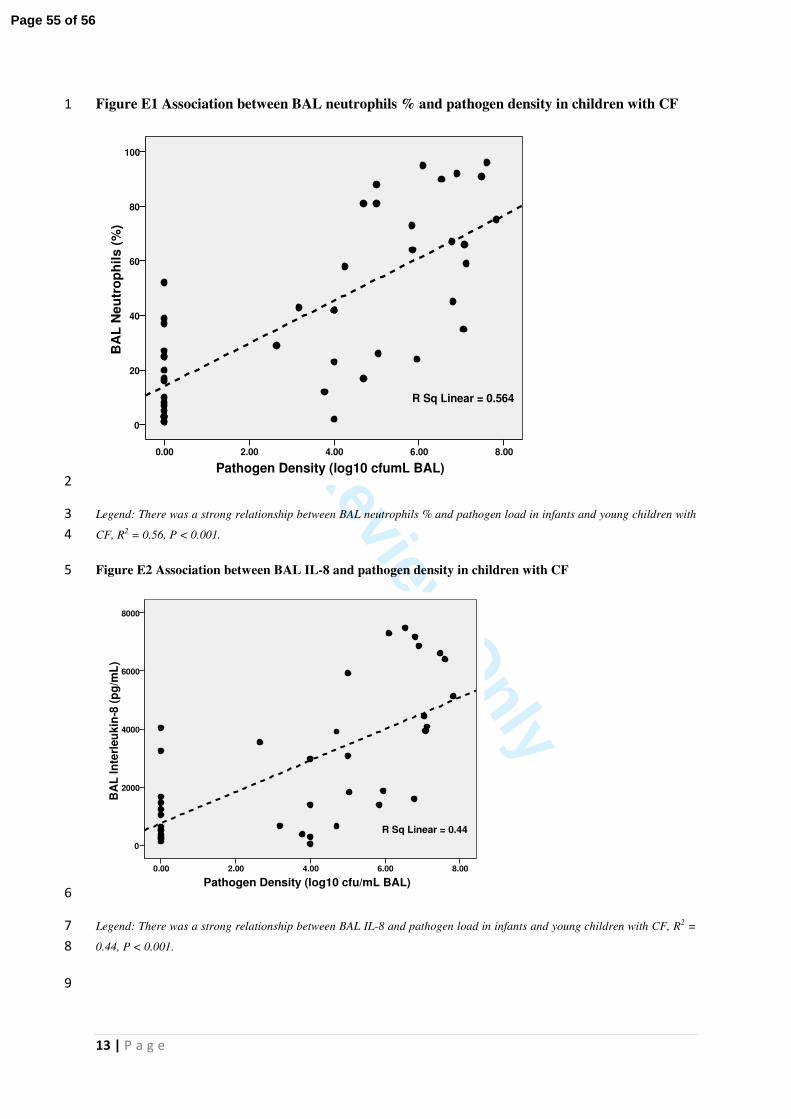
For Review O
nly
13 | P a g e
Figure E1 Association between BAL neutrophils % and pathogen density in children with CF 1
Pathogen Density (log10 cfumL BAL)
8.006.004.002.000.00
BA
L N
eu
tro
ph
ils (
%)
100
80
60
40
20
0
R Sq Linear = 0.564
2
Legend: There was a strong relationship between BAL neutrophils % and pathogen load in infants and young children with 3
CF, R2 = 0.56, P < 0.001. 4
Figure E2 Association between BAL IL-8 and pathogen density in children with CF 5
Pathogen Density (log10 cfu/mL BAL)
8.006.004.002.000.00
BA
L I
nte
rleu
kin
-8 (
pg
/mL
)
8000
6000
4000
2000
0
R Sq Linear = 0.44
6
Legend: There was a strong relationship between BAL IL-8 and pathogen load in infants and young children with CF, R2 = 7
0.44, P < 0.001. 8
9
Page 55 of 56

For Review O
nly
14 | P a g e
E Reference List 1
2
1. Aurora, P., P. Gustafsson, A. Bush, A. Lindblad, C. Oliver, C. E. Wallis, and J. Stocks. 2004. 3
Multiple breath inert gas washout as a measure of ventilation distribution in children with 4
cystic fibrosis. Thorax 59:1068-1073. 5
2. Aurora, P., A. Bush, P. Gustafsson, C. Oliver, C. Wallis, J. Price, J. Stroobant, S. Carr, J. Stocks, 6
and F. C. London Cystic. 2005. Multiple-breath washout as a marker of lung disease in 7
preschool children with cystic fibrosis. American Journal of Respiratory & Critical Care 8
Medicine 171:249-256. 9
3. Horsley, A. R., P. M. Gustafsson, K. A. Macleod, C. Saunders, A. P. Greening, D. J. Porteous, J. C. 10
Davies, S. Cunningham, E. W. Alton, and J. A. Innes. 2008. Lung clearance index is a sensitive, 11
repeatable and practical measure of airways disease in adults with cystic fibrosis. Thorax 12
63:135-140. 13
4. Fuchs, S. I., J. Eder, H. Ellemunter, and M. Gappa. 2009. Lung clearance index: normal values, 14
repeatability, and reproducibility in healthy children and adolescents. Pediatr.Pulmonol. 15
44:1180-1185. 16
5. Sinhal, S., J. Galati, D. N. Baldwin, J. Stocks, and J. J. Pillow. 2010. Reproducibility of multiple 17
breath washout indices in the unsedated preterm neonate. Pediatr.Pulmonol. 45:62-70. 18
19
20
21
22
Page 56 of 56