Embed Size (px)
Citation preview

• CHECKLIST FOR PHYSICAL EXAMINATION OF THE KNEE
• *This handout is for use as a "rough" guide and study aid. Your instructor may perform certain
maneuvers differently than depicted here. I acknowledge that this may be frustrating, but please try to
be understanding of this inter-examination variability.
A. Inspection (leg should be exposed to mid thigh) 1) Standing - alignment, foot structure, hip/pelvis 2) Gait - observe (e.g. is there a limp?) 3) Supine patient - effusion/swelling, erythema, quadriceps musculature (e.g. atrophy?)
Genu Genu Genu Varum Valgum Recurvatum
•
•
)
I
"Bowlegged"
Pelvic Obliquity
\
- /
..
"Knock Kneed"
Foot Structure
~..
Pes Planus "flat foot"
l J I.r
I
(updated 01/05/98)

...
• w
B. Palpation
• 1) Student should indicate that these were checked
a) Warmth b) Crepitus (What is it? What might it represent?) c) Effusion
Test for effusion .. Fluid wave
• Test for effusion .. Ballotable patella
\
• 2

2) Tenderness (landmarks) - Medial/lateral joint lines, MCl, lClJ patellar facets, quadriceps insertion, patellar tendon, tibial tubercle, iliotibial band, pes anserine
• (Patient is supine with the knee flexed to a comforable postion "" 200 - 900
)
Anatomy of the knee jOint (right) (anteromedial aspect)
TIbial Tu hAI~"'Ij£_
·....--Ijj;,HHI"'~-ME!CJ. Epicondyle of Femur
Medial Joint Line
Area of Pes Anserine Insertion
•
• 3

Points of orientation for palpation of the knee
• (Always start with the
t distal pole of patella.)
then ...
•
• 4
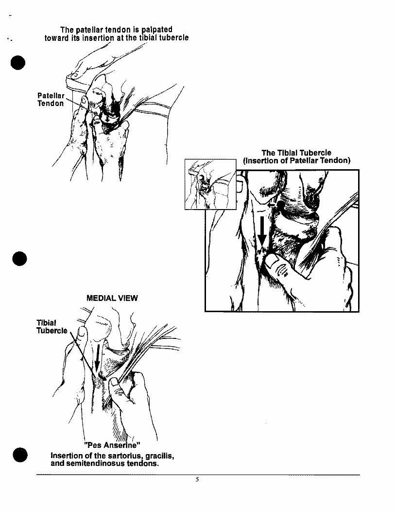
The patellar tendon is p'alpated toward 12"at the tibial tubercle
•
•
•
Tibial Tubercle
MEDIAL VIEW
I" ".';', y,' .
~ ~I
The Tibial Tubercle (Insertion of Patellar Tendon)
"Pes Anser nen
Insertion of the sartorius, gracilis,and semitendinosus tendons.
5

The lateral collateral ligament \\,\
•
(Donlt do this in acutely injured knee.) (KEYPOINT - find fibular head.)
• Palpation of the medial collateral ligament
• 6

C. Range of Motion
• 1) Hip (student only has to demonstrate passive) Flexion, Internal Rotation, External Rotation
(compare to other side)
(Remember: hip pathology can refer pain to the knee)
Hip Flexion
:-::-~::"I!!!I!lJ:==...;.t::::3::----:::===-:::,~o~oNeutral
Hip Rotation Measured with Hip in 90° of Flexion
• o
0°Neutral
•
•••••....
•• ••••• .•
7

2) Knee Flexion, Extension (Active, Passive - know the difference)
• Hyperextension 00
Neutral
• 3) Hamstring Flexibility
Assessment for hamstring tightness(compare to other side)
t
• 8
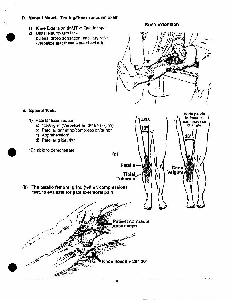
D. Manual Muscle Testing/Neurovascular Exam
Knee Extension
• 1) Knee Extension (MMT of Quadriceps) 2) Distal Neurovascular
pulses, gross sensation, capillary reWI (verbalize that these were checked)
E. Special Tests
1) Patellar Examination a) "Q-Angle" (Verbalize landmarks) (FYI) b) Patellar tethering/compression/grind* c) Apprehension* d) Patellar glide, tilt*
• *8e able to demonstrate (a)
Patella
Tibial Tubercle
(b) The patella femoral grind (tether, compression) test, to evaluate for patella-femoral pain
Wide pelvis In females
can Increase Qangle
• Knee flexed » 20°-30°
9
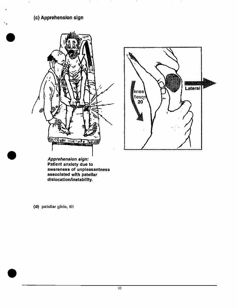
(c) Apprehension sign
•
• Apprehension sign: Patient anxiety due to awareness of unpleasantness associated with patellar dislocationlinstability.
(d) patellar glide, tilt
• 10

2. Ligamentous Testing
• a) Anterior cruciate ligament - Lachman } b) Posterior cruciate ligament - posterior drawer compare both knees c) Medial collateral ligament - Valgus stress at 30° d) Lateral collateral ligament - Varus stress at 30°
(a) Lachman test for anterior cruciate instability is at 20° of flexion .
(b) Posterior Drawer for posterior cruciate instability at 80° to 90° of flexion. •
Angle =800
~90·
• 11

(c) Valgus Stress testing: (b) Varus Stress testing: Medial Structures (MCl) lateral Structures (lCl)
•
• (a) (b)
end of table view
Knee is flexed 1'::1 30·
I I
• (Choose a technique that you feel most comfortable with.)
12

3. Meniscal Examination
• a) Joint line tenderness (demonstrate palpation of the joint lines) b) McMurray's - be able to demonstrate
(What is the sensitivity and specificity of this maneuver?)
•
MeM urray's Test
External rotation of tibia
Internal rotation of tibia
Flex the knee fully, with the fingers in joint line.
The lower leg (tib-fib) should be externally rotated when testing for medial meniscus tear, and internally rotated when testing for lateral meniscus tear. A distinct "pOpll or "clunkll felt in the joint line is suggestive of a meniscus tear.
"'If you don't get full flexion at the knee and you don't get your fingers in joint line you can't do a good McMurray's Test.
• 13