Embed Size (px)
Citation preview

For Peer Review O
nly
Bone growth, limb proportions and non-specific stress in
archaeological populations from Croatia
Journal: Annals of Human Biology
Manuscript ID: TAHB-2013-0038.R1
Manuscript Type: Research Paper
Keywords: children, growth pattern, long bones, medieval
URL: http://mc.manuscriptcentral.com/tahb E-mail: [email protected]
Annals of Human Biology

For Peer Review O
nly
1
Bone growth, limb proportions and non-specific stress in archaeological
populations from Croatia
Pinhasi R1,2,, Timpson, A3,5, Thomas, M3, Šlaus M4.
1 School of Archaeology, Newman Building, University College Dublin, Belfield,
Dublin 4, Ireland. Email: [email protected]
2 Smurfit Institute of Genetics, Trinity College Dublin, Dublin 2, Ireland
3 Department of Genetics, Evolution and Environment, University College London,
Gower Street, London WC1E 6BT. Emails: [email protected],
4 Anthropological Center of the Croatian Academy of Sciences and Arts, 10000
Zagreb, Croatia. Email: [email protected]
5 Institute of Archaeology, University College London, 31-34 Gordon Square, London,
WC1H 0PY United Kingdom
Abbreviated title: Bone growth in past Croatian populations
Poofs to be sent to: School of Archaeology, Newman Building, University College
Dublin, Belfield, Dublin 4, Ireland.
Key words: children, growth patterns, long bones, medieval
Page 1 of 45
URL: http://mc.manuscriptcentral.com/tahb E-mail: [email protected]
Annals of Human Biology
123456789101112131415161718192021222324252627282930313233343536373839404142434445464748495051525354555657585960
For Peer Review O
nly
2
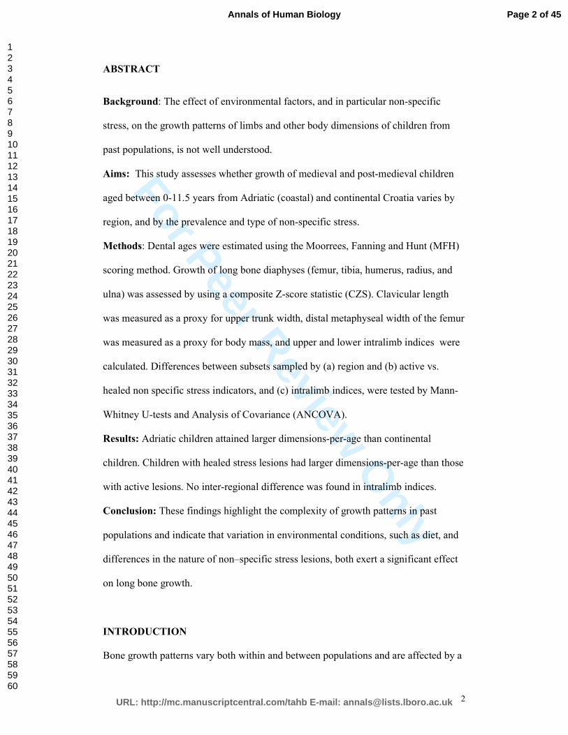
ABSTRACT
Background: The effect of environmental factors, and in particular non-specific
stress, on the growth patterns of limbs and other body dimensions of children from
past populations, is not well understood.
Aims: This study assesses whether growth of medieval and post-medieval children
aged between 0-11.5 years from Adriatic (coastal) and continental Croatia varies by
region, and by the prevalence and type of non-specific stress.
Methods: Dental ages were estimated using the Moorrees, Fanning and Hunt (MFH)
scoring method. Growth of long bone diaphyses (femur, tibia, humerus, radius, and
ulna) was assessed by using a composite Z-score statistic (CZS). Clavicular length
was measured as a proxy for upper trunk width, distal metaphyseal width of the femur
was measured as a proxy for body mass, and upper and lower intralimb indices were
calculated. Differences between subsets sampled by (a) region and (b) active vs.
healed non specific stress indicators, and (c) intralimb indices, were tested by Mann-
Whitney U-tests and Analysis of Covariance (ANCOVA).
Results: Adriatic children attained larger dimensions-per-age than continental
children. Children with healed stress lesions had larger dimensions-per-age than those
with active lesions. No inter-regional difference was found in intralimb indices.
Conclusion: These findings highlight the complexity of growth patterns in past
populations and indicate that variation in environmental conditions, such as diet, and
differences in the nature of non–specific stress lesions, both exert a significant effect
on long bone growth.
INTRODUCTION
Bone growth patterns vary both within and between populations and are affected by a
Page 2 of 45
URL: http://mc.manuscriptcentral.com/tahb E-mail: [email protected]
Annals of Human Biology
123456789101112131415161718192021222324252627282930313233343536373839404142434445464748495051525354555657585960

For Peer Review O
nly
3
range of environmental and genetic factors. Anthropological studies of growth
patterns in past populations are based on cross-sectional analyses of changes in bone
dimensions in relation to age (Johnston 1962; Merchant and Ubelaker 1977; Cook
1984; Jantz and Owsley 1984a,b; Mensforth 1985; Owsley and Jantz 1985; Lovejoy
et al. 1990; Hoppa 1992; Saunders 1992; Saunders and Hoppa 1993; Hoppa and
Gruspier 1996; Steyn and Henneberg 1996; Tanner 1998;Humphrey 1998; Hoppa and
Fitzgerald 1999; Jantz and Jantz 1999; Mays 1999; Humphrey 2000; Humphrey 2003;
Saunders 2000; Šereikienë and Jankauskas 2004; Floyd and Littletone 2006; Mays et
al. 2009). Most studies examined growth of long bone diaphyseal dimensions by
plotting each dimension as a function of dental age, based on the assessment of tooth
crown and root developmental charts (Moorrees et al. 1963a,b; Buikstra and Ubelaker
1994) and dental formation/eruption charts (see Ubelaker 1989; Buikstra and
Ubelaker 1994). These studies indicate inter-population variability in growth of
diaphyseal limb dimensions (Sundick 1978; Jantz and Owsley 1984a; Mensforth,
1985; Ribot and Roberts 1996; Humphrey 2000; Lewis 2002a,b; Pinhasi et al. 2005;
Pinhasi et al. 2006; Pinhasi et al. 2011). Studies of modern-day populations (Tanner
1986; Wall 1991) have also shown that inter-population differences can increase or
decrease in response to the extent to which a process of “catch-up” growth follows
recovery from one or more episodes of growth faltering (Tanner 1986).
Growth patterns reflect the overall health of a population (Johnston and Zimmer
1989) and dimensions-per-age from past populations can be compared to modern
growth data from various world regions (cf. Ice and James 2011) in order to provide
an indicator of the health of past populations. Anthropological studies typically assess
the health status of prehistoric populations by recording information about the
prevalence type and degree of expression of a series of skeletal markers of stress (c.f.
Page 3 of 45
URL: http://mc.manuscriptcentral.com/tahb E-mail: [email protected]
Annals of Human Biology
123456789101112131415161718192021222324252627282930313233343536373839404142434445464748495051525354555657585960

For Peer Review O
nly
4
Goodman et al. 1988). These include markers associated with a specific disease (e.g.
leprosy, tuberculosis, etc.), and a range of non-specific stress indicators which do not
have specific proximate causes, but their high prevalence in some archaeological
skeletal populations is known to be a contributor to poor health (Goodman and Rose
2002).
Only a handful of studies assessed whether there is a significant interaction
between skeletal stress indicators and growth in past populations (Lukacs 2001;
Sciulli and Oberly 2002; Floyd and Littleton 2006; Pinhasi et al. 2006; Temple 2008;
Schillaci et al. 2011). These studies highlighted the complexity of understanding the
effects of poor skeletal health on growth and stature trajectories in past population. In
the case of some disease markers there is a clear association between a specific stress
indicator and lower-than-average diaphyseal long bone dimensions. The
“Osteological Paradox” (Wood et al. 1992) asserts that in any population which
suffers from disease or nutritional stress the interpretation of “health’ is problematic.
Any skeletal population will include individuals that never experienced stress and
have none of the related skeletal lesions, individuals that experienced moderate stress
which lasted long enough to result in some skeletal lesion, and individuals that
suffered heavy stress resulting in death soon after the onset of the disease and which
may therefore have a few or no skeletal lesions. Wood et al. (1992) further point out
that the frequency of pathological lesions that appear to be active at the time of death,
without any indication for remodeling (i.e. healing) should be higher in individuals
exposed to high levels of stress, in particular during the first five years of life.
Therefore, an assessment of stress indicators in skeletal samples should take into
consideration both age-specific prevalence rates, and also whether the studied
condition was active or inactive at the time of death.
Page 4 of 45
URL: http://mc.manuscriptcentral.com/tahb E-mail: [email protected]
Annals of Human Biology
123456789101112131415161718192021222324252627282930313233343536373839404142434445464748495051525354555657585960

For Peer Review O
nly
5
Mixed-longitudinal studies of growth patterns of rural Guatemalan children with
suboptimal nutritional intake indicated that body measurements such as head and
chest circumferences provide nutritional information which is not evident from the
study of body length and weight. Undernourished Guatemalan children have smaller
head and chest circumferences compared to the well-nourished Denver children of the
same age, but the magnitude of difference between these two groups varied by age
and was not correlated with differences in overall limb lengths. Differences between
the two groups in chest circumference decreased at the age of 12 - 48 months and
increased beyond this age, while head circumference differences were minimal at
birth, become established at around 6 months and become progressively greater
through 24 months (Malina et al. 1975). This suggests that the assessment of growth
in relation to nutritional stress based only on the study of limb dimensions may
provide a limited perspective on growth trajectories.
In this study we focus on the analysis of inter-regional variation in growth of long
bone diaphyses (femur, tibia, humerus, radius, and ulna) by using a composite Z-score
statistic (CZS, see below), the clavicular length (a proxy for upper trunk width), the
distal metaphyseal width of the femur (a proxy for body mass), and the upper and
lower intralimb indices (brachial and crural indices, respectively). We also assess
whether there is a significant difference in long bone diaphyseal dimensions per age
between individuals with active versus healed non-specific stress indicators: cribra
orbitalia and periostitis. The four archaeological sites analyzed in this study are:
Stenjevec (1050–1250 AD) and Nova Rača (1400–1700 AD), located in continental
Croatia; and Koprivno (1500–1700 AD) and Dugopolje (1350–1500 AD), which are
located in the Adriatic region of Croatia (Figure 1). At present the two regions differ
in their ecology as continental Croatia has a central European climate with average
Page 5 of 45
URL: http://mc.manuscriptcentral.com/tahb E-mail: [email protected]
Annals of Human Biology
123456789101112131415161718192021222324252627282930313233343536373839404142434445464748495051525354555657585960

For Peer Review O
nly
6
winter temperature below 0° C (Goldstein 1999) while Adriatic Croatia has a
Mediterranean climate, with short, wet winters during which the temperature rarely falls
below 5° C. Estimated temperature variations from the Northern Hemisphere as a whole
indicates that during these periods, average temperatures were similar to those in present
day, prior to AD 1400 and on average 0.6° C colder during the Little Ice Age, AD 1400-
1900 (Mann 2003).
Growth patterns are dependent on both environmental and genetic factors.
Historical and archaeological data suggest that the four sites were 'ethnically'
heterogeneous. Dugopolje was populated by the descendants of Early Croats and a
heterogeneous group of peoples fleeing from Ottoman Turkish military intrusions
and/or conquests (Novak and Šlaus 2007). Koprivno was inhabited by the same
descendants of the Early Croats and an ethnically heterogeneous group of Turkish
subjects known as “Vlachs” (Kužić 2001; Gjurašin 2006). In continental Croatia,
Stenjevec was inhabited by heterogeneous Slavs (consisting of Early Croats and their
descendants, Slovaks, and Bulgars) and Hungarians (Demo 1996), while Nova Rača
(situated as it was on the fortified military border that separated Croatia from the
Ottoman Empire during the 16th to 18th centuries) was inhabited by a heterogeneous
group of peoples that included Croats, Bosnians and Serbs (Šlaus 2000). Hence, there
is no discernible genetic pattern that would imply a consistent or interpretable
difference between individuals from continental and coastal regions.
In contrast, historical, archaeological, and dental data suggest there may have been
dietary differences between individuals from continental and coastal regions. In
continental Croatia the subsistence spectrum was primarily based on agriculture.
Historical confirmation is provided by the urbarii –legal documents that defined
relationships between feudal lords and peasants. One of these, the urbarium from the
Page 6 of 45
URL: http://mc.manuscriptcentral.com/tahb E-mail: [email protected]
Annals of Human Biology
123456789101112131415161718192021222324252627282930313233343536373839404142434445464748495051525354555657585960

For Peer Review O
nly
7
monastery of Streza in continental Croatia dated to 15th century, shows that millet was
the staple crop which was a main component of the diet, possibly because it was
easily cultivated, had a short period of vegetation, and could be sown in dry and sandy
soils (Kolar-Dimitrijevic 2003). Additional crops were wheat, buckwheat, rye, and
barley, whilst poultry, pork and fish were rarely consumed (Adamček et al.
1980) .Thus crop failures may have had catastrophic impact on the nutrition and
health of these peasants. In the Nova Rača parish Book of the Dead (compiled from
1830–1848 listing the age, sex, and cause of death of its parishioners) the second most
common cause of death in children is listed as “emaceratio” or “ex debilitate”
suggesting undernourishment or starvation (Šlaus 2000). A diet based on products
rich in carbohydrates also resulted in high frequencies of caries and significant wear
on the occlusal surfaces of teeth recorded in individuals from the 11th to the 16th
century from continental Croatia (Vodanovic et al. 2005; Novak et al., 2010). In
contrast, lack of cultivable soil in the Adriatic hinterland resulted in a livelihood
largely dependent on livestock breeding and a diet that was based more on
consumption of meat and animal products than in continental Croatia. There is
historical information (Šaric 2008) that the primary occupation of the inhabitants of
Koprivno was transhumant pastoralism with average flocks consisting of between 200
and 600 sheep, along with some large cattle (Jurin-Starcevic 2008). This dietary
difference is further supported by the lower caries frequency in individuals from
Koprivno than from continental Croatia (Novak et al. 2007).
MATERIALS
Samples comprised a total of 198 skeletons of children from the following
archaeological cemeteries: Dugoplje - excavated from 2004 to 2005 (Gjurašin 2005),
Koprivno – excavated from 2001 to 2002 (Gjurašin 2002, 2006), Stenjevec –
Page 7 of 45
URL: http://mc.manuscriptcentral.com/tahb E-mail: [email protected]
Annals of Human Biology
123456789101112131415161718192021222324252627282930313233343536373839404142434445464748495051525354555657585960

For Peer Review O
nly
8
excavated from 1983 to 1997 (Simoni 2004), and Nova Rača – excavated from 1986
to 1995 (Jakovljević 1986). The analyzed skeletal material is thus grouped into two
composite series; coastal Adriatic, and Continental Croatia (Table I). The pooled
cemetery samples encompass fairly extensive time periods – particularly the
continental series that covers a time span of potentially 650 years, but can be regarded
as a single analytical period on the basis of the historical and archaeological data. The
analyzed cemeteries are related to small rural settlements and the analyses of the
recovered archaeological artifacts, grave architecture, and available historical sources,
suggest that the vast majority of the recovered individuals belonged to lower social
categories (Gjurašin 2002, 2005, 2006). Except for six of the adult individuals from
the Stenjevec cemetery (three males, and three females), who were buried with
jewelry that may be indicative of higher social status (Simoni 2004), there is no
evidence of social stratification in any of the analyzed cemeteries. Additionally,
during the time period encompassed by the analyzed sites no fundamental
technological, scientific, subsistence, or social changes occurred that could
significantly affect growth profiles – with one exception, the introduction of syphilis
in Croatia in 1500 AD (Gruber 2009). The presence of syphilis is noted in historical
documents in both continental Croatia, and on the Adriatic coast and its hinterland
(Bazala 1972; Glesinger 1978), and has also been recorded in osteological material
(Šlaus and Novak 2007). All skeletons were screened for the presence of congenital
syphilis according to criteria outlined in Ortner (2003). No such individuals were
detected. Therefore, we believe that that there are no diachronic factors that could
significantly affect the growth profiles of the two composite series.
METHODS
Metric measurements
Page 8 of 45
URL: http://mc.manuscriptcentral.com/tahb E-mail: [email protected]
Annals of Human Biology
123456789101112131415161718192021222324252627282930313233343536373839404142434445464748495051525354555657585960
For Peer Review O
nly
9
Maximum diaphyseal length dimensions for the humerus, ulna, radius, femur, and
tibia were measured directly or using a standard osteometric board following the
method described in Buikstra and Ubelaker (1994). Additionally, distal femoral
epiphyseal breadth and clavicular length measurements were taken using a standard
sliding caliper, following the methods provided in Buikstra and Ubalaker (1994). The
left bone was chosen whenever possible but was replaced with the right bone if it was
missing or poorly preserved. The brachial index is expressed as a percentage of the
relative length of the lower arm to the upper arm, and is calculated as the radius
length/humerus length x100 (Aiello and Dean 1990). The crural index expressed as
the percentage of the relative length of the lower leg to the upper leg as a percentage,
calculated as the tibial length/femoral length x 100 (Aiello and Dean 1990).
Age Assessment
The dental age of all specimens was estimated by applying the Moorrees, Fanning and
Hunt (MFH) scoring method for the permanent lower molars, premolars, canines and
incisors (Moorrees et al. 1963a) in order to obtain a dental formation rank. Age for
each rank was obtained from charts specified in Smith (1991) for each of the analyzed
teeth. A chronological age estimate was obtained for all teeth and the mean age for all
teeth from each individual was calculated. In cases in which an outlier age estimate
was obtained for a given tooth, the radiographs were re-examined by one of the
authors (MS) against the dental formation and eruption chart of Schour and Massler
(1941) and errors were corrected by re-applying the MFH method, to obtain new age
estimates for each of the mandibular teeth. In all cases the final dental age was based
on the separate assessment of at least 3 lower teeth, while the mean number of teeth
analyzed for the complete sample is 4.6.
Page 9 of 45
URL: http://mc.manuscriptcentral.com/tahb E-mail: [email protected]
Annals of Human Biology
123456789101112131415161718192021222324252627282930313233343536373839404142434445464748495051525354555657585960

For Peer Review O
nly
10
Diagnosis of stress indicators
Studies of non-specific stress in children generally involve assessment of the presence
of one or more of the following stress indicators: cribra orbitalia, porotic hyperostosis,
periostitis, maxillary sinusitis, dental enamel hypoplasia, Harris lines, and endocranial
lesions (Lewis 2007). In this study, extensive postmortem tooth loss precluded the
assessment of dental enamel hypoplasia. Therefore we considered only cribra orbitalia
and non-specific periostitis frequencies reliable enough to be incorporated in this
analysis. While the assessment of stress on the basis of only two indicators is not ideal,
it does provide the opportunity to directly examine the possible associations between
each of the two stress indicators and growth patterns in the analyzed populations.
Cribra orbitalia is a sieve-like expansion in the orbital plates of the frontal bone that is
considered to be part of similar bone changes that affect the skull vault (porotic
hyperostosis) (Carlson et al. 1974; Cybulski 1977; Stuart-Macadam 1985; Mittler and
van Gerven 1994; Fairgrieve and Molto 2000). Various hypotheses have been
suggested for the cause of porotic hyperostosis including anemia, metabolic diseases
such as scurvy and rickets, syphilis, cancer, and pressure from binding and carrying
(Williams 1929; Angel 1966; Mensforth et al. 1978; Stuart-Macadam 1985; Stuart-
Macadam 1992; Ortner 2003; Wapler et al. 2004; Papathanasiou 2005; Walker et al.
2009) Porotic hyperostosis and cribra orbitalia are the osseous outcome of marrow
hyperplasia (Trancho 1987; Stuart-Macadam 1991; Mittler and van Gerven 1994;
Larsen 1997; Fairgrieve and Molto 2000; Walker et al., 2009; but see Ortner et al.,
1999, 2001), which can be caused from various disease processes. Possible, although
rare, causes include renal osteodystrophy, hereditary spherocytosis, and cyanotic
congenital heart disease (Moseley 1966; Ortner 2003; Blom et al. 2005). Other causes
Page 10 of 45
URL: http://mc.manuscriptcentral.com/tahb E-mail: [email protected]
Annals of Human Biology
123456789101112131415161718192021222324252627282930313233343536373839404142434445464748495051525354555657585960

For Peer Review O
nly
11
include genetically inherited forms of anemia such as thalassemia and sickle–cell
anemia, and acquired iron–deficiency anemia (Tayles 1996; Hershkovitz et al. 1997;
Larsen 1997; Bocherens et al. 1999; Ortner 2003). Cumulative evidence from
numerous bioarchaeological studies worldwide resulted in a general consensus linking
porotic hyperostosis in archaeological skeletal populations with acquired iron
deficiency anemia (Larsen 1997). However, according to Walker (Walker et al. 2009),
hematological research shows that iron deficiency alone cannot sustain the massive
red blood cell production that causes marrow expansion responsible for these lesions.
He argues that the most likely cause of porotic hyperostosis is rather the accelerated
loss and compensatory overproduction of red blood cells seen in hemolytic and
megaloblastic anemias, and in addition states that porotic hyperostosis and cribra
orbitalia often have different etiologies.
In this study no attempts were made to discern the possible etiologies of cribra
orbitalia and the prevalence of this condition was simply assessed as an indicator of
stress in all children aged 1–11.5 years (as it was not possible to assess with
confidence the prevalence of the condition in newborns). No attempt was made to
evaluate the severity of the lesion but a distinction was made between active and
nonactive (healed) lesions according to criteria suggested by Mittler and Van Gerven
(1994). Because other pathological conditions including metabolic diseases, cancer,
and infectious diseases can mimic the osseous changes that result from acquired iron-
deficiency anemia, every effort was made to distinguish conditions that resulted in
marrow hyperplasia, from those that did not, according to criteria described by Ortner
(2003). Additionally, because genetic anemias may have been present– particularly
those from the Adriatic hinterland region where malaria was endemic – all skeletons
were carefully screened for the presence of thalassemia and sickle-cell anemia, once
Page 11 of 45
URL: http://mc.manuscriptcentral.com/tahb E-mail: [email protected]
Annals of Human Biology
123456789101112131415161718192021222324252627282930313233343536373839404142434445464748495051525354555657585960

For Peer Review O
nly
12
again according to criteria suggested by Ortner (2003). All cases potentially suspect
for genetic anemias, cancer, metabolic and infectious diseases were excluded from
this study. The presence of cribra orbitalia was assessed in all individuals with at least
one orbital roof preserved. Consequently, the number of individuals that were
assessed in regard to the prevalence of cribra orbitalia varied by site (see below).
Overall skeletal preservation and recovery of bones were similar at all four cemeteries.
Periostitis is defined as a basic inflammatory response that develops because of
non-specific bacterial infections and is macroscopically recognized as osseous
plaques with demarcated margins or irregular elevations of bone surfaces. Periostitis
can also be caused by trauma and specific infectious diseases such as leprosy,
tuberculosis, and treponemal disease (Larsen, 1997; Ortner, 2003). Both leprosy and
tuberculosis have been reported in osteological data from Croatia (Šlaus, 2006), and
cases of trauma are frequently found in almost all archaeological data. To ensure,
therefore, that only cases of non-specific periostitis were included, all skeletons with
osteological evidence of leprosy or tuberculosis were excluded from the periostitis
sample. Periostitis was not considered present if it was located on the same bone as a
fracture or other trauma. Nonspecific periostitis was diagnosed when two or more
skeletal elements, excluding the endocranial surfaces of the skull, exhibited active or
healed periostitis. Criteria for inclusion in the sample were the presence of least 50%
of all long bones (all of the long bones present – including both sides). No attempt
was made to evaluate the severity of the lesion but a distinction was made between
active and nonactive (healed) lesions according to criteria suggested by Mensforth et
al. (1978).
Statistical methods
The raw data included 777 measurements from 7 bone metrics from 198 skeletons.
Page 12 of 45
URL: http://mc.manuscriptcentral.com/tahb E-mail: [email protected]
Annals of Human Biology
123456789101112131415161718192021222324252627282930313233343536373839404142434445464748495051525354555657585960

For Peer Review O
nly
13
Since the skeletons were incomplete, the sample sizes for any particular bone metric
were small, ranging from n=151 (76%) to n=80 (40%) (Table SI 1). To enable the
detection of potential differences with greater sensitivity, we combined different
metrics from each individual skeleton. Combining diaphyseal metrics provides two
main advantages. Firstly it increased the sample sizes. This was particularly important
in this study since we were trying to detect small differences in subsets of an already
small dataset, which consisted of measurements from incomplete skeletons. Secondly,
it has the minor (but nonetheless useful) effect of reducing the impact of outliers. In
order to balance this increased sensitivity against the potential increase in variance
from combining different metrics we first assessed the correlation between each pair
of metrics. The correlation coefficients (Pearson's R) ranged from 0.931 between the
clavicle length and femur breadth, to 0.9943 between the femur length and tibia
length (Figure SI 1). However, within this narrow range there appears to be a cluster
of five metrics (diaphyseal lengths of the femur, tibia, humerus, radius and ulna) that
were particularly strongly correlated, whilst clavicle length, and femoral distal
metaphyseal breadth were less so. This difference can be visualized using an un-
rooted tree using a distance matrix of the correlation coefficients (Figure SI 2).
We created a single combined metric, or composite Z-score (CZS) (see e.g.
Relethford, 2001) for each skeleton by averaging Z-scores or each of the five long
bone diaphyseal metrics. This ensured that each CZS was constructed from a
minimum of one diaphyseal dimension and a maximum of five. In addition to using
the CZS, we also included the distal femoral metaphyseal breadth, clavicle length and
both upper and lower intralimb indices.
Considering age as a covariate. Since all bone length dimensions have a strong
Page 13 of 45
URL: http://mc.manuscriptcentral.com/tahb E-mail: [email protected]
Annals of Human Biology
123456789101112131415161718192021222324252627282930313233343536373839404142434445464748495051525354555657585960

For Peer Review O
nly
14
correlation with age (between age and the combined metric: Pearson's R=0.948,
P<2.2e-16), it was essential to adjust for age as a covariate (Pinhasi et al. 2005, 2006).
A model selection approach was used to establish a suitable relationship between CZS
and age. A cubic model was selected as most appropriate having both the lowest
Bayesian information criterion (BIC) (Burnham and Anderson, 2002) and the lowest
residual variance of the three tested models (Table SI 2 and Figure SI 3).
To a lesser extent intralimb indices also exhibited a correlation with age,
therefore tests were also performed on the residuals from a suitable model. In the case
of the crural index, the correlation with age was 0.365 (P<0.001), and a cubic model
had the lowest BIC. In the case of the brachial index the correlation with age was
0.275 (P=0.061), and a quadratic model had the lowest BIC.
The residuals were then used in two distinct statistical tests, firstly to test
whether or not there is any significant difference between two groups. If a significant
difference was found, a second test was applied to estimate the magnitude of that
difference.
Testing for a significant difference between groups, and the magnitude of that
difference. Firstly, Mann-Whitney U-tests were performed on the residuals to test if
there was a significant difference between the Adriatic and continental sets. Secondly,
the magnitude of the difference was estimated using Analysis of Covariance
(ANCOVA). Specifically, we calculated the sum of the squares of the residuals for
region A (SSA), repeated this procedure for region B (SSB), and for the total pooled
regions (SSTotal). The portion of variation attributable to inter-regional differences was
estimated by deducting the within-variation portion from the total variation: SSBetween
= SSTotal – (SSA+ SSB). The proportion of variation attributable to differences between
Page 14 of 45
URL: http://mc.manuscriptcentral.com/tahb E-mail: [email protected]
Annals of Human Biology
123456789101112131415161718192021222324252627282930313233343536373839404142434445464748495051525354555657585960

For Peer Review O
nly
15
regions was calculated as: SSBetween / SSTotal. The associated p-value was calculated
using a re-sampling with randomization test using 500,000 re-samples. The p-value
was calculated as the proportion of re-sampled SSbetween that were greater or equal to
the observed SSbetween (see Jobling et al. 2004; Pesarin and Salmaso 2010). Both tests
were applied using the combined metric, femoral distal breadth and clavicle length.
In addition to testing region as a factor (coastal vs. continental) we repeated the
analyses on the two non-specific stress indicators (periostitis, cribra orbitalia) using
only the combined metric, as sample sizes for femoral distal breadth and clavicle
length were smaller. The independence of these three factors was confirmed using a Χ
2 test (Table II).
RESULTS
Table III provides a summary of the results of the statistical tests for (1) regional
differences, using the combined metric, femoral distal breadth, and clavicle length, (2)
differences between active and healed periostitis, using the combined metric, (3)
differences between active and healed cribra orbitalia, using the combined metric, (4)
regional differences in the crural index, and (5) regional differences in the brachial
index.
Region
A highly significant difference between the two regions was found using CZS (Figure
2) and femoral distal breadth (Figure 3), and to a lesser extent clavicle length (Figure
4), with the Adriatic sub-set exhibiting larger dimensions-per-age (Mann-Whitney U-
test P-values are <0.001, <0.001 and 0.053, respectively). The magnitude of the
Page 15 of 45
URL: http://mc.manuscriptcentral.com/tahb E-mail: [email protected]
Annals of Human Biology
123456789101112131415161718192021222324252627282930313233343536373839404142434445464748495051525354555657585960

For Peer Review O
nly
16
difference between the two regions was estimated at 18.4% for CZS, 14.0% for
femoral distal breadth, and 11.6% for the clavicle length (P-values are <0.001, 0.016,
and 0.043, respectively).
Stress indicators
Sample sizes for individuals determined to have either active or healed stress
indicators were small (cribra orbitalia active N=21, healed N=61, periostitis active N
= 63, healed N = 52) compared with the previous regional analysis (continental N =
65, Adriatic N = 133), therefore analysis was only performed using the more sensitive
combined metric. Significant differences between active and healed were found for
both cribra orbitalia and periostitis, in both cases children with healed lesions
exhibited larger dimensions-per-age than those with active lesions (Figures 5 and 6,
Mann Whitney U-test P-values = 0.0037 and <0.001 respectively), and the magnitude
of the difference was estimated at 15.7% (P = 0.054) and 12.0% (P = 0.026)
respectively.
Intralimb indices
Mann-Whitney U-tests of the crural index of Adriatic vs. continental populations
revealed no significant differences (Adriatic N = 70, continental N = 24, P = 0.161).
Mann-Whitney U tests of the brachial index of Adriatic vs. continental populations
also revealed no significant differences (Adriatic N = 45, continental N = 19, P =
0.402).
DISCUSSION
This study reveals significant differences in the growth trajectories of Adriatic vs.
continental Croatian children, although p-values above 0.0073 should be treated with
Page 16 of 45
URL: http://mc.manuscriptcentral.com/tahb E-mail: [email protected]
Annals of Human Biology
123456789101112131415161718192021222324252627282930313233343536373839404142434445464748495051525354555657585960

For Peer Review O
nly
17
caution given the application of multiple tests (Sidak correction using 7 tests). The
Adriatic coastal hinterland populations generally attained higher limb length, femoral
distal breadth and clavicular length dimensions-per-age than the continental
populations. The difference in femoral and distal breadth dimensions implies that
body mass of children from continental Croatian was generally lower than that of
their peers in the Adriatic hinterland (see Ruff 2007 and references therein for
calculation of body size based on femoral distal epiphyseal breadth).
No significant regional differences in the brachial and crural indices were found,
suggesting that while the Adriatic children were larger overall, this did not translate
into significant differences in their limb proportions, although it should be noted that
the sample sizes for this test were small. Cowgill et al. (2012) analysed developmental
variation in body proportions in relation to latitudinal and climatic factors in a global
set of modern and archaeological subadult samples. They report that both indices are
established early in ontogeny in all populations, and that environmental effects on
these indices appear to be minimal. Similar results were reported by Temple et al.
(2011) in their study of the ontogeny of intralimb indices in relation to ecogoegraphic
differences in Jomon period foragers. The results reported here are in accord with
these studies as they indicate no significant difference in the ontogeny of these indices
between Adriatic and Continental regions. While this study did not assess the
potential interaction between growth for a given age category and the wide range of
environmentally confounding effects, it suggests genetic conservation of these indices
during ontogeny.
In terms of environmental variation, the two regions differ to some extent in their
mean annual temperatures and climates, but these differences are not strong enough to
exert a significant effect on the growth trajectories of limb dimensions, body mass
Page 17 of 45
URL: http://mc.manuscriptcentral.com/tahb E-mail: [email protected]
Annals of Human Biology
123456789101112131415161718192021222324252627282930313233343536373839404142434445464748495051525354555657585960

For Peer Review O
nly
18
and intralimb indices (see Katzmarzyk and Leonard 1998). The two regions are
located in a similar eco-geographical region with similar elevation. Available
historical documents indicate that while living conditions during the medieval and
post-medieval periods were harsh in both regions of Croatia, children from the
Adriatic hinterland had a better dietary status than those from continental Croatia. The
Nova Rača parish Book of the Dead additionally suggests that, at least some children
from continental Croatia died as a result of chronic malnutrition and previous studies
(Frisancho et al. 1980) have shown that in populations exposed to this type of
malnourishment the delay in skeletal growth is most noticeable during childhood.
Frisancho et al. (1980) also suggest the influence of environmental factors such as
nutrition is greater on younger children.
Ribot and Roberts (1996) assessed the growth of long bone dimensions in Early
(Raunds) and Late (Chichester) medieval populations from England with the
following non-specific stress indicators: dental enamel hypoplasia, porosity on the
ectocranial surface of the skull (both orbital and vault lesions), subperiosteal new
bone formation on long bones and the ectocranial surface of the skull, and Harris lines.
Individuals were categorized into those with high stress (those exhibiting two or more
stress indicators), stressed, or unstressed individuals (those exhibiting just one, or no
stress indicators) and the diaphyseal dimensions of their humerii, femora and tibiae
were compared. Their results showed no significant differences in growth according
to their levels of stress. In contrast, a recent study by Schillaci et al. (2011), reports
growth faltering starting soon after birth and continuing until about 5 years of age
among Pueblo Indians from the American Southwest, and that children without
porotic cranial lesions exhibited a higher degree of stunting than those with lesions. A
study by Pinhasi et al. (2006) examined the effects of rickets on long bone growth
Page 18 of 45
URL: http://mc.manuscriptcentral.com/tahb E-mail: [email protected]
Annals of Human Biology
123456789101112131415161718192021222324252627282930313233343536373839404142434445464748495051525354555657585960

For Peer Review O
nly
19
patterns in medieval and post-medieval populations from Britain and showed no
significant differences in long bone growth patterns between individuals with
osteological evidence for rickets, and those without the disease. In contrast, Mays et
al. (2009) studied the effects of rickets on the growth patterns of long bones in 19th
century children from Birmingham, England. They applied multiple regression
analyses to long bone dimensions of individuals with active rickets, healed rickets,
and no evidence of rickets for the 2–6 years old age cohort, and then compared partial
correlation coefficients in order to detect significant associations between growth
patterns and rickets. Their results showed that individuals with rickets in the 2–6 year
age group had shorter bones for their age than individuals without the disease,
however they found no difference between the long bone dimensions.
The apparent incongruities in the results of these various studies might be
explained as follows. Firstly, it has been shown that dental age underestimates the
chronological age in the case of malnourished children, by 10-12 months on average
(Lampl & Johnston 1996). It is therefore possible that the obtained charts undermine
the true discrepancies in growth between children with vs. without stress. Secondly,
there are at present only a few case studies which assess how bone growth trajectories
are interacting with stress and there is no reason to expect that all non specific and
specific stress, from a range of diseases and nutritional conditions, would have the
same impact on bone growth. This is especially the case when we take into
consideration the fact that non-specific stress indicators are associated with either a
range of diseases and nutritional conditions, or have unknown etiologies. Thirdly it is
expected that the differences between survivors with healed lesions and those with
active lesions, can provide insight about the actual correlation (if any) between a
given stress indicator and health in past populations.
Page 19 of 45
URL: http://mc.manuscriptcentral.com/tahb E-mail: [email protected]
Annals of Human Biology
123456789101112131415161718192021222324252627282930313233343536373839404142434445464748495051525354555657585960

For Peer Review O
nly
20
Future studies which assess a wider range of stress indicators may provide more
insight into the effects of stress and to what extent the size and proportions of children
with healed stress indicators manage to “catch up”.
CONCLUSION
This study examined differences in the growth patterns using skeletons of children
from four medieval and post-medieval cemeteries, comparing differences between
those from the Adriatic hinterland, and continental Croatia. Our results indicate that
(a) Adriatic children generally have larger bone dimensions per age, (b) children with
healed lesions of cribra orbitalia or periostitis attained significantly larger dimensions-
per-age than those with active lesions, and (c) no differences in brachial and crural
indices of children from the two regions were detected.
Our results indicate that environmental factors play a significant role in in growth
trajectories of past populations. Based on the historical documents and the ecological
settings we postulate that difference in diet was a substantial contributor to
differences in growth patterns of children from the two regions.
ACKNOWLEDGMENTS
We are grateful to Noreen von Cramon-Taubadel for her comments and advice. We
thank the Editor and two anonymous reviewers for their comments and suggestions
on earlier drafts. This research was supported by the European Research Council
Starting Grant (ERC-2010-StG 263,441).
Declaration of Interest
The authors report no conflicts of interest. The authors alone are responsible for the
content and writing of the paper.
Page 20 of 45
URL: http://mc.manuscriptcentral.com/tahb E-mail: [email protected]
Annals of Human Biology
123456789101112131415161718192021222324252627282930313233343536373839404142434445464748495051525354555657585960

For Peer Review O
nly
21
LITERATURE CITED
Adamček J. 1980. Agrarni Odnosi u Hrvatskoj od Sredine 15. do Kraja 17. Stoljeća.
Zagreb, Sveučilišna Naklada Liber.
Adamček J. 1980. Agrarni Odnosi u Hrvatskoj od Sredine XV do Kraja XVII
Stoljeća. Zagreb, Jugoslavenska Akademija Znanosti i Umjetnost.
Aiello LC, Dean C. 1990. An Introduction to Human Evolutionary Anatomy. London,
Elsevier Academic Press.
Angel JL. 1966. Porotic hyperostosis, anemias, malarias and marshes in the
prehistoric Eastern Mediterranean. Science 153:760–763.
Bazala V. 1972. Pregled Povijesti Zdrastvene Kulture Dubrovačke Republike. Zagreb,
Grafički Zavod Hrvatske.
Blom DE, Buikstra JE, Keng L, Tomczak PD, Shoreman E, Stevens–Tuttle D. 2005.
Anemia and childhood mortality: latitudinal patterning along the coast of Pre–
Columbian Peru. Am J Phys Anthropol 127:152–169.
Bocherens H, Billiou D, Mariotti A, Patou-Mathis M, Otte M, Bonjean D, Toussaint
M. 1999. Palaeoenvironmental and Palaeodietary Implications of Isotopic
Biogeochemistry of Last Interglacial Neanderthal and Mammal Bones in
Scladina Cave (Belgium). J Arch Sci 26:599–607.
Buikstra JE, Ubelaker DH. 1994. Standards for Data Collection from Human Skeletal
rRemains. Fayetteville, Arkansas Archeological Survey.
Burnham KP, Anderson DR. 2002. Model selection and multimodel inference: a
practical information-theoretic approach, 2nd Ed. New York, Springer.
Carlson DS, Armelagos GJ, van Gerven DP. 1974. Factors influencing the etiology of
cribra orbitalia in prehistoric Nubia. J Hum Evol 3:405–410.
Page 21 of 45
URL: http://mc.manuscriptcentral.com/tahb E-mail: [email protected]
Annals of Human Biology
123456789101112131415161718192021222324252627282930313233343536373839404142434445464748495051525354555657585960

For Peer Review O
nly
22
Cook DC. 1984. Subsistence and health in the lower Illinois Valley. In Cohen MN,
Armelagos GJ, editors. Paleopathology at the origins of agriculture. Orlando:
Academic Press. p 235-269.
Cowgill LW, Elezear C, Auerbach BM, Temple DH, Okazaki K. 2012.
Developmental variation in ecogeographic body proportions. Am J Phys
Anthropol 148:557-570.
Cybulski JS. 1977. Cribra orbitalia, a possible sign of anemia in early historic native
populations of the British Columbia Coast. Am J Phys Anthropol 47:31–40.
Demo Ž. 1996. Vukovar Lijeva Bara. Zagreb, Arheološki Muzej Zagreb.
Fairgrieve SI, Molto JE. 2000. Cribra orbitalia in two temporally disjunct population
samples from the Dakhleh Oasis, Egypt. Am J Phys Anthropol 111:319–331.
Floyd B, Littleton J. 2006. Linear enamel hypoplasia and growth in an Australian
Aboriginal community: not so small, but not so healthy either. Ann Hum Biol 33:424-
443.
Frisancho AR, Guire K, Babler W, Borkan G, Way A. 1980. Nutritional influence on
childhood development and genetic control of adolescent growth of Quechuas
and Mestizos from the Peruvian lowlands. Am J Phys Anthropol 52:367–375.
Goodman AH, Martin DL. 2002. Reconstructing Health Profiles from Skeletal
Remains. In: Steckel RH, Rose JC, editors. The Backbone of History: Health
and Nutrition in the Western Hemisphere. Cambridge: Cambridge University
Press. P. 11-60.
Gjurašin H. 2002. Nastavak zaštitnih istraživanja arheoloških lokaliteta u selu
Koprivno – općina Dugopolje – sjeveroistočno od Klisa u godini 2002.
Obavijesti 33:151–153.
Gjurašin H. 2005. Dugopolje - kasnosrednjovjekovno groblje. Obavijesti 37:78–80.
Page 22 of 45
URL: http://mc.manuscriptcentral.com/tahb E-mail: [email protected]
Annals of Human Biology
123456789101112131415161718192021222324252627282930313233343536373839404142434445464748495051525354555657585960

For Peer Review O
nly
23
Gjurašin H. 2006. Zaštitna arheološka istraživanja u selu Koprivno sjeveroistočno od
Klisa. Starohrvatska Prosvjeta 32:163–193.
Glesinger L. 1978. Povijest Medicine. Zagreb, Školska Knjiga.
Goldstein I. 1999. Croatia: A History. London, Hunt & Co.
Gruber F. 2009. History of venereology in Croatia. Acta Dermatovenerologia
Croatica 17:247–262.
Hershkovitz I, Rothschild BM, Latimer B, Dotour O, Léonetti G, Greenwald CM,
Rothschild C, Jellema LM. 1997. Recognition of sickle cell anemia in skeletal
remains of children. Am J Phys Anthropol 104:213–226.
Hoppa RD. 1992. Evaluating human skeletal growth: an Anglo-Saxon example. Int J
Osteoarch 2:275–288.
Hoppa RD, Fitzgerald CM. 1999. Human growth in the past : studies from bones and
teeth. Cambridge, Cambridge University Press.
Hoppa RD, Gruspier KL. 1996. Estimating diaphyseal length from fragmentary
subadult skeletal remains: implications for palaeodemographic reconstructions
of a southern Ontario ossuary. Am J Phys Anthropol 100:341–354.
Humphrey LT. 1998. Patterns of growth in the modern human skeleton. Am J Phys
Anthropol 105:57–72.
Humphrey LT. 2000. Growth studies of past populations: an overview and an
example. In: Mays S, Cox M, editors. Human Osteology in Archaeology and
Forensic Science. London: Greenwich Medical Media. p 25–38.
Humphrey LT. 2003. Linear growth variation in the archaeological record. In:
Thmpson JL, Krovitz GE, Nelson AJ, editors. Patterns of Growth and
Development in the Genus Homo. Cambridge: Cambridge University Press. p
144–169.
Page 23 of 45
URL: http://mc.manuscriptcentral.com/tahb E-mail: [email protected]
Annals of Human Biology
123456789101112131415161718192021222324252627282930313233343536373839404142434445464748495051525354555657585960

For Peer Review O
nly
24
Ice GH, James GD. 2011. Stress and human biology. In: Stnson S, Bogin B,
O’Rourke D, edtitors. Human Biology: An Evolutionary and Biocultural
Prespective. New Jersey: Wiley and Sons. P. 459–512.
Jakovljević G. 1986. Arheološka istraživanja u Novoj Rači. Obavijesti 18:18–19.
Jantz LM, Jantz RL. 1999. Secular change in long bone length and proportion in the
United States, 1800-1970. Am J Phys Anthropol 110:57–67.
Jantz RL, Owsley DW. 1984a. Long bone growth among Arikara skeletal populations.
Am J Phys Anthropol 63:13–20.
Jantz RL, Owsley DW. 1984b. Temporal changes in limb proportionality among
skeletal samples of Arikara Indians. Ann Hum Biol 11:157–163.
Jobling MA, Hurles ME, Tyler-Smith C. 2004. Human Evolutionary Genetics:
Origins, Peoples and Disease. New York, Garland Science Publishers.
Johnston F, Zimmer L. 1989. Assessment of growth and Age in the immature
skeleton. In: Iscan MY, Kennedy KAR, editors. Reconstruction of life from
the skeleton. New York: Alan R. Liss. p 11–21.
Johnston FE. 1962. Growth of long bones of infants and young children at Indian
Knoll. Am J Phys Anthropol 20:249–254.
Johnston FE, Wainer H, Thissen D, R. M. 1976. Hereditary and environmental
determinants of growth in height in a longitudinal sample of children and
youth of Guatemalan and European ancestry. Am J Phys Anthropol 44:469–
475.
Jurin-Starcevic K. 2008. Zemlja, seljaštvo i agrikultura u razdoblju osmanske vlasti.
In: Kusin V, editor. Dalmatinska Zagora – Nepoznata Zemlja. Zagreb:
Ministarstvo kulture Republike Hrvatske. p 221–231.
Page 24 of 45
URL: http://mc.manuscriptcentral.com/tahb E-mail: [email protected]
Annals of Human Biology
123456789101112131415161718192021222324252627282930313233343536373839404142434445464748495051525354555657585960

For Peer Review O
nly
25
Katzmarzyk PT, Leonard WR. 1998. Climatic influences on human body size and
proportions: Ecological adaptations and secular trends. Am J Phys Anthropol
106:483–503.
Kolar-Dimitrijevic M. 2003. Urbar pavlinskog samostana u Strezi 1477. Podravina
3:103–123.
Kužić K. 2001. Povijest Dugopolja i njegovih sela. In: Gulin A, editor. Zbornik
radova općine Dugopolje. Zagreb-Dugopolje: Općina Dugopolje. p 149–248.
Lambert PM. 1993. Health in prehistoric populations of the Santa Barbara Channel
Islands. Am Antiquity 58:509-522.
Lampl, M. and F. E. Johnston (1996). "Problems in the aging of skeletal juveniles:
Perspectives from maturation assessments of living children." American
Journal of Physical Anthropology 101(3): 345-355.
Larsen CS. 1997. Bioarchaeology. Cambridge, Cambridge University Press.
Lewis ME. 2007. The bioarchaeology of children : Perspectives from Biological and
Forensic Anthropology. Cambridge, Cambridge University Press.
Lovejoy CO, Russell KF, Harrison ML. 1990. Long bone growth velocity in the
Libben population. Am J Hum Biol 2:533–542.
Lukacs JR, Walimbe SR, Floyd B. 2001. Epidemiology of enamel hypoplasia in
deciduous teeth: explaining variation in prevalence in western India. Am J
Hum Biol 13:788-807.
Malina RM, Habicht JP, Martorell R, Lechtig A, Yarbrough C, Klein RE. 1975. Head
and chest circumferences in rural Guatemalan Ladino children, birth to seven
years of age. The Am J Clin Nutr 28Arch Pathol:1061–1070.
Page 25 of 45
URL: http://mc.manuscriptcentral.com/tahb E-mail: [email protected]
Annals of Human Biology
123456789101112131415161718192021222324252627282930313233343536373839404142434445464748495051525354555657585960

For Peer Review O
nly
26
Mann EM. 2003. Little ice age. In: MacCracken MC, Perry JS, editors. Encyclopedia
of Global Environmental Change, Vol. 1. Chichester, UK: Wiley-Liss. p 504–
509.
Mays S, Brickley M, Ives R. 2009. Growth and vitamin D deficiency in a population
from 19th century Birmingham, England. Int J Osteoarch 19:406–415.
Mays SA. 1999. Linear and appositional long bone growth in earlier human
popultaions: a case study from medieval England. In: Hoppa RD, Fitzgerald C,
editors. Human Growth in the Past: Studies from Bones and Teeth.
Cambridge: Cambridge University Press. p 290–312.
Mensforth RP. 1985. Relative tibia long bone growth in the Libben and Bt-5
prehistoric skeletal populations. Am J Phys Anthropol 68:247–262.
Mensforth RP, Lovejoy CO, Lallo JW, Armelagos GJ. 1978. The role of
constitutional factors, diet and infectious disease in the etiology of porotic
hyperostosis and periosteal reactions in prehistoric infants and children. Med
Anthropol 2:1–59.
Merchant VL, Ubelaker DH. 1977. Skeletal growth in the protohistoric Arikara. Am J
Phys Anthropol 46:61–72.
Mittler DM, van Gerven DP. 1994. Developmental, diachronic and demographic
analysis of cribra orbitalia in the medieval Christian populations of Kulubnarti.
Am J Phys Anthropol 93:287–297.
Moorrees CFA, Fanning EA, Hunt EE. 1963a. Age variation of formation stages for
ten permanent teeth. J Dent Res 42:1490–1502.
Moorrees CFA, Fanning EA, Hunt EE. 1963b. Formation and resorption of three
deciduous teeth in children. Am Phys Anthropol 21:205–213.
Page 26 of 45
URL: http://mc.manuscriptcentral.com/tahb E-mail: [email protected]
Annals of Human Biology
123456789101112131415161718192021222324252627282930313233343536373839404142434445464748495051525354555657585960

For Peer Review O
nly
27
Moseley JE. 1966. Radiographic studies in hematological bone disease: implications
for paleopathology. In: Jarcho S, editor. Human Paleopathology. New Haven:
Yale University Press. p 121–130.
Novak M, Šlaus M. 2007. Učestalost i distribucija cribrae orbitaliae u
kasnosrednjovjekovnoj populaciji iz Dugopolja. Starohrvatska Prosvjeta
34:451–475.
Novak M, Šlaus M, Pasaric M. 2007. Bioarchaeological characteristics of the Early
Modern population from the Koprivno-Kod križa site near Klis. Opuscula
Archaeologica 31:303-346.
Novak M, Šlaus M, Vyroubal V, Bedić Z. 2010. Dental pathologies in rural
mediaeval populations from continental Croatia. Anthrop Közl 51:11–21.
Ortner DJ. 2003. Identification of Pathological Conditions in Human Skeletal
Remains. Boston, Academic Press.
Ortner DJ, Butler W, Cafarella J, Milligan L. 2001. Evidence of probable scurvy in
subadults from archaeological sites in North America. Am J Phys Anthropol
114:343–351.
Ortner DJ, Kimmerle E, Diez M. 1999. Probable evidence of scurvy in subadults from
archaeological sites in Peru. Am J Phys Anthropol 108:321–331.
Owsley DW, Jantz RL. 1985. Long bone lengths and gestational age distributions of
post-contact period Arikara Indian perinatal infant skeletons. Am J Phys
Anthropol 68:321–328.
Papathanasiou A. 2005. Health status of the Neolithic population of Alepotrypa Cave,
Greece. Am J Phys Anthropol 126:377–390.
Pesarin F, Salmaso L. 2010. Permutation Tests for Complex Data:Theory,
Applicationas and Software. New York, John Wiley & Sons.
Page 27 of 45
URL: http://mc.manuscriptcentral.com/tahb E-mail: [email protected]
Annals of Human Biology
123456789101112131415161718192021222324252627282930313233343536373839404142434445464748495051525354555657585960

For Peer Review O
nly
28
Pinhasi R, Shaw R, White B, Ogden D. 2006. Morbidity, rickets, and long bone
growth in post-medieval Britain- a cross-population analysis. Ann Hum Biol
33:372–389.
Pinhasi R, Stefanovic S, Papathanasiou A, Stock JT. 2011. Variability in long bone
growth patterns and limbproportions within and amongst Mesolithic and
Neolithic populations from Southeast Europe. In: Pinhas R, Stock J, editors.
Human Bioarchaeology of the Transition to Agricultuee. New York: Wiley-
Blackwell. p 177–202.
Pinhasi R, Teschler-Nicola M, Knaus A, Shaw P. 2005. Cross-population analysis of
the growth of long bones and the os coxae of three early medieval Austrian
populations. Am J Hum Biol 17:470–488.
Relethford JH. 2001. Global Analysis of Regional Differences in Craniometric
Diversity and Population Substructure. Hum Biol 73:29–636.
Ribot I, Roberts C. 1996. A study of non-specific stress indicators and skeletal growth
in two medieval subadult populations. J Anthrop Sci 23:67–79.
Ruff C. 2007. Body size prediction from juvenile skeletal remains. Am J Phys
Anthropol 133:698–716.
Šaric M. 2008. Ekohistorijski osvrt na planine i morlacki svijet (Land, peasantry and
agriculture in the hinterland of Dalmatia ). In: Kusin V, editor. Dalmatinska
Zagora – Nepoznata Zemlja. Zagreb: Ministarstvo kulture Republike Hrvatske.
p 221–231.
Saunders SR. 1992. Sunadult skeletons and growth related studies. In: Saunders SR,
Katzenberg MA, editors. Skeletal biology of past peoples: rtesearch methods.
New York: Wiley-Liss. p 1–19.
Page 28 of 45
URL: http://mc.manuscriptcentral.com/tahb E-mail: [email protected]
Annals of Human Biology
123456789101112131415161718192021222324252627282930313233343536373839404142434445464748495051525354555657585960

For Peer Review O
nly
29
Saunders SR. 2000. Subadult skeletons and growth-related studies. In: Katzenberg
MA, Saunders SR, editors. Biological Anthropology of the Human Skeleton.
New York: Wiley-Liss. p 135–161.
Saunders SR, Hoppa RD. 1993. Growth deficit in survivors and non-survivors:
biological correlates of mortality bias in subadult skeletal samples. Yrbk Phys
Anthropol 36:127–151.
Schillaci MA, Nikitovic D, Akins NJ, Tripp L, Palkovich AM. 2011. Infant and
juvenile growth in ancestral Pueblo Indians. Am J Phys Anthropol 14:318–326.
Schour I, Massler M. 1941. The development of the human dentition. J Am Dent
Assoc 26:1153–1160.
Sciulli PW, Oberly J. 2002. Native Americans in Eastern North America: the southern
Great Lakes and Upper Ohio Valley. In Steckel RH, Rose JC, editors. The
backbone of history: Health and nutrition in the Western Hemisphere.
Cambridge: Cambridge University Press. p 440-480.
Šereikienë I, Jankauskas R. 2004. Lithuanian children's growth patterns in teh past: an
updated medieval sample. Papers on Anthropology 13:226–238.
Simoni K. 2004. Stenjevec Starohrvatsko groblje. Zagreb: rheološki Muzej u Zagrebu.
Šlaus M. 2000. Biocultural analysis of sex differences in mortality profiles and stress
levels in the Late medieval population from Nova Rača, Croatia. Am J Phys
Anthropol 111:193–209.
Šlaus M. 2006. Bioarheologija. Demografija, Zdravlje, Traume i Prehrana
Starohrvatskih Populacija. Zagreb, Školska Knjiga.
Šlaus M, Novak M. 2007. A case of venereal syphilis in the Modern Age horizon of
graves near the church of St. Lawrence in Crkvari. Prilozi Instituta za
Arheologiju 24:503–510.
Page 29 of 45
URL: http://mc.manuscriptcentral.com/tahb E-mail: [email protected]
Annals of Human Biology
123456789101112131415161718192021222324252627282930313233343536373839404142434445464748495051525354555657585960

For Peer Review O
nly
30
Smith BH. 1991. Standards of human tooth formation and dental age assessment. In:
Kelley MA, Larsen CS, editors. Advances in dental anthropology. New York:
Wiley-Liss. p 143–168.
Steyn M, Henneberg M. 1996. Skeletal growth of children from the Iron Age site at
K2 (South Africa). Am J Phys Anthropol 100:389–396.
Stuart-Macadam P. 1985. Porotic hyperostosis: representative of a childhood
condition. Am J Phys Anthropol 66:391–398.
Stuart-Macadam P. 1991. Anaemia in Roman Britain: Poundbury Camp. In: Bush H,
Zvelebil M, editors. Health in past societies Biocultural interpretations of
human skeletal remains in archaeological contexts. Oxford: British
Archaeological Reports International Series 567. p 101–113.
Stuart-Macadam P. 1992. Porotic hyperostosis: a new perspective. Am J Phys
Anthropol 87:39–47.
Sundick RI. 1978. Human skeletal growthand age determination. Homo 29:228-248.
Tanner JM. 1986. Growth as a target-seeking function: Catch-up and catch-down
growth in man. In: Falkner F, Tanner JM, editors. Human growth: a
comprehensive treatise. New York: Plenum Press. p 167–179.
Tanner JM. 1998. A brief history of the study of human growth. In: Ulijaszek S,
Johnston FE, Preece MA, editors. The Cambridge encyclopedia of human
growth and development. Cambridge : Cambridge University Press. p 2–7.
Tayles N. 1996. Anemia, genetic diseases, and malaria in prehistoric mainland
Southeast Asia. Am J Phys Anthropol 101:11–27.
Temple DH. 2008. What can stature variation reveal about environmental differences
between prehistoric Jomon foragers? Understanding the impact of stress on
developmental stability. Am J Hum Biol 20: 431-439.
Page 30 of 45
URL: http://mc.manuscriptcentral.com/tahb E-mail: [email protected]
Annals of Human Biology
123456789101112131415161718192021222324252627282930313233343536373839404142434445464748495051525354555657585960

For Peer Review O
nly
31
Temple DH, Okazaki K, Cowgill LW. 2011. Ontogeny of limb proportions among
Late through Final Jomon period foragers. Am J Phys Anthropol 145:415-425.
Trancho G. 1987. Cribra orbitalia in the remains of ancient Nubians. Am J Phys
Anthropol 74:511–520.
Ubelaker DH. 1989. Human skeletal remains: excavation, analysis, interpretation.
Washington, D.C.: Taraxacum Press.
Vodanovic M, Brkic H, Slaus M, Demo Z. 2005. The frequency and distribution of
caries in the mediaeval population of Bijelo Brdo in Croatia (10th-11th
century). Arch Oral Biol 50:669–680.
Walker PL, Bathurst RR, Richman R, Gjerdrum T, Andrushko VA. 2009. The causes
of porotic hyperostosis and cribra orbitalia: A reappraisal of the iron-
deficiency-anemia hypothesis. Am J Phys Anthropol 139:109–125.
Wall CE. 1991. Evidence of weaning stress and catch-up growth in the long bones of
a central California Amerindian sample. Ann Hum Biol 18:9–22.
Wapler, U., E. Crubézy, Schultz M. 2004. "Is cribra orbitalia synonymous with
anemia? Analysis and interpretation of cranial pathology in Sudan." American
Journal of Physical Anthropology 123(4): 333-339.
Williams HU. 1929. Human paleopathology with some original observations on
symmetrical osteoporosis of the skull. Arch Pathol 7:839–902.
Wood, J.W. Milner, G.R. Harpending, H.C. Weiss, K.M.. The osteological paradox.
Problems of inferring prehistoric health from skeletal samples." Current
Anthropology 33: 343-370.
FIGURE LEGENDS
Figure 1. Geographic location of Croatian samples
Page 31 of 45
URL: http://mc.manuscriptcentral.com/tahb E-mail: [email protected]
Annals of Human Biology
123456789101112131415161718192021222324252627282930313233343536373839404142434445464748495051525354555657585960

For Peer Review O
nly
32
Figure 2. A bivariate plot of CZS vs. dental age of (a) Adriatic and (b) continental
Croatians
Figure 3. A bivariate plot of femur distal metaphyseal breadth vs. dental age of (a)
Adriatic and (b) continental Croatians
Figure 4. A bivariate plot of clavicular length vs. dental age of (a) Adriatic and (b)
continental Croatians
Figure 5 A bivariate plot of CZS vs. dental age of chidren with (a) active vs. (b)
healed cribra orbitalia
Figure 6. A bivariate plot of CZS vs. dental age of chidren with (a) active vs. (b)
healed periostitis
SI Figures
SI Fig 1. Pearson's R correlation coefficients of the long bone and clavicular
measurements.
SI Fig 2. An un-rooted tree constructed from a distance matrix of the correlation
coefficients
SI Fig 3. Model selection of CZS by age and coresponding residuals
SI Fig. 4. A bivariate plot of the crural index of (a) Adriatic and (b) continental
Croatians
SI Fig. 5. A bivariate plot of the brachial index of (a) Adriatic and (b) continental
Croatians
Page 32 of 45
URL: http://mc.manuscriptcentral.com/tahb E-mail: [email protected]
Annals of Human Biology
123456789101112131415161718192021222324252627282930313233343536373839404142434445464748495051525354555657585960

For Peer Review O
nly
Figure 1. Geographic location of Croatian samples 124x99mm (300 x 300 DPI)
Page 33 of 45
URL: http://mc.manuscriptcentral.com/tahb E-mail: [email protected]
Annals of Human Biology
123456789101112131415161718192021222324252627282930313233343536373839404142434445464748495051525354555657585960

For Peer Review O
nly
Figure 2. A bivariate plot of CZS vs. dental age of (a) Adriatic and (b) continental Croatians 177x88mm (300 x 300 DPI)
Page 34 of 45
URL: http://mc.manuscriptcentral.com/tahb E-mail: [email protected]
Annals of Human Biology
123456789101112131415161718192021222324252627282930313233343536373839404142434445464748495051525354555657585960

For Peer Review O
nly
Figure 3. A bivariate plot of femur distal metaphyseal breadth vs. dental age of (a) Adriatic and (b) continental Croatians
177x88mm (300 x 300 DPI)
Page 35 of 45
URL: http://mc.manuscriptcentral.com/tahb E-mail: [email protected]
Annals of Human Biology
123456789101112131415161718192021222324252627282930313233343536373839404142434445464748495051525354555657585960

For Peer Review O
nly
Figure 4. A bivariate plot of clavicular length vs. dental age of (a) Adriatic and (b) continental Croatians 177x88mm (300 x 300 DPI)
Page 36 of 45
URL: http://mc.manuscriptcentral.com/tahb E-mail: [email protected]
Annals of Human Biology
123456789101112131415161718192021222324252627282930313233343536373839404142434445464748495051525354555657585960

For Peer Review O
nly
Figure 5 A bivariate plot of CZS vs. dental age of chidren with (a) active vs. (b) healed cribra orbitalia 166x80mm (300 x 300 DPI)
Page 37 of 45
URL: http://mc.manuscriptcentral.com/tahb E-mail: [email protected]
Annals of Human Biology
123456789101112131415161718192021222324252627282930313233343536373839404142434445464748495051525354555657585960

For Peer Review O
nly
Figure 6. A bivariate plot of CZS vs. dental age of chidren with (a) active vs. (b) healed periostitis 177x88mm (300 x 300 DPI)
Page 38 of 45
URL: http://mc.manuscriptcentral.com/tahb E-mail: [email protected]
Annals of Human Biology
123456789101112131415161718192021222324252627282930313233343536373839404142434445464748495051525354555657585960

For Peer Review O
nly
Table 1. Contextual details about the skeletal subadult series from Croatia
Site N Period Location
Dugopolje 74 1350-1700 AD
Adriatic
hinterland Koprivno 59
Stenjevec 40 1100-1700 AD
Continental
Croatia Nova Rača 25
Table 2. Chi-square test results for the three factors.
Factors d.f Chi-
squared
P-value
Region vs Periostitis 1 d.f. 0.17 0.682
Region vs Cribra Orbitalia 1 d.f. 2.73 0.099
Periostitis vs Cribra Orbitalia 1 d.f. 0.02 0.901
Table 3. Summary of statistical results for test of growth differences by (1) CZS,
femoral breadth, clavicle length, (2) limb proportion by region and (3) stress
indicators (active vs. healed) for CZS.
Test
Statistic Variable
1 N Variable 2 N
MWW
p-
value
Difference
(p-value)
Region CZS Adriatic 133 Continental 65 <0.001
18.4%
(<0.001)
Femur
breadth Adriatic 90 Continental 32 <0.001
14.0%
(0.016)
Clavicle
length Adriatic 69 Continental 30 0.053
11.6%
(0.043)
Limb
proportions
Crural
index Adriatic 70 Continental 24 0.161
Brachial
index Adriatic 45 Continental 19 0.402
Stress
indicators CZS
CO
active 21 CO healed 61 0.0037
15.7%
(0.054)
CZS
Per
active 63 Per healed 52 <0.001
12.0%
(0.026)
CO= Cribra orbitali; Per= Periostitis; MWW- Mann- Whitney Wilcoxon test
Page 39 of 45
URL: http://mc.manuscriptcentral.com/tahb E-mail: [email protected]
Annals of Human Biology
123456789101112131415161718192021222324252627282930313233343536373839404142434445464748495051525354555657585960

For Peer Review O
nly
Page 40 of 45
URL: http://mc.manuscriptcentral.com/tahb E-mail: [email protected]
Annals of Human Biology
123456789101112131415161718192021222324252627282930313233343536373839404142434445464748495051525354555657585960

For Peer Review O
nly
Page 41 of 45
URL: http://mc.manuscriptcentral.com/tahb E-mail: [email protected]
Annals of Human Biology
123456789101112131415161718192021222324252627282930313233343536373839404142434445464748495051525354555657585960

For Peer Review O
nly
Page 42 of 45
URL: http://mc.manuscriptcentral.com/tahb E-mail: [email protected]
Annals of Human Biology
123456789101112131415161718192021222324252627282930313233343536373839404142434445464748495051525354555657585960

For Peer Review O
nly
SI Fig. 4. A bivariate plot of femur length vs. tibia length of (a) Adriatic and (b) continental Croatians 215x279mm (200 x 200 DPI)
Page 43 of 45
URL: http://mc.manuscriptcentral.com/tahb E-mail: [email protected]
Annals of Human Biology
123456789101112131415161718192021222324252627282930313233343536373839404142434445464748495051525354555657585960

For Peer Review O
nly
209x297mm (200 x 200 DPI)
Page 44 of 45
URL: http://mc.manuscriptcentral.com/tahb E-mail: [email protected]
Annals of Human Biology
123456789101112131415161718192021222324252627282930313233343536373839404142434445464748495051525354555657585960

For Peer Review O
nly
Supplementary Tables
Table SI 1. Number of specimens and percentage of total skeletal sample by
measurement.
Measurement Sample size % of all 198 skeletons
Ulna length 87 44
Humerus length 129 65
Tibia length 109 55
Femur length 151 76
Radius length 80 40
Clavicle length 99 50
Femur breadth 122 62
Table SI 2. Model selection based on lowest Bayesian Information Criteria(BIC) and
lowest variance of residuals
Model Variance in
residuals BIC
Linear 0.0106 -337
Quadratic 0.0075 -404
Cubic 0.0067 -424
Page 45 of 45
URL: http://mc.manuscriptcentral.com/tahb E-mail: [email protected]
Annals of Human Biology
123456789101112131415161718192021222324252627282930313233343536373839404142434445464748495051525354555657585960