Embed Size (px)
Citation preview
For Medical Practitioners
For Colon
ESD Endoscopic Submucosal Dissection
- Techniques for Colon and Rectum -
Shinji Tanaka Hiroshima University
Konodai Hospital, International Medical Center of Japan
Fukuoka University Chikushi Hospital
Yoshiro Tamegai
Sumio Tsuda
Yutaka Saito National Cancer Center Hospital
Naohisa Yahagi Toranomon Hospital
Hiro-o Yamano Akita Red Cross Hospital
Supported by Olympus Medical Systems Corp.
Introduction
Towards the Standardisation of Colorectal ESD
Shinji Tanaka, Hiroshima University Suggested Indications for Colorectal ESD
Endoscopic submucosal dissection (ESD) is capable of en-bloc resection of lesions regardless of their size. This has not
only enabled accurate histopathological examination, but also preservation of organs, already making it a standard
treatment modality for early gastric cancer, particularly in Japan. ESD has also been listed as an insured treatment in Japan
for superficial oesophageal cancer, as well as gastric cancer. However, ESD for colorectal cancer has not yet been
established as a standard treatment modality, due to the difficulty of the techniques and because the pathological
characteristics of colorectal cancer are radically different from those of oesophageal and gastric cancers. In view of this,
the Working Group for Standardisation of Colorectal ESD was started as a subordinate organisation of the Japanese
Gastrointestinal Endoscopy Promotion Liaison Committee in April 2006. The committee is presently clarifying the
colorectal tumours for which ESD can be indicated, improving the endotherapy devices, endoscopes and ancillary
equipment (focusing on those released by Olympus).
The majority of colorectal tumours are benign adenomatous lesions, and most early colorectal carcinomas have diameters
of no more than 2 cm that can be resected by en-bloc EMR using a snare. Laterally spreading tumours (LSTs) are more
difficult to resect with this technique. Since most granular-type LSTs (LST-G) are adenomatous even when they are large
and accurate preoperative diagnosis is possible using magnifying observation, they can be cured completely with
well-planned piecemeal EMR. It is important to have a good understanding of the particularities of colorectal anatomy and
colorectal tumours and to avoid performing unnecessary ESD because of an inability to perform accurate qualitative
diagnosis of tumours prior to the treatment. Since the colon is a long hollow organ, dysfunctions caused by local resection
of regions other than the lower rectum are much less problematic than in the oesophagus or stomach, and the lower rectum
can be also approved by transanal surgical resection. In large lesions the value of the ESD procedure, due to the operation
time being too long, may also be questioned compared to laparoscopic colectomy.
At present, ESD can only be indicated for a small number of lesions due to the fact that the majority of colorectal tumours
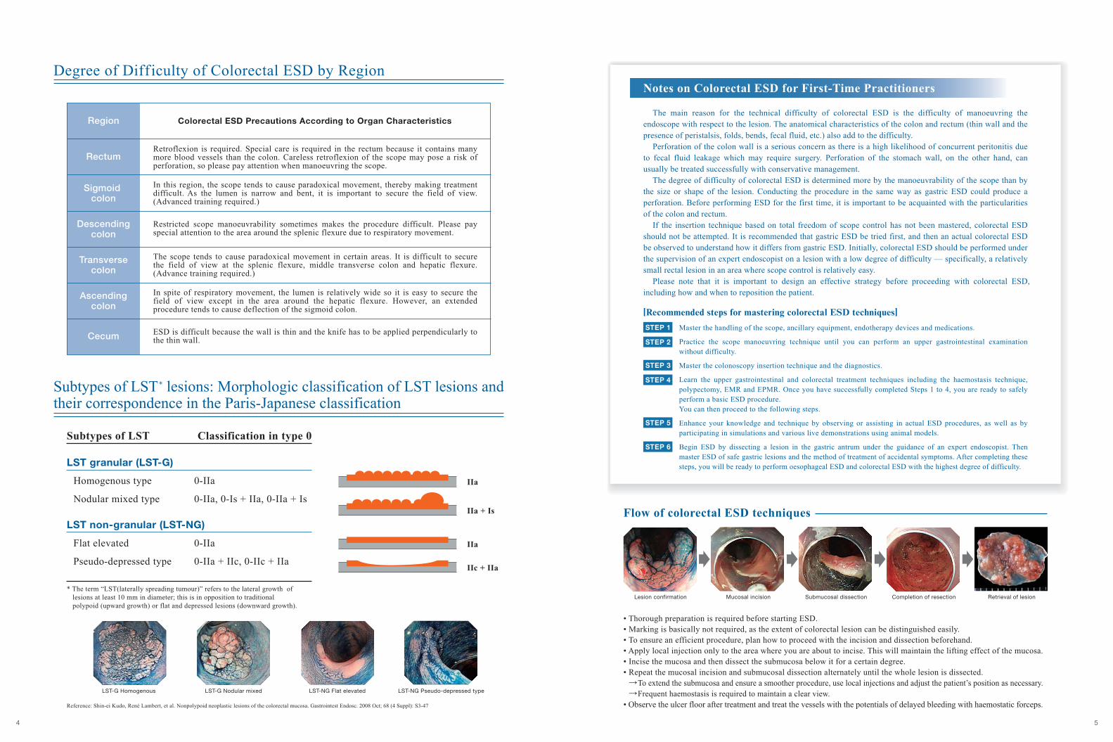
are adenomatous lesions. In addition, the technical difficulty of colorectal ESD is still quite high because of the difficulty
in manoeuvring an endoscope with respect to the lesion, as well as the anatomic characteristics of the colon and rectum
(thin wall and the presence of peristalsis, folds, bends, fecal fluid, etc.). For example, if the colon wall is perforated, there
is a very high likelihood of peritonitis due to fecal fluid leakage — a complication that may require surgical treatment.
Perforation of the stomach wall, on the other hand, can usually be cured with conservative management. The difficulty of
colorectal ESD is determined more by how difficult the scope is to manoeuvre than by the size or visual shape of the lesion.
It should be kept in mind that it is not acceptable to risk ESD in a situation where the scope is difficult to manoeuvre.
In closing, we hope that this booklet can assist the clinical studies of colorectal ESD that are being performed at an
increasing number of hospitals. It is our wish that progress in ESD continues until it is standardised and that, by providing
readers with the knowledge they need and discouraging them from attempting procedures that are too difficult, this booklet
will support that progress by helping to prevent medical accidents that could hinder the development of colorectal ESD.
[Working Group for Standardisation ofColorectal ESD]
Yoshiro Tamegai Konodai Hospital, International Medical Center of Japan
Yutaka Saito National Cancer Center Hospital
Shinji Tanaka Hiroshima University
Sumio Tsuda Fulruoka University Chikushi Hospital
Naohisa Yahagi ToranomonHospital Hiro-o Yamano AkitaRedCrossHospital
Among lesions that require en-bloc endoscopic resection,
the following are considered to be indications for colorectal ESD.
1. Lesions that are hard to resect en bloc using a snare:
• Laterally spreading tumours of the non-granular type (LST-NG),particularly the pseudo-depressed type.
• Lesions presenting Type V1 pit pattern.
• SM carcinomas with scanty invasion.
• Large depressed type tumours.
• Large elevated lesions suspected to be carcinoma .
2. Intramucosal lesions accompanied by submucosal fibrosis .
3. Sporadic local tumours located at chronic inflammations such asulcerative colitis .
4. Local residual early carcinoma after endoscopic resection .
Notes) *1. Includes tall laterally spreading tumours of granular type (LST-G). *2. Lesions caused by prolapse due to biopsy or lesion peristalsis.*3. Technical difficulty is high.
Supplement
1. Both magnifying observation results as well as ordinary endoscopic observation should be used to determine
whether or not colorectal ESD is indicated.
2. In principle, SM carcinomas should be eliminated from the indication when invasion is clearly massive.
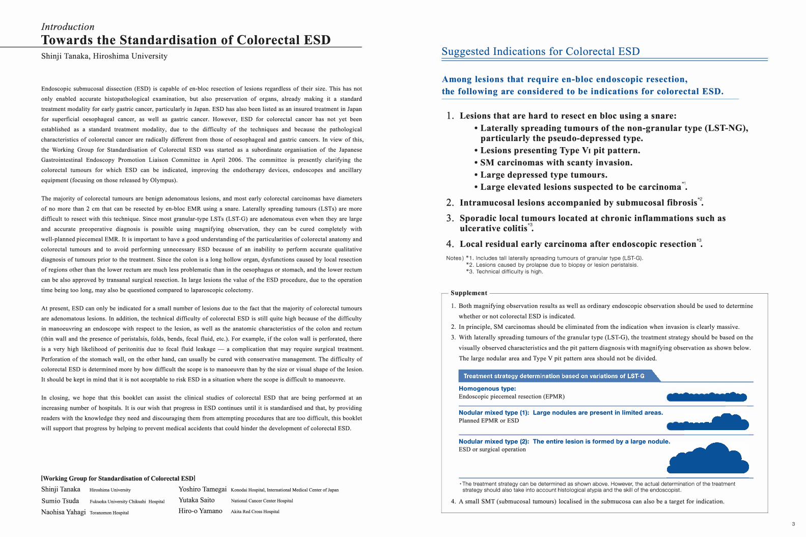
3. With laterally spreading tumours of the granular type (LST-G), the treatment strategy should be based on the
visually observed characteristics and the pit pattern diagnosis with magnifying observation as shown below.
The large nodular area and Type V pit pattern area should not be divided.
Homogenous type: Endoscopic piecemeal resection (EPMR)
Nodular mixed type (1): Large nodules are present in limited areas. Planned EPMR or ESD
Nodular mixed type (2): The entire lesion is formed by a large nodule. ESD or surgical operation
·The treatment strategy can be determined as shown above. However, the actual determination of the treatmentstrategy should also take into account histological atypia and the skill of the endoscopist.
4. A small SMT (submucosal tumours) localised in the submucosa can also be a target for indication.
3
Printed in Japan F1296SE-0309
Disclaimer: Any content or information (“Content”) presented herein is illustrative in nature and does not guarantee or represent specific information, outcomes, or results. Olympus Corporation, its subsidiaries, affiliates, directors, officers, employees, agents, and representatives (collectively “Olympus”) does not represent to or warrant the accuracy or applicability of the Content. Under no circumstances shall Olympus be liable for any costs, expenses, losses, claims, liabilities, or other damages (whether direct, indirect, special, incidental, consequential, or otherwise) that may arise from, or be incurred in connection with, the Content or any use thereof.