Embed Size (px)
Citation preview
Biology 12 - The Digestive System - Chapter Notes
Food is mechanically and chemically broken down into
molecules during digestion, after which they can be taken up
by body cells through the separate process of absorption.
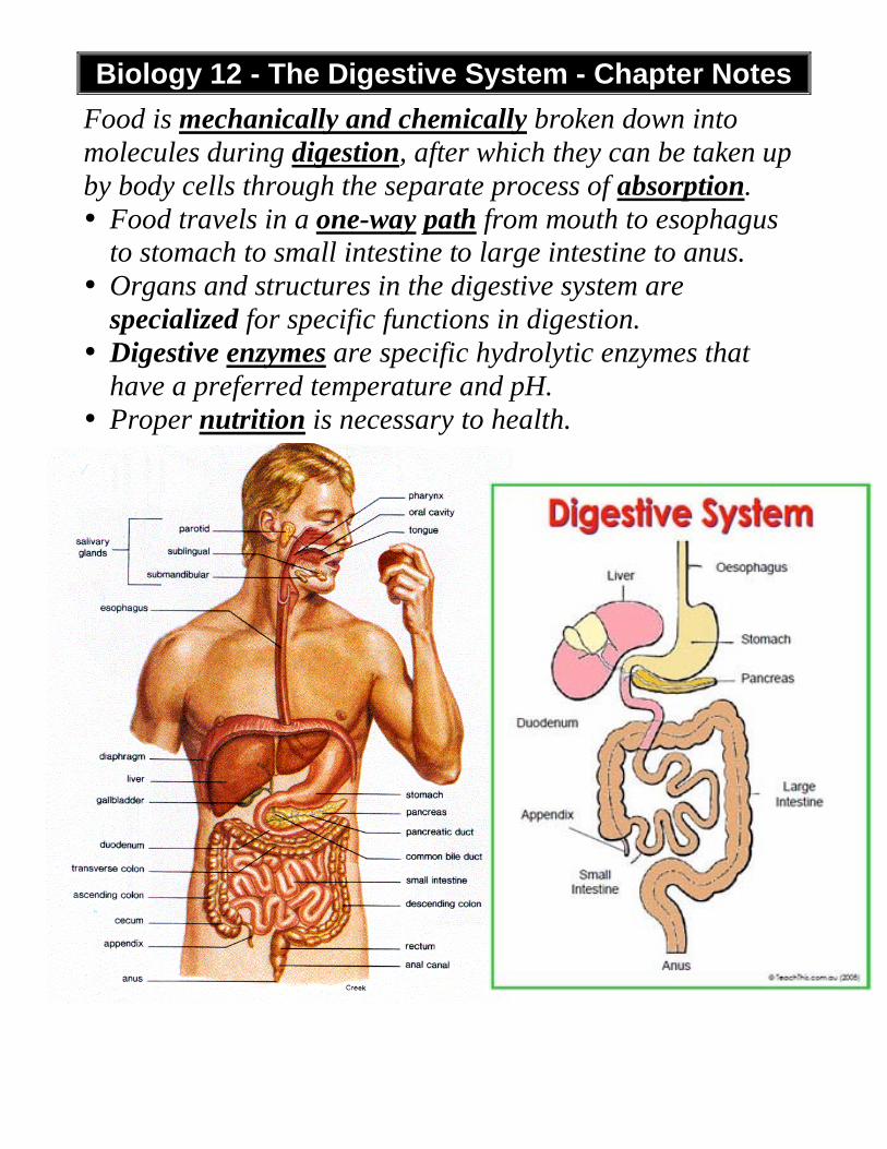
• Food travels in a one-way path from mouth to esophagus
to stomach to small intestine to large intestine to anus.
• Organs and structures in the digestive system are
specialized for specific functions in digestion.
• Digestive enzymes are specific hydrolytic enzymes that
have a preferred temperature and pH.
• Proper nutrition is necessary to health.
DIGESTION: the mechanical and chemical breaking down of ingested food into particles, then into molecules small enough to move into the blood. ABSORPTION: the passage of digested nutrients from the intestines into the blood or lymph, which distributes them through the body. ELIMINATION: the expulsion of indigestible residues from the body. (POOP!)
We will look at DIGESTION first. During digestion, proteins are broken down into amino acids, carbohydrates into glucose, fat to glycerol and fatty acids, nucleic acids to nucleotides.
Mouth
• your mouth is where digestion begins. • the mouth receives food, chews it up, moistens it, and
starts to digest any starch in the food. Structure
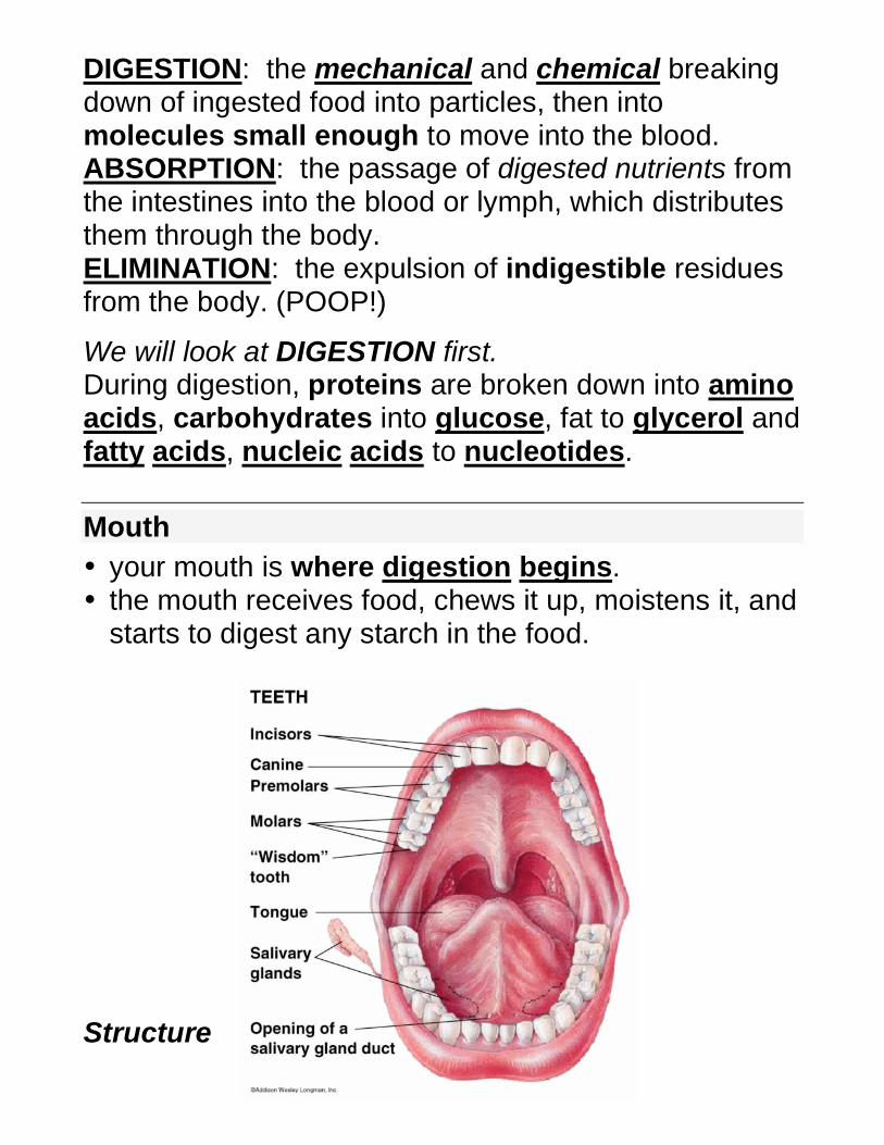
Teeth • a normal adult mouth has 32 teeth to chew food into
pieces that can be swallowed easily. • different teeth types are incisors for biting,
canines for tearing, premolars for grinding, and 12 molars for crushing.
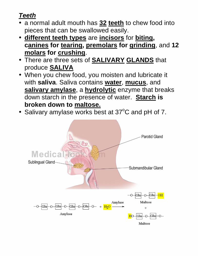
• There are three sets of SALIVARY GLANDS that produce SALIVA
• When you chew food, you moisten and lubricate it with saliva. Saliva contains water, mucus, and salivary amylase, a hydrolytic enzyme that breaks down starch in the presence of water. Starch is broken down to maltose.
• Salivary amylase works best at 37oC and pH of 7.
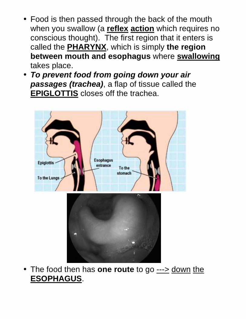
• Food is then passed through the back of the mouth when you swallow (a reflex action which requires no conscious thought). The first region that it enters is called the PHARYNX, which is simply the region between mouth and esophagus where swallowing takes place.
• To prevent food from going down your air passages (trachea), a flap of tissue called the EPIGLOTTIS closes off the trachea.
• The food then has one route to go ---> down the
ESOPHAGUS.
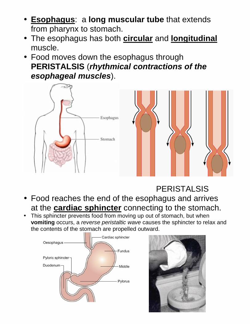
• Esophagus: a long muscular tube that extends from pharynx to stomach.
• The esophagus has both circular and longitudinal muscle.
• Food moves down the esophagus through PERISTALSIS (rhythmical contractions of the esophageal muscles).
PERISTALSIS • Food reaches the end of the esophagus and arrives
at the cardiac sphincter connecting to the stomach. • This sphincter prevents food from moving up out of stomach, but when
vomiting occurs, a reverse peristaltic wave causes the sphincter to relax and the contents of the stomach are propelled outward.
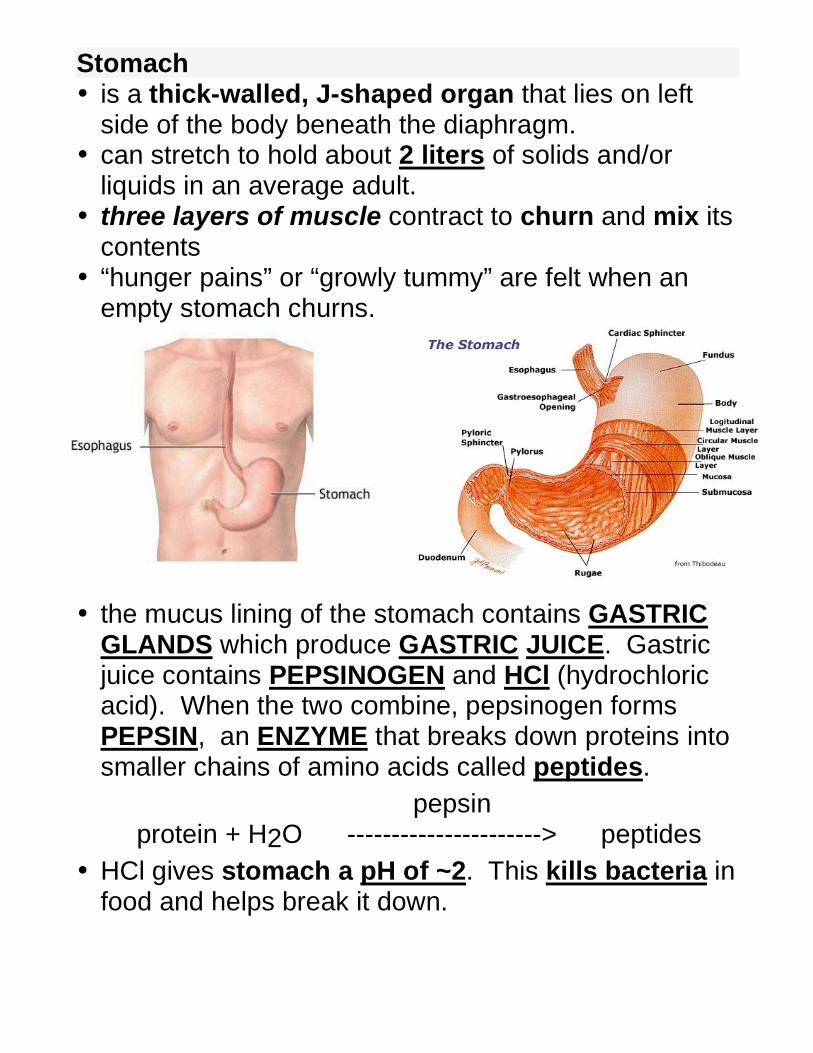
Stomach • is a thick-walled, J-shaped organ that lies on left
side of the body beneath the diaphragm. • can stretch to hold about 2 liters of solids and/or
liquids in an average adult. • three layers of muscle contract to churn and mix its
contents • “hunger pains” or “growly tummy” are felt when an
empty stomach churns. • the mucus lining of the stomach contains GASTRIC
GLANDS which produce GASTRIC JUICE. Gastric juice contains PEPSINOGEN and HCl (hydrochloric acid). When the two combine, pepsinogen forms PEPSIN, an ENZYME that breaks down proteins into smaller chains of amino acids called peptides.
pepsin protein + H2O ----------------------> peptides
• HCl gives stomach a pH of ~2. This kills bacteria in food and helps break it down.
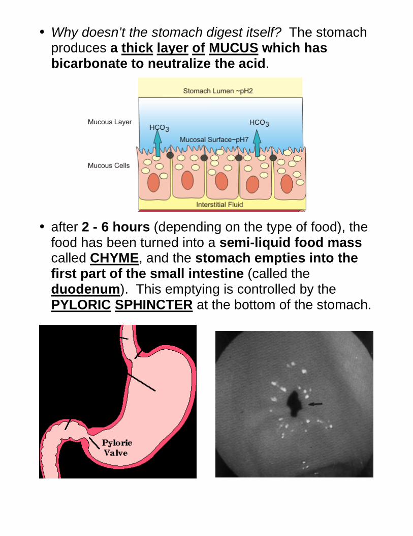
• Why doesn’t the stomach digest itself? The stomach produces a thick layer of MUCUS which has bicarbonate to neutralize the acid.
• after 2 - 6 hours (depending on the type of food), the food has been turned into a semi-liquid food mass called CHYME, and the stomach empties into the first part of the small intestine (called the duodenum). This emptying is controlled by the PYLORIC SPHINCTER at the bottom of the stomach.
Small Intestine: The Food Processor
• Most of digestion and absorption of most nutrients occur in the small intestine.
• Divided into three zones: the DUODENUM, JEJUNUM, and ILIUM.
• The small intestines are about 6 meters long compared to 1.5 m for large intestine.
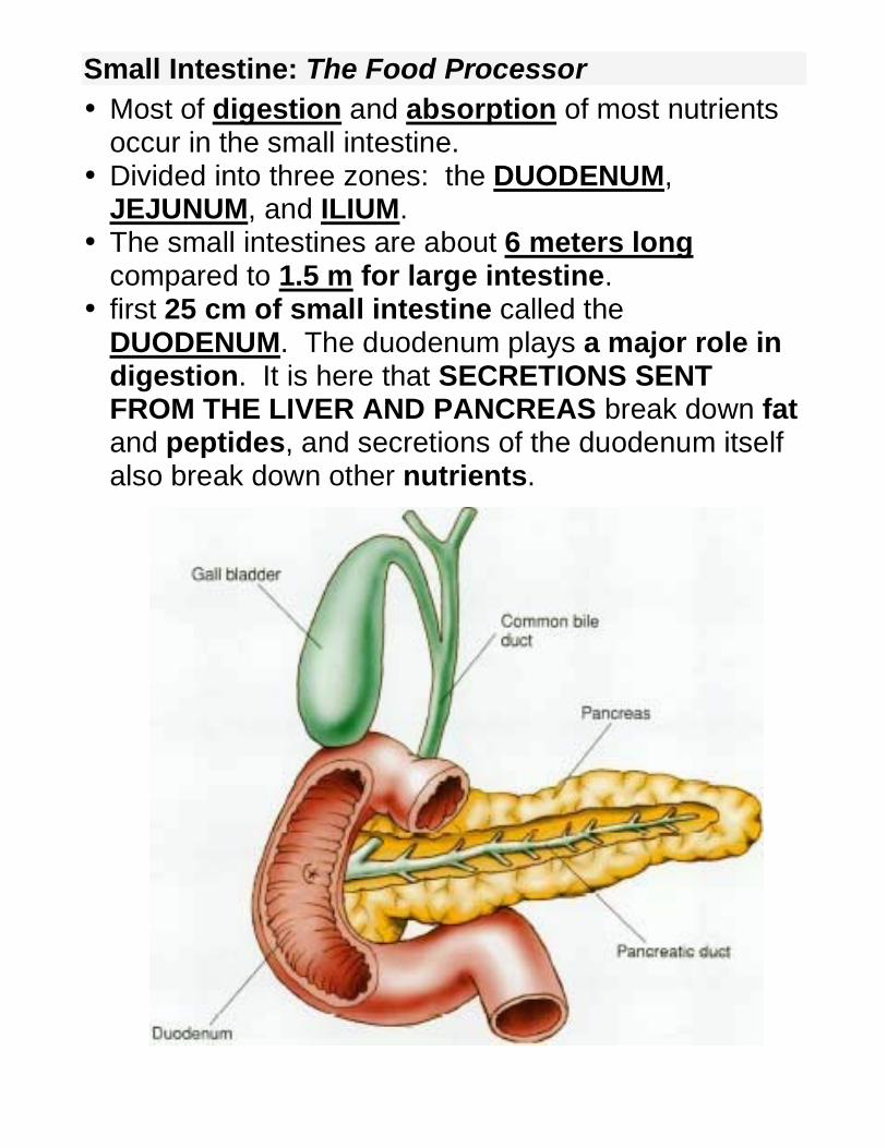
• first 25 cm of small intestine called the DUODENUM. The duodenum plays a major role in digestion. It is here that SECRETIONS SENT FROM THE LIVER AND PANCREAS break down fat and peptides, and secretions of the duodenum itself also break down other nutrients.
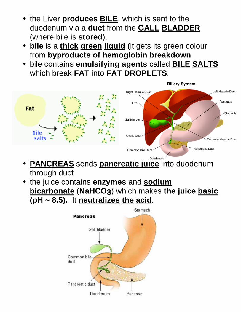
• the Liver produces BILE, which is sent to the duodenum via a duct from the GALL BLADDER (where bile is stored).
• bile is a thick green liquid (it gets its green colour from byproducts of hemoglobin breakdown
• bile contains emulsifying agents called BILE SALTS which break FAT into FAT DROPLETS.
• PANCREAS sends pancreatic juice into duodenum
through duct • the juice contains enzymes and sodium
bicarbonate (NaHCO3) which makes the juice basic (pH ~ 8.5). It neutralizes the acid.
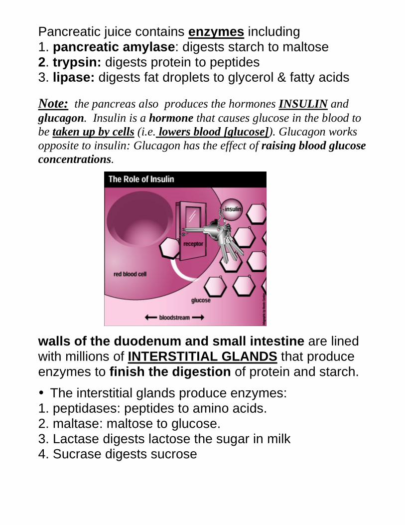
Pancreatic juice contains enzymes including 1. pancreatic amylase: digests starch to maltose 2. trypsin: digests protein to peptides 3. lipase: digests fat droplets to glycerol & fatty acids
Note: the pancreas also produces the hormones INSULIN and
glucagon. Insulin is a hormone that causes glucose in the blood to
be taken up by cells (i.e. lowers blood [glucose]). Glucagon works
opposite to insulin: Glucagon has the effect of raising blood glucose
concentrations.
walls of the duodenum and small intestine are lined with millions of INTERSTITIAL GLANDS that produce enzymes to finish the digestion of protein and starch.
• The interstitial glands produce enzymes: 1. peptidases: peptides to amino acids. 2. maltase: maltose to glucose. 3. Lactase digests lactose the sugar in milk 4. Sucrase digests sucrose
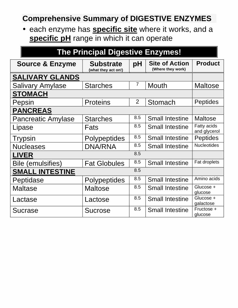
Comprehensive Summary of DIGESTIVE ENZYMES
• each enzyme has specific site where it works, and a specific pH range in which it can operate
The Principal Digestive Enzymes!
Source & Enzyme Substrate (what they act on!)
pH Site of Action (Where they work)
Product
SALIVARY GLANDS
Salivary Amylase Starches 7 Mouth Maltose
STOMACH
Pepsin Proteins 2 Stomach Peptides
PANCREAS
Pancreatic Amylase Starches 8.5 Small Intestine Maltose
Lipase Fats 8.5 Small Intestine Fatty acids and glycerol
Trypsin Polypeptides 8.5 Small Intestine Peptides
Nucleases DNA/RNA 8.5 Small Intestine Nucleotides
LIVER 8.5
Bile (emulsifies) Fat Globules 8.5 Small Intestine Fat droplets
SMALL INTESTINE 8.5
Peptidase Polypeptides 8.5 Small Intestine Amino acids
Maltase Maltose 8.5 Small Intestine Glucose + glucose
Lactase Lactose 8.5 Small Intestine Glucose + galactose
Sucrase Sucrose 8.5 Small Intestine Fructose + glucose
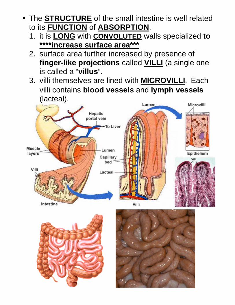
• The STRUCTURE of the small intestine is well related to its FUNCTION of ABSORPTION. 1. it is LONG with CONVOLUTED walls specialized to
****increase surface area*** 2. surface area further increased by presence of
finger-like projections called VILLI (a single one is called a “villus”.
3. villi themselves are lined with MICROVILLI. Each
villi contains blood vessels and lymph vessels (lacteal).
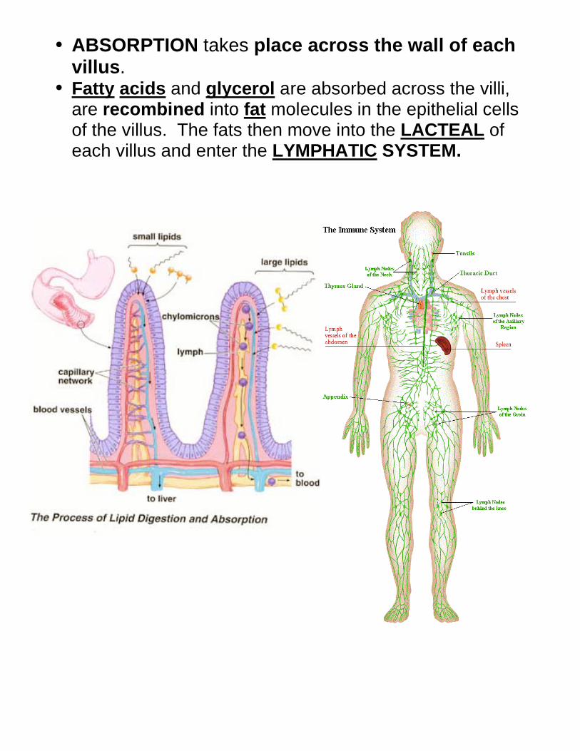
• ABSORPTION takes place across the wall of each villus.
• Fatty acids and glycerol are absorbed across the villi, are recombined into fat molecules in the epithelial cells of the villus. The fats then move into the LACTEAL of each villus and enter the LYMPHATIC SYSTEM.
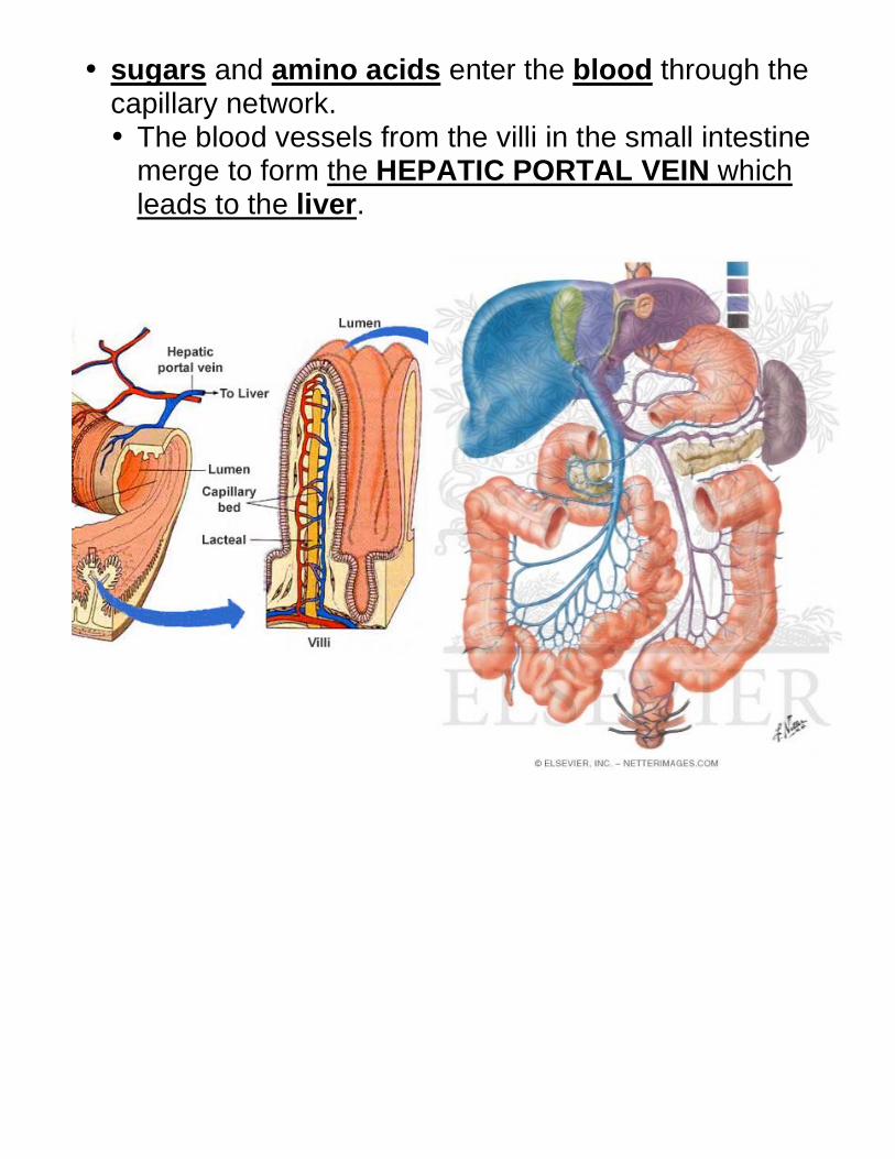
• sugars and amino acids enter the blood through the capillary network. • The blood vessels from the villi in the small intestine
merge to form the HEPATIC PORTAL VEIN which leads to the liver.
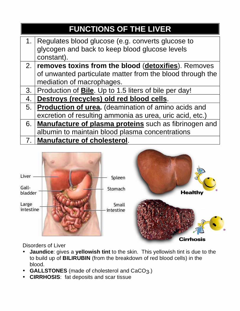
FUNCTIONS OF THE LIVER
1. Regulates blood glucose (e.g. converts glucose to glycogen and back to keep blood glucose levels constant).
2. removes toxins from the blood (detoxifies). Removes of unwanted particulate matter from the blood through the mediation of macrophages.
3. Production of Bile. Up to 1.5 liters of bile per day!
4. Destroys (recycles) old red blood cells.
5. Production of urea. (deamination of amino acids and excretion of resulting ammonia as urea, uric acid, etc.)
6. Manufacture of plasma proteins such as fibrinogen and albumin to maintain blood plasma concentrations
7. Manufacture of cholesterol.
Disorders of Liver • Jaundice: gives a yellowish tint to the skin. This yellowish tint is due to the
to build up of BILIRUBIN (from the breakdown of red blood cells) in the blood.
• GALLSTONES (made of cholesterol and CaCO3.) • CIRRHOSIS: fat deposits and scar tissue
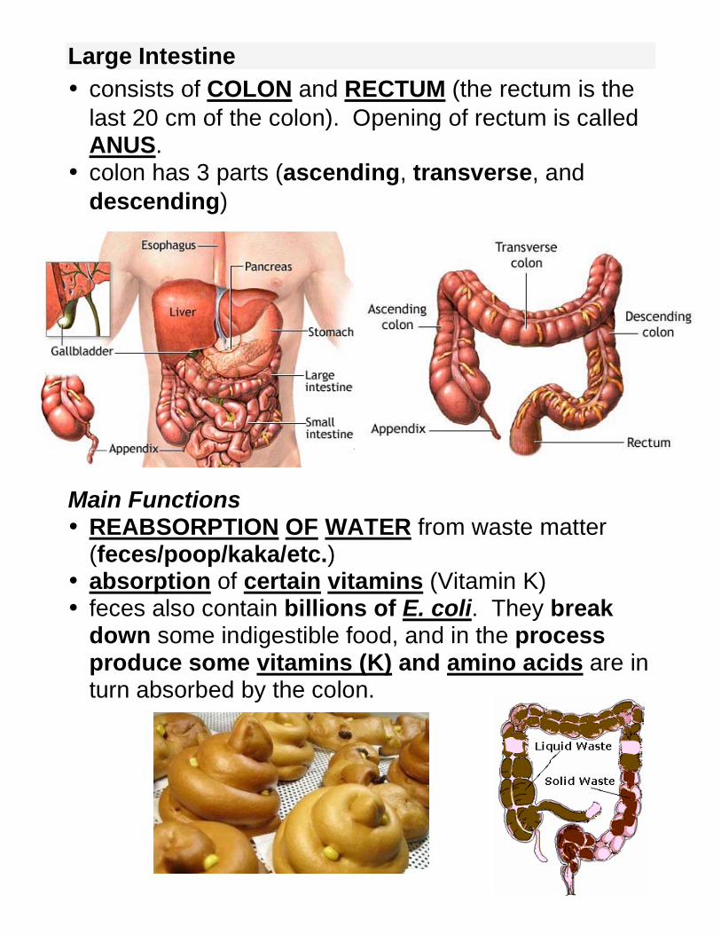
Large Intestine
• consists of COLON and RECTUM (the rectum is the
last 20 cm of the colon). Opening of rectum is called ANUS.
• colon has 3 parts (ascending, transverse, and
descending) Main Functions • REABSORPTION OF WATER from waste matter
(feces/poop/kaka/etc.) • absorption of certain vitamins (Vitamin K) • feces also contain billions of E. coli. They break
down some indigestible food, and in the process produce some vitamins (K) and amino acids are in turn absorbed by the colon.
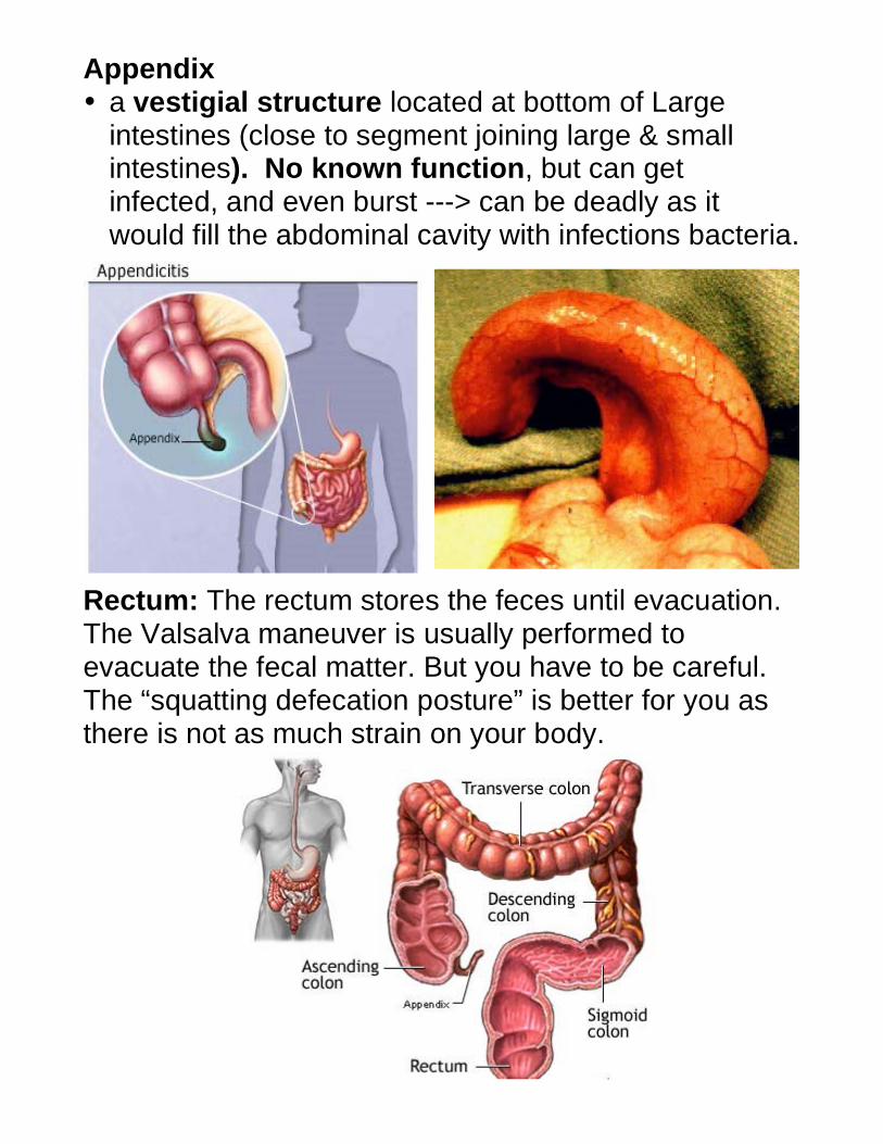
Appendix • a vestigial structure located at bottom of Large
intestines (close to segment joining large & small intestines). No known function, but can get infected, and even burst ---> can be deadly as it would fill the abdominal cavity with infections bacteria.
Rectum: The rectum stores the feces until evacuation. The Valsalva maneuver is usually performed to evacuate the fecal matter. But you have to be careful. The “squatting defecation posture” is better for you as there is not as much strain on your body.
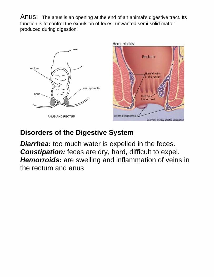
Anus: The anus is an opening at the end of an animal's digestive tract. Its
function is to control the expulsion of feces, unwanted semi-solid matter produced during digestion.
Disorders of the Digestive System
Diarrhea: too much water is expelled in the feces. Constipation: feces are dry, hard, difficult to expel. Hemorroids: are swelling and inflammation of veins in the rectum and anus