Embed Size (px)
Citation preview

P1: SFK/UKS P2: SFK
BLBS102-c45 BLBS102-Simpson March 21, 2012 14:38 Trim: 276mm X 219mm Printer Name: Yet to Come
45Biosensors for Sensitive Detection of
Agricultural Contaminants, Pathogensand Food-Borne Toxins
Barry Byrne, Edwina Stack, and Richard O’Kennedy
IntroductionContaminant Monitoring
Traditional Means of AssessmentInstrumentation-Based Analysis
BiosensorsBiacoreSensor SurfacesAssay Configuration
AntibodiesAntibody Production StrategiesOptical Immunosensors for Quality DeterminationElectrochemical SensorsAlternative Biosensor Formats
PathogensBacterial PathogensFungal Pathogens
ToxinsMycotoxinsWater and Marine Toxins
LegislationConclusionAcknowledgementsWebsites of InterestReferences
Abstract: Immunosensors permit the rapid and sensitive analysisof a range of analytes. Here, we provide a critical assessment of howsuch formats can be implemented, with emphasis on the detectionof bacterial and fungal pathogens, agricultural contaminants (e.g.pesticides and herbicides) and toxins.
INTRODUCTIONThe monitoring of quality of food destined for human consump-tion is a key consideration for farmers, the food industry, leg-islators and, most importantly, for consumers (Karlsson 2004).Hence, it is an absolute necessity to ensure that any contaminants
that may have a deleterious effect on human health are moni-tored qualitatively and quantitatively in a sensitive and reliablemanner. For example, herbicides and pesticides are extremelyeffective at suppressing the growth of plant and insect popula-tions on agricultural produce, such as tomatoes and strawberries.However, the extensive use of such potentially toxic compoundsmay compromise the quality of the product, and prolonged ex-posure may manifest itself as chronic toxicity in human hosts(Keay and McNeil 1998). Furthermore, bacterial strains suchas Salmonella typhimurium and Listeria monocytogenes, whichare causative agents of salmonellosis and listeriosis, respectively,can act as opportunistic pathogens and cause death through theingestion of contaminated produce. Consequently, the develop-ment of suitable methods for their rapid detection is an absolutenecessity. Finally, there is also an urgent need to accuratelymonitor the distribution of toxins, including fungal (e.g. myco-toxins) and water-borne toxins (e.g. phycotoxins), which causesevere illness through the consumption of contaminated food(e.g. nuts, shellfish meat). In summary, rapid, sensitive and ac-curate methodologies are essential for the evaluation of productquality and for satisfying legislative requirements.
CONTAMINANT MONITORINGTraditional Means of Assessment
There are several standard methods that are currently used tomonitor the quality of food. As an example, fruit and vegetableproduce may be inspected by monitoring the colour, gloss, firm-ness, shape and size of the product, as well as noting the presenceor absence of visible defects. This visual inspection may be per-formed alongside more invasive methods, including the analysisof the soluble solid content of the product and determining theacidity, and is particularly useful for produce such as apples,pears and berries. The main advantage of such tests relates tothe fact that they may be carried out immediately post-harvest
Food Biochemistry and Food Processing, Second Edition. Edited by Benjamin K. Simpson, Leo M.L. Nollet, Fidel Toldra, Soottawat Benjakul, Gopinadhan Paliyath and Y.H. Hui.C© 2012 John Wiley & Sons, Inc. Published 2012 by John Wiley & Sons, Inc.
858
P1: SFK/UKS P2: SFK
BLBS102-c45 BLBS102-Simpson March 21, 2012 14:38 Trim: 276mm X 219mm Printer Name: Yet to Come
45 Biosensors for Sensitive Detection of Agricultural Contaminants, Pathogens and Food-Borne Toxins 859
by the farmer, and at a minimal cost (Mitchum et al. 1996).As further examples, grain and nuts may be inspected for thepresence of fungal contamination, while bacterial spoilage maybe indicated by the presence of an uncharacteristically strongodour, such as in coleslaw and milk. However, these tests arenot sufficient for providing confirmation to the consumer thatthe product satisfies regulations with respect to acceptable max-imum residue limits (MRLs). Furthermore, it is not possible toprovide accurate quantitative or qualitative analysis of individualcontaminants through these methodologies, including those thatcannot be seen by visual inspection (e.g. mycotoxins). Hence,it is common practice for food samples to be removed and sentto an external laboratory where comprehensive in situ analysismay be performed (Giraudi and Baggiani 1994).
Instrumentation-Based Analysis
There is a selection of different methodologies available forquality determination, and some of the more relevant exam-ples are discussed in this section. Product firmness can be de-termined through the implementation of the Magness-Taylortest, namely a destructive method that assesses the maximumforce required to perforate the product in a specific way (Abbott2004). This has been applied for the analysis of fruit, includingpears (Gomez et al. 2005). The non-destructive determinationof elasticity may also be permitted through the measurementof acoustic responses, with the signal being interrogated usingfast Fourier transform-based analysis. Shmulevich et al. (2003)demonstrated the efficacy of this approach for evaluating thefirmness of apples, and monitored product softening over timein a controlled atmosphere environment. While these methodsare suitable for monitoring the structural properties of the prod-uct in question, they do not permit rigorous quality evaluation.
More suitable analytical methods include near-infrared (IR)spectroscopy, implemented by Berardo et al. (2005) for the de-tection of mycotoxigenic fungi and associated toxic metabolites,and scanning electron microscopy, applied for the inspectionof ultrastructural changes of the epicuticular layer of oranges
treated with fludioxonil, a pesticide used to control the growth oftwo Penicillium species (Penicillium digitatum and Penicilliumitalicum) (Schirra et al. 2005). Furthermore, gas chromatog-raphy (GS) or mass spectrometry (MS) and high-performanceliquid chromatography (HPLC) are accurate and highly sen-sitive methods for the detection of an array of contaminants,including pesticide, herbicide and toxins residues. The lattermethod may also be used to detect the presence of indicatormolecules that are representative of product freshness, includingflavonoids (MacLean et al. 2006), while liquid chromatographycoupled with mass spectrometry (LC-MS) can accurately mon-itor product bitterness. This was demonstrated by Dourtoglouet al. (2006) for the analysis of olives (Olea europaea). In spiteof the efficacy of these analytical platforms, the instrumentationneeded to perform this analysis is expensive and bulky and mayrequire extensive operator training. In addition, analysis timesmay also be extensive, as many contaminants require lengthysample pre-treatment prior to assessment.
Here, we focus on the application of biosensor-based plat-forms that are rapid, sensitive and reliable and are frequentlyused for the detection of herbicide and pesticide residues, bacte-rial and fungal pathogens and toxins (fungal and water-borne).
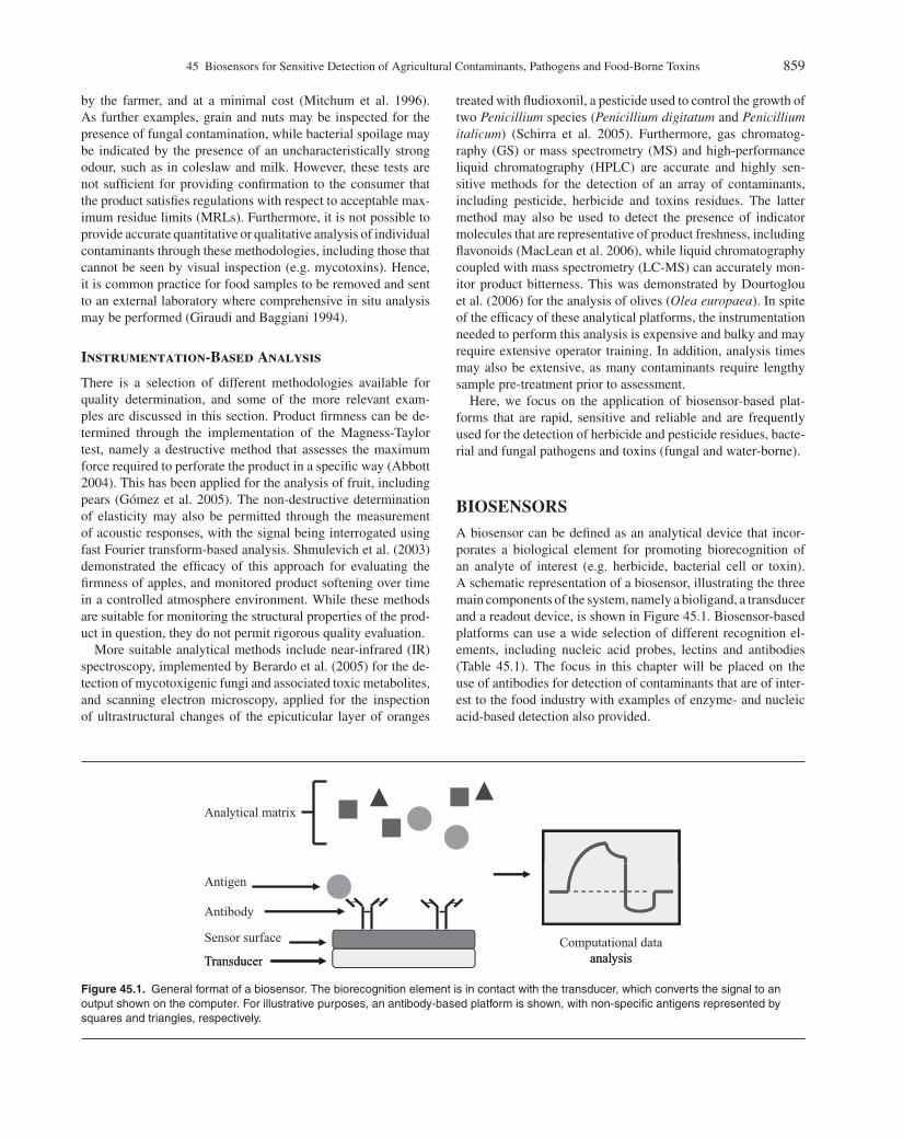
BIOSENSORSA biosensor can be defined as an analytical device that incor-porates a biological element for promoting biorecognition ofan analyte of interest (e.g. herbicide, bacterial cell or toxin).A schematic representation of a biosensor, illustrating the threemain components of the system, namely a bioligand, a transducerand a readout device, is shown in Figure 45.1. Biosensor-basedplatforms can use a wide selection of different recognition el-ements, including nucleic acid probes, lectins and antibodies(Table 45.1). The focus in this chapter will be placed on theuse of antibodies for detection of contaminants that are of inter-est to the food industry with examples of enzyme- and nucleicacid-based detection also provided.
Analytical matrix
Transducer
Sensor surface
Antibody
Antigen
Computational data analysisTransducer analysis
Figure 45.1. General format of a biosensor. The biorecognition element is in contact with the transducer, which converts the signal to anoutput shown on the computer. For illustrative purposes, an antibody-based platform is shown, with non-specific antigens represented bysquares and triangles, respectively.
P1: SFK/UKS P2: SFK
BLBS102-c45 BLBS102-Simpson March 21, 2012 14:38 Trim: 276mm X 219mm Printer Name: Yet to Come
860 Part 8: Food Safety and Food Allergens
Table 45.1. The Recognition Elements CommonlyUsed in Sensor Systems
1. Antibodies and antibody fragments: Derived by enzymedigestion or genetic engineering (Fab fragment, scFvand diabody).
2. Lectins, namely carbohydrate-binding proteins. Onlysuitable for the detection of glycosylated entities, suchas glycoproteins.
3. Enzymes: e.g. those specific for one particular substrate(i.e. horseradish peroxidase for hydrogen peroxide).
4. Cell membrane receptors.5. A living cell: eukaryotic or prokaryotic.6. Nucleic acid-based probes: DNA and RNA or peptide
nucleic acid.7. Aptamers.8. Chemically generated recognition surfaces: Including
plastibodies (artificial antibodies or molecularlyimprinted polymers).
Fab, fragment antigen binding; scFv, single-chain variable fragment.
A transducer is a device, such as a piezoelectric crystal orphotoelectric cell that converts input energy of one form intooutput energy of another, with the output signal generated pro-portional to the concentration of the target analyte (Luong et al.1995). There are many different types of transducers that canbe used in biosensor-based platforms, and a selection of these isshown in Table 45.2. Finally, a readout device typically consistsof a computer-linked monitor that presents the data in a formthat can easily be interpreted by the end-user.
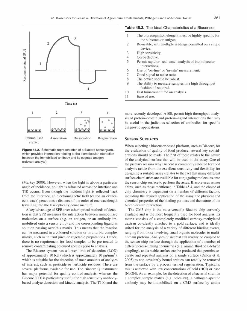
Many different biosensor platforms have dedicated softwarepackages that can facilitate in the interpretation of biomolecu-lar interactions. For example, Biacore, a frequently used com-
mercial optical biosensor based on surface plasmon resonance(SPR), which is discussed in more detail later, uses Biaevalua-tion software that presents data in the form of a sensorgram (Fig.45.2). Here, interactions between immobilised and free entitiescan be easily visualised by monitoring changes in refractive in-dex (RI). This correlates to a change in mass imparted by theinteraction between the two binding elements (e.g. antibody andcognate antigen), with units of measurement referred to as re-sponse units (RU). In Biacore analysis, a response of 1000 RUis representative of a change in resonance angle of 0.1◦, whichcorresponds to an alteration in the surface coverage of the sensorsurface of approximately 1 ng/mm2.
While Biacore is an excellent example of a biosensor thatis commonly used for the evaluation of quality of food (whichis discussed in more detail later) other biosensor platforms arealso applicable that are based on electrochemical, piezoelectric,magnetic and thermal detection (Byrne et al. 2009). Many ofthese platforms are developed ‘in-house’ and have their owndedicated software packages for the interpretation of data, andkey examples of these novel sensors are also described later.Finally, for a biosensor to be applicable in any field, including inthe monitoring of quality of food produce, it must have certaincharacteristics. These are listed in Table 45.3.
Biacore
Biacore (GE Healthcare) uses an optical-based transducer sys-tem for the measurement of analytes based on the principle ofSPR. SPR works on the principle of total internal reflection(TIR), a phenomenon that occurs at the interface between twonon-absorbing materials, such as water and a solid. When asource of light is directed at such an interface from a mediumwith a higher RI to a medium of lower RI (such as light travellingthrough glass and water), the light is refracted to the interface
Table 45.2. Examples of Transducers Commonly Used in Biosensor Systems
Type Example Principle of Use
Electrochemical Conductimetric Solutions containing ions conduct electricity. Depending on the reaction,the change in conductance is measured.
Potentiometric Measurement of the potential of a cell when there is no current flowing todetermine the concentration of an analyte.
Voltammetric A changing potential is applied to a system and the resulting change incurrent is measured.
Field effecttransistor-based
Field effect transistor A current flows along a semi-conductor from a source gate to a drain. Asmall change in gate voltage can cause a large variation in the currentfrom the source to the drain.
Optical Surface plasmon resonance Surface plasmon resonance (a detailed explanation is given in thischapter).
Thermal Calorimetry Heat exchange is detected by thermistors and related to the rate of areaction.
Surface acousticwave
Rayleigh surface wave An immobilised sample on the surface of a crystal affects thetransmission of a wave to a detector.
Piezoelectric Electrochemical quartzcrystal microbalance
A vibrating crystal generates current that is affected by a materialadsorbed onto its surface.
P1: SFK/UKS P2: SFK
BLBS102-c45 BLBS102-Simpson March 21, 2012 14:38 Trim: 276mm X 219mm Printer Name: Yet to Come
45 Biosensors for Sensitive Detection of Agricultural Contaminants, Pathogens and Food-Borne Toxins 861
3
1
2
Res
onan
ce s
igna
l (R
U)
4
Time (s)
4321
RegenerationDissociationAssociationImmobilised surface
Figure 45.2. Schematic representation of a Biacore sensorgram,which provides information relating to the biomolecular interactionbetween the immobilised antibody and its cognate antigen(relevant analyte).
(Markey 2000). However, when the light is above a particularangle of incidence, no light is refracted across the interface andTIR occurs. Even though the incident light is reflected backfrom the interface, an electromagnetic field (called an evanes-cent wave) penetrates a distance of the order of one wavelengthtravelling into the less optically dense medium.
A key advantage of SPR over other optical methods of detec-tion is that SPR measures the interaction between immobilisedmolecules on a surface (e.g. an antigen, or an antibody im-mobilised onto a sensor chip) and the corresponding ligand insolution passing over this matrix. This means that the reactioncan be measured in a coloured solution or in a turbid complexmatrix, such as in fruit juice or vegetable preparations. Hence,there is no requirement for food samples to be pre-treated toremove contaminating coloured species prior to analysis.
The Biacore system has a lower limit of detection (LOD)of approximately 10 RU (which is approximately 10 pg/mm2),which is suitable for the detection of trace amounts of analytesof interest, such as pesticide or herbicide residues. There areseveral platforms available for use. The Biacore Q instrumenthas major potential for quality control analysis, whereas theBiacore 3000 is particularly useful for high sensitivity antibody-based analyte detection and kinetic analysis. The T100 and the
Table 45.3. The Ideal Characteristics of a Biosensor
1. The biorecognition element must be highly specific forthe substrate or antigen.
2. Re-usable, with multiple readings permitted on a singledevice.
3. High sensitivity.4. Cost-effective.5. Permit rapid or ‘real-time’ analysis of biomolecular
interactions.6. Use of ‘on-line’ or ‘in-situ’ measurement.7. Good signal to noise ratio.8. The device should be robust.9. The ability to measure samples in a high throughput
fashion, if required.10. Fast turnaround time on analysis.11. Ease of use.
more recently developed A100, permit high-throughput analy-sis of protein–protein and protein–ligand interactions that maybe useful in the judicious selection of antibodies for specificdiagnostic applications.
Sensor Surfaces
When selecting a biosensor-based platform, such as Biacore, forthe evaluation of quality of food produce, several key consid-erations should be made. The first of these relates to the natureof the analytical surface that will be used in the assay. One ofthe primary reasons why Biacore is commonly selected for foodanalysis (aside from the excellent sensitivity and flexibility fordesigning a suitable assay) relates to the fact that many differentsurface chemistries are available for conjugating molecules ontothe sensor chip surface to perform the assay. Biacore uses sensorchips, such as those mentioned in Table 45.4, and the choice ofchip chemistry is dependent on a number of different factors,including the desired application of the assay, the physical andchemical properties of the binding partners and the nature of thebiomolecular interaction.
The CM5 chip is the most versatile Biacore chip currentlyavailable and is the most frequently used for food analysis. Itsmatrix consists of a completely modified carboxy-methylateddextran covalently attached to a gold surface, and is ideallysuited for the analysis of a variety of different binding events,ranging from those involving small organic molecules to multi-domain proteins. Analytes of interest can readily be coupled tothe sensor chip surface through the application of a number ofdifferent cross-linking chemistries (e.g. amine, thiol or aldehydecoupling), and a stable surface can be produced that permits ac-curate and repeated analysis on a single surface (Dillon et al.2005) as non-covalently bound entities can readily be removedfrom the surface by a process termed regeneration. Typically,this is achieved with low concentrations of acid (HCl) or base(NaOH). As an example, for the detection of a bacterial strain ina complex sample matrix (e.g. coleslaw), a pathogen-specificantibody may be immobilised on a CM5 surface by amine

P1: SFK/UKS P2: SFK
BLBS102-c45 BLBS102-Simpson March 21, 2012 14:38 Trim: 276mm X 219mm Printer Name: Yet to Come
862 Part 8: Food Safety and Food Allergens
Table 45.4. The Surface Chemistries of Available Biacore Chips
Chip Type Modification Type Applications
CM5 100% carboxylation of dextran surface. General use. Routinely selected for antibody-antigen andprotein-protein interaction analysis.
CM4 30% carboxylation of dextran surface. Serum, cell extracts.CM3 100% carboxylation of dextran surface. Serum, cell extracts.C1 100% carboxylation of dextran surface. Permits binding events to occur closer to the sensor surface, which is
advantageous in situations where multivalent interactions occur, orwhere analytes are large.
L1 Lipophilic. Lipid capturing.SA Streptavidin surface. Detection of biotin-containing molecules. Useful for immobilisation.NTA Nickel – nitrilotriacetic acid. Detection of histidine-tagged molecules.HPA Flat hydrophobic surface. Used for membrane-associated interactions.Au and SIA None. Useful for self-assembled monolayer-based interactions analysis.
coupling. This covalently captured immunoglobulin may thenbe used as a bioligand to capture the pathogen from the foodsample, which is injected over the sensor surface and, therefore,is in solution. The change in mass resulting from the biomolecu-lar interaction introduces a change in RI, as seen by an increasein RU, and regeneration can subsequently be used to liberate thebacterial cell(s), so that the immobilised antibody is availablefor subsequent analysis. This method of analysis can be used fordetection purposes and can be used to establish LODs. Further-more, where required, kinetic analysis can be used to determinethe affinity of the antibody for its cognate antigen.
Assay Configuration
For food-based biosensor analysis, analytes of interest rangein size from large (intact bacterial and fungal cells) to small(pesticide and toxin residues), requiring that the assay formatselected must be capable of providing accurate and quantitativedetection. Biacore-based assays that monitor interactions be-tween large biomolecules, such as antibodies and proteinaceousantigens, can be developed by immobilising either the antibodyor antigen on the surface, as the mass change introduced by thebinding of the other entity is sufficient to cause a recordablechange in RU that can be seen on a sensorgram. However, ascontaminants such as herbicides, pesticides and toxins have lowmolecular weights, it is often necessary to employ an indirectmeasurement method where the analyte is immobilised directlyon the sensor surface, and the larger of the binding elementsis subsequently introduced. The resultant change in mass intro-duced by the binding of the larger element (in solution) to thesmaller, immobilised ligand can therefore be easily seen as achange in RU. If the assay was to be performed in reverse (e.g.the larger entity is immobilised and the small hapten or toxin isfree in solution), a small change in mass would be introducedduring an interaction event, which may be difficult to detect andquantify reliably.
Many of the examples that are discussed in this chapter employantibody-based competition or inhibition assay formats (Fig.45.3). An inhibition assay involves the combination of the sam-
ple of interest with the specific antibody before injection ontoa biosensor chip containing an immobilised target molecule.There is competition between the immobilised and free antigen(from the sample to be analysed) for antibody binding. A changein signal (e.g. RU) is recorded, and this is inversely proportionalto the amount of target analyte that remains free in solution.An alternative method to detect analytes of interest requires acompetitive assay format. In this format, the antibody, specificto the analyte of interest, is immobilised on the surface. Thesample, containing the analyte to be determined, is mixed with aknown concentration of standard consisting of the target analytethat has been conjugated to a large carrier protein. This resultsin the analyte and the conjugated standard competing for theimmobilised antibody on the surface of the biochip. An increasein signal is caused by the binding of the large analyte-carrierconjugate and the data generated are similar to the inhibitionassay since the signal recorded is inversely proportional to theamount of target analyte present in the sample. However, to per-form these assays, it is necessary to have a suitable antibody,and methods for antibody production are discussed in Section‘Antibody Production Strategies’.
ANTIBODIESAntibodies are the key biorecognition elements of the immunesystem. A large variety of antibodies and antibody-derived frag-ments have been produced with the capability of detecting anarray of structurally diverse analytes, ranging from proteins tohaptens, and these have been implemented in a number of dif-ferent biosensor formats. Antibodies are globular glycoproteins(sugar-containing proteins), and five main classes (or serotypes)exist in nature, namely IgA, IgM, IgE, IgD and IgG. Single an-tibody molecules typically have molecular weights of approx-imately 150–200 kilodaltons (kDa). IgG antibodies (Fig. 45.4)have a Y-shaped backbone with four polypeptide chains locatedin two identical chains that are covalently attached throughdisulphide bonds. The innermost chains are referred to as theheavy chains because they are approximately double the molec-ular weight of the outer arms (termed the light chains). The

P1: SFK/UKS P2: SFK
BLBS102-c45 BLBS102-Simpson March 21, 2012 14:38 Trim: 276mm X 219mm Printer Name: Yet to Come
45 Biosensors for Sensitive Detection of Agricultural Contaminants, Pathogens and Food-Borne Toxins 863
Inhibition assay Competition assay
Gold layer
Linker layer
Glass layer
Dextran matrix
Antigen Antibody Conjugated antigen
Figure 45.3. Formats for inhibition and competition assays. For illustration, a Biacore surface is represented.Inhibition: Free analyte inhibits binding of the antibody to the immobilised analyte on the chip. The signal generated when the antibody bindsto the immobilised analyte is inversely proportional to the concentration of free analyte in the sample.
Competition: Free ( ) and conjugated ( ) analyte compete for binding to the immobilised antibody. The signal generated is inverselyproportional to the amount of free analyte in the sample.
Complementarity determining regions Heavy chain
Light chain
y
V
VH
Intrachain disulphide bond
CL
VL
CH1
Hinge region
Carbohydrate
CH
CH
VH
2
Interchaindisulphide bond
CH3
Figure 45.4. Structure of an IgG antibody. CH1–3 refers to constant heavy regions 1 to 3, respectively. VH, variable heavy; VL, variable light.

P1: SFK/UKS P2: SFK
BLBS102-c45 BLBS102-Simpson March 21, 2012 14:38 Trim: 276mm X 219mm Printer Name: Yet to Come
864 Part 8: Food Safety and Food Allergens
recognition sites of the antibody, which interact with an epitopeon an antigen of interest, are located at the ends of the variableheavy (VH) and variable light (VL) regions of the heavy andlight chains, respectively. They are commonly referred to as thecomplementarity determining regions, or CDRs. Each arm ofan antibody can bind to one antigen, so one IgG molecule cantheoretically bind to two antigens.
Antibody Production Strategies
There are three main methods for generating antibodies that maybe used in biosensor-based platforms for quality evaluation, andthese are discussed in this section. Polyclonal antibodies are pro-duced through the immunisation of animal hosts with a particularantigen. The immunogen is typically administered in the pres-ence of a suitable adjuvant, which elicits an immune response inthe host. Small molecules, such as toxins or haptens, may have tobe conjugated to larger carrier molecules, such as bovine serumalbumin (BSA), to enhance immunorecognition. Serum titresare typically analysed by enzyme-linked immunosorbent assay(ELISA) to quantify the host-based response. If this is deemedto be suitable, blood samples are subsequently collected and theantibodies generated are purified from the serum. Polyclonal an-tibodies typically consist of a variety of different serotypes withvarying affinities/specificities towards the analyte in question.Animals often used for polyclonal antibody generation includeguinea pigs, rabbits, goats, sheep and donkeys (Leenaars andHendriksen 2005).
The second method involves the use of hybridoma technologyto produce monoclonal antibodies (Kohler and Milstein 1975,Nelson et al. 2000, Hudson and Souriau 2003). These are gen-erated by immunising an animal (typically, a mouse) with theantigen of interest in the presence of a suitable adjuvant. Oncea sufficient immune response is detected by ELISA, the spleen,bone marrow from long bones (femur and humerus) or primarylymphoid organs (such as lymph nodes) are removed from thesacrificed animal and the antibody-producing B-cells are har-vested. These cells can then be fused to immortal myeloma cellsby using an electrical current or polyethylene glycol. The result-ing hybrid cells (hybridomas), which secrete antibodies that aredirected towards the desired antigen, are then selected and clonedout to ensure monoclonality. The advantage of this approach isthat there is a constant supply of the antibody that is requiredfor analysis. However, there is a significant cost involved in theproduction and the screening of these antibodies.
Recombinant antibodies are the third form of antibodies thatare increasingly being used. They are often produced in bac-terial strains such as Escherichia coli and are expressed in aphage display format. Libraries, with the capacity to express alarge number of antibodies, are generated, and they are referredto as naive, synthetic or immune depending on their mode ofproduction (Bradbury and Marks 2004). Immune libraries areconstructed though the administration of the immunogen of in-terest to a suitable host (e.g. mouse, rabbit, chicken), which ismonitored for antibody production. Antibody-encoding nucleicacid is then purified from lymphoid organs, such as the spleen,and cloned into a phage or phagemid vector that, in turn, is
V
VH
VH
CL
VL
CH1
VH
VH
VL
scFv Fab
Figure 45.5. Different antibody fragments available for biosensor-based analysis. CH1–3 refers to constant heavy region 1; VH,variable heavy; VL, variable light; scFv, single-chain variablefragment; Fab, fragment antigen binding.
propagated in E. coli. Phage display libraries can subsequentlybe screened against targets of interest by biopanning. This isfeasible since the phage express the active recombinant antibod-ies on their surface and those that bind to the specific antigenimmobilised on a capture surface can be differentiated from non-binding antibodies, and characterised further to determine theiraffinity for the target antigen. A key advantage with recombinantantibodies is the ability to increase their affinity for an antigen ofinterest through site-directed mutagenesis or other approachessuch as chain shuffling or error-prone PCR, which is not possi-ble for monoclonal or polyclonal antibodies (Conroy et al. 2009,O’Kennedy et al. 2010).
The main types of antibody fragments produced by phagedisplay are the fragment antigen binding (Fab) and single-chainvariable fragment (scFv), whose structures are shown in Fig-ure 45.5. The presence of the constant regions in a Fab is thoughtto aid in the stabilisation of the antibody variable regions, whichmight not function efficiently when expressed in the monomericscFv format (Rothlisberger et al. 2005). It was previously shownin our laboratory that the Fab antibody format is the most reliableand sensitive for use in small molecule competition biosensorassays involving haptens. The strict monovalency of this formatcan lead to a significant enhancement in assay sensitivity in bothELISA and competition SPR assays (Townsend et al. 2006).
The scFv is the most widely used antibody fragment. Thevariable regions (VH and VL) of the antibody are linked by aflexible peptide linker. The most frequently used are based onglycine-serine repeat structures, with the length of the linkerrelated to the intended valency of the molecule. When a shortlinker is used, the stability and folding of the scFv does not occurproperly. This is caused by the insufficient juxtaposing of the VH
and VL regions in the single chain for the monomer to function.ScFvs selected in this format invariably form bivalent dimers,or diabodies, which often have increased avidity for an antigenover the monomeric forms typically observed when long-linkersystems are used (Holliger et al. 1993, Kortt et al. 1997, Atwell
P1: SFK/UKS P2: SFK
BLBS102-c45 BLBS102-Simpson March 21, 2012 14:38 Trim: 276mm X 219mm Printer Name: Yet to Come
45 Biosensors for Sensitive Detection of Agricultural Contaminants, Pathogens and Food-Borne Toxins 865
et al. 1999). Long linkers (ranging from 18–21 amino acids)favour the production of scFv formats, which are predominantlymonomeric (Holliger et al. 1993, Perisic et al. 1994, McGuin-ness et al. 1996). In summary, recombinant antibodies are anexcellent alternative to polyclonal and monoclonal antibodiesfor the detection of analytes of interest, and have great potentialfor application in the monitoring of quality in the food industry(Hudson and Souriau 2003, O’Kennedy et al. 2010).
Optical Immunosensors for QualityDetermination
There have been several excellent examples of biosensor-basedanalysis for the detection of pesticides, herbicides, toxins andbacterial cells, and some of the more pertinent observations thatemploy antibody-based recognition (immunosensors) are dis-cussed in this section. One of the earliest examples demonstrat-ing the use of antibody-based biosensing for detecting herbicideresidues was described by Minunni and Mascini (1993). Here,Biacore was selected as a platform to facilitate the detectionof traces (50 pg/mL) of the herbicide atrazine in water sam-ples. Another optical biosensor was developed to detect andquantify carbamate residues in vegetables. It was observed thatchanges in the concentration of carbamate could be monitoredusing chlorophenol red (Xavier et al. 2000). Moran et al. (2002)used SPR to characterise antibodies that were subsequentlyused to detect the presence of 2-(4-thiazolyl)benzimidazole,a molecule that is used as a food preservative and an agri-cultural fungicide. Schlecht et al. (2002) implemented a C1four-channel sensor chip in a study to quantifiably detectthe presence of 2,4-dichlorophenoxyacetic acid (2,4-D), anorganochlorine herbicide, and monitor cross-reactivity of poly-clonal antibodies with a structurally related analogue, namely2,4,5-trichlorophenoxyacetic acid (2,4,5-T). This assay was per-mitted by immobilising 2,4-D analogues onto the surface of a C1sensor chip surface through a thiol-carboxyl group reaction, andhad a sensitivity of 0.1µg/mL. Finally, Caldow et al. (2005) useda Biacore Q instrument to detect the bacteriostatic antibiotic, ty-losin, in bees’ honey. This polyketide is active against mostgram-positive bacteria, mycoplasma, and certain gram-negativebacteria. They were able to detect tylosin at the level of 2.5µg/kgin honey, demonstrating the ability of biosensor platforms, suchas Biacore, to detect analytes in complex sample matrices. Thesefive key examples demonstrate early applications of using im-munosensors for the detection of low-molecular weight ana-lytes that have a deleterious effect on the quality of agriculturalproduce.
More recently, miniaturised biosensor platforms have been de-veloped for in situ analysis of pesticide and herbicide residues. Aportable immunosensor for the detection of 2,4-D was describedby Kim et al. (2007). Here, murine hosts were immunised with a2,4-D-BSA conjugate, and the resultant monoclonal antibodieswere implemented for detection purposes. When tested on spikedriver water samples, this assay format had excellent sensitivity(0.1 ppb of 2,4-D) and permitted the parallel analysis of multiplesamples. This example also demonstrates how modification ofthe assay format can greatly improve sensitivity, which is a key
consideration for the detection of analytes, such as 2,4-D, whichmay reside in food or water samples in trace amounts. A sand-wich assay format, incorporating a second biotinylated antibody,was subsequently developed whose sensitivity was significantlyenhanced (0.1 ppt of 2,4-D).
Competitive and inhibition assay formats can be utilised effec-tively in a number of different biosensor formats for the detectionof pesticide and herbicide residues. In some cases, this can beapplied where the direct monitoring of the interaction betweenan antigen and its cognate antibody is not sufficiently sensitive.To illustrate this, Gouzy et al. (2009) developed a Biacore-basedSPR competition assay for the detection of the herbicide iso-proturon, as employing a direct detection assay was deemed tobe inadequate. Here, a rat-derived anti-isoproturon monoclonalantibody was selected for biorecognition, and the competitionassay had a good LOD (0.1µg/L). Salmain et al. (2008) de-veloped an indirect competition immunoassay format for thedetection of atrazine. The biosensor format implemented was anIR optical platform that had nanomolar sensitivity.
Salmain et al. (2008) subsequently performed comparativesensitivity analysis in an ELISA-based assay format, and simi-lar observations were made. ELISA assays are routinely selectedfor immunodetection purposes and, depending on the quality ofthe antibody, have excellent sensitivity. However, a major draw-back relates to the fact that analysis times are often lengthy, withmultiple incubation and washing stages required for assay com-pletion. This is in contrast to biosensor-based assays that permitrapid analysis and facilitate the detection of multiple analyteson a single sensor surface, as opposed to using multiple wells.Therefore, ELISA formats may be initially used to validate an as-say format before transferring this to a biosensor platform. Thishas been demonstrated by Herranz et al. (2008) for the eval-uation of leporine polyclonal antibodies specific for simazinederivatives prior to their implementation in an immunosensorplatform. The resultant assay had a LOD of 1.3 ng/L in contam-inated water samples, and results were obtained in 30 minutes.
In the biosensor assay described by Herranz et al. (2008),it was also possible to monitor for cross-reactivity with othermolecules, including structurally-related triazines (propazineand atrazine), and demonstrate the absence of non-specific bind-ing to unrelated entities, such as 2,4-D. Biosensors permit rapidcross-reactivity analysis to be performed, where multiple ana-lytes may be tested on a single antibody-immobilised surface.Alternatively, where small analytes are to be detected, numer-ous structural analogues may be immobilised on different sur-faces (e.g. in a CM5 sensor chip, four individual flow cells areavailable) and tested with a panel of antibodies. This parallelimmunosensing approach was recently demonstrated by Gaoet al. (2009) for the detection of atrazine and four additionalchemicals, namely paraverine, 17-β-estradiol, chlorampheni-col and nonylphenol. The biorecognition elements implementedduring this analysis were either monoclonal (anti-atrazine, anti-17-β-estradiol and anti-chloramphenicol) or polyclonal (anti-nonylphenol and anti-paraverine) antibodies generated in murineand leporine hosts, respectively. Optical biosensor-based plat-forms that can sensitively detect multiple contaminants, suchas herbicide or pesticide residues, suggest the way forward for

P1: SFK/UKS P2: SFK
BLBS102-c45 BLBS102-Simpson March 21, 2012 14:38 Trim: 276mm X 219mm Printer Name: Yet to Come
866 Part 8: Food Safety and Food Allergens
Substrate
Product
Enzyme
e-
Transducer
Output signal
Figure 45.6. Schematic diagram of an enzyme-transducer biosensor. When the redox enzyme goes through its catalytic cycle (going from anoxidised to reduced state and back to its resting state) the redox action of the enzyme is detected by the transducer and the change inelectrical state is recorded as a change in the output signal. An electron is represented by e−.
monitoring the quality of agricultural produce. Furthermore, thepossibility of translating these methodologies onto portable mi-crodevices will permit ‘on-site’ analysis to be performed in arapid, reliable and sensitive manner.
Electrochemical Sensors
Electrochemical sensors have also been used extensively to de-tect analytes of interest in agricultural produce. These platformsare based on four different transducer types, namely amperomet-ric, impedimetric, potentiometric and conductimetric (Conroyet al. 2009, Byrne et al. 2009). The biorecognition element inthese sensors is in direct contact with a transducer, and the re-sulting signal that is generated is converted from a biochemicalsignal to an electrical signal. Similar to optical immunosensors,
antibodies and enzymes are commonly used for biorecognitionpurposes. Enzymes that belong to the oxidoreductase class (en-zyme classification (EC) 1) are frequently selected as they alter-nate between oxidised and reduced states that can be measuredelectrochemically and, therefore, can be exploited in these ana-lytical devices.
Equation 1 illustrates the generation of an electron through theredox cycling of an enzyme. When the enzyme is located in closeproximity to the surface of the transducer, electron transfer canoccur directly (as shown in Fig. 45.6). However, as is the casewith many naturally occurring enzymes, they are surroundedby a layer of carbohydrate or lipid. This increases the distancethat electrons have to traverse, thus, causing a decrease in thesignal recorded by the transducer. In situations where this occurs,electron mediators, such as ferrocene, are used (Fig. 45.7).
e-
Substrate
Product
Transducer
R
R
Electrical signal
Enzyme
Figure 45.7. An enzyme-based electrochemical biosensor with an electron mediator. The mediator shuttles the electron (e−) from theenzyme to the surface of the transducer where it is converted from a chemical signal to an electrical signal. R and R′ represent the oxidisedand reduced forms, respectively, of an electron mediator.

P1: SFK/UKS P2: SFK
BLBS102-c45 BLBS102-Simpson March 21, 2012 14:38 Trim: 276mm X 219mm Printer Name: Yet to Come
45 Biosensors for Sensitive Detection of Agricultural Contaminants, Pathogens and Food-Borne Toxins 867
Table 45.5. The Ideal Characteristics and Properties ofan Electron Mediator
1. Exhibits reversible kinetics.2. Reacts readily with the reduced form of an enzyme.3. Has a low oxidation potential and is mediator activity is
pH independent.4. Is stable in both redox forms.5. Easily retained at the surface of an electrode.6. Unreactive towards oxygen.7. Chemically unreactive with the immobilised biological
material.
Table 45.5 illustrates the characteristics that are favourable inchoosing a specific mediator (Cassidy et al. 1998).
Mediators are usually low molecular redox couples that shut-tle the electrons from the enzyme’s active site to the surfaceof the transducer. Equation 2 illustrates the reaction of the me-diator and the subsequent generation of an electron resultingelectrochemical cycling.
H2O2 + 2H+ + 2e− → 2H2O (1)
Mediatorred → Mediatorox + e− (2)
Equations 3, 4, 5 and 6 illustrate a peroxidase-mediated reac-tion on a biosensor surface. Equation 3 shows the resting stateperoxidase (in its reduced form) reacting with hydrogen perox-ide and two hydrogen ions to form an intermediary oxidised-peroxidase compound and two water molecules. Equation 4shows the reaction of the intermediary oxidised-peroxidase com-pound with the reduced form of a mediator to produce the restingreduced-state peroxidase and an oxidised mediator. Equation 5shows the oxidised mediator reacting with two electrons, thus,reverting to the reduced form of the mediator. It is these electronsthat the transducer detects and converts into an electrical signal.
The final Equation 6 shows the overall reaction, whereby onehydrogen peroxide molecule in the presence of two electrons andtwo hydrogen ions is converted to two water molecules (Ryanet al. 2006).
H2O2 + 2H+ + POred → POox + 2H2O (3)
POox + Mediatorred → POred + Mediatorox (4)
Mediatorox + 2e− → Mediatorred (5)
H2O2 + 2e− + 2H+ → 2H2O (6)
It is often possible to use a multi-enzyme electrochemicalbiosensor system to detect the presence and determine the con-centration of a particular compound in a matrix of interest. Thismethod employs two or more enzymes that are in proximity witheach other and in contact with a mediator or directly in contactwith the transducer. As illustrated in Figure 45.8, enzyme 1 (glu-cose oxidase (Gox)) generates hydrogen peroxide (H2O2) as aby-product through the catalysis of glucose by Gox. The H2O2
generated is catalysed by a peroxidase enzyme and the redoxcycling of the mediator. The cycling of the peroxidase enzymein the presence of H2O2 generates electrons that are detectedby the transducer. This in turn enables the concentration of thetarget analyte to be determined, thereby providing quantitativedetermination.
There are several well-characterised electrochemical biosen-sor devices that have been applied in the fruit and vegetableindustry for the detection of a number of structurally di-verse compounds, including pesticides, herbicides, insecticides,organophosphates, organochlorines and carbamates. With ref-erence to the detection of pesticide residues, there are manyavailable options that can be implemented by the end user. Morespecifically, enzymes that are directly affected by the presenceof a pesticide can be incorporated into a biosensor-based plat-form permitting detection (Amine et al. 2006). As an example,acetylcholinesterase (AChE) presents in muscles, red blood cells
Substrate
Gox Enzyme
H2O2
Product
e-
R
R'
Transducer
Electrical signal
Figure 45.8. A representation of a sensor with two enzymes operating together. The first enzyme catalyses the substrate of interest and, as aby-product, H2O2 is generated. This is then catalysed by a peroxidase enzyme. The redox activity of the peroxidase enzyme is detected bythe transducer. Gox is glucose oxidase; H2O2 is hydrogen peroxide; R and R′ represent the oxidised and reduced forms, respectively, of anelectron mediator and e− is an electron.

P1: SFK/UKS P2: SFK
BLBS102-c45 BLBS102-Simpson March 21, 2012 14:38 Trim: 276mm X 219mm Printer Name: Yet to Come
868 Part 8: Food Safety and Food Allergens
and nerve tissue, catalyses the hydrolysis of the acetylcholine, aneurotransmitter, to yield choline and acetic acid. This enzyme isassociated with cognition in mammalian hosts. Compounds thateffect AChE activity include organophosphates and parathion,which is routinely used as an acaricide and an insecticide. Bo-tulinum toxin, produced by the bacterial strain Clostridium bo-tulinum, also suppresses the release mechanism of AChE (Ranget al. 1998). The suppression of activity of this enzyme results inthe accumulation of acetylcholine in the host, which can causean excessive over-stimulation of the cholinergic nerves. Deathusually results from the failure of the circulatory and respira-tory systems (Timbrell 1991). Schulze et al. (2002) developedan amperometric AChE biosensor that was used for the detec-tion of carbamate and organophosphate residues in a numberof fruit-containing baby foods and fruit and vegetable samplestaken from four different geographical locations. The protocolinvolved the printing of thick film electrodes onto sheets ofpolyvinylchloride and ‘curing’ for 30 minutes at 90◦C prior tothe introduction of AChE by glutaraldehyde coupling. The ac-tivity of AChE was analysed by monitoring the formation ofthiocholine by the enzymatic hydrolysis of acetylcholine chlo-ride. This sensor format permitted the detection of trace levels ofthese analytes (lower than 5 µg/kg), which included carbofuran,carbaryl and chlorpyrifos.
Wheat is a common component of food produce that shouldbe monitored for the presence of pesticide residues. Del Carloet al. (2005) quantified the amount of a phosphothionate in-secticide (pirimiphos-methyl) present in durum wheat using anelectrochemical biosensor. They used an AChE-inhibition as-say and obtained a calibration curve between 25–1000 ng/mLwith a detection limit of 38 ng/mL. When real samples of du-rum wheat were analysed, the LOD increased to 65–133 ng/mL,presumably because of the complexity of the sample matrix.
Cell-based biosensors that are also based on the inhibitionof AChE are useful alternative formats for detecting chemi-cal contaminants. Here, interactions between mammalian cellsand chemicals, such as pesticides, are conducive to the disrup-tion of cell membrane permeability, which can be quantified.To demonstrate how this assay format may be applied, Flam-pouri et al. (2010) recently prepared neuroblastoma and fibrob-last mammalian cells by standard cell culture methodologies andimmobilised these on a working electrode through entrapment insodium alginate beads. Two chemicals of interest were selectedfor investigation, namely diazinon (an insecticide) and propineb(a fungicide), and were detected at low nanomolar levels onthis platform through the monitoring of changes in membranepotential. Of particular interest in this study was the ability ofthe sensor format to subsequently differentiate between organic(n = 5) and pesticide-treated (n = 8) tomato samples that had14 detectable pesticide residues, including diazinon and thiaben-dazole. The principle behind this particular assay was the mon-itoring of cytosolic calcium accumulation in the neuroblastomacells, with pesticide residues inducing cell membrane depolar-isation. This interesting observation demonstrates the potentialof using biosensors to differentiate between organic and non-organic agricultural produce, which is of particular interest tothe consumer. Furthermore, with the recent trends in biosensor
miniaturisation, it is likely that future sensors will permit thisdifferentiation to be performed ‘on-site’.
Many other examples demonstrate the use of electrochemicalbiosensors for monitoring the presence of indicator moleculesthat are representative of contamination. In an early example,Voss and Galensa (2000) used an electrochemical biosensorto monitor the presence and concentration of amino acids infruit juice. Amino acids were separated on a lithium cation-exchange HPLC column, and an amperometric platform wassubsequently used to monitor the production of H2O2. This assaycould detect amino acid concentrations ranging from 0.1 mg/mLto 5 mg/mL (d-proline and d-methionine to l-alanine, respec-tively) in a range of different analytical samples, including fruitjuice, wine and beer. d-alanine was detected at a concentrationof 0.5 mg/mL, and the authors suggested that the presence of thisamino acid was a result of bacterial contamination. Kriz et al.(2002) used a SIRE (sensors based on injection of the recog-nition element)-based biosensor to monitor l-lactate content inbaby food and tomato paste, whereby a small amount of enzyme(lactate oxidase) was injected into an internal delivery flow sys-tem and was held in direct spatial contact with an amperometrictransducer by a semi-permeable membrane. Measurements weredetermined by the enzymatic conversion of l-lactate to pyruvateand H2O2, and the range of detection of l-lactate was between0.10 and 2.51 mM. Here, all assay measurements were com-pared to an established spectrophotometric assay, with the con-tributors stating that the biosensor method employed during thisanalysis had a distinct advantage, as the measurement could beperformed in less than 3 minutes (as opposed to 30–35 minutesfor spectrophotometry-based analysis).
Additional examples of electrochemical sensor-based de-tection of contaminants include the development of an im-munosensor for the detection of 2,4-D based on electrochemicalimpedance spectroscopy (Navratilova and Skadal 2004) and,more recently, a sensor implementing a molecularly-imprintedconducting polymer, namely poly(3,4-ethylenedioxythiopene-co-thiopene-acetic acid), for the detection of atrazine (Pardieuet al. 2009). These platforms had sensitivities of 45 nmol/L and10−7 mol/L for 2,4-D and atrazine, respectively, and demonstratethe efficacy of using electrochemical detection as an alternativeto optical platforms, such as Biacore, for quality determination.
In recent years, there is a steadily increasing trend for thedevelopment of biosensors in an array format, where multipleanalytes can be measured simultaneously on a single device.Examples of optical platforms that permit this analysis havebeen discussed earlier by Herranz et al. (2008) and Gao et al.(2009), and many electrochemical platforms are also available.An enzyme-based three-electrode biosensor for detecting thepresence of markers of maturity and quality in tropical fruitswas developed by Jawaheer et al. (2003). The assay was de-veloped to quantifiably determine the presence of β-d-glucose,total d-glucose, sucrose and ascorbic acid in pineapples, mangosand papaya fruit. These markers are indicative of maturity andquality in selected fruit products and were detected in pectin(a natural polysaccharide present in plant cells) extracted fromthese samples. A fabrication format was developed that permit-ted the integration of the individual sensors into a multi-sensor

P1: SFK/UKS P2: SFK
BLBS102-c45 BLBS102-Simpson March 21, 2012 14:38 Trim: 276mm X 219mm Printer Name: Yet to Come
45 Biosensors for Sensitive Detection of Agricultural Contaminants, Pathogens and Food-Borne Toxins 869
array, and analytes were measured in this matrix to enhance theenzymatic responses over analyte ranges of 0–7 mM. Interfer-ences normally related to electrochemically active compoundspresent in fruits were minimised by including a membrane madeout of cellulose acetate.
Electrochemical assay formats may be further enhanced byelectrochemiluminescence (ECL). ECL reactions are of interestbecause of their versatility for a range of different types of im-munoassay. The principle of ECL is the co-oxidation of luminoland a substrate (called an enhancer) by H2O2 in the presence ofthe enzyme horseradish peroxidase (HRP). The resulting amper-ometric signal is detected by the transducer and a quantifiableelectrical signal is generated. ECL as a detection system hasadvantages over other methods, including high sensitivity anda reduced assay time. In experiments carried out by Rubtsovaet al. (1998), specific antibodies against atrazine were covalentlyimmobilised on photo-activated nylon. A chemiluminescence-based assay was subsequently used to measure 2,4-D with adetection limit of 0.2 µg/L.
Portable electrochemical biosensors are also applicable forthe detection of herbicide and pesticide residues. A portable anddisposable immunomembrane-based electrochemical biosensorwas developed for the detection of picloram in spiked lettuce,rice and water samples, and the latter extracted from paddyfields. The competitive assay format developed here by Tanget al. (2008) utilised a polyclonal anti-picloram antibody, puri-fied from serum extracted from immunised rabbits, and had asensitivity of 5 ng/mL.
Alternative Biosensor Formats
Other types of biosensors have also been used for post-harvestanalysis. Pogacnik and Franko (2003) developed a photother-mal biosensor to measure organophosphate and carbamate com-pounds in salads, lettuce and onions. This approach used thermallens spectrometry, a technique that depends upon the adsorp-tion of optical radiation in the sample generating heat. Thisintroduces a change in the RI, and, through this analysis, con-centrations can be determined. Paraoxon was detected in all ofthe samples tested by this protocol. Recently, a piezoelectricantibody-based sensor for the detection of pesticide residuesin fruit juice matrices was described by March et al. (2009).Piezoelectric, or mass-based, sensors operate on the principlethat a biorecognition event, such as the interaction between anantibody and an agricultural contaminant, results in a change
in mass. This is conducive to a change in, for example, reso-nance frequency that can readily be detected by the end-user.Piezoelectric sensors typically use quartz crystals as transduc-ers, and are a cost-effective alternative to optical sensors, whichare often expensive (Byrne et al. 2009, Conroy et al. 2009).The immunosensor described by March et al. was based on theimplementation of a quartz-crystal microbalance for detectingcarbaryl and 3,5,6-trichloro-2-pyridinol (TCP) residues. Here,a rapid (20 minute) inhibition assay format was adopted, us-ing antigen-specific monoclonal antibodies for biorecognition,and the piezoelectric sensor had good detection limits for car-baryl (11 µg/L) and TCP (7 µg/L) in spiked fruit juice samples,although the authors suggested that assay sensitivity could beimproved by incorporating crystals with enhanced resonance fre-quencies. However, this example does emphasise the efficiencyof using biosensors for detecting analytes of interest in com-plex and coloured sample matrices without the need for samplepre-preparation, which significantly reduces analysis times.
PATHOGENSBacterial Pathogens
Several bacterial strains are of great significance for the foodand horticultural industries, including L. monocytogenes, S. ty-phimurium and E. coli O157:H7, and Table 45.6 show someof the earlier examples of using SPR instruments to detect thepresence of bacterial contaminants in food. The bacterial strainE. coli 0157:H7 is a major pathogen of interest in fruit and veg-etables, as it causes severe illness and can be fatal in the infants,the elderly and the immunocompromised (Byrne et al. 2009).Hence, the detection and enumeration of this and other bacterialpathogens are absolute requirements for the food industry. A dis-posable conductometric electrochemical immunosensor, basedon a lateral flow strip connected to an ohmmeter, was describedby Muhammad-Tahir and Alocilja (2004). Anti-E. coli 0157:H7antibodies, labelled with polyaniline (PANI), were immobilisedonto the nitrocellulose strip and a sample was allowed to migrateup the strip. PANI is an excellent conducting polymer frequentlyselected for use in chemical sensor platforms. A drop in resis-tance, proportional to the concentration of E. coli 0157:H7 cellsbinding to the antibodies, indicated a decrease in the electrontransfer from the PANI-conjugated antibody.
L. monocytogenes is a highly infectious pathogen that has veryserious implications if present in food or food products due to the
Table 45.6. Examples of Surface Plasmon Resonance-Based Analysis of Bacterial Pathogens
Analyte Limit of Detection Reference
Salmonella enteritidis, Listeria monocytogenes 106 cells/mL Koubova et al. 2001.Salmonella groups B, D and E 1.7 × 10³ CFU/mL Bokken et al. 2003.Staphylococcal enterotoxin B 0.5 ng/mL Homola 2003.Staphylococcal enterotoxin B 1.0 ng/mL Nedelkov et al. 2000.Salmonella typhimurium 102–109 CFU/mL Oh et al. 2004.
CFU, colony forming unit.
P1: SFK/UKS P2: SFK
BLBS102-c45 BLBS102-Simpson March 21, 2012 14:38 Trim: 276mm X 219mm Printer Name: Yet to Come
870 Part 8: Food Safety and Food Allergens
associated risk of fatality (Hearty et al. 2006). While the naturalecosystem of this bacterial strain includes soil, water, plant ma-terial and decaying plant detritus (Suihko et al. 2002), numerousfoods, such as raw vegetables, fruits, and horticultural samples,are also prone to infection. Furthermore, the psychrophilic natureof this strain, conferring the ability to grow at refrigeration tem-peratures, and the ability of this bacterium to withstand high saltconcentrations and tolerate a wide pH range suggest that contam-ination of foodstuffs such as coleslaw is a frequent occurrence.Leonard et al. (2005) devised a rapid SPR-based immunosensorassay for the detection of L. monocytogenes, which incorpo-rated a polyclonal antibody specific for internalins. InternalinB (InlB), a protein on the surface of L. monocytogenes, partic-ipates with Internalin A (InlA) in the invasion of mammaliancells. In this assay format, the antibody was immobilised on aCM5 sensor chip and varying concentrations of cells were theninjected over the surface. A detection limit level of less than 2 ×10⁵ cells/mL of sample was reported. Hearty et al. (2006) gen-erated a monoclonal antibody that specifically interacted withthe InlA surface protein, and demonstrated the efficacy of thisantibody by developing a Biacore-based assay capable of detect-ing 1 × 10⁷ cells/mL. Finally, Tully et al. (2006) described theuse of quantum dot-labelled antibodies, specific for InlA, for theimmunostaining of L. monocytogenes cells, and demonstratedthat these have major potential for use with fluorescence-basedsensor formats. These three examples demonstrate the efficacyof detecting bacterial pathogens through the identification of cellsurface epitopes and subsequent generation of antibodies. Bac-terial cells, in particular, present an array of different antigenicdeterminants, including capsular, flagellar and surface antigens.While carbohydrate elements may also, in theory, be targeted,these typically have lower immunogenic potential than theirproteinaceous counterparts, and therefore are rarely used forbiorecognition.
Many of the more recent examples of biosensor-based bacte-rial pathogen detection have focused on the use of electrochemi-cal platforms, including impedimetric assays. Tully et al. (2008)described an impedance-based assay for the analysis of the L.monocytogenes InB protein, which used a leporine host-derivedpolyclonal antibody as a bioligand. The assay format developed,which used planar screen-printed carbon electrodes modifiedwith PANI for antibody capture, had an excellent LOD for InlB(4.1 pg/mL). Additional electrochemical platforms have alsobeen developed for the detection of S. typhimurium, a pathogentypically transmitted to human hosts through the ingestion ofcontaminated animal produce, such as meat, eggs or milk. Nan-dakumar et al. (2008) devised an antibody-based impedimetricassay capable of detecting 500 colony forming unit (CFU)/mLof S. typhimurium in less than 10 minutes. Interestingly, the au-thors suggested that the developed methodology would be ofparticular use in portable pathogen detectors, due to the rela-tively low complexity of the sensor format and the necessityfor a small number of data samples (30) to provide accuratepathogen detection.
Biosensors are particularly useful for the rapid detection ofanalytes of interest in complex sample matrices, and this alsoapplies to the detection of bacterial pathogens where additional
methodologies may be used in conjunction with immunosensingfor pathogen retrieval. Immunomagnetic separation (IMS) maybe applied to initially pre-concentrate bacterial cells from a foodsample prior to biosensor-based analysis (Byrne et al. 2009)and, as with all immunodetection-based strategies, the efficacyof this approach is dependent on the quality of the antibody thatis selected for biorecognition. Liebana et al. (2009) used a com-bination of IMS and magnetic electrode-based electrochemicalbiosensing for detecting S. typhimurium cells in milk, with theantibody capable of selectively differentiating between targetand E. coli cells in this matrix. Detection was verified throughthe introduction of a second HRP-labelled anti-Salmonella an-tibody, thereby creating a sandwich assay format on the sensorsurface that could be completed in less than one hour. Interest-ingly, the sensitivity of this assay was influenced by the natureof the samples selected for analysis. When S. typhimurium cellswere propagated in Luria Bertani (LB) medium, the LOD was5 × 103 CFU/mL. In spiked milk samples diluted tenfold in LBmedium, the sensitivity was 7.5 × 10³ CFU/mL. However, whenthe milk was pre-enriched for six and eight hours, the limits ofdetection could be significantly reduced to 1.4 CFU/mL and0.1 CFU/mL, respectively, suggesting that sample preparationhas a significant impact on assay sensitivity.
Salam and Tothill (2009) also developed an electrochemicalimmunosensor for S. typhimurium detection that was applied forthe analysis of chicken breast samples. The assay format con-sisted of a murine monoclonal antibody captured on a screen-printed gold electrode via amine coupling. Similarly to the ex-ample described above by Liebana et al. (2009), a secondaryHRP-labelled polyclonal antibody was selected for enhancingspecificity. The assay format permitted rapid cross-reactivityanalysis to be performed with additional bacterial strains, in-cluding Staphylococcus aureus, Pseudomonas spp. and Kleb-siella pneumonia (all at a concentration of 1 × 10⁹ CFU/mL).This is an important consideration for pathogen detection, andthe ability to develop a species-specific immunoassay and sub-sequently, screen for non-specific binding to additional bacterialstrains/pathogens is essential.
Liao and Ho (2009) describe a novel electrochemical sen-sor for the detection of E. coli O157:H7. Instead of selectingan epitope on the bacterial cell for immunorecognition, a geneencoding an enzyme that participates in O-antigen assembly(rfbE) was targeted by a single-stranded DNA probe, and the re-sultant assay had excellent sensitivity (0.75 amol) for the targetgene. In another excellent example, Koo et al. (2009) devel-oped an ‘antibody-free’ biosensor platform for the detection ofL. monocytogenes. Here, the biomolecular interaction betweenthe Listeria adhesion protein (LAP, also referred to as alcohol ac-etaldehyde dehydrogenase) and biotin-tagged heat shock protein60 (HSP60) was used to facilitate the detection of this oppor-tunistic pathogen in a matrix containing additional pathogenicbacterial strains, including Bacillus cereus and Proteus vulgaris.Of particular interest in this study was the observation that theefficiency of detection observed through the use of the HSPbiorecognition element was over 80-fold higher than for an anti-L. monocytogenes monoclonal antibody previously developedin the same laboratory.

P1: SFK/UKS P2: SFK
BLBS102-c45 BLBS102-Simpson March 21, 2012 14:38 Trim: 276mm X 219mm Printer Name: Yet to Come
45 Biosensors for Sensitive Detection of Agricultural Contaminants, Pathogens and Food-Borne Toxins 871
Fungal Pathogens
The sensitive detection of fungal strains is also of great impor-tance, due to their association with crop spoilage and inherentability to act as human pathogens. A key consideration whenanalysing fungal cells and spores by optical biosensing relatesto the fact that they are significantly larger than their bacterialcounterparts. Fungal spores are often over 40 µM in diame-ter, which complicates analysis in platforms, such as Biacore,where system blockages may result from their direct injection.Hence, it is possible to utilise modified assay formats, such asa subtraction inhibition assay (SIA), for the detection of theselarge fungal spores (Byrne et al. 2009). The principle behind theSIA involves the pre-incubation of antigen with a target-specificantibody. Unbound antibody is subsequently retained (e.g. bycentrifugation or by filtration) and enumerated by capturing ona biosensor surface immobilised with an antibody-specific cap-ture immunoglobulin. The observed response is inversely pro-portional to the amount of pathogen present in the original sam-ple or culture, and the major benefit of the SIA assay format isthat the system is not exposed to potentially harmful pathogens.This Biacore SIA format is also applicable for the monitoringof bacterial cells, including L. monocytogenes (Leonard et al.2004).
SIA-based detection of fungal pathogens was demonstrated bySkottrup et al. (2007a), who pre-incubated a murine monoclonalantibody with sporangia of the fungal strain Phytophthora in-festans and used a centrifugation-based protocol for separatingfree and bound antibodies. Free immunoglobulin was subse-quently passed over a Biacore CM5 surface containing immo-bilised goat anti-mouse polyclonal antibody, which was used toquantitate free antibody over a pre-determined dilution series.The LOD was 2.2 × 10⁶ sporangia/mL, and no cross-reactivitywas seen when other fungal strains, such as Botrytis cinereaand Melampsora euphorbiae were assayed in parallel. A similarassay was also selected for detecting spores of Puccinia stri-iformis, a well-characterised plant pathogen. Here, a polyclonalrabbit anti-mouse IgM antibody was used for the capture of amonoclonal antibody (IgM), whose production was elicited ina murine host immunised with whole urediniospores. The re-sultant assay took approximately 45 minutes to complete andpermitted the detection of 3.1 × 10⁵ urediniospores/mL (Skot-trup et al. 2007b). These two important examples demonstratethe applicability of using biosensor-based analysis for the detec-tion of fungal spores, which is another key consideration for theevaluation of quality.
TOXINSMycotoxins
Mycotoxins are toxic compounds that are synthesised as sec-ondary metabolites by fungal strains. For example, the fungusFusarium moniliforme produces the carcinogenic mycotoxin,fumonisin B1. Mullett et al. (1998) developed a rapid (approx-imately 10 minutes) SPR method to detect this mycotoxin inspiked samples. Here, polyclonal fumonisin B1-specific anti-serum (2 mg/mL) was diluted (1:5,000, 1:10,000, 1:1,500 and
1:2,000) and immobilised onto gold layers. Next, fumonisin B1,in concentrations of 0.0–100µg/mL, was subsequently passedover each surface, prior to measuring the change in SPR. Theirassay had a detection limit of 50 ng/mL. This research groupstate that the ‘lab-made’ SPR device used in their research couldbe implemented as an early stage method in the screening oflarge numbers of potentially contaminated food samples. Pos-itive samples could then be analysed further by more sensitivemethods of analysis, such as liquid chromatography or elec-trospray ionisation mass spectrometry (Hartl and Humpf 1999).More recently, van der Gaag et al. (2003) developed a biosensor-based assay capable of detecting fumonisin B1 and three otheranalytes, namely zearalenone, ochratoxin A and aflatoxin B1.
The monitoring of the presence of aflatoxins, which are nat-urally occurring mycotoxins produced by several strains of As-pergillus spp., in fruit, vegetable and food produce, is also of crit-ical significance. Produce prone to contamination includes nuts(almonds, walnuts), cereals (rice, wheat, maize) and oilseeds(soybean and peanuts). Aflatoxin monitoring is also importantas consumption of infected produce is implicated in carcinoma ofthe liver (Bhatnagar et al. 2002, Bennett and Klich 2003). Whileapproximately 16 structurally diverse aflatoxins have been re-ported, aflatoxins B1, B2, G1 and G2 and M represent the great-est danger to human health (Keller et al. 2005). Many of thesecompounds may be generated in Aspergillus parasiticus, whileAspergillus flavus can synthesise aflatoxins B1 and B2 (Yu et al.2004).
Several biosensor-based platforms have been developed topermit the detection of trace levels of different aflatoxins (Lacyet al. 2006). Daly et al. (2000) used a rabbit-derived polyclonalantibody to detect AFB1, which was conjugated to BSA andimmobilised onto a CM5 Biacore chip. A competition assaybetween free and bound AFB1 had a linear range of detection(3–98 ng/mL). Daly et al. (2002) subsequently generated murinescFvs against AFB1 by using a phage display format and incor-porated these antibodies into a Biacore-based inhibition assay.Dunne et al. (2005) developed a unique SPR-based inhibitionassay that incorporated monomeric and dimeric scFv antibodyfragments that could detect AFB1. Monomeric scFvs could de-tect between 390 and 12,000 ppb, while the dimeric scFv wasmore sensitive, detecting between 190 and 24,000 ppb.
Several other research groups have developed similar rapid an-alytical biosensor-based platforms for aflatoxin detection. Saps-ford et al. (2006) developed an indirect competitive immunoas-say on a fluorescence-based biosensor that permitted rapid de-tection of AFB1 in spiked corn (cornflakes, cornmeal) and nut(peanuts, peanut butter) products. Mouse monoclonal antibodieswere labeled with the fluorescent dye CY5, with detectable sig-nals inversely proportional to the concentration of AFB1 present.Limits of detection for nut and corn products were 0.6–1.4 ng/gand 1.5–5.1 ng/g, respectively. Adanyi et al. (2007) also recentlydescribed a unique protocol for permitting aflatoxin detectionby using optical wavelength lightmode spectroscopy (abbrevi-ated to OWLS). Integrated optical wavelength sensors were se-lected for use in this experiment with the detection range for acompetitive assay being between 0.5 and 10 ng/mL. An indi-rect screening protocol was subsequently applied to wheat and

P1: SFK/UKS P2: SFK
BLBS102-c45 BLBS102-Simpson March 21, 2012 14:38 Trim: 276mm X 219mm Printer Name: Yet to Come
872 Part 8: Food Safety and Food Allergens
barley that permitted the detection of AFB1 and ochratoxin A. Amore recent example of biosensor-based detection of the lattertoxin is provided by Yuan et al. (2009), who selected a Biacore Q-based platform for their analysis. In this study, a murine-derivedmonoclonal antibody was used for the detection of ochratoxinresidues in spiked cereal, beverages, juices (apple and grape)and wine samples in a competition assay format. Sensitivity wassignificantly enhanced by using gold nanoparticles.
Not all biosensor-based technologies for the evaluation ofquality use antibodies for biorecognition, and this is also appli-cable to the detection of aflatoxins. An example is the develop-ment of an enzyme-based SPR assay for the detection of AFB1
and AFG1. The principle behind this assay was the ability ofthese aflatoxins to bind to porcine neutrophil elastase. The effi-cacy of using this approach was demonstrated by the excellentsensitivity that was observed (low ppb for both analytes), whichdemonstrates the potential of using enzymatic biosensors as al-ternatives to immunosensors for detection purposes (Cuccioloniet al. 2008).
Water and Marine Toxins
Marine toxins pose a considerable threat to human health be-cause of their potential to cause respiratory, neurological andgastrointestinal problems, including mortalities at very low toxinconcentrations. This is primarily linked to the consumption ofcontaminated shellfish meat. Hence, a number of countries haveestablished regulations and specific concentration limits to con-trol the acceptable levels of these phycotoxins in seafood (FAO2004).
Phycotoxins include a variety of toxins from different phyto-plankton groups. They vary widely in their physical properties,chemical structure and toxic mechanisms. The type of poison-ing and, therefore, the phycotoxin groups are classified accord-ing to the associated symptoms: paralytic shellfish poisoning(PSP), neurotoxic shellfish poisoning, amnesic shellfish poison-ing (ASP), diarrheic shellfish poisoning (DSP) and azaspiracidshellfish poisoning associated with shellfish; and ciguatera poi-soning associated with finfish. ASP results from contaminationwith the amino acid molecule domoic acid and its derivatives.Domoic acid is a potent kainoid, neuroexcitatory toxin pro-duced by the marine diatom Pseudo-nitzschia pungens (Panet al. 2001). Many species of shellfish bioaccumulate domoicacid through the consumption of toxin-containing phytoplank-ton. Furthermore, shellfish known to have been contaminatedwith domoic acid include anchovies, crabs, mussels, clams, scal-lops, gastropods, mackerel and oysters. It was shown that thefirst gastrointestinal (nausea, vomiting, diarrhoea and abdom-inal cramps) and neurological symptoms (memory loss) willappear 15 minutes post-consumption of contaminated shellfish.Vision problems may also occur, including diplopia, disconju-gate and ophthalmoplegia. Severe intoxication will cause sig-nificant neurological issues, including confusion and seizures,sometimes leading to coma and death.
SPR has been applied for the detection of domoic acid inan indirect competitive immunosensor format (Yu et al. 2005).Domoic acid was covalently linked onto a mixed self-assembled
monolayer (SAM)-modified SPR chip and competition occurredbetween free domoic acid and anti-domoic acid monoclonal an-tibodies in solution. The detection limit was 0.1 ng/mL, whichwas lower than what was initially obtained when normal col-orimetric ELISA analysis was performed. This was due to thelack of non-specific antibody adsorption on the SAM and thehigh sensitivity of the SPR instrument. Stevens et al. (2007)subsequently developed a portable six-channel SPR system fordomoic acid detection in clam extracts with a LOD of 3 ng/mLand a quantitative detection range of 4–60 ng/mL. The portabil-ity of this biosensor is a key advantage over other SPR-basedsensors, as both researchers and governmental shellfish moni-toring programmes have highlighted the need for rapid in situmethods for monitoring biotoxin contamination (Kroger et al.2002).
DSP results from the consumption of molluscs contaminatedwith the polycyclic ether toxins okadaic acid (OA), dinophysistoxin-1 (DTX1) and pectenotoxins (PTX). Yessotoxin (YTX) isalso classified under this grouping as it was first isolated in 1987from scallops associated with a DSP poisoning event, althoughthe pharmacological activity of YTXs is different to that of DSPtoxins (Bowden 2006). OA, DTX and PTX are all producedby dinoflagellates belonging to the Dinophysis and Prorocen-trum species, whereas YTXs are produced by Protoceratiumreticulatum (Satake et al. 1999). The known marine organismssusceptible to infection by Dinophysis and Prorocentrum in-clude clams, mussels, oysters and scallops. The main symptomassociated with DSP poisoning is diarrhoea, but other symptomssuch as abdominal pain, nausea and vomiting may also occur.Hospitalisation is not normally required, and no human deathshave been documented to date.
Llamas et al. (2007) developed a unique SPR-based im-munosensor for the detection of OA in shellfish extracts. Theyutilised a polyclonal antibody generated through repeated immu-nisation of an OA-bovine thyroglobulin (BTG) conjugate into arabbit host and an amine sensor chip surface functionalised withan OA derivative, OA-N-hydroxysuccinimide. In this competi-tion assay, 20 mussel samples, negative for OA, were spiked with160 ng/g of OA. When compared to LC-MS, the immunosen-sor demonstrated a slight overestimation of OA. Furthermore,the polyclonal antibody was found to have 40% cross-reactivitywith DTX-1, but when applied to a matrix environment no cross-reactivity was observed. Subsequently, a monoclonal antibodywas developed against an OA-BTG conjugate (Stewart et al.2009) that showed a cross-reactivity profile for both DTX-1 andDTX-2 that corresponded with the toxin potency of all the OAtoxins. The antibody was selected using a novel approach inwhich hybridoma screening was carried out in an ELISA com-petition format, a direct ELISA format and a SPR biosensorformat, using an OA-immobilised CM5 sensor chip.
PSP causes a significant risk to public health due to its highmortality rate and considerable economic damage. PSPs arewater soluble, thermostable, tetrehydropurine molecules, whichare subdivided into four structural categories; carbamates, N-sulfo-carbamoyls, decarbamoyls and deoxydecarbamoyls. Themain phycotoxins associated with PSP include the neurotoxins,gonyatoxin (GTX) and saxitoxin (STX). The carbamate STX is

P1: SFK/UKS P2: SFK
BLBS102-c45 BLBS102-Simpson March 21, 2012 14:38 Trim: 276mm X 219mm Printer Name: Yet to Come
45 Biosensors for Sensitive Detection of Agricultural Contaminants, Pathogens and Food-Borne Toxins 873
the most toxic and over 20 different analogues have been de-scribed (Fusetani and Kern 2009), with a range of associatedtoxicity levels. PSPs are known to be produced by Alexandrium,Gymnodinium and Pyrodinium species. Infected organisms caninclude clams, crabs, cockles, cods, gastropods, herrings, lob-sters, mackerel, mussels, oysters, pufferfish and scallops. Symp-toms can be neurological and, less frequently, gastrointestinal,including nausea, vomiting and diarrhoea. These symptoms canappear from 15 minutes to 10 hours after ingestion of contam-inated shellfish, any may also be associated with the onset of atingling, a prickly feeling in the toes and fingertips, a burningsensation of the lips and skin, dizziness, numbness in the mouthand extremities, headache, ataxia and fever. Individuals may alsoexperience a feeling of calmness and serenity. Severe poisoningcan lead to lack of coordination and respiratory distress that canlead to death within 2–25 hours of intoxication.
Fonfrıa et al. (2007) developed a SPR inhibition assay usingan anti-GTX2/3 antibody and a STX-immobilised CM5 Biacoresurface. Here, STX in solution was detected by competition forbinding to the anti-GTX2/3 antibody. The biosensor detecteda number of different saxitoxin analogues, including dcSTX,GTX2/3, dcGTX2/3, GTX5 and C1/2 (with slightly higher sen-sitivity than STX). Significantly, the antibody had low cross-reactivity for neoSTX, GTX1/4 or decarbamoyl neosaxitoxin.The biosensor platform was subsequently shown to be effectivein a number of different shellfish matrices, including mussels,clams, cockles, scallops and oysters, with a detection limit be-tween 2 and 50 ng/mL observed. More recently, Taylor et al.(2008) devised a novel SPR immunosensor for the low molec-ular weight toxin, tetrodotoxin (TTX), using a polyclonal anti-TTX antibody as a biorecognition element. TTX was chemicallyimmobilised on a gold film with a mixed SAM, which reducednon-specific binding at the surface. The inhibitory concentra-tion causing 20% displacement (IC20) for the assay was approxi-mately 0.3 ng/mL, and the corresponding concentration for 50%displacement (IC50) was approximately 6 ng/mL. In summary,these examples clearly demonstrate how biosensors can be usedto monitor potentially fatal contaminants in water and marinelife, which has a direct impact on the food industry.
LEGISLATIONIn this chapter, a variety of different biosensor-based platformsused to detect a range of different molecules such as pesti-cides, herbicides, mycotoxins and phycotoxins are described.It is important to consider the legislation associated with themonitoring of quality. The Food and Agriculture Organisationof the United Nations (FAOSTAT) (http://faostat.fao.org/) is aninternational body whose overall aim is to protect the healthof consumers. FAOSTAT has an online database (The CodexAlimentarius: Pesticide Residues in Food Maximum ResidueLimits; see ‘Websites of Interest’), which details the MRLs forpesticides in commodities such as fruit and vegetables. The es-tablishment of a MRL (usually expressed as mg/kg) is dependenton good agricultural practice for pesticides. This relates to wherethe highest detectable residues anticipated (when a pesticide-containing product is applied to a commodity to remove con-
taminants) are intended to be toxicologically acceptable. Underthese arrangements, the important point is that residue levels donot pose unacceptable risks for consumers by being present invalues that exceed these levels.
Similar legislation exists within the European Union (EU).Within the EU Directive (91/414/EEC), the Plant ProtectionProducts Directive (‘The Authorisations Directive’: 1991) wasproposed with the aim of harmonising the overall arrangementsfor authorisation of plant protection products within the EU.However, legislation relating to MRLs of pesticides in cere-als, fruit, vegetables, foods of animal origin and feeding stuffswas substantially amended several times since this legislationwas developed. A single act has replaced the amended originalDirective Regulation (EC) No. 396/2005. This establishes max-imum levels of pesticide residues permitted in or on food andfeed of plant and animal origin. These MRLs include levels thatare specific to particular foodstuffs that are intended for humanor animal consumption, and a general limit that applies whereno specific MRL has been established.
As infants have developing immune systems, fruit andvegetable-based baby foods need to be rigorously monitored,as the presence of low concentrations of pesticides could be ofsignificance. The EU has set an MRL for any given pesticide ininfant foods of a concentration not exceeding 10 µg/mL. Thisinformation is very significant for the use and future applicationsof biosensor-based detection methods.
The EU has established an upper safe limit of 20 mg/kgfor total ASP toxin content in the edible part of the mollusc(EEC 1991), with HLPC selected as a reference method. TheEuropean Commission have set tolerable levels for OA, DTXsand PTXs present in edible tissues at 160 µg OA equivalent/kgshellfish and a maximal level of YTXs of 1 mg YTX equiv-alent/kg shellfish (European Commission Decision 2002). Themouse or rat bioassay is the reference method for DSP detec-tion. The EU directive 91/492/EEC allows a maximum level ofPSP toxin of 80 µg STX eq/100g shellfish meat. The EuropeanCommission also decided in 2002 (2002/226/EC) to allow a con-ditional harvesting of scallops with a concentration of domoicacid (DA) in the whole body exceeding 20 mg/kg but lower than250 mg/kg, based on the limit of ASP, imparted by Council Di-rective 91/492/EEC (1991). More recently (2009), the EuropeanFood Safety Authority (EFSA) recommended that it was safe toconsume shellfish containing less than 4.5 mg domoic acid/kg,so as not to exceed the acute reference dose of 30 µg DA/kgbody weight (equivalent to 1.8 mg DA per portion for a 60 kgadult) (EFSA 2009a, EFSA 2009b).
CONCLUSIONThis chapter has provided a concise review of how biosensorsmay be applied to monitoring the quality of fruit and vegetableproduce, and also for the detection of pathogens (bacterial andfungal) and associated toxins. Examples are given where suchapplications have been successfully employed, while recent de-velopments in the area have focused on the development of lowand high-density arrays capable of multiple analyte monitoringand portable or disposable platforms for in situ monitoring. With

P1: SFK/UKS P2: SFK
BLBS102-c45 BLBS102-Simpson March 21, 2012 14:38 Trim: 276mm X 219mm Printer Name: Yet to Come
874 Part 8: Food Safety and Food Allergens
the advent of such technology in a user-friendly format, the scopefor analysis will be greatly expanded and simplified, leading toa very significant improvement in food quality analysis.
ACKNOWLEDGEMENTSThe financial support of the Centre for Bioanalytical Sciencesand Biomedical Diagnostics Institute, Dublin City University,the Industrial Developmental Agency, Ireland, Science Founda-tion Ireland (SFI, grant no: 05/CE3/B754), Enterprise Ireland,Safefood Biotoxin Research Network, the EU 7th FrameworkProgrammes (Biolisme, research for benefit of Small MediumEnterprises, grant no. 232037 and the PHARMATLANTICknowledge transfer Network, project number 2009–1/117), theUS-Ireland partnership program, Biosafety for EnvironmentalContaminants using Novel Sensors (BEACONS; 08/US/1512),supported by SFI and the Beaufort Marine Award carriedout under the Sea Change Strategy and the Strategy forScience Technology and Innovation (2006–2013), are gratefullyacknowledged.
WEBSITES OF INTEREST1. Food and Agriculture Organisation of the United Nations.
Available at http://faostat.fao.org/2. Pesticide Residues in Food (MRLs/EMRLs). Available at
http://www.codexalimentarius.net/mrls/pestdes/jsp/pest_q-e.jsp?language = EN&version = ext&hasbulk = 0
3. The Pesticides Safety Directorate. Available athttp://www.pesticides.gov.uk/
4. Irish National Food Residue Database. Available athttp://nfrd.teagasc.ie/db_search_crit.asp
5. Environmental Health and Toxicology Database. Availableat http://sis.nlm.nih.gov/enviro.html
6. Toxicology Data Network. Available athttp://toxnet.nlm.nih.gov/cgi-bin/sis/htmlgen?TOXLINE
7. Marine Biotoxin Database. Available at http://www.fao.org/docrep/007/y5486e/y5486e00.htm#Contents
REFERENCES
Abbott JA. 2004. Textural quality assessment for fresh fruits andvegetables. Adv Exp Biol Med 542: 265–279.
Adanyi N et al. 2007. Development of immunosensor based onOWLS technique for determining Aflatoxin B1 and OchratoxinA. Biosens Bioelectron 22(6): 797–802.
Amine A et al. 2006. Enzyme inhibition-based biosensors for foodsafety and environmental monitoring. Biosens Bioelectron 21(8):1405–1423.
Atwell JL et al. 1999. ScFv multimers of the anti-neuraminidaseantibody NC10: length of the linker between VH and VL domainsdictates precisely the transition between diabodies and triabodies.Protein Eng 12(7): 597–604.
Bennett JW, Klich M. 2003. Mycotoxins. Clin Microbiol Rev 16(3):497–516.
Berardo N et al. 2005. Rapid detection of kernel rots and mycotoxinsin maize by near-infrared reflectance spectroscopy. J Agric FoodChem 53(21): 8128–8134.
Bhatnagar D et al. 2002. Toxins of filamentous fungi. Chem Immunol81: 167–206.
Bokken GC et al. 2003. Immunochemical detection of Salmonellagroup B, D and E using an optical surface plasmon resonancebiosensor. FEMS Microbiol Lett 222(1): 75–82.
Bowden BF. 2006. Yessotoxins–polycyclic ethers from dinoflagel-lates: relationships to diarrheic shellfish toxins. Toxin Rev 25(2):137–157.
Bradbury AR, Marks JD. 2004. Antibodies from phage antibodylibraries. J Immunol Methods 290(1–2): 29–49.
Byrne B et al. 2009. Antibody-based sensors: principles, problemsand potential for detection of pathogens and associated toxins.Sensors 9: 4407–4445.
Caldow M et al. 2005. Development and validation of an opticalSPR biosensor assay for tylosin residues in honey. J Agric FoodChem 53(19): 7367–7370.
Cassidy JF et al. 1998. Principles of chemical and biological sensors.In: D Diamond (ed.) Amperometric Methods of Detection. Wiley-Interscience, New York, pp. 73–132.
Conroy PJ et al. 2009. Antibody production, design and use forbiosensor-based applications. Semin Cell Dev Biol 20(1): 10–26.
Cuccioloni M et al. 2008. Biosensor-based screening method for thedetection of aflatoxins B1-G1. Anal Chem 80(23): 9250–9256.
Daly SJ et al. 2002. Production and characterisation of murine sin-gle chain Fv antibodies to aflatoxin B-1 derived from a pre-immunised antibody phage display library system. Food AgricImmunol 14(4): 255–274.
Daly SJ et al. 2000. Development of surface plasmon resonance-based immunoassay for aflatoxin B1. J Agric Food Chem 48(11):5097–5104.
Del Carlo M et al. 2005. Determining pirimiphos-methyl in durumwheat samples using an acetylcholinesterase inhibition assay.Anal Bioanal Chem 381(7): 1367–1372.
Dillon PP et al. 2005. Novel assay format allowing the prolongeduse of regeneration-based sensor chip technology. J ImmunolMethods 296(1–2): 77–82.
Dourtoglou VG et al. 2006. Storage of olives (Olea europaea L.)under CO2 atmosphere: liquid chromatography-mass spectrome-try characterisation of indices related to changes in polyphenolicmetabolism. J Agric Food Chem 54(6): 2211–2217.
Dunne L et al. 2005. Surface plasmon resonance-based immunoas-say for the detection of alfatoxin B-1 using single-chain antibodyfragments. Spectrosc Lett 38(3): 229–245.
EEC Commission Decision. 1991. Laying down the health con-ditions for the production and the placing on the market offishery products. Official Journal of the European Communities.(91/493/EEC) No. L. 268, 1–15. (PSP, ASP).
EFSA. 2009a. Scientific opinion of the panel on contaminants inthe food chain on a request from the European Commission onmarine biotoxins in shellfish – summary on regulated marinebiotoxins. EFSA J 1306: 1–23.
EFSA. 2009b. Scientific opinion of the panel on contaminants inthe food chain on a request from the European Commission onmarine biotoxins in shellfish – domoic acid. EFSA J 1181: 1–61.
European Commission Decision. 2002. Laying down rules for theimplementation of Council Directive 91/492/EEC as regards themaximum levels and the methods of analysis of certain marinebiotoxins in bivalve molluscs, echinoderms, tunicates and ma-rine gastropods. Official Journal of the European Communities(2002/225/EC) No. L.75, 62–63. (DSP).

P1: SFK/UKS P2: SFK
BLBS102-c45 BLBS102-Simpson March 21, 2012 14:38 Trim: 276mm X 219mm Printer Name: Yet to Come
45 Biosensors for Sensitive Detection of Agricultural Contaminants, Pathogens and Food-Borne Toxins 875
FAO 2004. Marine biotoxins: FAO food and nutrition. 2004. Paper80.
Flampouri K et al. 2010. Development and validation of a cellularbiosensor detecting pesticide residues in tomatoes. Talanta 80(5):1799–1804.
Fonfrıa ES et al. 2007. Paralytic shellfish poisoning detection by sur-face plasmon resonance-based biosensors in shellfish matrixes.Anal Chem 79(16): 6303–6311.
Fusetani N, Kern W. 2009. Marine toxins: an overview. Prog MolSubcell Biol 46: 1–44.
Gao Z et al. 2009. Immunochip for the detection of five kinds ofchemicals: atrazine, nonylphenol, 17-beta estradiol, paraverineand chloramphenicol. Biosens Bioelectron 24(5): 1445–1450.
Giraudi G, Baggiani C. 1994. Immunochemical methods for envi-ronmental monitoring. Nucl Med Biol 21(3): 557–572.
Gomez AH et al. 2005. Impulse response of pear fruit and its rela-tion to Magness-Taylor firmness during storage. Postharvest BiolTechnol 35(2): 209–215.
Gouzy MF et al. 2009. A SPR-based immunosensor for the detectionof isoproturon. Biosens Bioelectron 24(6): 1563–1568.
Hartl M, Humpf HU. 1999. Simultaneous determination of fumon-isin B1 and hydrolyzed fumonisin B1 in corn products by liq-uid chromatography/electrospray ionization mass spectrometry.J Agric Food Chem 47(12): 5078–5083.
Hearty S et al. 2006. Production, characterisation and potential ap-plication of a novel monoclonal antibody for rapid identificationof virulent Listeria monocytogenes. J Microbiol Methods 66(2):294–312.
Herranz S et al. 2008. Preparation of antibodies and development ofa sensitive immunoassay with fluorescence detection for triazineherbicides. Anal Bioanal Chem 391(5): 1801–1812.
Holliger P et al. 1993. “Diabodies”: small bivalent and bispecificantibody fragments. Proc Natl Acad Sci USA 90(14): 6444–6448.
Homola J. 2003. Present and future of surface plasmon resonancebiosensors. Anal Bioanal Chem 377(3): 528–539.
Hudson PJ, Souriau C. 2003. Engineered antibodies. Nat Med 9(1):129–134.
Jawaheer S et al. 2003. Development of a common biosensor formatfor an enzyme-based biosensor array to monitor fruit quality.Biosens Bioelectron 18(12): 1429–1437.
Karlsson R. 2004. SPR for molecular interaction analysis: a reviewof emerging application areas. J Mol Recognit 17(3): 151–161.
Keay RW, McNeil CJ. 1998. Separation-free electrochemical im-munosensor for rapid determination of atrazine. Biosens Bioelec-tron 13(9): 963–970.
Keller NP et al. 2005. Fungal secondary metabolism – from bio-chemistry to genomics. Nat Rev Microbiol 3(12): 937–947.
Kim SJ et al. 2007. Novel miniature SPR immunosensor equippedwith all-in-one multi-microchannel sensor chip for detecting low-molecular-weight analytes. Biosens Bioelectron 23(5): 701–707.
Kohler G, Milstein C. 1975. Continuous cultures of fused cellssecreting antibody of predefined specificity. Nature (Lond)256(5517): 495–497.
Koo OK et al. 2009. Targeted capture of pathogenic bacteria usinga mammalian cell receptor coupled with dielectrophoresis on abiochip. Anal Chem 81(8): 3094–3101.
Kortt AA et al. 1997. Single-chain Fv fragments of anti-neuraminidase antibody NC10 containing five- and ten-residuelinkers form dimers and with zero-residue linker a trimer. ProteinEng 10(4): 423–433.
Koubova V et al. 2001. Detection of foodbourne pathogens us-ing surface plasmon resonance biosensors. Sensors Actuators74(1–3): 100–105.
Kriz K et al. 2002. Amperometric determination of L-lactate basedon entrapment of lactate oxidase on a transducer surface witha semi-permeable membrane using a SIRE technology-basedbiosensor. Application: tomato paste and baby food. J Agric FoodChem 50(12): 3419–3424.
Kroger S et al. 2002. Biosensors for marine pollution research,monitoring and control. Mar Pollut Bull 45(1–12): 24–34.
Lacy A et al. 2006. Rapid analysis of coumarins using surfaceplasmon resonance. J AOAC Int 89(3): 884–892.
Leenaars M, Hendriksen CFM. 2005. Critical steps in the produc-tion of polyclonal and monoclonal antibodies: evaluation andrecommendations. ILAR J 46(3): 269–279.
Leonard P et al. 2004. A generic approach for the detection ofwhole Listeria monocytogenes cells in contaminated samples us-ing surface plasmon resonance. Biosensors Bioelectron 19(10):1331–1335.
Leonard P et al. 2005. Development of a surface plasmon resonance-based immunoassay for Listeria monocytogenes. J Food Prot68(4): 728–735.
Liao WC, Ho JA. 2009. Attomole DNA electrochemical sensor forthe detection of Escherichia coli O157:H7. Anal Chem 81(7):2470–2476.
Liebana S et al. 2009. Rapid detection of Salmonella in milk byelectrochemical magneto-immunosensing. Biosens Bioelectron25(2): 510–513.
Llamas NM et al. 2007. Development of a novel immunobiosensormethod for the rapid detection of okadaic acid contamination inshellfish extracts. Anal Bioanal Chem 389(2): 581–587.
Luong JHT et al. 1995. Enzyme reactions in the presence of cy-clodextrins: biosensors and enzyme assays. Trends Biotechnol13(11): 457–463.
MacLean DD et al. 2006. Postharvest variation in apple (Malusx domestica Borkh.) flavonoids following harvest, storage, and1-MCP treatment. J Agric Food Chem 54(3): 870–878.
March C et al. 2009. A piezoelectric immunosensor for the de-termination of pesticide residues and metabolites in fruit juices.Talanta 78(3): 827–833.
Markey F. 2000. Principles of surface plasmon resonance. In: KNagata, KH Handa (eds.) Real-Time Analysis of BiomolecularInteractions. Springer, Tokyo, pp. 13–22.
McGuinness BT et al. 1996. Phage diabody repertoires for selectionof large numbers of bispecific antibody fragments. Nat Biotechnol14(9): 1149–1154.
Minunni M, Mascini M. 1993. Detection of pesticides in drinkingwater using real-time biospecific interaction analysis (BIA). AnalLett 26(7): 1441–1460.
Mitchum B et al. 1996. Methods for determining quality of freshcommodities. Perishables Handle Newsletters 85: 1–5.
Moran E et al. 2002. Methods for generation of monoclonal anti-bodies to the very small drug hapten, 5-benzimidazolecarboxylicacid. J Immunol Methods 271(1–2): 65–75.
Muhammad-Tahir Z, Alocilja EC. 2004. A disposable biosensor forpathogen detection in fresh produce samples. Biosyst Eng 88(2):145–151.
Mullett W et al. 1998. Immunoassay of fumonisins by a sur-face plasmon resonance biosensor. Anal Biochem 258(2): 161–157.

P1: SFK/UKS P2: SFK
BLBS102-c45 BLBS102-Simpson March 21, 2012 14:38 Trim: 276mm X 219mm Printer Name: Yet to Come
876 Part 8: Food Safety and Food Allergens
Nandakumar V et al. 2008. A methodology for rapid detec-tion of Salmonella typhimurium using label-free electrochemi-cal impedance spectroscopy. Biosens Bioelectron 24(4): 1045–1048.
Navratilova I, Skadal P. 2004. The immunosensors for measure-ment of 2,4-dichlorophenoxyacetic acid based on electrochem-ical impedance spectroscopy. Bioelectrochemistry 62(1): 11–18.
Nedelkov D et al. 2000. Multitoxin biosensor–mass spectrometryanalysis: a new approach for rapid, real-time, sensitive analysis ofstaphylococcal toxins in food. Int J Food Microbiol 60(1): 1–13.
Nelson PN et al. 2000. Monoclonal antibodies. Mol Pathol 53(3):111–117.
O’Kennedy R et al. 2010. Speedy, small, sensitive and specific –reality of myth for future analytical methods. Anal Lett 43:1630–1648.
Oh B et al. 2004. Surface plasmon resonance immunosensor forthe detection of Salmonella typhimurium. Biosens Bioelectron19(11): 1497–1504.
Pan Y et al. 2001. Pseudo-nitzschia sp. of Pseudodelicatissima—aconfirmed producer of domoic acid from the northern Gulf ofMexico. Mar Ecol Prog Ser 220: 83–92.
Pardieu E et al. 2009. Molecularly imprinted conducting polymer-based electrochemical sensor for detection of atrazine. Anal ChimActa 649(2): 236–245.
Perisic O et al. 1994. Crystal structure of a diabody, a bivalentantibody fragment. Structure 2(12): 1217–1226.
Pogacnik L, Franko M. 2003. Detection of organophosphate andcarbamate pesticides in vegetable samples by a photothermalbiosensor. Biosens Bioelectron 18(1): 1–9.
Rang HP et al. 1998. Cholinergic transmission. In: Pharmacology,3rd edn. Churchill Livingstone, Edinburgh.
Rothlisberger D et al. 2005. Domain interactions in the Fab frag-ment: a comparative evaluation of the single-chain Fv and Fabformat engineered with variable domains of different stability. JMol Biol 347(4): 773–789.
Rubtsova MY et al. 1998. Chemiluminescent biosensors based onporous supports with immobilised peroxidase. Biosens Bioelec-tron 13(1): 75–85.
Ryan BJ et al. 2006. Horseradish and soybean peroxidases: com-parable tools for alternative niches? Trends Biotechnol 24(8):355–363.
Salam F, Tothill IE. 2009. Detection of Salmonella typhimuriumusing an electrochemical immunosensor. Biosens Bioelectron24(8): 2630–2636.
Salmain M et al. 2008. Infrared optical immunosensor: applica-tion to the measurement of the herbicide atrazine. Anal Biochem373(1): 61–70.
Sapsford KE et al. 2006. Indirect competitive immunoassay fordetection of aflatoxin B1 in corn and nut products using the arraybiosensor. Biosens Bioelectron 21(12): 2298–2305.
Satake M et al. 1999. Confirmation of yessotoxin and 45, 46, 47-trinoryessotoxin production by Protoceratium reticulatum col-lected in Japan. Nat Toxins 7(4): 147–150.
Schirra M et al. 2005. Residue level, persistence, and storage ofcitrus fruit treated with fludioxonil. J Agric Food Chem 53(17):6718–6724.
Schlecht U et al. 2002. Reversible surface thiol immobilization ofcarboxyl group containing haptens to a BIAcore biosensor chip
enabling repeated usage of a single sensor surface. BioconjugateChem 13(2): 188–193.
Schulze H et al. 2002. Development, validation, and applicationof an acetylcholinesterase-biosensor test for the direct detec-tion of insecticide residues in infant food. Biosens Bioelectron17(11–12): 1095–1105.
Shmulevich I et al. 2003. Sensing technology for quality assessmentin controlled atmospheres. Postharv Biol Technol 29(2): 145–154.
Skottrup P et al. 2007a. Rapid determination of Phytophthora infes-tans sporangia using a surface plasmon resonance immunosensor.J Microbiol Methods 68(3): 507–515.
Skottrup P et al. 2007b. Detection of fungal spores using genericsurface plasmon resonance immunoassay. Biosens Bioelectron22(11): 2724–2729.
Stevens RC et al. 2007. Detection of the toxin domoic acid from clamextracts using a portable surface plasmon resonance biosensor.Harmful Algae 6(2): 166–174.
Stewart LD et al. 2009. Development of a monoclonal antibodybinding okadaic acid and dinophysistoxins-1, -2 in proportion totheir toxicity equivalence factors. Toxicon 54(4): 491–498.
Suihko ML et al. 2002. Characterisation of Listeria monocytogenesfrom the meat, poultry and seafood industries by automated ri-botyping. Int J Food Microbiol 72(1–2): 137–146.
Tang L et al. 2008. Rapid detection of picloram in agriculturalfield samples using a disposable immunomembrane-based elec-trochemical sensor. Environ Sci Technol 42(4): 1207–1212.
Taylor AD et al. 2008. Quantitative detection of tetrodotoxin (TTX)by a surface plasmon resonance (SPR) sensor. Sensors and Actu-ators B: Chemical. 130(1): 120–128.
Timbrell JA. 1991. Biochemical mechanisms of toxicity: specificexamples. In: Principles of Biochemical Toxicology, 2nd edn.Taylor and Francis, London.
Townsend S et al. 2006. Optimizing recombinant antibody func-tion in SPR immunosensing. The influence of antibody structuralformat and chip surface chemistry on assay sensitivity. BiosensBioelectron 22(2): 268–274.
Tully E et al. 2006. The development of rapid fluorescence-basedimmunoassays, using quantum dot-labelled antibodies for thedetection of Listeria monocytogenes cell surface proteins. Int JBiol Macromol 39(1–3): 127–134.
Tully E et al. 2008. The development of a ‘labeless’ immunosensorfor the detection of Listeria monocytogenes cell surface protein,Internalin B. Biosens Bioelectron 23(6): 906–912.
Van der Gaag B et al. 2003. Biosensors and multiple mycotoxinanalysis. Food Control 14: 251–254.
Voss K, Galensa R. 2000. Determination of L- and D-amino acidsin foodstuffs by coupling of high-performance liquid chromatog-raphy with enzyme reactors. Amino Acids 18(4): 339–352.
Xavier MP et al. 2000. Fiber optic monitoring of carbamate pesti-cides using porous glass with covalently bound chlorophenol red.Biosens Bioelectron 14(12): 895–905.
Yu J et al. 2004. Clustered pathway genes in aflatoxin biosynthesis.Appl Environ Microbiol 70(3): 1253–1262.
Yu Q et al. 2005. Detection of low-molecular-weight domoic acid us-ing surface plasmon resonance sensor. Sensors Actuators 107(1):193–201.
Yuan J et al. 2009. Surface plasmon resonance biosensor for thedetection of ochratoxin A in cereals and beverages. Anal ChimActa 656(1–2): 63–71.