Embed Size (px)
Citation preview
This journal is c The Royal Society of Chemistry 2011 Chem. Soc. Rev., 2011, 40, 221–232 221
Cite this: Chem. Soc. Rev., 2011, 40, 221–232
Food analysis and food authentication by peptide nucleic acid
(PNA)-based technologies
Stefano Sforza,* Roberto Corradini, Tullia Tedeschi and Rosangela Marchelli
Received 7th April 2010
DOI: 10.1039/b907695f
This tutorial review will address the issue of DNA determination in food by using Peptide Nucleic
Acid (PNA) probes with different technological platforms, with a particular emphasis on the
applications devoted to food authentication. After an introduction aimed at describing PNAs
structure, binding properties and their use as genetic probes, the review will then focus specifically
on the use of PNAs in the field of food analysis. In particular, the following issues will be
considered: detection of genetically modified organisms (GMOs), of hidden allergens, of microbial
pathogens and determination of ingredient authenticity. Finally, the future perspectives for the
use of PNAs in food analysis will be briefly discussed according to the most recent developments.
1. Introduction
The research on new and selective methods and technologies
for fast, reliable and sensitive detection of specific DNA
sequences has important applications in many fields. In food
analysis, DNA detection is increasingly applied as an answer
to different needs, such as for GMO detection,1 microbial
pathogen determination,2 assessment of the presence of
undeclared allergenic ingredients.3
The detection of DNA sequences in any matrix always starts
with its extraction and several kits are available on the market.
Extraction procedures have to be carefully optimized, since
this step can be very difficult in food, due to the complexity of
the matrices and in some cases, such as oils or lecithin, to the
low abundance of DNA.4 Amplification of the target is
normally performed by Polymerase Chain Reaction (PCR)
using specific primers in order to amplify selectively the region
of interest of the DNA. The presence of an amplified DNA
product of the expected size, normally assessed by gel electro-
phoresis, is usually considered a sufficient proof of the
presence of the sequence of interest. Real time PCR techniques
are used for DNA quantification, and also in this case the
appearance of a fluorescent signal, linked to the presence of an
amplified DNA sequence, is usually considered sufficient for
proving the presence of the DNA sequence of interest. In these
cases, the selectivity and the specificity of DNA detection
heavily relies on the selectivity and the specificity of the
primers used for the DNA amplification.5
Department of Organic and Industrial Chemistry, University ofParma, Parco Area delle Scienze 17a, University Campus, I-43124,Parma, Italy. E-mail: [email protected];Fax: +39 0521-905472; Tel: +39 0521-905406
Stefano Sforza
Stefano Sforza is an AssociateProfessor of Organic andFood Chemistry at the Facultyof Agriculture of the Univer-sity of Parma. His mainresearch fields involve thesynthesis and study of new chiralmodified Peptide Nucleic Acids(PNAs) and their bindingproperties to DNA and RNA,the use of PNAs for foodanalysis, the development ofnew mass spectrometry-basedmethods for the structuraldetermination and analysis ofbiomolecules (amino acids,
peptides, toxins, etc.) in foods, the assessment of food authenti-city through the use of DNA and peptides as molecular markers.
Roberto Corradini
Roberto Corradini is anAssociate Professor in Organicand Bioorganic Chemistry inthe Biotechnology Degree atthe University of Parma. Heis a member of the Editorialboard of Chirality (Wiley,VCH). His research workhas been mainly in the field ofmolecular recognition andenantiomer discriminationof biological molecules. Inparticular, he has contributedto the development of cyclo-dextrin based sensors andpeptide nucleic acid (PNA)
monomers and oligomers and of PNA-based methods for identifica-tion of DNA sequences, both in biomedicine and in food analysis.
Chem Soc Rev Dynamic Article Links
www.rsc.org/csr TUTORIAL REVIEW
Dow
nloa
ded
on 0
8 O
ctob
er 2
012
Publ
ishe
d on
30
Sept
embe
r 20
10 o
n ht
tp://
pubs
.rsc
.org
| do
i:10.
1039
/B90
7695
FView Online / Journal Homepage / Table of Contents for this issue

222 Chem. Soc. Rev., 2011, 40, 221–232 This journal is c The Royal Society of Chemistry 2011
Nevertheless, in order to avoid false positives or when
similar DNA sequences have to be discriminated, in many
cases the confirmation of the identity of the amplified DNA is
considered necessary.6 This can be achieved by ‘‘nested PCR’’,
which consists in a further amplification of a shorter sequence
within the former strand.5 Alternatively, recognition of
the presence of the target sequence can be performed by
hybridization with specific probes via Watson–Crick base
pairing. Recognition should be as specific as possible, and
must be followed by a change in some measurable property
either in solution or on a sensing surface. This can be achieved
by subsequent reactions (mostly enzymatic) or by changes in
some physico-chemical property.
In the field of the molecular probes, Peptide Nucleic Acids
(PNAs) can be considered very promising tools due to their
unique properties. PNAs are DNA mimics in which the
negatively charged sugar–phosphate backbone is replaced by
a neutral polyamide backbone composed of N-(2-aminoethyl)
glycine units (Fig. 1).7 In the PNA structure, in spite of the
different chemical functionalities, the backbone length
and the distance of the nucleobases from the backbone are
the same as in natural DNA. As a consequence, PNAs can
bind complementary DNA or RNA sequences following
standard Watson–Crick rules.8 Due to the lack of electrostatic
repulsions, given the uncharged nature of the PNA backbone,
PNA/DNA and PNA/RNA complexes have improved
thermal stability as compared to DNA/DNA and DNA/
RNA duplexes. The high affinity for the DNA target allows
to decrease the limits of detection in many applications
devoted to DNA recognition.
Another interesting feature of PNA probes is their selectivity: a
single base-pairing mismatch in PNA/DNA duplexes is more
destabilizing than in DNA/DNA complexes of the same length.
Moreover, since the increased affinity allows us to obtain stable
PNA–DNA and PNA–RNA complexes even with short PNA
probes (10-mer or less), as compared to DNA probes, the
possibility to use short probes in post-PCR applications favours
an even improved selectivity, which in some applications allows
for an easy detection of single-point mutations and single
nucleotide polymorphisms.9
A further outstanding advantage of PNA probes arises from
the fact that, because of the neutrality of the PNA backbones,
PNA–DNA hybridization is less sensitive to the presence of salts,
which are necessary to attenuate electrostatic repulsions in
duplex DNA. Actually, PNA–DNA binding can be efficiently
achieved even under very low salt concentrations, a condition
that promotes the destabilization of RNA and DNA secondary
structures, resulting in an improved access to target sequences.10
Low ionic strength conditions are essential when targeting a
double-strand DNA, in order to disfavour DNA–DNA duplex
formation, and to allow PNA probes to invade the double
helix, displacing the homologous DNA strand. The relative
independence of their performance from the environment makes
the analytical procedures more robust, especially in the case of
the analysis of complex matrices such as foods.
Finally, another interesting property of PNAs, which is
useful in many biological applications, is their stability to
both nucleases and peptidases, since their ‘‘unnatural’’ skeleton
prevents recognition by natural enzymes, making them highly
persistent in biological fluids.11 A major drawback is that
enzymatic reactions, which are often used in combination with
DNA probes, are not possible using PNA substitutes. Therefore,
detection schemes involving e.g. DNA-ligases or DNA-
polymerases cannot be performed with PNAs. However,
PNAs can easily be modified with recognition elements for
proteins (such as biotin) and then coupled with enzymatic
assays. As a further consequence of their enzymatic and
chemical stability, kits and sensory systems based on PNA
probes are superior to DNA-based ones for long-term storage.
Fig. 1 Chemical structures of DNA and PNA.
Tullia Tedeschi
Tullia Tedeschi is a Researcherin Organic Chemistry at theFaculty of Agriculture of theUniversity of Parma. Herresearch interests are focusedon the synthesis of chiralPeptide Nucleic Acids(PNA) with high opticalpurity, study of their inter-action with complementaryDNA and RNA by spectro-photometrical techniques anddevelopment of new methodo-logies (microarrays, capillaryelectrophoresis and gel-electrophoresis) for usingPNAs as highly specific geneticprobes.
Rosangela Marchelli
Rosangela Marchelli is a fullprofessor of Organic Chemistryat the Faculty of Agricultureof the University of Parma,and delegate of the ItalianChemical Society in theDivision of Food Chemistryof EuCheMS (EuropeanChemical and MolecularSciences). She is also amember of the NDA(Nutrition, Dietetics and FoodAllergy) Panel of EFSA(European Food SafetyAuthority). Her researchactivity focuses on DNA and
RNA recognition by means of chiral peptide nucleic acids(PNAs), determination of peptides in food by HPLC/MS andthe development of new detection methods of mycotoxins in food.
Dow
nloa
ded
on 0
8 O
ctob
er 2
012
Publ
ishe
d on
30
Sept
embe
r 20
10 o
n ht
tp://
pubs
.rsc
.org
| do
i:10.
1039
/B90
7695
F
View Online
This journal is c The Royal Society of Chemistry 2011 Chem. Soc. Rev., 2011, 40, 221–232 223
The excellent hybridization properties of PNA oligomers,
combined with their unique chemistry, have been exploited in
a variety of molecular biology tools, biomedical applications
and diagnostic techniques.12 The scarce commercial availability
of PNA probes, in the last years a limiting step, is being
overcome by the fact that custom-made PNA probes are now
commercially available, being easily synthesized by the
standard procedures usually adopted in peptide chemistry.
The high chemical stability of PNAs allows the use of several
different protecting groups for the terminal nitrogen (including
Fmoc and Boc) and for the nucleobases (amides, Cbz or
acid labile protecting groups), fully exploiting the different
strategies developed in the last years in peptide synthesis.13
In food analysis, although still not exploited routinely,
several promising applications have been published in the last
years. PNA probes have been used in different applications
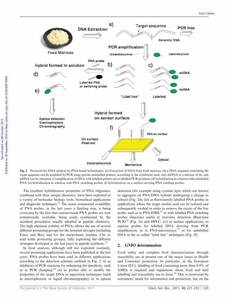
according to the detection schemes outlined in Fig. 2: (i) as
inhibitors of PCR reactions for enhancing the specificity, such
as in PCR clamping;14 (ii) as probes able to modify the
properties of the target DNA in separation techniques (such
as electrophoresis or liquid chromatography) or in optical
detection (for example using cyanine dyes which are known
to aggregate on PNA:DNA hybrids undergoing a change in
colour) (Fig. 2d); (iii) as fluorescently labelled PNA probes in
applications where the target nucleic acid can be isolated and
subsequently washed in order to remove the excess of the free
probe, such as in PNA-FISH,15 or with labelled PNA switching
probes (beacons) useful in real-time detection (Real-time
PCR)16 (Fig. 2e) and HPLC; (iv) in surface applications, as
capture probes for labelled DNA deriving from PCR
amplification, as in PNA-microarrays,17 or for unlabelled
DNA in the so called ‘‘label free’’ techniques (Fig. 2f).
2. GMO determination
Food safety and complete food characterization through
traceability are at present one of the major issues in Health
and Consumer protection. In particular, in the European
Union (EU), labelling of food containing more than 0.9% of
GMOs is required and regulations about food and feed
labelling and traceability are in force.18 This is motivated by
consumers’ needs for information and protection, but on the
Fig. 2 Protocols for DNA analysis by PNA-based technologies. (a) Extraction of DNA from food matrices; (b) a DNA sequence containing the
target sequence can be amplified by PCR using specific unlabelled primers; according to the conditions used, only dsDNA or a mixture of ds- and
ssDNA can be obtained; (c) amplification of DNA with labelled primers gives labelled PCR products; (d) hybridization in solution with unlabelled
PNA; (e) hybridization in solution with PNA switching probes; (f) hybridization on a surface carrying PNA catching probes.
Dow
nloa
ded
on 0
8 O
ctob
er 2
012
Publ
ishe
d on
30
Sept
embe
r 20
10 o
n ht
tp://
pubs
.rsc
.org
| do
i:10.
1039
/B90
7695
F
View Online

224 Chem. Soc. Rev., 2011, 40, 221–232 This journal is c The Royal Society of Chemistry 2011
other side it implies more onerous and cost-effective procedures
for the food industry. Technical implementation of tools and
procedures for product traceability can greatly help to
simplify analysis and reduce costs, thus making the labelling
procedures more ‘‘producers’ friendly’’.
A very important achievement of analytical systems, which
can be used for GMO traceability, is the possibility to recognize
living organisms through the identification of DNA, a technique
which has received in the past decade a tremendous momentum
from the scientific efforts linked to the research in genomics.
There has been an increasing interest in the research for the
application of genetic tools to the traceability of components
of interest in the food chain, and standardized procedures are
now available for the detection of specific DNA sequences, as
well as many laboratories can now provide validated methods
for the detection of a wide range of GMOs, based on
semiquantitative or quantitative polymerase chain reaction
(PCR) procedures.19 Several private laboratories are currently
running thousands of DNA analyses per year and this type of
market is constantly increasing, not only to comply with the
European regulations, but also to provide proper information
to consumers. Actually, although PCR methods have been
validated for several matrices, the ease of applicability of this
type of analyses can further be improved. For the quantitative
determination of GMOs the proper standard material is
required, which is not always available. The use of highly
specific probes for the confirmation of the identity of PCR
products is particularly important where the presence of
GMOs has immediate industrial or legal consequences (for
example, several years ago, hundreds of hectares of maize
contaminated with small percentages of GM varieties were
destroyed in Northern Italy).20 Thus, the use of highly specific
probes either in the post-PCR assessment or as components
of biosensors is highly desirable in these cases. On account of
their properties, PNAs have been used for the detection of
specific sequences in advanced diagnostic methods for the
detection of GMOs.
One very simple and immediate method of assessing
electrophoretic band identity was the use of the so called
‘‘PCR clamping’’ technique, which was originally proposed for
the detection of point mutations.14 By adding a PNA able to bind
next to the primer site on the target DNA, the specific inhibition
of the PCR product, as revealed by agarose gel analysis,
was obtained, allowing qualitative and semi-quantitative
determination of the GMO presence.21 This method is
particularly suitable when no other equipment except the simple
and cheap gel electrophoresis apparatus is available.
In a more complex approach, PNA probes linked to micro-
titre plates were used to specifically capture biotin-labelled
target DNA sequences, which were then used as template in
Fig. 3 IE HPLC profiles obtained using: (A) an unlabelled PNA probe and labeled DNA; (B) a PNA beacon and unlabelled DNA. (A) HPLC
profile of a Cy5-labelled PCR amplicon specific for RR soybean (79 bp) obtained from a soy burger labelled ‘‘GMO-free’’: (b) the PCR product
(dsDNA) crude; (c) dsDNA after purification, (d) after digestion with l-exonuclease (ssDNA); (e) after hybridization with a specific PNA probe
(ssDNA–PNA). Column: TSK gel DEAE–NPR (4.6 mm id � 3.5 cm). T = 35 1C. Eluent A: Tris 20 mM in H2O at pH 9; eluent B: NaCl 1 M in
eluent A at pH 9. Gradient: from 50% A to 30% A in 10 min. Flow rate: 0.5 mL min�1. Fluorescence detector: lex = 646 nm, lem = 664 nm.
(reproduced with permission from ref. 24). (B) HPLC profile of the same PCR product as in (A) unlabelled (from soy flour containing RR-soy).
(a) PCR product alone; (b) PNA beacon (1 mM) alone; (c) PNA beacon (1 mM) + a non-specific PCR product; (d) PNA beacon (1 mM) + the
specific PCR product. Attribution: (i) beacon and components of the PCR reaction; (iv) PNA beacon–DNA hybrid. T= 25 1C. Column: TSK-gel
DNA NPR (4.6 mm id � 7.5 cm); eluent A: Tris 0.02 M, pH = 9.0, eluent B: NaCl 1 M in eluent A. Linear gradient: from 100% A to 100% B in
20 min; flow rate: 0.5 mL min�1. lex = 497 nm, lem = 520 nm (reproduced with permission from ref. 26).
Dow
nloa
ded
on 0
8 O
ctob
er 2
012
Publ
ishe
d on
30
Sept
embe
r 20
10 o
n ht
tp://
pubs
.rsc
.org
| do
i:10.
1039
/B90
7695
F
View Online
This journal is c The Royal Society of Chemistry 2011 Chem. Soc. Rev., 2011, 40, 221–232 225
real time immuno-PCR reactions. High sensitivity (LOD in
the attomolar range) was reported for MON810 maize using
this method.22
Another common equipment present in many laboratories is
HPLC. Hybridization of oligonucleotides with PNA can
be detected using an anion exchange column, operating
under non-denaturing conditions.23 A protocol using PNA
hybridization and anion exchange HPLC was developed for
the assessment of the identity of PCR products derived from
genetically modified soybean and maize. PNAs of different
lengths aimed at evaluating the effect of the PNA structure on
signal intensity and specificity were used for gene sequences
corresponding to a region of the CaMV 35S-CTP construct of
Roundup Ready (RR) GM-soybean and to the CryIA
sequence of the Bt-176 GM-maize. The results showed that
it was possible to perform a clear identification of the PCR
product based on the presence of the chromatographic peak of
the PNA:DNA hybrid (Fig. 3A).24 In this study, the PCR
product was fluorescently labelled and, in order to allow access
of the PNA to its target, an excess of one of the two strands
had to be generated either by an enzymatic digestion or by
asymmetric PCR.
PNA labelled with fluorophores can be alternatively used to
trace DNA with no need of labelling the target DNA.
However, the background fluorescence of the free probe
usually prevents the specific detection and quantitation of
the PNA:DNA hybrid. In an impressive demonstration of
the PNA properties, a single-molecule detection of transgenic
DNA was performed in early studies by means of PNA probes
and double wavelength fluorescence analysis.25 In this study
two PNA probes complementary to two different sequences of
the transgene, labelled with different fluorophores, were used
in connection with an advanced apparatus composed of two
single-photon counting detectors. The simultaneous detection
of the PNA probes combined with the target DNA was
performed, thus allowing to discard signals due to the free
probes. Subsequent studies however were devoted to develop
methods which could be used with more common equipment
available in analytical laboratories.
PNA beacons and the related ‘‘light up probes’’ have also
been produced, in analogy with molecular beacon oligo-
nucleotides. They are modified with a fluorophore and a
quencher (or a quenching surface) at each end, held together
by hydrophobic interactions in aqueous solution; upon inter-
action with the target DNA a fluorescence ‘‘switch-on’’ occurs
(Fig. 3B). PNA beacons present, relatively to DNA beacons, the
advantages of a higher selectivity and a simpler design. One of
the major limitations in diagnostics is represented by the
eventual fluorescence background of the free (uncomplexed)
beacon, which, though lower than that of a fluorescently
labelled probe, can, however, interfere with the signal obtained
by the PNA:DNA duplex with the analyte sequence at low
concentrations, especially for DNA amplified from complex
matrices such as food. A way for overcoming this problem was
the combined use of PNA beacons and IE-HPLC for the
selective label-free detection of DNA, taking advantage of the
very specific signal generated by the duplex DNA:PNA
beacon, which allows to avoid the presence of unspecific
peaks. This approach was used to detect label-free DNA
amplified from Roundup Ready soy in a pilot experiment
(Fig. 3B).26 A PNA beacon containing a chiral monomer
modified at C5 with a lysine side chain was shown to perform
a better label-free DNA recognition in a model system.27
Microarray technology is a very powerful tool for the
simultaneous detection of several DNA sequences and
multi-samples analysis. The use of PNA probes by microarray
platforms with fluorescence read-out has been successfully
used in the detection of GMOs. Since the detection limit of
this technique is in the nanomolar range, multiple PCR
amplification, with Cy3 or Cy5 labelling of the target DNA,
is performed prior to hybridization. Procedures for obtaining
a single stranded DNA have to be used in this case, either by
enzymatic digestion of one strand or by asymmetric PCR
(i.e. using an excess of one primer).
A PCR protocol allowing for the simultaneous detection of
five transgenes and two endogenous controls in food and feed
matrices was developed28 and procedures for the deposition of
PNAs on microarrays were optimized and proved to
be suitable for GMO detection.29 The two methods were
combined in order to develop a PNA array device for the
screening of GMOs in food. PNA probes complementary to
the GMO sequences were opportunely designed, synthesized,
and deposited on commercial slides. The best performances
were obtained with 15-mer probes and by means of a
sufficiently long spacer allowing the PNA probes to be accessible
for hybridization with the target DNA. The device was tested
on a model system constituted by flour samples containing a
mixture of standards at known concentrations of transgenic
material, in particular Roundup Ready soybean and Bt11,
Bt176, Mon810, and GA21 maize. The DNA was amplified
using the specific multiplex PCR method and tested on the
PNA array. Every GMO present in the tested samples (Fig. 4)
was correctly identified by subsequent fluorescence measurements
by a microarray reader.30
An advanced technology for the detection of transgenic
DNA using fluorescence detection was described by Knoll
and coworkers using Surface Plasmon enhanced Fluorescent
Spectroscopy (SPFS), which enables us to measure selectively
the fluorescence of molecules captured on the sensor surface
by excitation through an evanescent field generated in the
SPR experiment.31 High sensitivity and excellent mismatch
recognition was demonstrated in these studies.32
Sandwich hybridization assays with gold nanoparticles and
Surface Plasmon Resonance Imaging (SPRI) readout were
used for improving detection sensitivity for oligonucleotide
hybridization down to low femtomolar concentrations. This
method uses a nanoparticle-enhanced SPRI detection scheme
based on PNA probes (Fig. 5).33 The method was successfully
used for the discrimination between oligonucleotides matching
the PNA probe sequence and oligonucleotides carrying a single-
base mismatch. The same method was subsequently used for the
direct detection of GMO in a ‘‘PCR-free’’ analysis, using
genomic DNA extracted from soy flour. Avoiding amplification
by PCR would be a great advantage, since in this way less
steps are required in the overall procedure with less occasions
for contaminations. This methodology gave striking
performances, since quantities as low as 0.1% of GMO
material in samples containing 10 pg mL�1 gave significant
Dow
nloa
ded
on 0
8 O
ctob
er 2
012
Publ
ishe
d on
30
Sept
embe
r 20
10 o
n ht
tp://
pubs
.rsc
.org
| do
i:10.
1039
/B90
7695
F
View Online

226 Chem. Soc. Rev., 2011, 40, 221–232 This journal is c The Royal Society of Chemistry 2011
signals in the presence of a large excess of non-target DNA.34
Targeting of large dsDNA was facilitated by sonication, which
broke the DNA into smaller fragments, and by a hybridization
protocol which used denaturation at high temperature, followed
by freezing of the sample in order to avoid reannealing of DNA.
The use of microfluidic channels allowed then to capture the
target sequence and wash out the non-complementary strand.
The high specificity was due to a combination of different events:
(a) capture of DNA by the highly specific PNA probes;
(b) efficient capture of the oligonucleotide-labelled gold nano-
particles which catalyzed the subsequent aggregation of other
nanoparticles, thus amplifying the optical signal.
The possibility to avoid PCR and to detect directly the
genomic DNA can be very useful in food as well as in
biomedical applications when the target DNA is present at
very low concentrations.
3. Hidden allergen determination
Food allergy is an expanding concern in western countries, as
severe reactions to foods have become more common in the
recent years.35 Recently, in order to protect the consumers, the
EU introduced a directive which lists 14 common allergenic
ingredients to be declared on the label when present in a given
food (Directive 2007/68/EC). Nevertheless, of particular
concern are the so called ‘‘hidden allergens’’, i.e. allergenic
ingredients accidentally present in a food, and thus not
declared on the label, which may trigger severe allergic
reactions if inadvertently consumed by susceptible subjects.
Although direct detection methods of allergenic proteins are
usually the first choice for screening purposes, indirect
methods, based on the detection of DNA sequences specific
for a given allergenic ingredient, are also becoming popular.36
Actually, direct methods can fail when applied to complex
food mixtures or to severely processed foods, in which proteins
may be heavily modified and, therefore, not detectable by
antibodies directed to recognize their native forms. In
contrast, DNA detection is usually more feasible in these
cases, DNA being more resistant to drastic thermal treatments.
However, the low amount of DNA present implies that a very
specific confirmatory analysis is usually mandatory. Peptide
Nucleic Acids, thanks to their very specific DNA binding
properties, their chemical and enzymatic stability and the
possibility to be used in connection with several detection
methods, are ideal candidates for the task.
The possibility to use PNAs as confirmatory probes
post-PCR analysis has been first demonstrated in combination
with HPLC.37 A PCR analysis was developed aimed at
amplifying a 156 bp region of the gene coding for Cor a 1,
the major hazelnut allergen. Simultaneously, a 15-mer PNA
probe complementary to an internal region of the amplicon
was designed and synthesized, and used in anion exchange
HPLC. When targeting a double stranded DNA produced in a
PCR reaction, the major problem hampering the detection by
PNA probes is represented by the necessity to invade the DNA
double helix. In fact, since DNA recognition by PNAs takes
place via standard Watson–Crick hydrogen bonds, the PNA
Fig. 4 Specificity assessment of the PNA array. Each slide was
hybridized with DNA and amplified twice by multiplex PCR,
previously extracted from flour containing: (1) GMO free soybean;
(2) GMO free maize; (3) 5% MON810 maize; (4) 5% RR soybean;
(5) 5% Bt11 maize; (6) 5% Bt176 maize; and (7) 5% GA21 maize. The
PNA probes were spotted, at a concentration of 30 mM, as follows: SL
(soybean lectin), MZ (maize zein), MON810 (MON810 maize), RR
(RR soybean), Bt11 (Bt11 maize), Bt176 (Bt176 maize), and GA21
(GA21 maize) (reproduced with permission from ref. 30).
Fig. 5 Description of the strategy used for the ultrasensitive
nanoparticle-enhanced SPRI detection of the target DNA sequence.
PNA 1: surface immobilized specific PNA capture probe; DNA-FM:
DNA full match to be detected; DNA 12-mer: specific DNA capture
probe linked to gold nanoparticles; AuNp: gold nanoparticles
(reproduced with permission from ref. 33).
Dow
nloa
ded
on 0
8 O
ctob
er 2
012
Publ
ishe
d on
30
Sept
embe
r 20
10 o
n ht
tp://
pubs
.rsc
.org
| do
i:10.
1039
/B90
7695
F
View Online
This journal is c The Royal Society of Chemistry 2011 Chem. Soc. Rev., 2011, 40, 221–232 227
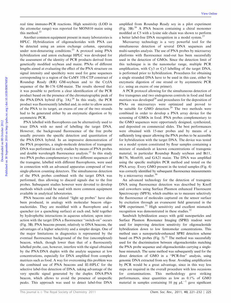
probe must be able to compete with the homologous DNA
strand for binding to the target region. In order to circumvent
this problem, the single strand PCR product was obtained, by
using a suitable PCR primer, labelled at the 50 terminus in the
strand homologous to PNA (the one not to be targeted by the
PNA probe) with a phosphate group. The post-PCR selective
enzymatic digestion of the strand functionalized with the
phosphate with Lambda exonuclease allowed PNA to bind
to the remaining single strand DNA. The second major
problem, i.e. the sensitivity of the detection system for the
PNA–DNA binding event, was solved by labelling, again with
the use of a suitable PCR primer, the remaining strand with a
fluorophore, such as the Cy5 dye. The definitive confirmatory
experiment was then obtained by a set of four injections in the
HPLC-FLD system, as represented in Fig. 6. First, the double
strand PCR product was injected and detected (Fig. 6a). Then,
the PCR product after enzymatic digestion was analysed: as it
can be seen, the single strand DNA showed a chromatographic
shift as compared to the double strand DNA (still present in
small amounts) (Fig. 6b). In the third experiment, the single
strand DNA was injected after hybridization with the
complementary PNA: the formation of a PNA–DNA duplex
could be visualized by a further shift, much more marked, of
the retention times (Fig. 6c). Finally, as a final confirmation,
the single strand DNA was injected after mixing with a PNA
non-complementary to any region of the single strand
amplified DNA: in this case the DNA did not change its
retention time as compared to the free DNA in the second
experiment, clearly showing that no hybridization had
taken place (Fig. 6d).37 It is to be remarked that, by using
fluorescence detection, the free PNA is not detectable with this
system, and its presence can be evidenced only by the effect
exerted on the chromatographic behavior of DNA. In this
way, excess PNA can be used in the test without any
interference with the analysis. By using this methodology,
the accidental presence of hazelnut DNA in food matrices
not containing hazelnut as ingredient was detected.37
The possibility of detecting hidden allergens by PNA-based
post-PCR confirmatory analysis was subsequently tackled
with a different technique, i.e. by using PNA microarrays,
demonstrating that multiple allergen detection is feasible by
this platform.38 In this case PNA array platforms were
designed by synthesizing two different probes complementary
to the gene regions coding for Cor a 1 (the major hazelnut
allergen) and Ara h 2 (the major peanut allergen). These PNA
probes were synthesized with two 2-(2-aminoethoxy)ethoxyacetyl
spacers at the amino terminus, in order to have an unhindered
free amino group to be used for linking to the array surfaces.
Coupling conditions to the surface were carefully optimized
according to the characteristics of PNA chemistry. In particular,
given the low solubility of PNAs in aqueous solvents, several
washing cycles with different solvents were introduced in order
to avoid PNA dragging during deposition. After slide deposition,
the sensitivity of the PNA microarrays was tested with
synthetic oligonucleotides, allowing to define a limit of detection
down to 1 nMDNA (in the reported experimental conditions).
Then, a duplex PCR (i.e. a PCR simultaneously amplifying
both the genomic region of hazelnuts and peanuts) was
developed. The arrays were first tested with amplicons from
pure hazelnut and peanut, demonstrating the specificity of the
system (Fig. 7). Finally, food products commercially available,
purchased on the market, were screened. PCR amplicons
obtained from the samples were tested with the PNA micro-
arrays, confirming in some samples the presence of hazelnuts
and/or peanuts, in some cases even undeclared either as
ingredients or possible contaminants (Fig. 8).38 It must be
said that these experiments were performed before the
implementation of the actual EU regulation. Thus, PNA
microarrays were shown to be suitable platforms for the fast
and reliable post-PCR confirmation of the DNA identity in
food analysis.
A quite unconventional method using PNA probes for
post-PCR confirmation of DNA identity based on circular
Fig. 6 AE-HPLC identity assessment of the PCR amplified DNA of
hazelnut (Cor a 1) by a PNA probe: (a) double stranded DNA from
PCR; (b) single strand DNA after enzymatic digestion; (c) hybridization
of the single strand DNA after enzymatic digestion with the specific
PNA probe complementary to the hazelnut DNA sequence;
(d) hybridization of the single strand DNA after enzymatic digestion
with a PNA probe non-complementary to any hazelnut DNA
sequence (reproduced with permission from ref. 37).
Dow
nloa
ded
on 0
8 O
ctob
er 2
012
Publ
ishe
d on
30
Sept
embe
r 20
10 o
n ht
tp://
pubs
.rsc
.org
| do
i:10.
1039
/B90
7695
F
View Online

228 Chem. Soc. Rev., 2011, 40, 221–232 This journal is c The Royal Society of Chemistry 2011
dichroism (CD) was also proposed.39 The method relies on the
properties of a particular aza dye, the diethylthiadicarbocyanine
dye [DiSC2(5)], which has a strong tendency to aggregate on
PNA–DNA duplexes. This aggregation gives rise to a
characteristic band in the visible spectrum at 540 nm, with
the appearance of a typical purple color. This feature, which
could potentially lead to the development of colorimetric tests
for directly visualizing the specific PNA–DNA interaction, is
nevertheless made less specific from the tendency of the
DiSC2(5) dye to aggregate even on free PNA molecules,
introducing a strong bias to the colorimetric analysis which
can lead to many false positives, especially when excess PNA is
to be used, as it is frequent in the case of post-PCR
confirmative analyses.40 However, when observed by spectro-
polarimetry, the dye aggregate on the PNA–DNA duplex gives
rise to a very strong exciton coupling effect at the same
wavelength, which could be easily visualized by CD, since
the helical chirality of the duplex is transferred to the dye
aggregate. The dye being achiral, the free dye in solution does
not give rise to any CD signal and, what is most interesting
for the robustness of the method, even the dye-free PNA
aggregate is ‘‘invisible’’ when observed by the CD technique,
standard PNAs also being non-chiral molecules. The method
was first optimized with oligonucleotides, then applied to the
identification of DNA extracted and amplified by PCR from
peanuts, peanut-containing and peanut-free foods, allowing
for a very sensitive detection. Typical results are shown in
Fig. 9. The PNA recognizes and binds the DNA amplified
from pure peanuts, inducing the dye aggregation on the
PNA–DNA duplex which gives rise to an intense exciton
coupling effect. Also the peanut DNA present in cereal
snacks is easily detectable, albeit with a lower signal,
by this method. A chocolate wafer without peanut gave no
CD signal: in the absence of any PNA–DNA duplex, the dye
aggregation does not take place nor is visible on the free
PNA.39
4. Microbial contaminant detection
The identification of microbial contaminants is a primary issue
for food safety. As a typical example, among the 2008 data
concerning the total alerts in food and feed, as included
in the Rapid Alert System, 24% were about potentially
pathogenic microorganisms, the highest relative category
of risk.41 The ideal analysis for assessing food microbial
contaminants should be fast, sensitive and selective, in order
to avoid false positive and false negative results and to
yield data on the eventual contamination in the shortest
possible time.
In order to respond to these issues, several methods for
rapid microbiology based on PNA probes appeared in the last
years and several examples of detection of microbial agents
having food relevance were also reported.42 One of the
mostly used methods is PNA-based Fluorescence In situ
Hybridization (PNA-FISH). In this assay fluorophore-labelled
PNAs targeted to rRNA are used for the direct detection of
microorganisms in tissues, biological fluids, culture media,
filters, on slides or in solution.
The advantages of PNA-FISH methods, rather than the
more common DNA-FISH, were outlined in a work aimed at
Fig. 7 PNA microarrays tested with PCR products deriving from
amplification of DNA extracted from pure hazelnut (a) and peanut
(b). H: PNA complementary to hazelnut DNA; P: PNA complementary
to peanut DNA; B: blank; CP: control probe (reproduced with
permission from ref. 38).
Fig. 8 PNA array analysis for the detection of hazelnut and peanut in
commercial foodstuffs. (a): Breakfast cereals, possible traces of tree
nuts and peanuts declared; (b): Muesli snack with chocolate, peanut
declared, hazelnut NOT declared. H: PNA complementary to hazelnut
DNA; P: PNA complementary to peanut DNA; B: blank; CP: control
probe (reproduced with permission from ref. 38).
Fig. 9 A CD-based experiment with a PNA probe specific for peanut
DNA together with Disc2(5) dye for the post-PCR confirmation of the
DNA identity (reproduced with permission from ref. 39).
Dow
nloa
ded
on 0
8 O
ctob
er 2
012
Publ
ishe
d on
30
Sept
embe
r 20
10 o
n ht
tp://
pubs
.rsc
.org
| do
i:10.
1039
/B90
7695
F
View Online

This journal is c The Royal Society of Chemistry 2011 Chem. Soc. Rev., 2011, 40, 221–232 229
developing PNA probes targeted at the detection of whole cells
of Listeria spp. (Fig. 10).43 PNA probes turned out to be more
able than DNA probes to penetrate recalcitrant biological
structures such as the membranes of gram-positive cells.
Moreover, due to their high affinity for complementary
RNA sequences, combined to the independence from ionic
strength of PNA–RNA stability, PNAs were able to bind
regions on the ribosomes which were inaccessible for
DNA probes, exploiting hybridization conditions which can
denature RNA. Actually, when using DNA probes, any attempt
of destabilizing RNA secondary structures on the target
unavoidably yields also a loss of the probe affinity for the
target itself; in contrast, with PNA probes, as in the reported
example, recognition took place under low salt, high temperature
and high pH conditions, in which RNA is likely to be in a
non-native form.
In a similar example, PNA probes developed to detect
Campylobacter jejuni, Campylobacter coli and Campylobacter
lari were able to detect C. coli spiked in drinking water
samples, after membrane filtration to concentrate the
microorganisms.44 A PNA-FISH procedure targeted to
Acinetobacter spp. and Pseudomonas aeruginosa was recently
applied for detection, after fixation on slides, showing
100% specificity and 100% selectivity towards the former
and 100% specificity and 95% selectivity towards the
latter.45 A commercial version of PNA-FISH was developed
in order to detect both gram-negative and gram-
positive bacteria. In a particular striking example four
differently labelled PNA probes were used for the
simultaneous detection of Escherichia coli, Salmonella
enterica, P. aeruginosa and Staphylococcus aureus.42 In a
similar variant, named PNA Chemiluminescent In situ
Hybridization (PNA-CISH), PNAs labelled with soybean
peroxidase are hybridized with the target and then treated
with a chemiluminescent substrate, generating light captured
by a camera system. Combination of this system with previous
membrane filtration, in order to concentrate the micro-
organisms to be detected, allowed detection of P. aeruginosa
in bottled water, E. coli in tap water and Dekkera bruxellensis
in wine.42
5. Ingredient authentication
The authenticity of food products is an important issue which
is recently gaining increasing attention: correct labelling and
traceability of the ingredients through all stages of production,
processing, and distribution has become of primary importance
in many western countries. Among many labelling declarations
which claim ‘‘quality’’ characteristics of a given food, and
often difficult to be proved objectively, most concern varieties
of vegetables or particular breeds of animals used as ingredients.
Typical examples include, just to name a few, cheeses made
only from sheep milk, wines produced from defined grape
varieties, monocultivar olive oils, minced meat or fish declared
from a given breed, and so on. DNA markers are well suited
for traceability purposes, due to the remarkable persistence of
DNA, even in the hostile environments found during many
processing steps used for food production. The use of DNA
markers as diagnostic tools for ingredient authenticity in food
matrices has been investigated in an increasing number of
projects worldwide.46 However, some processed foods contain
highly degraded DNA which may affect the subsequent PCR
used for the amplification of the diagnostic DNA sequence. In
these cases, a very sensitive and specific method for the
detection of small amounts of DNA has become therefore
highly desirable. Even if the literature in the field is still quite
scarce, it is obvious that all the PNA-based techniques so
far described might be very useful in order to assess food
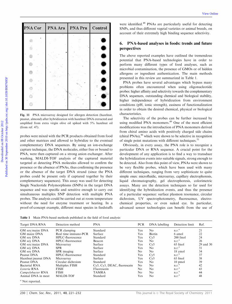
authenticity and avoid frauds. As an example, the authors
assayed the presence of hazelnut oil in extra virgin olive oil by
using the PNA microarray system already reported for the
detection of hazelnut as a hidden allergen.38 In a preliminary
test model, down to 5% hazelnut oil in extra virgin olive oil
could be detected by this method (Fig. 11).47 Hazelnut olive oil
may be used to adulterate extra virgin olive oil on account of
the similarity of the lipophilic components, which prevent the
discrimination. In this case, the adulteration is not only a
fraud, but can be dangerous to people allergic to hazelnuts.
Recently, in a mass spectrometry-based approach, modified
pyrrolidinyl-PNA probes were used for targeting DNA after
extraction and amplification from food samples. The PNA
Fig. 10 Typical PNA hybridization results in PNA-FISH applications. (A) Cells of Bacillus cereus ATCC 11778 hybridized with the universal
bacterial PNA probe. (B) Cells of Listeria monocytogenes FSL-C1-122 hybridized with the Listeria-specific PNA probe (reproduced with
permission from ref. 43).
Dow
nloa
ded
on 0
8 O
ctob
er 2
012
Publ
ishe
d on
30
Sept
embe
r 20
10 o
n ht
tp://
pubs
.rsc
.org
| do
i:10.
1039
/B90
7695
F
View Online
230 Chem. Soc. Rev., 2011, 40, 221–232 This journal is c The Royal Society of Chemistry 2011
probes were mixed with the PCR products obtained from food
and other matrices and allowed to hybridize to the eventual
complementary DNA sequences. By using an ion-exchange
capture technique, the DNAmolecules, either free or bound to
PNA, were then captured on a strong anion exchanger. After
washing, MALDI-TOF analysis of the captured material
targeted at detecting PNA molecules allowed to confirm the
presence or the absence of PNAs, thus confirming the presence
or the absence of the target DNA strand (since the PNA
probes could be present only if captured together by their
complementary sequences). This assay was used for detecting
Single Nucleotide Polymorphisms (SNPs) in the target DNA
sequence and was specific and sensitive enough to carry out
simultaneous multiplex SNP detection with multiple PNA
probes. The analysis could be carried out at room temperature
without the need for enzyme treatment or heating. In a
proof-of-concept example, different meat species in feedstuffs
were identified.48 PNAs are particularly useful for detecting
SNPs, and thus different vegetal varieties or animal breeds, on
account of their extremely high binding sequence selectivity.
6. PNA-based analyses in foods: trends and future
perspectives
The above reported examples have outlined the tremendous
potential that PNA-based technologies have in order to
perform many different types of food analyses, such as
microbial contamination, the presence of GMOs or of hidden
allergens or ingredient authentication. The main methods
presented in this review are summarized in Table 1.
PNA probes have several advantages which bypass many
problems often encountered when using oligonucleotide
probes: higher affinity and selectivity towards the complementary
DNA sequences, outstanding chemical and biological stability,
higher independence of hybridization from environment
conditions (pH, ionic strength), easiness of functionalization
in order to obtain the desired chemical, physical or biological
characteristics.
The selectivity of the probes can be further increased by
using modified PNA monomers.49 One of the most efficient
modifications was the introduction of PNA monomers derived
from chiral amino acids with positively charged side chains
(chiral PNAs),50 which were shown to be selective in recognition
of single point mutations with different techniques.51
Obviously, in every assay, the PNA role is to recognize a
particular DNA or RNA sequence. A crucial point for the
development of any application is to find a way to transduce
the hybridization events into suitable signals, strong enough to
be detected. Also from this point of view, PNAs were shown to
be very flexible probes, which have been used with many
different techniques, ranging from very sophisticate to quite
simple ones: microfluidic, microarray, capillary electrophoresis,
liquid chromatography, gel electrophoresis, colorimetric
assays. Many are the detection techniques so far used for
identifying the hybridization events, and thus the presence
of a particular sequence: surface plasmon resonance, circular
dichroism, UV spectrophotometry, fluorescence, electro-
chemical properties, or even naked eye. In particular,
advanced sensor technologies can benefit from the use of
Fig. 11 PNA microarray designed for allergen detection (hazelnut,
peanut, almond) after hybridization with hazelnut DNA extracted and
amplified from extra virgin olive oil spiked with 5% hazelnut oil
(from ref. 47).
Table 1 Main PNA-based methods published in the field of food analysis
Target DNA/RNA Detection method PNA PCR DNA labelling Detection limit Ref.
GM soy/maize DNA PCR clamping Standard Yes No n.r.a 21GM maize DNA Real time immuno-PCR Surface Yes Biotin 6 amol 22GM soy DNA HPLC-fluorescence Standard Yes Cy5 200 fmol 24GM soy DNA HPLC-fluorescence Beacon Yes No n.r.a 26GM soy/maize DNA Microarray Surface Yes Cy5 65 fmol 29 and 30GM soy DNA SPR Surface Yes No n.r.a 31GM soy DNA SPR imaging Surface No No 18 ymol 34Peanut DNA HPLC-fluorescence Standard Yes Cy5 n.r.a 37Hazelnut/peanut DNA Microarray Surface Yes Cy5 65 fmol 38Peanut DNA Circular dichroism Standard Yes No 10 pmol 39Bacterial RNA Multiplex FISH Cy5, Cy3, DEAC, fluorescein No No n.r.a 42Listeria RNA FISH Fluorescein No No n.r.a 43Campylobacter RNA FISH TAMRA No No n.r.a 44Animal DNA in meat MALDI-TOF Standard Yes No n.r.a 48
a Not reported.
Dow
nloa
ded
on 0
8 O
ctob
er 2
012
Publ
ishe
d on
30
Sept
embe
r 20
10 o
n ht
tp://
pubs
.rsc
.org
| do
i:10.
1039
/B90
7695
F
View Online
This journal is c The Royal Society of Chemistry 2011 Chem. Soc. Rev., 2011, 40, 221–232 231
PNA probes in combination with different detection methods.
The capture of a negatively charged DNA sequence by a
neutral PNA probe deposited on the surface of an electrode
(PNA can be adsorbed onto carbon electrodes) induces a
dramatic change in the potential, which is transformed into
an electrochemical signal;52,53 miniaturized sensing systems
can be achieved using impedance measurements on surfaces
modified with PNAs;54,55 mechanical detection can be
achieved by a change in the frequency of quartz crystal
microbalance (QCM),56 but future applications can also be
envisaged in the use of micro- and nanocantilevers.57 All the
above mentioned techniques have greatly benefited by the use
of PNA probes in terms of sensitivity and selectivity.
On account of the high flexibility, PNAs open wide
possibilities in the field of DNA detection in foods. Thus, it
is to be expected that they will be more and more useful tools
in food analysis and food authentication. In this field what is
often needed can be summarized in two words: fast and
reliable. In this context a likely evolution of the current state
of the art is in the direction of making the plethora of already
existing PNA-based assays simpler and more robust.
In particular, a likely evolution, not only in the PNA field,
but concerning DNA analysis in general, is the possibility to
detect DNA without any need of labelling it (label-free)
or even, in the most advanced applications, without pre-
amplification by PCR (PCR-free), a technique often prone
to many bias and errors. In this case it is not incorrect to state
that, as far as PNAs are concerned, the future is already here:
several applications presented in this review already are an
example, although preliminary, of label-free and PCR-free
DNA detection (Table 1). Techniques which are promising
for the label-free detection of DNA are those based
on plasmonics, such as surface plasmon resonance (SPR)
techniques58,59 and photonics60 (i.e. read out of the properties
of an electromagnetic field confined in a microstructured
medium as a result of the events occurring at the interfaces),
using optical devices such as waveguides or photonic crystal
fibers,61 but also clever examples of PCR-free colorimetric
detection of bacterial DNA with enzymatic assays using
suitably derivatized PNAs have already been preliminarily
presented in the literature.62
What is now needed is the implementation of these first
steps to routinary DNA detection techniques, since PNAs
have already been proven to be well suited to the task.
Acknowledgements
Italian Ministry of Education, University and Research
(MIUR) is gratefully acknowledged for fundings through the
Projects of Relevant National Interest 2007 scheme (PRIN
2007, contract number 2007F9TWKE).
References
1 M. Hernandez, D. Rodriguez-Lazaro and A. Ferrando, Curr.Anal. Chem., 2005, 1, 203–221.
2 A. Lauri and P. O. Mariani, Genes Nutr., 2009, 4, 1–12.3 R. E. Poms, E. Anklam and M. Kuhn, J. AOAC Int., 2004, 87,1391–1397.
4 G. Di Bernardo, S. Del Gaudio, U. Galderisi, A. Cascino andM. Cipollaro, Biotechnol. Prog., 2007, 23, 297–301.
5 PCR Methods in Foods, ed. J. Maurer, Springer, Berlin, Germany,2006.
6 F. Weighardt, Nat. Biotechnol., 2007, 25, 1213–1214.7 P. E. Nielsen, M. Egholm, R. H. Berg and O. Buchardt, Science,1991, 254, 1497–1500.
8 M. Egholm, O. Buchardt, L. Christensen, C. Behrens, S. M. Freier,D. A. Driver, R. H. Berg, S. K. Kim, B. Norden and P. E. Nielsen,Nature, 1993, 365, 566–568.
9 (a) B. Ren, J. M. Zhou and M. Komiyama, Nucleic Acids Res.,2004, 32, 42e; (b) B. S. Gaylord, M. R. Massie, S. C. Feinstein andG. C. Bazan, Proc. Natl. Acad. Sci. U. S. A., 2005, 102, 34–39.
10 L. S. Yilmaz, H. E. Okten and D. R. Noguera, Appl. Environ.Microbiol., 2006, 72, 733–744.
11 V. A. Demidov, V. N. Potaman, M. D. Frank-Kamenetskii,M. Egholm, O. Buchardt, S. H. Sonnichsen and P. E. Nielsen,Biochem. Pharmacol., 1994, 48, 1310–1313.
12 N. Sahu, G. Shilakari, A. Nayak and D. H. Kohli, Curr. Chem.Biol., 2008, 2, 110–121.
13 P. E. Nielsen and M. Egholm, Peptide Nucleic Acids: Protocols andApplications, Horizon Press, Wymondham, Norfolk, UK, 2nd edn,2004.
14 (a) H. Orum, P. E. Nielsen, M. Egholm, R. H. Berg, O. Buchardtand C. Stanley, Nucleic Acids Res., 1993, 21, 5332–5336;(b) H. Orum, Curr. Issues Mol. Biol., 2000, 2, 27–30.
15 L. Cerqueira, N. F. Azevedo, C. Almeida, T. Jardim, C. W. Keeviland M. J. Vieira, Int. J. Mol. Sci., 2008, 9, 1944–1960.
16 C. Tapparel, S. Cordey, S. Van Belle, L. Turin, W. M. Lee,N. Regamey, P. Meylan, K. Muhlemann, F. Gobbini andL. Kaiser, J. Clin. Microbiol., 2009, 47, 1742–1749.
17 O. Brandt and J. D. Hoheisel, Trends Biotechnol., 2004, 22,617–622.
18 (a) Regulation (EC) No 1829/2003 The European Parliament andthe Council of the European Union on genetically modified foodand feed. Off. J. Eur. Union 2003, L268, 1–23; (b) The EuropeanParliament and the Council of the European Union Regulation(EC) No 1830/2003 concerning the traceability and labelling ofgenetically modified organisms and the traceability of food andfeed products produced from genetically modified organisms andamending Directive 2001/18/EC. Off. J. Eur. Union 2003, L268,24–28; (c) Commission Regulation (EC) No 65/2004 establishing asystem for the development and assignment of unique identifiersfor genetically modified organisms. Off. J. Eur. Union 2004, L10,5–10.
19 Validation of methods for GMO detection is extensivelycarried out in the Ispra Joint Research Center of the EuropeanCommission. See http://gmo-crl.jrc.ec.europa.eu/.
20 Regione Piemonte, Ordinanza Presidente Giunta Regionale n. 63,11/7/2003; Regione Lombardia DGR13848/2003.
21 C. Peano, F. Lesignoli, M. Gulli, R. Corradini, M. C. Samson,R. Marchelli and N. Marmiroli, Anal. Biochem., 2005, 344,174–182.
22 L. Roth, J. Zagon, A. Ehlers, L. W. Kroh and H. Broll, Anal.Bioanal. Chem., 2009, 394, 529–537.
23 F. Lesignoli, A. Germini, R. Corradini, S. Sforza, G. Galaverna,A. Dossena and R. Marchelli, J. Chromatogr., A, 2001, 922,177–185.
24 S. Rossi, F. Lesignoli, A. Germini, A. Faccini, S. Sforza,R. Corradini and R. Marchelli, J. Agric. Food Chem., 2007, 55,2509–2516.
25 A. Castro and J. G. K. Williams, Anal. Chem., 1997, 69,3915–3920.
26 F. Totsingan, S. Rossi, R. Corradini, T. Tedeschi, S. Sforza,A. Juris and E. Scaravelli, Org. Biomol. Chem., 2008, 6, 1232–1237.
27 F. Totsingan, T. Tedeschi, S. Sforza, R. Corradini andR. Marchelli, Chirality, 2009, 21, 245–253.
28 A. Germini, A. Zanetti, C. Salati, S. Rossi, C. Forre, S. Schmid,C. Fogher and R. Marchelli, J. Agric. Food Chem., 2004, 52,3275–3280.
29 A. Germini, A. Mezzelani, F. Lesignoli, R. Corradini,R. Marchelli, R. Bordoni, C. Consolandi and G. De Bellis,J. Agric. Food Chem., 2004, 52, 4535–4540.
30 A. Germini, S. Rossi, A. Zanetti, R. Corradini, C. Fogher andR. Marchelli, J. Agric. Food Chem., 2005, 53, 3958–3962.
Dow
nloa
ded
on 0
8 O
ctob
er 2
012
Publ
ishe
d on
30
Sept
embe
r 20
10 o
n ht
tp://
pubs
.rsc
.org
| do
i:10.
1039
/B90
7695
F
View Online

232 Chem. Soc. Rev., 2011, 40, 221–232 This journal is c The Royal Society of Chemistry 2011
31 H. Park, A. Germini, S. Sforza, R. Corradini, R. Marchelli andW. Knoll, Biointerphases, 2007, 2, 80–88.
32 H. Park, A. Germini, S. Sforza, R. Corradini, R. Marchelli andW. Knoll, Biointerphases, 2006, 1, 113–122..
33 R. D’Agata, R. Corradini, G. Grasso, R. Marchelli and G. Spoto,ChemBioChem, 2008, 9, 2067–2070.
34 R. D’Agata, R. Corradini, C. Ferretti, L. Zanoli, M. Gatti,R. Marchelli and G. Spoto, Biosens. Bioelectron., 2010, 25,2095–2100.
35 S. Cochrane, K. Beyer, M. Clausen, M. Wjst, R. Hiller,C. Nicoletti, Z. Szepfalusi, H. Savelkoul, H. Breiteneder,Y. Manios, R. Crittenden and P. Burney, Allergy (Oxford,zU. K.), 2009, 64, 1246–1265.
36 A. van Hengel, Anal. Bioanal. Chem., 2007, 389, 111–118.37 A. Germini, E. Scaravelli, F. Lesignoli, S. Sforza, R. Corradini and
R. Marchelli, Eur. Food Res. Technol., 2005, 220, 619–624.38 S. Rossi, E. Scaravelli, A. Germini, R. Corradini, C. Fogher and
R. Marchelli, Eur. Food Res. Technol., 2006, 223, 1–6.39 S. Sforza, E. Scaravelli, R. Corradini and R. Marchelli, Chirality,
2005, 17, 515–521.40 T. Tedeschi, S. Sforza, S. Ye, R. Corradini, A. Dossena,
M. Komiyama and R. Marchelli, J. Biochem. Biophys. Methods,2007, 70, 735–741.
41 The Rapid Alert System for Food and Feed (RASFF), AnnualReport 2008, European Commission.
42 (a) H. Perry-O’Keefe and J. J. Hyldig-Nielsen, in Environmental:Molecular Microbiology Protocols and Applications, ed.P. Rochelle, Horizon Scientific Press, Wymondham, UK, 2001;(b) H. Stender, M. Fiandaca, J. J. Hyldig-Nielsen and J. Coull,J. Microbiol. Methods, 2002, 48, 1–17.
43 B. F. Brehm-Stecher, J. J. Hyldig-Nielsen and E. A. Johnson, Appl.Environ. Microbiol., 2005, 71, 5451–5457.
44 M. J. Lehtola, C. J. Loades and C. W. Keevil, J. Microbiol.Methods, 2005, 62, 211–219.
45 A. Y. Peleg, Y. Tilahun, M. J. Fiandaca, E. M. C. D’Agata,L. Venkataraman, R. C. Moellering Jr and G. M. Eliopoulos,J. Clin. Microbiol., 2009, 47, 830–832.
46 S. Doveri and D. Lee, J. Agric. Food Chem., 2007, 55, 4640–4644.
47 S. Rossi, PhD thesis, University of Parma, 2007.48 B. Boontha, J. Nakkuntod, N. Hirankarn, P. Chaumpluk and
T. Vilaivan, Anal. Chem., 2008, 80, 8178–8186.49 K. N. Ganesh and P. E. Nielsen, Curr. Org. Chem., 2000, 4,
931–943.50 (a) S. Sforza, R. Corradini, S. Ghirardi, A. Dossena and
R. Marchelli, Eur. J. Org. Chem., 2000, 2905–2913;(b) V. Menchise, G. De Simone, T. Tedeschi, R. Corradini,S. Sforza, R. Marchelli, D. Capasso, M. Saviano andC. Pedone, Proc. Natl. Acad. Sci. U. S. A., 2003, 100,12021–12026.
51 (a) R. Corradini, G. Feriotto, S. Sforza, R. Marchelli andR. Gambari, J. Mol. Recognit., 2004, 17, 76–84; (b) M. Chiari,G. Galaverna, S. Sforza, M. Cretich, R. Corradini andR. Marchelli, Electrophoresis, 2005, 26, 4310–4316.
52 J. Wang, Biosens. Bioelectron., 1998, 13, 757–762.53 P. Kara, K. Kerman, D. Ozkan, B. Meric, A. Erdem, P. E. Nielsen
and M. Ozsoz, Bioelectrochemistry, 2002, 58, 119–126.54 A. Macanovic, Nucleic Acids Res., 2004, 32, 20e.55 J. Liu, S. Tian, P. E. Nielsen and W. Knoll, Chem. Commun., 2005,
2969–2971.56 J. Wang, P. E. Nielsen, M. Jiang, X. Cai, J. R. Fernandes,
D. H. Grant, M. Ozsoz, A. Beglieter and M. Mowat, Anal. Chem.,1997, 69, 5200–5202.
57 K. S. Hwang, S. M. Lee, S. K. Kim, J. H. Lee and T. S. Kim, Annu.Rev. Anal. Chem., 2009, 2, 77–98.
58 C. Ananthanawat, T. Vilaivan, V. P. Hoven and X. Su, Biosens.Bioelectron., 2010, 25, 1064–1069.
59 J. Homola, Chem. Rev., 2008, 108, 462–493.60 X. Fan, I. M. White, S. I. Shopova, H. Zhu, J. D. Suter and
Y. Sun, Anal. Chim. Acta, 2008, 620, 8–26.61 (a) F. Poli, A. Cucinotta and S. Selleri, Photonic crystal fibers, in
Material Science, Springer-Verlag, Dordrecht, The Netherlands,vol. 102, 2007; (b) A. Coscelli, M. Sozzi, F. Poli, D. E. Passaro,A. Cucinotta, S. Selleri, R. Corradini and R. Marchelli, IEEE J.Sel. Top. Quantum Electron., 2010, 16, 967–972.
62 N. Zhang and D. H. Appella, J. Am. Chem. Soc., 2007, 129,8424–8425.
Dow
nloa
ded
on 0
8 O
ctob
er 2
012
Publ
ishe
d on
30
Sept
embe
r 20
10 o
n ht
tp://
pubs
.rsc
.org
| do
i:10.
1039
/B90
7695
F
View Online


![Splicing isoform-specific functional genomic in cancer cells · 2018. 12. 14. · morpholinos, peptide nucleic acid (PNA), locked nucleic acid (LNA)] confer resistance to various](https://img.pdfslide.us/doc/110x75/614a7e2a12c9616cbc697388/splicing-isoform-specific-functional-genomic-in-cancer-cells-2018-12-14-morpholinos.jpg)