Embed Size (px)
Citation preview
fMRI of Simultaneous Interpretation Reveals the Neural Basis of Extreme Language Control
Alexis Hervais-Adelman1, Barbara Moser-Mercer2, Christoph M. Michel3 and Narly Golestani1
1Brain and Language Lab, Department of Clinical Neuroscience, 2Faculty of Translation and Interpreting and 3Functional BrainMapping Lab, Department of Fundamental Neuroscience, University of Geneva, Geneva, Switzerland
Address correspondence to Dr Alexis Hervais-Adelman, Brain and Language Lab, Campus Biotech, Neuroscience Department, 9 Chemin desMines, Case Postale 60, Geneva 20, Switzerland. Email: [email protected]
We used functional magnetic resonance imaging (fMRI) to examinethe neural basis of extreme multilingual language control in a groupof 50 multilingual participants. Comparing brain responses arisingduring simultaneous interpretation (SI) with those arising during sim-ultaneous repetition revealed activation of regions known to beinvolved in speech perception and production, alongside a networkincorporating the caudate nucleus that is known to be implicated indomain-general cognitive control. The similarity between the net-works underlying bilingual language control and general executivecontrol supports the notion that the frequently reported bilingualadvantage on executive tasks stems from the day-to-day demands oflanguage control in the multilingual brain. We examined neural corre-lates of the management of simultaneity by correlating brain activityduring interpretation with the duration of simultaneous speaking andhearing. This analysis showed significant modulation of the putamenby the duration of simultaneity. Our findings suggest that, during SI,the caudate nucleus is implicated in the overarching selection andcontrol of the lexico-semantic system, while the putamen is impli-cated in ongoing control of language output. These findings providethe first clear dissociation of specific dorsal striatum structures inpolyglot language control, roles that are consistent with previouslydescribed involvement of these regions in nonlinguistic executivecontrol.
Keywords: caudate, cognitive control, language control, multilingualism,putamen
Introduction
Bilingualism confers a variety of cognitive advantages (Abuta-lebi et al. 2009; Diamond 2010), including improved non-linguistic executive skills (Bialystok et al. 2012), and delayingthe appearance of symptoms of Alzheimer’s disease (Schwei-zer et al. 2012). It has been suggested that the observed bene-fits of bilingualism stem from enhanced inhibitory control,required for continual selection between multiple languages inbilinguals (Green 1998). More recently, it has been proposedthat the bilingual advantage arises from increased use of amore general “conflict monitoring” system in bilinguals com-pared with monolinguals (Costa et al. 2009; Hilchey and Klein2011; Abutalebi et al. 2012b). Consistent with these sugges-tions, brain-imaging evidence indicates that bilingual languagecontrol depends upon a cortico-subcortical network incorpor-ating the dorsal striatum, the anterior cingulate cortex (ACC)and the supplementary motor area (SMA) (Abutalebi andGreen 2008; Kroll et al. 2008; Hervais-Adelman et al. 2011;Abutalebi et al. 2012a). These brain regions are known to par-ticipate in nonlinguistic executive-control processes such asresponse selection (Grahn et al. 2008), response inhibition
(Aron 2008), and conflict monitoring (Botvinick et al. 2004;Abutalebi et al. 2012b).
Electroencephalographic (EEG) research on languagecontrol has principally focused on the time course of event-related potentials, and has provided some results that arecomplementary to those of the functional magnetic resonanceimaging (fMRI) and positron emission tomography (PET)literature (Hervais-Adelman et al. 2011). The EEG literature issomewhat heterogeneous; it includes studies of perceptionand production with focuses on several different evoked com-ponents related to both language and executive function. Thisliterature has been thoroughly reviewed and synthesized byMoreno et al. (2008). Among the domain-general componentsstudied is the N200, which has been observed in several bilin-gual tasks (Rodriguez-Fornells et al. 2006); it is related to re-sponse suppression and has been localized to the ACC(Nieuwenhuis et al. 2003; Huster et al. 2010). Moreno et al.(2008) conclude that the event-related potential (ERP) evi-dence indicates that language switching requires active inhib-ition, and that the EEG signatures of language switching bearsome similarity to those underlying withholding of responsesin nonlinguistic paradigms.
Other ERP studies have allowed researchers to draw conclu-sions about the time course of language selection duringspeech production (for reviews see Costa 2005; Kroll et al.2008). The weight of the evidence points toward simultaneousactivation of a bilingual’s two languages (e.g., Hoshino andThierry 2011), in support of bilingual language models such asthe bilingual interactive activation +model (Dijkstra and vanHeuven 2002) that posit an integrated bilingual lexicon whichis accessed in a language nonselective manner (van Heuvenand Dijkstra 2010).
While great strides have been made in our understanding ofpolyglot language control, much of the existing evidence re-garding the neural basis of bilingual language control comesfrom the study of tasks requiring only punctate applications oflanguage control, such as language switching (Khateb et al.2007; Wang et al. 2009; Garbin et al. 2011) and translation(Klein et al. 1995; Price et al. 1999; Quaresima et al. 2002; Leh-tonen et al. 2005). Distinct neural bases for general, executivetask set maintenance, and for moment-to-moment cognitivecontrol have previously been established (Dosenbach et al.2008), and this distinction likely applies to polyglot languagecontrol. Given the mounting interest in the role of languagecontrol networks and the beneficial nonlinguistic conse-quences of their daily use, it is timely to explore the neuralbases of extreme multilingual language control, using a taskthat places exceptional demands on both continuous andmoment-to-moment control. We therefore examined the neuralbasis of SI in a group of 50 multilingual participants havinghad no previous SI experience. Successful execution of SI
© The Author 2014. Published by Oxford University Press. All rights reserved. For Permissions, please e-mail: [email protected]
Cerebral Cortexdoi:10.1093/cercor/bhu158
Cerebral Cortex Advance Access published July 17, 2014 at U
niversity of Geneva on A
ugust 7, 2014http://cercor.oxfordjournals.org/
Dow
nloaded from
depends heavily upon verbal working memory, simultaneousspeech perception and articulation, and switching and divid-ing of attention between languages and between input andoutput modalities (Moser-Mercer et al. 2000). The existing neu-roimaging literature on SI is very sparse; only 1 PET study on 8professional interpreters revealed activation in the left inferiorfrontal gyrus and the SMA during interpretation (Rinne et al.2000; Tommola et al. 2000).
Fabbro (1999) proposes a “neurofunctional” account of SI,which attempts to break down the task of SI into its cognitivesubcomponents, and which incorporates a broad network ofrelatively left-lateralized brain areas. Fabbro proposes thatacoustic analyses and motor output are handled bilaterally bythe auditory and sensorimotor cortices, respectively. He sug-gests that phonological decoding takes place in left hemi-sphere subcortical and temporo-parietal structures, thatsemantic associations required to convert the message fromone language to another occur in the left anterior inferiorfrontal cortex, and that articulatory processing for productionis effected in left-lateralized subcortical structures, SMA andpremotor regions, as well as in Broca’s area. The right hemi-sphere is characterized as being implicated in prosodic, emo-tional, and pragmatic decoding as well as in attentionalcontrol, for both production and perception. This account,however, focuses primarily on the mechanistic aspects of lan-guage perception and production implicated in SI, and doesnot directly concern itself with the question of the basis of lan-guage control or the executive components (such as workingmemory, planning, error monitoring, etc.) of the task. As out-lined above, in addition to producing and comprehendingspeech, successful SI depends heavily on verbal workingmemory, simultaneous speech perception and production, andswitching of attention between languages and between inputand output modalities (Moser-Mercer 2000; Moser-Mercer et al.2000).
Neuroimaging of SI provides a unique insight into the me-chanisms of language control as it allows us both to examinethe global network implicated by the task (task set mainten-ance), and to specifically test for brain regions whose activity ismodulated by the duration of simultaneous speaking and lis-tening (moment-to-moment control).
Materials and Methods
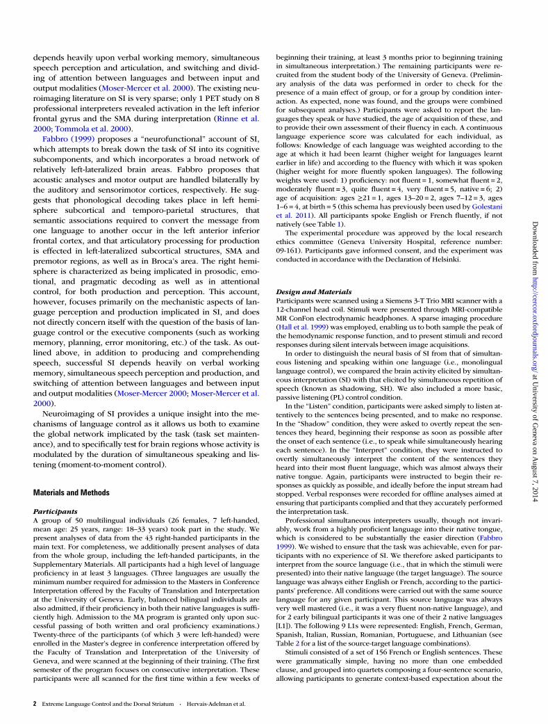
ParticipantsA group of 50 multilingual individuals (26 females, 7 left-handed,mean age: 25 years, range: 18–33 years) took part in the study. Wepresent analyses of data from the 43 right-handed participants in themain text. For completeness, we additionally present analyses of datafrom the whole group, including the left-handed participants, in theSupplementary Materials. All participants had a high level of languageproficiency in at least 3 languages. (Three languages are usually theminimum number required for admission to the Masters in ConferenceInterpretation offered by the Faculty of Translation and Interpretationat the University of Geneva. Early, balanced bilingual individuals arealso admitted, if their proficiency in both their native languages is suffi-ciently high. Admission to the MA program is granted only upon suc-cessful passing of both written and oral proficiency examinations.)Twenty-three of the participants (of which 3 were left-handed) wereenrolled in the Master’s degree in conference interpretation offered bythe Faculty of Translation and Interpretation of the University ofGeneva, and were scanned at the beginning of their training. (The firstsemester of the program focuses on consecutive interpretation. Theseparticipants were all scanned for the first time within a few weeks of
beginning their training, at least 3 months prior to beginning trainingin simultaneous interpretation.) The remaining participants were re-cruited from the student body of the University of Geneva. (Prelimin-ary analysis of the data was performed in order to check for thepresence of a main effect of group, or for a group by condition inter-action. As expected, none was found, and the groups were combinedfor subsequent analyses.) Participants were asked to report the lan-guages they speak or have studied, the age of acquisition of these, andto provide their own assessment of their fluency in each. A continuouslanguage experience score was calculated for each individual, asfollows: Knowledge of each language was weighted according to theage at which it had been learnt (higher weight for languages learntearlier in life) and according to the fluency with which it was spoken(higher weight for more fluently spoken languages). The followingweights were used: 1) proficiency: not fluent = 1, somewhat fluent = 2,moderately fluent = 3, quite fluent = 4, very fluent = 5, native = 6; 2)age of acquisition: ages ≥21 = 1, ages 13–20 = 2, ages 7–12 = 3, ages1–6 = 4, at birth = 5 (this schema has previously been used by Golestaniet al. 2011). All participants spoke English or French fluently, if notnatively (see Table 1).
The experimental procedure was approved by the local researchethics committee (Geneva University Hospital, reference number:09-161). Participants gave informed consent, and the experiment wasconducted in accordance with the Declaration of Helsinki.
Design andMaterialsParticipants were scanned using a Siemens 3-T Trio MRI scanner with a12-channel head coil. Stimuli were presented through MRI-compatibleMR ConFon electrodynamic headphones. A sparse imaging procedure(Hall et al. 1999) was employed, enabling us to both sample the peak ofthe hemodynamic response function, and to present stimuli and recordresponses during silent intervals between image acquisitions.
In order to distinguish the neural basis of SI from that of simultan-eous listening and speaking within one language (i.e., monolinguallanguage control), we compared the brain activity elicited by simultan-eous interpretation (SI) with that elicited by simultaneous repetition ofspeech (known as shadowing, SH). We also included a more basic,passive listening (PL) control condition.
In the “Listen” condition, participants were asked simply to listen at-tentively to the sentences being presented, and to make no response.In the “Shadow” condition, they were asked to overtly repeat the sen-tences they heard, beginning their response as soon as possible afterthe onset of each sentence (i.e., to speak while simultaneously hearingeach sentence). In the “Interpret” condition, they were instructed toovertly simultaneously interpret the content of the sentences theyheard into their most fluent language, which was almost always theirnative tongue. Again, participants were instructed to begin their re-sponses as quickly as possible, and ideally before the input stream hadstopped. Verbal responses were recorded for offline analyses aimed atensuring that participants complied and that they accurately performedthe interpretation task.
Professional simultaneous interpreters usually, though not invari-ably, work from a highly proficient language into their native tongue,which is considered to be substantially the easier direction (Fabbro1999). We wished to ensure that the task was achievable, even for par-ticipants with no experience of SI. We therefore asked participants tointerpret from the source language (i.e., that in which the stimuli werepresented) into their native language (the target language). The sourcelanguage was always either English or French, according to the partici-pants’ preference. All conditions were carried out with the same sourcelanguage for any given participant. This source language was alwaysvery well mastered (i.e., it was a very fluent non-native language), andfor 2 early bilingual participants it was one of their 2 native languages[L1]). The following 9 L1s were represented: English, French, German,Spanish, Italian, Russian, Romanian, Portuguese, and Lithuanian (seeTable 2 for a list of the source-target language combinations).
Stimuli consisted of a set of 156 French or English sentences. Thesewere grammatically simple, having no more than one embeddedclause, and grouped into quartets composing a four-sentence scenario,allowing participants to generate context-based expectation about the
2 Extreme Language Control and the Dorsal Striatum • Hervais-Adelman et al.
at University of G
eneva on August 7, 2014
http://cercor.oxfordjournals.org/D
ownloaded from
content of subsequent phrases. The thematic content of the French andEnglish sets was highly overlapping, though nonidentical, due tomatching constraints. (An example of an English quartet: “The cargo
ship has been found several weeks after disappearing at sea,” “Thecrew are all alive and well, and are currently answering questions,”“The few details that have emerged have left more questions thananswers,” “There are rumors of secret cargoes and ransom demands”;and an example of a French quartet: “Il y a beaucoup de spéculationsur les circonstances de l’incident,” “Nous soupçonnons que le requina pu confondre le surfeur avec un poisson blessé,” “Le jeune homme aété grièvement blessé dans l’attaque,” “Les médecins devront l’opérertoute la nuit pour sauver ses jambes” [in English: “There is a lot ofspeculation about the circumstances of the incident,” “We suspect thatthe shark mistook the surfer for an injured fish,” “The young man wasseriously injured in the attack,” “Doctors will have to operate all nightto save his legs”].) Sentences were recorded by a bilingual malespeaker of French and of Southern British English. Participants heardsentences in all conditions exclusively in one of the 2 available lan-guages (see Table 2). Sentence quartets were presented in randomorder, and were randomly allocated to conditions.
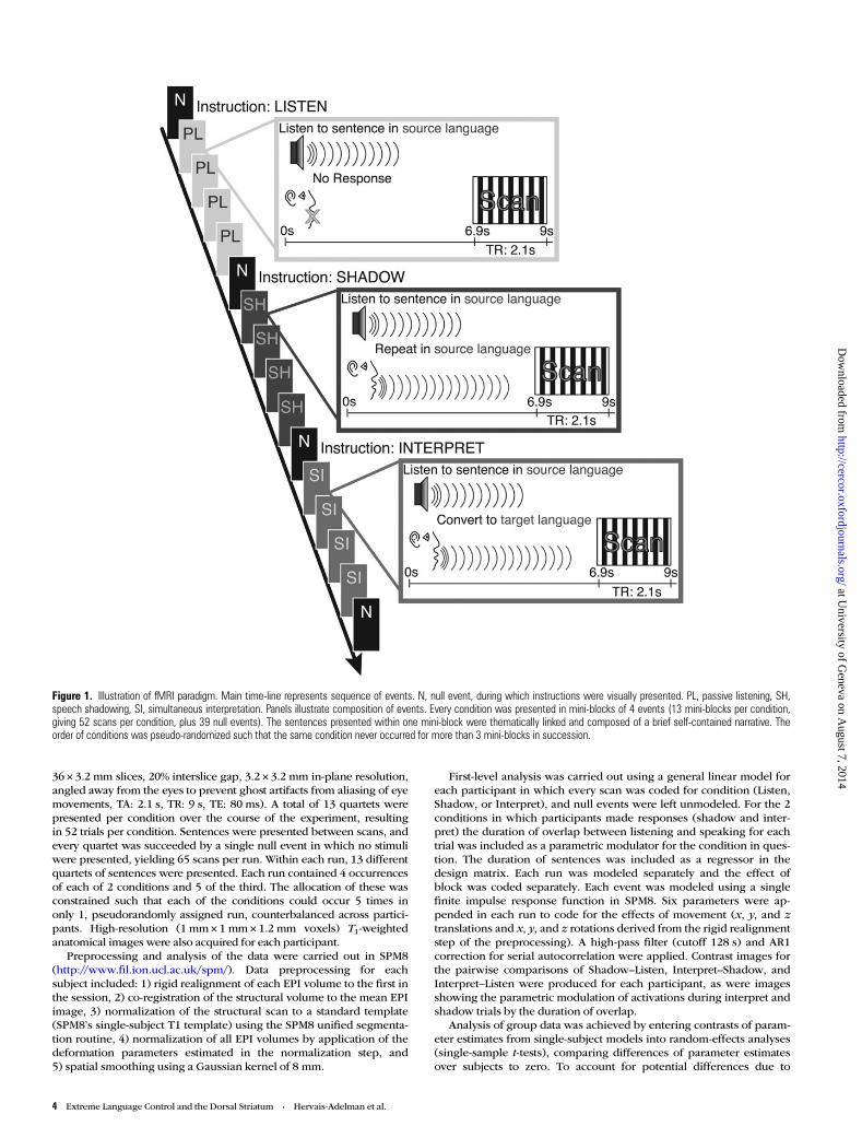
Immediately prior to the onset of each quartet, participants werepresented with an on-screen cue for the duration of the preceding MRIacquisition (2.1 s), consisting of an instruction: “Listen,” “Shadow,” or“Interpret,” which they carried out for the subsequent 4 sentences.Conditions were pseudorandomly ordered such that the same instruc-tion occurred for no more than 3 quartets in succession. The design isillustrated in Figure 1.
Prior to the experiment, participants underwent a brief practicesession inside the scanner during which they were familiarized with thetasks and with the speaker’s voice and accent. The experimenters moni-tored performance during practice to ensure that participants werecapable of carrying out the interpretation task in a simultaneousfashion. We specifically verified that participants began their responseswhile the stimuli were ongoing, and that they were capable of produ-cing relatively fluent output in the target language during interpretation.
In order to provide an adequate control for brain activations asso-ciated with non-native language perception and comprehension, aswell as within-language output monitoring, and division of attentionbetween speaking and listening, we elected to use SH in the non-nativelanguage (which would be the source language during the SI trials).This is potentially somewhat different to L1 SH, it may, for example,induce co-activation of L1 (as would be suggested by the BIA+ modelof Dijkstra and van Heuven 2002). SH in the non-native language (thesource language for SI) thus allows us to control for any associatedautomatic co-activation of L1 induced when hearing the non-native lan-guage, since this likely also occurs during interpretation. It additionallyprovides the best available control for the perception, comprehension,monitoring, and switching components of a SI task from the samesource language, enabling us to separate those from the additionalcerebral activations elicited by the multilingual nature of the interpret-ation task.
fMRI Data Acquisition, Processing, and AnalysisA total of 195 functional volumes were acquired; divided over 3 runsthat lasted ∼10 min each (T2*-weighted echo-planar imaging (EPI),
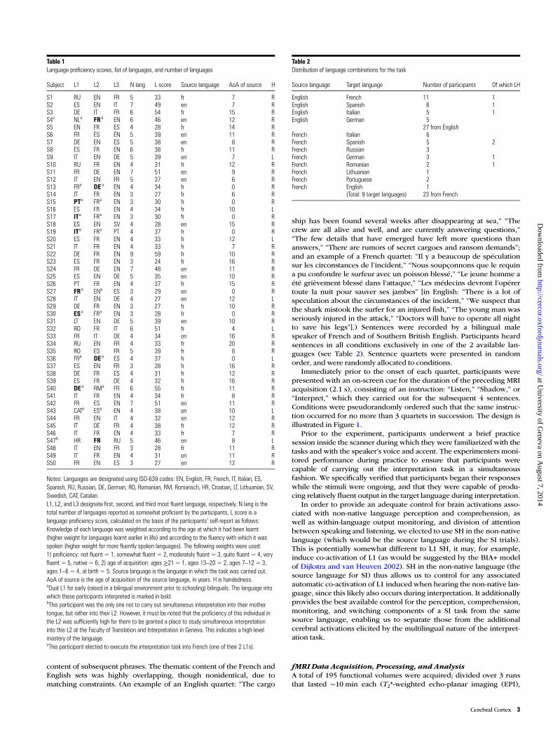
Table 2Distribution of language combinations for the task
Source language Target language Number of participants Of which LH
English French 11 1English Spanish 6 1English Italian 5 1English German 5
27 from EnglishFrench Italian 6French Spanish 5 2French Russian 3French German 3 1French Romanian 2 1French Lithuanian 1French Portuguese 2French English 1
(Total: 9 target languages) 23 from French
Table 1Language proficiency scores, list of languages, and number of languages
Subject L1 L2 L3 N lang L score Source language AoA of source H
S1 RU EN FR 5 33 fr 7 RS2 ES EN IT 7 49 en 7 RS3 DE IT FR 6 54 fr 15 RS4c NLa FR a EN 6 46 en 12 RS5 EN FR ES 4 28 fr 14 RS6 FR ES EN 5 39 en 11 RS7 DE EN ES 5 38 en 8 RS8 ES FR EN 6 38 fr 11 RS9 IT EN DE 5 39 en 7 LS10 RU FR EN 4 31 fr 12 RS11 FR DE EN 7 51 en 9 RS12 IT EN FR 5 37 en 6 RS13 FRa DE a EN 4 34 fr 0 RS14 IT FR EN 3 27 fr 6 RS15 PTa FRa EN 3 30 fr 0 RS16 ES FR EN 4 34 fr 10 LS17 ITa FRa EN 3 30 fr 0 RS18 ES EN SV 4 28 en 15 RS19 ITa FRa PT 4 37 fr 0 RS20 ES FR EN 4 33 fr 12 LS21 IT FR EN 4 33 fr 7 RS22 DE FR EN 9 59 fr 10 RS23 ES FR EN 3 24 fr 16 RS24 FR DE EN 7 48 en 11 RS25 ES EN DE 5 35 en 10 RS26 PT FR EN 4 37 fr 15 RS27 FR a ENa ES 3 29 en 0 RS28 IT EN DE 4 27 en 12 LS29 DE FR EN 3 27 fr 10 RS30 ES a FRa EN 3 28 fr 0 RS31 LT EN DE 5 39 en 10 RS32 RO FR IT 6 51 fr 4 LS33 FR IT DE 4 34 en 16 RS34 RU EN FR 4 33 fr 20 RS35 RO ES FR 5 39 fr 8 RS36 FRa DE a ES 4 37 fr 0 LS37 ES EN FR 3 28 fr 16 RS38 DE FR ES 4 31 fr 12 RS39 ES FR DE 4 32 fr 16 RS40 DE a RMa FR 6 55 fr 11 RS41 IT FR EN 4 34 fr 8 RS42 FR ES EN 7 51 en 11 RS43 CATa ESa EN 4 38 en 10 LS44 FR EN IT 4 32 en 12 RS45 IT DE FR 4 38 fr 12 RS46 IT FR EN 4 33 fr 7 RS47b HR FR RU 5 46 en 8 LS48 IT EN FR 3 28 fr 11 RS49 IT FR EN 4 31 en 11 RS50 FR EN ES 3 27 en 12 R
Notes: Languages are designated using ISO-639 codes: EN, English, FR, French, IT, Italian, ES,Spanish, RU, Russian, DE, German, RO, Romanian, RM, Romansch, HR, Croatian, LT, Lithuanian, SV,Swedish, CAT, Catalan.L1, L2, and L3 designate first, second, and third most fluent language, respectively. N lang is thetotal number of languages reported as somewhat proficient by the participants. L score is alanguage proficiency score, calculated on the basis of the participants’ self-report as follows:Knowledge of each language was weighted according to the age at which it had been learnt(higher weight for languages learnt earlier in life) and according to the fluency with which it wasspoken (higher weight for more fluently spoken languages). The following weights were used:1) proficiency: not fluent = 1, somewhat fluent = 2, moderately fluent = 3, quite fluent = 4, veryfluent = 5, native = 6; 2) age of acquisition: ages ≥21 = 1, ages 13–20 = 2, ages 7–12 = 3,ages 1–6 = 4, at birth = 5. Source language is the language in which the task was carried out.AoA of source is the age of acquisition of the source language, in years. H is handedness.aDual L1 for early (raised in a bilingual environment prior to schooling) bilinguals. The language intowhich these participants interpreted is marked in bold.bThis participant was the only one not to carry out simultaneous interpretation into their mothertongue, but rather into their L2. However, it must be noted that the proficiency of this individual inthe L2 was sufficiently high for them to be granted a place to study simultaneous interpretationinto this L2 at the Faculty of Translation and Interpretation in Geneva. This indicates a high-levelmastery of the language.cThis participant elected to execute the interpretation task into French (one of their 2 L1s).
Cerebral Cortex 3
at University of G
eneva on August 7, 2014
http://cercor.oxfordjournals.org/D
ownloaded from
36 × 3.2 mm slices, 20% interslice gap, 3.2 × 3.2 mm in-plane resolution,angled away from the eyes to prevent ghost artifacts from aliasing of eyemovements, TA: 2.1 s, TR: 9 s, TE: 80 ms). A total of 13 quartets werepresented per condition over the course of the experiment, resultingin 52 trials per condition. Sentences were presented between scans, andevery quartet was succeeded by a single null event in which no stimuliwere presented, yielding 65 scans per run. Within each run, 13 differentquartets of sentences were presented. Each run contained 4 occurrencesof each of 2 conditions and 5 of the third. The allocation of these wasconstrained such that each of the conditions could occur 5 times inonly 1, pseudorandomly assigned run, counterbalanced across partici-pants. High-resolution (1 mm× 1 mm× 1.2 mm voxels) T1-weightedanatomical images were also acquired for each participant.
Preprocessing and analysis of the data were carried out in SPM8(http://www.fil.ion.ucl.ac.uk/spm/). Data preprocessing for eachsubject included: 1) rigid realignment of each EPI volume to the first inthe session, 2) co-registration of the structural volume to the mean EPIimage, 3) normalization of the structural scan to a standard template(SPM8’s single-subject T1 template) using the SPM8 unified segmenta-tion routine, 4) normalization of all EPI volumes by application of thedeformation parameters estimated in the normalization step, and5) spatial smoothing using a Gaussian kernel of 8 mm.
First-level analysis was carried out using a general linear model foreach participant in which every scan was coded for condition (Listen,Shadow, or Interpret), and null events were left unmodeled. For the 2conditions in which participants made responses (shadow and inter-pret) the duration of overlap between listening and speaking for eachtrial was included as a parametric modulator for the condition in ques-tion. The duration of sentences was included as a regressor in thedesign matrix. Each run was modeled separately and the effect ofblock was coded separately. Each event was modeled using a singlefinite impulse response function in SPM8. Six parameters were ap-pended in each run to code for the effects of movement (x, y, and ztranslations and x, y, and z rotations derived from the rigid realignmentstep of the preprocessing). A high-pass filter (cutoff 128 s) and AR1correction for serial autocorrelation were applied. Contrast images forthe pairwise comparisons of Shadow–Listen, Interpret–Shadow, andInterpret–Listen were produced for each participant, as were imagesshowing the parametric modulation of activations during interpret andshadow trials by the duration of overlap.
Analysis of group data was achieved by entering contrasts of param-eter estimates from single-subject models into random-effects analyses(single-sample t-tests), comparing differences of parameter estimatesover subjects to zero. To account for potential differences due to
Figure 1. Illustration of fMRI paradigm. Main time-line represents sequence of events. N, null event, during which instructions were visually presented. PL, passive listening, SH,speech shadowing, SI, simultaneous interpretation. Panels illustrate composition of events. Every condition was presented in mini-blocks of 4 events (13 mini-blocks per condition,giving 52 scans per condition, plus 39 null events). The sentences presented within one mini-block were thematically linked and composed of a brief self-contained narrative. Theorder of conditions was pseudo-randomized such that the same condition never occurred for more than 3 mini-blocks in succession.
4 Extreme Language Control and the Dorsal Striatum • Hervais-Adelman et al.
at University of G
eneva on August 7, 2014
http://cercor.oxfordjournals.org/D
ownloaded from
participants’ different language expertise, their language experiencescores were included as a covariate. The anatomical location of peakswas determined with reference to the automatic anatomical labeling(Eickhoff et al. 2009) and Brodmann area (BA) templates providedwith the MRIcron software package (http://www.mccauslandcenter.sc.edu/mricro/mricron/). All data reported reach a statistical significancethreshold of whole-brain familywise error (FWE) corrected P < 0.05 atthe voxel level.
Behavioral ResponsesParticipants’ verbal responses were recorded and checked to ensurecompliance with the instructions. Audio recordings were preprocessedto achieve noise-reduction and speech enhancement using Audacity(http://audacity.sourceforge.net/). Onsets and offsets of responseswere determined using an automated detection procedure, achievedusing an in-house Praat (http://praat.org, Boersma and Weenink 2011)script to scan wav files for increases in amplitude in combination withincreases in zero-crossings of the waveform relative to baseline (similarto that implemented by Kello and Kawamoto (1998)). The duration ofsimultaneous speech listening and speaking (overlap) was calculatedon the basis of the onsets and offsets of verbal responses and theknown onsets and durations of the stimulus files.
Responses were scored by a panel of accredited professional simul-taneous interpreters trained to work in the language combinationsused during the interpretation and SH tasks by the participants (onerater per language). Raters were asked to evaluate SH responses on abinary scale to indicate failure to comply with instructions (score of 0)and compliance (score of 1). They were asked to assess the interpret-ation trials on a five-point scale as follows: 0 = no output, 1 = 1 contentword, 2 = 2 content words (minimally a subject and object), 3 = 3content words or more to make a meaningful interpretation, 4 =complete interpretation.
Results
Results presented here are for the 43 right-handed participants.For the sake of completeness, an analysis of all 50 participants,incorporating the 7 left-handed individuals, is provided in theSupplementary Materials.
Ratings of PerformanceDue to equipment failure (microphone preamplifier glitches),only 46 complete sets of recordings out of 50 (includingleft-handed participants) were assessed. For the 43 right-handed participants included here, 41 complete datasets wereevaluated. For these, interpretation and SH performance werehighly satisfactory. Participants on average responded correct-ly and completely to over 90% of the SH trials (standard devi-ation [SD] over participants: 13.8), and attempted responses inover 95% of the interpretation trials (i.e., 5% missed trials ornonresponses). On average, participants’ interpretations wererated at 3.16 (SD over participants: 0.49) on the scale describedin the methods, indicating that average performance laybetween completely accurate interpretations and highly mean-ingful responses. These results demonstrate that participantsexecuted the task with a high degree of compliance and agood level of accuracy.
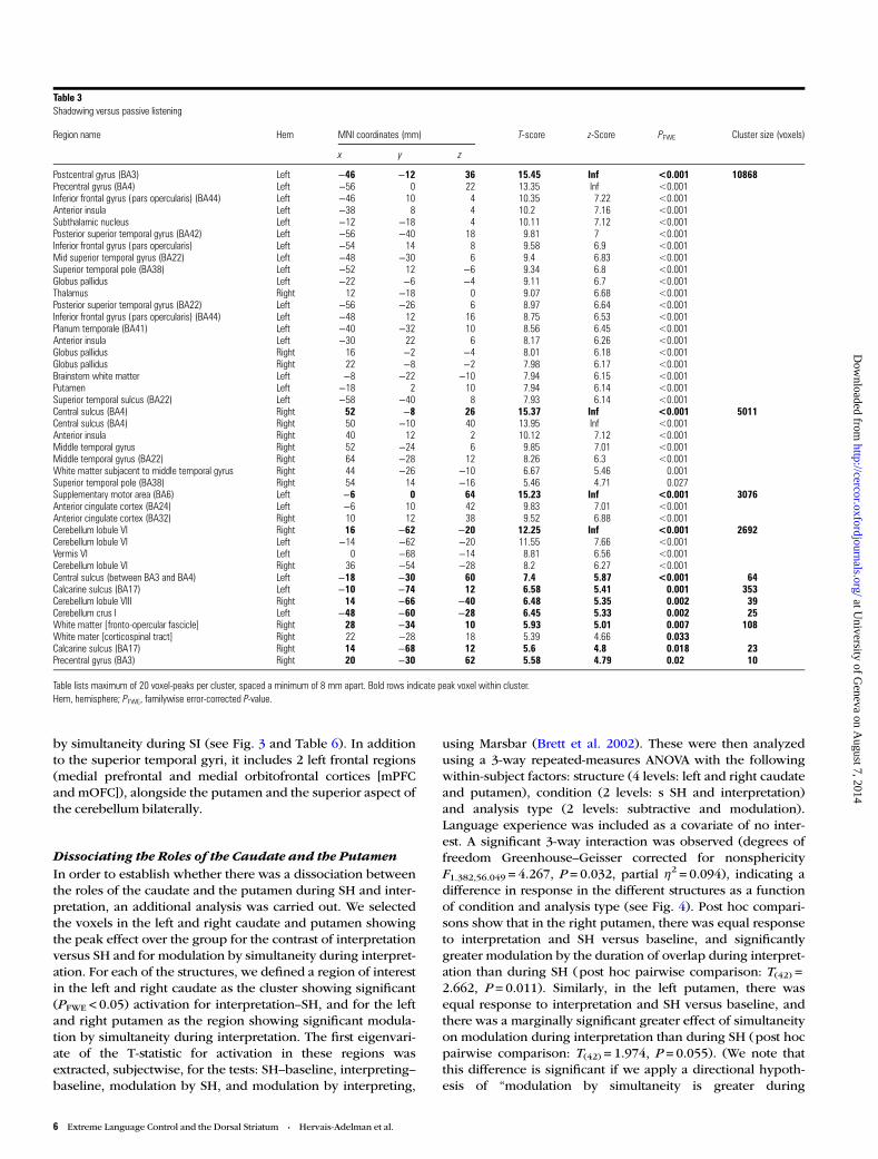
Neural Basis of SH: Monolingual Language ControlComparing the activation elicited by SH with that elicited byPL allowed us to examine the neural systems that supportmonolingual language control arising from speech productionand self-monitoring while simultaneously attending to an ex-ogenous speech stream. The results of this contrast are shownin Table 3 and Figure 2, and are broadly concordant with
previously reported data on SH (Peschke et al. 2009) and onspeech production (Peeva et al. 2010). We note that despite theSH having been in a language other than the participants’ L1,the activation pattern does not seem qualitatively differentfrom that previously reported for SH tasks. Further, weobserve no significantly elevated activity in the caudate nucleior in the putamen, subcortical structures that have previouslybeen associated with L2 repetition tasks (Klein et al. 1994,2006). For the sake of brevity, we will not further discuss thecomparison of SH and listening in this article.
Neural Basis of SI: Bilingual Language ControlAn initial comparison of activation during SI with that duringPL revealed that SI recruits all the brain regions implicated inSH. Brain areas recruited by SI over and above those recruitedby SH are involved in handling the additional demands of in-terpretation, which can be superficially characterized as thereal-time conversion of the input stream from one language toanother. Achieving this successfully depends upon multiplecognitive processes: deep but rapid semantic and syntacticanalysis of the input, retrieval of appropriate lexical, syntacticand stylistic alternatives in the other language (i.e., transla-tion), planning of the corresponding linguistic and semanticoutput, execution of speech output (speech production), mon-itoring of the output, error correction, and, finally, continuousattention to the incoming speech stream (Moser 1978). A com-parison of responses observed during SI with those observedduring SH revealed involvement of the left anterior SMA andpre-SMA, the left anterior insula, the left premotor cortex, thecaudate nuclei, crus I of the right cerebellum and the dorsalACC (dACC) [Fig. 2, Table 4]. Further significant increases in ac-tivation were also found in the left pars triangularis (BA 45, theanterior portion of Broca’s area) and pars orbitalis (BA 47) ofthe inferior frontal gyrus.
Neural Correlates of SimultaneityWewanted to not only examine the network that is engaged bythe overall task requirements, but also to examine networks in-volved in the execution of moment-to-moment control duringtask performance. The above subtractive analyses summarizethe brain activity associated with the relative demands of therespective experimental tasks, including maintaining and con-trolling the appropriate language set (a prerequisite of carryingout the task correctly), and the mechanisms implicated in ef-fecting the task. In order to better understand the neural basesunderlying moment-to-moment linguistic and cognitivecontrol required during SI and also during SH, we performedanalyses complementary to the above subtractive ones bytesting for brain regions whose activation was modulated bythe duration of simultaneous speaking and listening. Duringthese periods of overlap, participants must concurrentlyprocess input and produce output, while monitoring theoutput for errors. The brain regions whose response is modu-lated by the duration of these periods of simultaneity are likelyto be those that are implicated in the moment-to-momentcontrol demanded by either SH or interpreting. The mean dur-ation of overlap was 2.1 s (SD = 1.04 s) during SI, and 2.46 s(SD = 0.98 s) during SH.
During SH, the brain areas modulated by simultaneity areprincipally the superior temporal gyri, and a portion of the leftSMA (see Fig. 3 and Table 5). A broader network is modulated
Cerebral Cortex 5
at University of G
eneva on August 7, 2014
http://cercor.oxfordjournals.org/D
ownloaded from
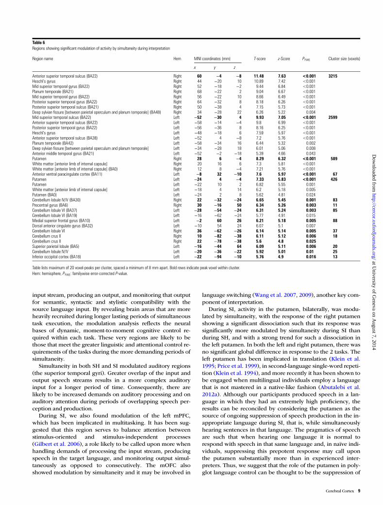
by simultaneity during SI (see Fig. 3 and Table 6). In additionto the superior temporal gyri, it includes 2 left frontal regions(medial prefrontal and medial orbitofrontal cortices [mPFCand mOFC]), alongside the putamen and the superior aspect ofthe cerebellum bilaterally.
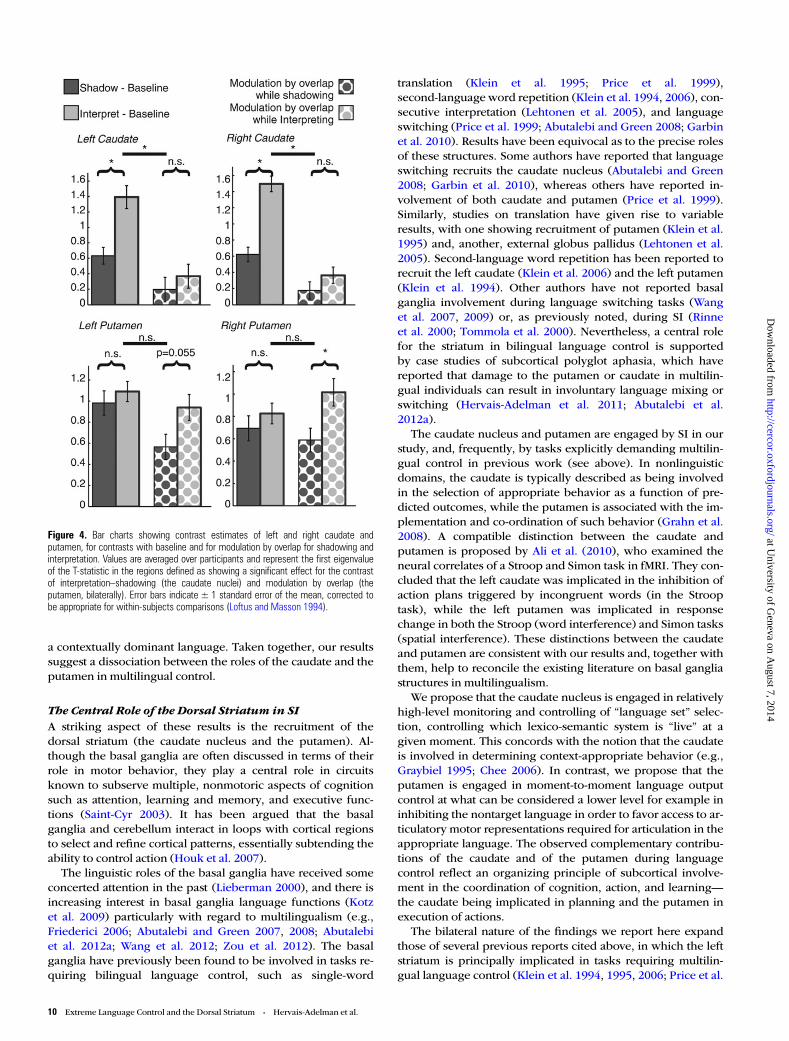
Dissociating the Roles of the Caudate and the PutamenIn order to establish whether there was a dissociation betweenthe roles of the caudate and the putamen during SH and inter-pretation, an additional analysis was carried out. We selectedthe voxels in the left and right caudate and putamen showingthe peak effect over the group for the contrast of interpretationversus SH and for modulation by simultaneity during interpret-ation. For each of the structures, we defined a region of interestin the left and right caudate as the cluster showing significant(PFWE < 0.05) activation for interpretation–SH, and for the leftand right putamen as the region showing significant modula-tion by simultaneity during interpretation. The first eigenvari-ate of the T-statistic for activation in these regions wasextracted, subjectwise, for the tests: SH–baseline, interpreting–baseline, modulation by SH, and modulation by interpreting,
using Marsbar (Brett et al. 2002). These were then analyzedusing a 3-way repeated-measures ANOVA with the followingwithin-subject factors: structure (4 levels: left and right caudateand putamen), condition (2 levels: s SH and interpretation)and analysis type (2 levels: subtractive and modulation).Language experience was included as a covariate of no inter-est. A significant 3-way interaction was observed (degrees offreedom Greenhouse–Geisser corrected for nonsphericityF1.382,56.049 = 4.267, P = 0.032, partial η2 = 0.094), indicating adifference in response in the different structures as a functionof condition and analysis type (see Fig. 4). Post hoc compari-sons show that in the right putamen, there was equal responseto interpretation and SH versus baseline, and significantlygreater modulation by the duration of overlap during interpret-ation than during SH (post hoc pairwise comparison: T(42) =2.662, P = 0.011). Similarly, in the left putamen, there wasequal response to interpretation and SH versus baseline, andthere was a marginally significant greater effect of simultaneityon modulation during interpretation than during SH (post hocpairwise comparison: T(42) = 1.974, P = 0.055). (We note thatthis difference is significant if we apply a directional hypoth-esis of “modulation by simultaneity is greater during
Table 3Shadowing versus passive listening
Region name Hem MNI coordinates (mm) T-score z-Score PFWE Cluster size (voxels)
x y z
Postcentral gyrus (BA3) Left −46 −12 36 15.45 Inf <0.001 10868Precentral gyrus (BA4) Left −56 0 22 13.35 Inf <0.001Inferior frontal gyrus (pars opercularis) (BA44) Left −46 10 4 10.35 7.22 <0.001Anterior insula Left −38 8 4 10.2 7.16 <0.001Subthalamic nucleus Left −12 −18 4 10.11 7.12 <0.001Posterior superior temporal gyrus (BA42) Left −56 −40 18 9.81 7 <0.001Inferior frontal gyrus (pars opercularis) Left −54 14 8 9.58 6.9 <0.001Mid superior temporal gyrus (BA22) Left −48 −30 6 9.4 6.83 <0.001Superior temporal pole (BA38) Left −52 12 −6 9.34 6.8 <0.001Globus pallidus Left −22 −6 −4 9.11 6.7 <0.001Thalamus Right 12 −18 0 9.07 6.68 <0.001Posterior superior temporal gyrus (BA22) Left −56 −26 6 8.97 6.64 <0.001Inferior frontal gyrus (pars opercularis) (BA44) Left −48 12 16 8.75 6.53 <0.001Planum temporale (BA41) Left −40 −32 10 8.56 6.45 <0.001Anterior insula Left −30 22 6 8.17 6.26 <0.001Globus pallidus Right 16 −2 −4 8.01 6.18 <0.001Globus pallidus Right 22 −8 −2 7.98 6.17 <0.001Brainstem white matter Left −8 −22 −10 7.94 6.15 <0.001Putamen Left −18 2 10 7.94 6.14 <0.001Superior temporal sulcus (BA22) Left −58 −40 8 7.93 6.14 <0.001Central sulcus (BA4) Right 52 −8 26 15.37 Inf <0.001 5011Central sulcus (BA4) Right 50 −10 40 13.95 Inf <0.001Anterior insula Right 40 12 2 10.12 7.12 <0.001Middle temporal gyrus Right 52 −24 6 9.85 7.01 <0.001Middle temporal gyrus (BA22) Right 64 −28 12 8.26 6.3 <0.001White matter subjacent to middle temporal gyrus Right 44 −26 −10 6.67 5.46 0.001Superior temporal pole (BA38) Right 54 14 −16 5.46 4.71 0.027Supplementary motor area (BA6) Left −6 0 64 15.23 Inf <0.001 3076Anterior cingulate cortex (BA24) Left −6 10 42 9.83 7.01 <0.001Anterior cingulate cortex (BA32) Right 10 12 38 9.52 6.88 <0.001Cerebellum lobule VI Right 16 −62 −20 12.25 Inf <0.001 2692Cerebellum lobule VI Left −14 −62 −20 11.55 7.66 <0.001Vermis VI Left 0 −68 −14 8.81 6.56 <0.001Cerebellum lobule VI Right 36 −54 −28 8.2 6.27 <0.001Central sulcus (between BA3 and BA4) Left −18 −30 60 7.4 5.87 <0.001 64Calcarine sulcus (BA17) Left −10 −74 12 6.58 5.41 0.001 353Cerebellum lobule VIII Right 14 −66 −40 6.48 5.35 0.002 39Cerebellum crus I Left −48 −60 −28 6.45 5.33 0.002 25White matter [fronto-opercular fascicle] Right 28 −34 10 5.93 5.01 0.007 108White mater [corticospinal tract] Right 22 −28 18 5.39 4.66 0.033Calcarine sulcus (BA17) Right 14 −68 12 5.6 4.8 0.018 23Precentral gyrus (BA3) Right 20 −30 62 5.58 4.79 0.02 10
Table lists maximum of 20 voxel-peaks per cluster, spaced a minimum of 8 mm apart. Bold rows indicate peak voxel within cluster.Hem, hemisphere; PFWE, familywise error-corrected P-value.
6 Extreme Language Control and the Dorsal Striatum • Hervais-Adelman et al.
at University of G
eneva on August 7, 2014
http://cercor.oxfordjournals.org/D
ownloaded from

interpretation than during shadowing” [i.e. using a one-tailedtest].)
Discussion
Simultaneous InterpretationAll the regions involved in SH are also involved in SI, reflectingthe common linguistic and executive demands of the tasks.Left inferior frontal gyrus regions additionally recruited duringinterpretation over SH include pars triangularis, known for itsrole in semantic processing (Dapretto and Bookheimer 1999;Bookheimer 2002), and pars orbitalis, implicated in semantic
memory and cognitive control of memory (Badre and Wagner2007). Pre-SMA and dACC were also additionally recruitedduring SI. Pre-SMA activation has been associated with tasks inwhich there is response conflict, and generation of complexmotor acts (Picard and Strick 1996; Nachev et al. 2008).Pre-SMA and dACC are thought to play complementary roles inaction selection: The pre-SMA is involved in the selection andinitiation of action sets, and the dACC in monitoring theoutcome for errors (Rushworth et al. 2004). During SI, there isa high level of competition between languages, and the en-gagement of the pre-SMA is consistent with the need to handlethe resulting response conflict. This analysis of the results issupported by previous studies on language switching, whichhave implicated the pre-SMA (Abutalebi et al. 2008).
The caudate nuclei were also engaged specifically during SI.The left caudate nucleus has previously been reported to be re-cruited in multilingual paradigms (Crinion et al. 2006; Abutale-bi and Green 2008; Garbin et al. 2010), and has been shown tobe implicated in multilingual language control by direct elec-trical stimulation (Wang et al. 2012).
The left anterior insula and SMA have been associated witha speech preparatory loop (Riecker et al. 2005). SI calls onspeech preparation to a greater extent than SH due to the re-quirement to formulate the output ab initio rather than simplyrepeating the heard speech segments, and due to more effort-ful speech output due to the competition between 2 languagesduring the former, but not the latter, condition. The increasedanterior insula and dACC activation may also reflect the in-creased attentional demands and difficulty of SI comparedwith SH, managed by the cingulo-opercular system (Petersenand Posner 2012).
The network of regions that we identified during SI isbroader than previously reported. This could be due to ourlarger sample size, or may be due to the fact that participantsin the present study were untrained, whereas the previouslystudied participants were professional simultaneous inter-preters with between 5 and 20 years of experience (Rinne et al.2000; Tommola et al. 2000). Trained interpreters’ expertisemay enable them to carry out the task efficiently and effectivelywhile recruiting fewer brain regions than naïve participants,consistent with many studies showing decreased brain activa-tion when a task is more rehearsed and automated comparedwith when it is more effortful and novel (Ericsson et al. 2006).
The regions we identify include many of those proposed byFabbro (1999) to underlie SI. In addition to these, our investi-gation reveals the involvement of brain regions involved in thelanguage- and executive-control requirements of the task.
The Neural Correlates of SimultaneityAn important distinction exists between the cognitive controlrequired to meet the overall demands of specific task instruc-tions, and the exercise of control on a moment-to-momentbasis while carrying out that task. This distinction is borne outin the existing neuroimaging literature, which shows differentbrain networks associated with task set maintenance over thefull length of experimental trials, or associated with the initi-ation of responses and the adjustment of control on amoment-to-moment basis (Dosenbach et al. 2008). Our para-metric modulation analysis aimed to reveal the latter. Duringperiods of overlapping speech input and speech production,the participants are concurrently engaged in processing the
Figure 2. Significant differences in BOLD response for the contrastsshadowing > passive listening (blue) and simultaneous interpreting > shadowing(red). Regions where both contrasts are significant (i.e., simultaneous interpreting >shadowing > passive listening) are also shown (magenta). Results are projected on acanonical single-subject MNI brain. Shadowing induces activation in a broad bilateralnetwork encompassing temporal, inferior frontal, inferior parietal, motor, andsubcortical regions including thalamus and globus pallidus. Interpretation additionallyrecruits further inferior frontal, motor, and basal ganglia regions. Bar charts indicateT-statistics averaged over the group at selected peak voxels in the named activationclusters, and error bars indicate standard error of the mean corrected to be appropriatefor repeated-measures designs (Loftus and Masson 1994). Coordinates indicate planeof section (in MNI space) of the background images. pOp, pars opercularis; pOr, parsorbitalis; PTr, pars triangularis; aIns, anterior insula; lCd, left caudate; pSMA,presupplementary motor area; SMA, supplementary motor area; ACC, anteriorcingulate cortex; Cru1, Crus 1 of cerebellum; CerVI, Cerebellum lobule VI; rCd, rightcaudate; STN, subthalamic nucleus; GP, globus pallidus.
Cerebral Cortex 7
at University of G
eneva on August 7, 2014
http://cercor.oxfordjournals.org/D
ownloaded from
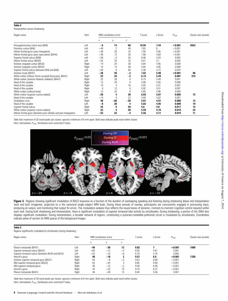
Figure 3. Regions showing significant modulation of BOLD response as a function of the duration of overlapping speaking and listening during shadowing (blue) and interpretation(red) and both (magenta), projected on a the canonical single-subject MNI brain. During these periods of overlap, participants are concurrently engaged in processing input,producing an output, and monitoring that output for errors. This modulation analysis thus reflects the neural bases of dynamic, moment-to-moment cognitive control required withineach task. During both shadowing and interpretation, there is significant modulation of superior temporal lobe activity by simultaneity. During shadowing, a portion of the SMA alsodisplays significant modulation. During interpretation, a broader network of regions, constituting a putamen-cerebellar-prefrontal circuit is modulated by simultaneity. Coordinatesindicate plane of section (in MNI space) of the background images.
Table 5Regions significantly modulated by simultaneity during shadowing
Region name Hem MNI coordinates (mm) T-score z-Score PFWE Cluster size (voxels)
x y z
Planum temporale (BA41) Left −40 −30 12 9.82 7 <0.001 1090Superior temporal sulcus (BA22) Left −60 −12 −2 6.93 5.61 0.001Superior temporal sulcus (between BA20 and BA22) Left −52 −10 −6 6.18 5.16 0.005Heschl’s gyrus Right 48 −18 2 9.57 6.9 <0.001 1339Anterior superior temporal gyrus (BA21) Right 64 −8 −2 9.53 6.88 <0.001Mid superior temporal gyrus (BA22) Right 62 −20 4 8.85 6.58 <0.001Mid superior temporal gyrus Right 56 −12 −2 8.58 6.46 <0.001Heschl’s gyrus Right 44 −22 10 8.19 6.27 <0.001Planum temporale (BA41) Right 48 −30 12 6.84 5.56 0.001
Table lists maximum of 20 voxel-peaks per cluster, spaced a minimum of 8 mm apart. Bold rows indicate peak voxel within cluster.Hem, hemisphere; PFWE, familywise error-corrected P-value.
Table 4Interpretation versus shadowing
Region name Hem MNI coordinates (mm) T-score z-Score PFWE Cluster size (voxels)
x y z
Presupplementary motor area (BA6) Left −6 14 60 10.94 7.44 <0.001 2833Premotor cortex (BA6) Left −40 2 48 7.65 6 <0.001Inferior frontal gyrus (pars triangularis) Left −48 20 24 7.56 5.95 <0.001Inferior frontal gyrus (pars opercularis) (BA44) Left −46 12 30 7.08 5.69 <0.001Superior frontal sulcus (BA6) Left −26 −2 54 6.46 5.34 0.002Inferior frontal sulcus (BA45) Left −42 32 32 6.07 5.1 0.005Anterior cingulate cortex (BA32) Right 14 24 38 5.84 4.95 0.009Anterior cingulate cortex (BA32) Right 14 12 48 5.84 4.95 0.009Superior frontal sulcus (between BA6 and BA8) Left −30 6 64 5.48 4.72 0.024Anterior insula (BA47) Left −28 26 −2 7.62 5.98 <0.001 86White matter [inferior fronto-occipital fasciculus] (BA47) Right 22 34 −2 6.73 5.49 0.001 375White matter [anterior thalamic radiation] (BA47) Right 22 28 6 6.73 5.49 0.001Head of the caudate Right 12 20 6 5.99 5.05 0.006Head of the caudate Right 10 4 16 5.93 5.01 0.007Head of the caudate Right 8 12 8 5.92 5.01 0.007White matter [callosal body] Right 12 32 8 5.85 4.96 0.009White matter [superior corona radiata] Left −20 4 30 6.03 5.07 0.005 72Head of the caudate Left −14 0 22 5.94 5.02 0.007Cerebellum crus I Right 48 −66 −28 5.87 4.97 0.008 12Head of the caudate Left −8 20 4 5.83 4.95 0.009 19Superior frontal sulcus Right 24 0 50 5.6 4.8 0.017 14White matter [superior corona radiata] Right 22 4 28 5.59 4.79 0.018 22Inferior frontal gyrus (between pars orbitals and pars triangularis) Left −52 22 −4 5.56 4.77 0.019 11
Table lists maximum of 20 voxel-peaks per cluster, spaced a minimum of 8 mm apart. Bold rows indicate peak voxel within cluster.Hem, hemisphere; PFWE, familywise error-corrected P-value.
8 Extreme Language Control and the Dorsal Striatum • Hervais-Adelman et al.
at University of G
eneva on August 7, 2014
http://cercor.oxfordjournals.org/D
ownloaded from
input stream, producing an output, and monitoring that outputfor semantic, syntactic and stylistic compatibility with thesource language input. By revealing brain areas that are moreheavily recruited during longer lasting periods of simultaneoustask execution, the modulation analysis reflects the neuralbases of dynamic, moment-to-moment cognitive control re-quired within each task. These very regions are likely to bethose that meet the greater linguistic and attentional control re-quirements of the tasks during the more demanding periods ofsimultaneity.
Simultaneity in both SH and SI modulated auditory regions(the superior temporal gyri). Greater overlap of the input andoutput speech streams results in a more complex auditoryinput for a longer period of time. Consequently, there arelikely to be increased demands on auditory processing and onauditory attention during periods of overlapping speech per-ception and production.
During SI, we also found modulation of the left mPFC,which has been implicated in multitasking. It has been sug-gested that this region serves to balance attention betweenstimulus-oriented and stimulus-independent processes(Gilbert et al. 2006), a role likely to be called upon more whenhandling demands of processing the input stream, producingspeech in the target language, and monitoring output simul-taneously as opposed to consecutively. The mOFC alsoshowed modulation by simultaneity and it may be involved in
language switching (Wang et al. 2007, 2009), another key com-ponent of interpretation.
During SI, activity in the putamen, bilaterally, was modu-lated by simultaneity, with the response of the right putamenshowing a significant dissociation such that its response wassignificantly more modulated by simultaneity during SI thanduring SH, and with a strong trend for such a dissociation inthe left putamen. In both the left and right putamen, there wasno significant global difference in response to the 2 tasks. Theleft putamen has been implicated in translation (Klein et al.1995; Price et al. 1999), in second-language single-word repeti-tion (Klein et al. 1994), and more recently it has been shown tobe engaged when multilingual individuals employ a languagethat is not mastered in a native-like fashion (Abutalebi et al.2012a). Although our participants produced speech in a lan-guage in which they had an extremely high proficiency, theresults can be reconciled by considering the putamen as thesource of ongoing suppression of speech production in the in-appropriate language during SI, that is, while simultaneouslyhearing sentences in that language. The pragmatics of speechare such that when hearing one language it is normal torespond with speech in that same language and, in naïve indi-viduals, suppressing this prepotent response may call uponthe putamen substantially more than in experienced inter-preters. Thus, we suggest that the role of the putamen in poly-glot language control can be thought to be the suppression of
Table 6Regions showing significant modulation of activity by simultaneity during interpretation
Region name Hem MNI coordinates (mm) T-score z-Score PFWE Cluster size (voxels)
x y z
Anterior superior temporal sulcus (BA22) Right 60 −4 −8 11.48 7.63 <0.001 3215Heschl’s gyrus Right 44 −20 10 10.89 7.42 <0.001Mid superior temporal gyrus (BA22) Right 52 −18 −2 9.44 6.84 <0.001Planum temporale (BA21) Right 68 −22 2 9.04 6.67 <0.001Mid superior temporal gyrus (BA22) Right 56 −22 10 8.66 6.49 <0.001Posterior superior temporal gyrus (BA22) Right 64 −32 8 8.18 6.26 <0.001Posterior superior temporal sulcus (BA21) Right 50 −38 4 7.15 5.73 <0.001Deep sylvian fissure [between parietal operculum and planum temporale] (BA48) Right 34 −28 22 6.26 5.22 0.004Mid superior temporal sulcus (BA22) Left −52 −30 4 9.93 7.05 <0.001 2599Anterior superior temporal sulcus (BA22) Left −58 −14 −4 9.8 6.99 <0.001Posterior superior temporal gyrus (BA22) Left −56 −36 8 8.16 6.25 <0.001Heschl’s gyrus Left −48 −18 6 7.59 5.97 <0.001Anterior superior temporal sulcus (BA38) Left −52 4 −8 7.2 5.76 <0.001Planum temporale (BA42) Left −58 −34 16 6.44 5.32 0.002Deep sylvian fissure [between parietal operculum and planum temporale] Left −34 −28 18 6.01 5.06 0.008Anterior middle temporal gyrus (BA21) Left −52 −2 −18 5.39 4.66 0.043Putamen Right 28 6 −4 8.29 6.32 <0.001 589White matter [anterior limb of internal capsule] Right 20 16 6 7.3 5.81 <0.001White matter [anterior limb of internal capsule] (BA0) Right 12 8 −4 7.21 5.76 <0.001Anterior ventral paracingulate cortex (BA11) Left −8 32 −10 7.6 5.97 <0.001 67Putamen Left −24 4 −4 7.33 5.83 <0.001 426Putamen Left −22 10 2 6.82 5.55 0.001White matter [anterior limb of internal capsule] Left −18 4 14 6.2 5.18 0.005Putamen (BA0) Left −24 2 8 5.62 4.81 0.024Cerebellum lobule IV/V (BA30) Right 22 −32 −24 6.65 5.45 0.001 83Precentral gyrus (BA6) Right 30 −16 50 6.34 5.26 0.003 11Cerebellum lobule VI (BA37) Left −28 −54 −24 6.31 5.24 0.003 85Cerebellum lobule VI (BA19) Left −16 −62 −24 5.77 4.91 0.015Medial superior frontal gyrus (BA10) Left −2 60 26 6.21 5.18 0.005 88Dorsal anterior cingulate gyrus (BA32) Left −10 54 24 6.07 5.1 0.007Cerebellum lobule VI Right 36 −62 −26 6.14 5.14 0.005 37Cerebellum crus II Right 10 −82 −38 6.11 5.12 0.006 18Cerebellum crus II Right 22 −78 −38 5.6 4.8 0.025Superior parietal lobule (BA5) Left −16 −44 64 6.09 5.11 0.006 20Cerebellum lobule IV/V Left −20 −36 −22 5.92 5.01 0.01 25Inferior occipital cortex (BA18) Left −22 −94 −10 5.76 4.9 0.016 13
Table lists maximum of 20 voxel-peaks per cluster, spaced a minimum of 8 mm apart. Bold rows indicate peak voxel within cluster.Hem: hemisphere, PFWE: familywise error-corrected P-value.
Cerebral Cortex 9
at University of G
eneva on August 7, 2014
http://cercor.oxfordjournals.org/D
ownloaded from
a contextually dominant language. Taken together, our resultssuggest a dissociation between the roles of the caudate and theputamen in multilingual control.
The Central Role of the Dorsal Striatum in SIA striking aspect of these results is the recruitment of thedorsal striatum (the caudate nucleus and the putamen). Al-though the basal ganglia are often discussed in terms of theirrole in motor behavior, they play a central role in circuitsknown to subserve multiple, nonmotoric aspects of cognitionsuch as attention, learning and memory, and executive func-tions (Saint-Cyr 2003). It has been argued that the basalganglia and cerebellum interact in loops with cortical regionsto select and refine cortical patterns, essentially subtending theability to control action (Houk et al. 2007).
The linguistic roles of the basal ganglia have received someconcerted attention in the past (Lieberman 2000), and there isincreasing interest in basal ganglia language functions (Kotzet al. 2009) particularly with regard to multilingualism (e.g.,Friederici 2006; Abutalebi and Green 2007, 2008; Abutalebiet al. 2012a; Wang et al. 2012; Zou et al. 2012). The basalganglia have previously been found to be involved in tasks re-quiring bilingual language control, such as single-word
translation (Klein et al. 1995; Price et al. 1999),second-language word repetition (Klein et al. 1994, 2006), con-secutive interpretation (Lehtonen et al. 2005), and languageswitching (Price et al. 1999; Abutalebi and Green 2008; Garbinet al. 2010). Results have been equivocal as to the precise rolesof these structures. Some authors have reported that languageswitching recruits the caudate nucleus (Abutalebi and Green2008; Garbin et al. 2010), whereas others have reported in-volvement of both caudate and putamen (Price et al. 1999).Similarly, studies on translation have given rise to variableresults, with one showing recruitment of putamen (Klein et al.1995) and, another, external globus pallidus (Lehtonen et al.2005). Second-language word repetition has been reported torecruit the left caudate (Klein et al. 2006) and the left putamen(Klein et al. 1994). Other authors have not reported basalganglia involvement during language switching tasks (Wanget al. 2007, 2009) or, as previously noted, during SI (Rinneet al. 2000; Tommola et al. 2000). Nevertheless, a central rolefor the striatum in bilingual language control is supportedby case studies of subcortical polyglot aphasia, which havereported that damage to the putamen or caudate in multilin-gual individuals can result in involuntary language mixing orswitching (Hervais-Adelman et al. 2011; Abutalebi et al.2012a).
The caudate nucleus and putamen are engaged by SI in ourstudy, and, frequently, by tasks explicitly demanding multilin-gual control in previous work (see above). In nonlinguisticdomains, the caudate is typically described as being involvedin the selection of appropriate behavior as a function of pre-dicted outcomes, while the putamen is associated with the im-plementation and co-ordination of such behavior (Grahn et al.2008). A compatible distinction between the caudate andputamen is proposed by Ali et al. (2010), who examined theneural correlates of a Stroop and Simon task in fMRI. They con-cluded that the left caudate was implicated in the inhibition ofaction plans triggered by incongruent words (in the Strooptask), while the left putamen was implicated in responsechange in both the Stroop (word interference) and Simon tasks(spatial interference). These distinctions between the caudateand putamen are consistent with our results and, together withthem, help to reconcile the existing literature on basal gangliastructures in multilingualism.
We propose that the caudate nucleus is engaged in relativelyhigh-level monitoring and controlling of “language set” selec-tion, controlling which lexico-semantic system is “live” at agiven moment. This concords with the notion that the caudateis involved in determining context-appropriate behavior (e.g.,Graybiel 1995; Chee 2006). In contrast, we propose that theputamen is engaged in moment-to-moment language outputcontrol at what can be considered a lower level for example ininhibiting the nontarget language in order to favor access to ar-ticulatory motor representations required for articulation in theappropriate language. The observed complementary contribu-tions of the caudate and of the putamen during languagecontrol reflect an organizing principle of subcortical involve-ment in the coordination of cognition, action, and learning—the caudate being implicated in planning and the putamen inexecution of actions.
The bilateral nature of the findings we report here expandthose of several previous reports cited above, in which the leftstriatum is principally implicated in tasks requiring multilin-gual language control (Klein et al. 1994, 1995, 2006; Price et al.
Figure 4. Bar charts showing contrast estimates of left and right caudate andputamen, for contrasts with baseline and for modulation by overlap for shadowing andinterpretation. Values are averaged over participants and represent the first eigenvalueof the T-statistic in the regions defined as showing a significant effect for the contrastof interpretation–shadowing (the caudate nuclei) and modulation by overlap (theputamen, bilaterally). Error bars indicate ± 1 standard error of the mean, corrected tobe appropriate for within-subjects comparisons (Loftus and Masson 1994).
10 Extreme Language Control and the Dorsal Striatum • Hervais-Adelman et al.
at University of G
eneva on August 7, 2014
http://cercor.oxfordjournals.org/D
ownloaded from
1999; Lehtonen et al. 2005; Abutalebi and Green 2008; Garbinet al. 2010). There is also some indication from direct electricalstimulation that the dominant striatum (typically the left stri-atum in right-handed individuals) is involved in the control ofspeech production, with the putamen more implicated in thecoordination of speech articulation, and the caudate involvedin inhibition and selection, in monolingual patients (Gil Robleset al. 2005). A recent meta-analysis of neuroimaging studies oflanguage switching, however, indicates that both right and leftstriatal structures are implicated in language switching (Luket al. 2012). Consistent with this, our data indicate that thebilateral caudate nuclei and putamen are implicated in SI. Ourbilateral findings may be due to the fact that owing to the largenumber of participants that we tested, we had relatively higherstatistical power, or it may be because in contrast to most previ-ous reports, we used a task that makes large, ongoing (asopposed to momentary) demands on language control. Thefindings herein reported demonstrate bilateral striatal involve-ment in a demanding, continuous language control task. (It isof particular interest to note that when including 7 left-handedparticipants in our analysis [see Supplementary Tables 1–4 andSupplementary Figures 1–3], the lateralization of these resultsdoes not qualitatively change, suggesting that the languagecontrol functions of these structures may not be substantiallyaffected by handedness. This finding merits further investiga-tion in the future.)
Our results provide new insights into the profound overlapbetween the neural bases of extreme language control and thoseof domain-general control of cognition and action. Indeed,recent evidence suggests that experienced simultaneous inter-preters display enhanced cognitive flexibility compared evenwith bilingual individuals (Yudes et al. 2011; Stavrakaki et al.2012). The recruitment of similar fronto-subcortical-cerebellarcircuits during language and executive control provides power-ful evidence that the continuous demands of language control inthe multilingual brain, and associated experience-dependentplasticity, could underlie the nonlinguistic, executive advantagesthat have been observed in bilingual individuals, advantages thatmay also be protective in defying challenges posed by aging andeven disease.
Supplementary MaterialSupplementary material can be found at: http://www.cercor.oxfordjournals.org/.
Funding
This work was supported by the Swiss National Science Foun-dation (grant nos. PP00P3_133701 and 320030_122085).
NotesWe thank Alain Dagher for helpful comments on the manuscript, aswell as 3 anonymous reviewers for their thoughtful suggestions. Wealso thank Frédéric Grouiller and Maria Pefkou for their assistancewith data collection, and Sophie Hengl, Violeta Seretan, MagdalenaOlivera-Tovar, and Carmen Delgado Luchner for their efforts in ratingthe participants’ output. Conflict of Interest: None declared.
ReferencesAbutalebi J, Annoni JM, Zimine I, Pegna AJ, Seghier ML, Lee-Jahnke H,
Lazeyras F, Cappa SF, Khateb A. 2008. Language control and lexicalcompetition in bilinguals: an event-related fMRI study. CerebCortex. 18:1496–1505.
Abutalebi J, Della Rosa PA, Castro Gonzaga AK, Keim R, Costa A,Perani D. 2012a. The role of the left putamen in multilingual lan-guage production. Brain Lang. 125:307–315.
Abutalebi J, Della Rosa PA, Green DW, Hernandez M, Scifo P, Keim R,Cappa SF, Costa A. 2012b. Bilingualism tunes the anterior cingulatecortex for conflict monitoring. Cereb Cortex. 22:2076–2086.
Abutalebi J, Green D. 2007. Bilingual language production: the neuro-cognition of language representation and control. J Neurolinguis-tics. 20:242–275.
Abutalebi J, Green D. 2008. Control mechanisms in bilingual languageproduction: neural evidence from language switching studies. LangCogn Process. 23:557–582.
Abutalebi J, Tettamanti M, Perani D. 2009. The bilingual brain: linguis-tic and non-linguistic skills. Brain Lang. 109:51–54.
Ali N, Green DW, Kherif F, Devlin JT, Price CJ. 2010. The role of the lefthead of caudate in suppressing irrelevant words. J Cogn Neurosci.22:2369–2386.
Aron AR. 2008. Progress in executive-function research: from tasks tofunctions to regions to networks. Curr Dir Psychol Sci. 17:124–129.
Badre D, Wagner AD. 2007. Left ventrolateral prefrontal cortex and thecognitive control of memory. Neuropsychologia. 45:2883–2901.
Bialystok E, Craik FI, Luk G. 2012. Bilingualism: consequences formind and brain. Trends Cogn Sci. 16:240–250.
Boersma P, Weenink D. 2011. Praat: doing phonetics by computer.Version 5.2.23.
Bookheimer S. 2002. Functional MRI of language: new approaches tounderstanding the cortical organization of semantic processing.Annu Rev Neurosci. 25:151–188.
Botvinick MM, Cohen JD, Carter CS. 2004. Conflict monitoring and an-terior cingulate cortex: an update. Trends Cogn Sci. 8:539–546.
Brett M, Anton JL, Valabregue R, Poline J-B. 2002. Region of interestanalysis using an SPM toolbox [abstract]. In. 8th International Con-ference on Functional Mapping of the Human Brain. Sendai, Japan.Available on CD-ROM in NeuroImage, Vol 16, No 2.
Chee MW. 2006. Dissociating language and word meaning in the bilin-gual brain. Trends Cogn Sci. 10:527–529.
Costa A. 2005. Lexical access in bilingual production. In: Kroll J, DeGroot AMB, editors. Handbook of bilingualism: psycholinguisticapproaches. Oxford, UK: Oxford University Press. p. 308–325.
Costa A, Hernandez M, Costa-Faidella J, Sebastian-Galles N. 2009. Onthe bilingual advantage in conflict processing: now you see it, nowyou don’t. Cognition. 113:135–149.
Crinion J, Turner R, Grogan A, Hanakawa T, Noppeney U, Devlin JT,Aso T, Urayama S, Fukuyama H, Stockton K et al. 2006. Languagecontrol in the bilingual brain. Science. 312:1537–1540.
Dapretto M, Bookheimer SY. 1999. Form and content: dissociatingsyntax and semantics in sentence comprehension. Neuron.24:427–432.
Diamond J. 2010. The benefits of multilingualism. Science.330:332–333.
Dijkstra T, van Heuven WJB. 2002. The architecture of the bilingualword recognition system: from identification to decision. BilingLang Cogn. 5:175–197.
Dosenbach NU, Fair DA, Cohen AL, Schlaggar BL, Petersen SE. 2008.A dual-networks architecture of top-down control. Trends CognSci. 12:99–105.
Eickhoff SB, Laird AR, Grefkes C, Wang LE, Zilles K, Fox PT. 2009.Coordinate-based activation likelihood estimation meta-analysis ofneuroimaging data: a random-effects approach based on empiricalestimates of spatial uncertainty. Hum Brain Mapp. 30(9):2907–2926.
Ericsson KA, Charness N, Feltovich PJ, Hoffman RR. 2006. The Cam-bridge handbook of expertise and expert performance. New York(NY): Cambridge University Press.
Fabbro F, editor. 1999. The neurolinguistics of bilingualism: an intro-duction. Hove: Psychology Press.
Cerebral Cortex 11
at University of G
eneva on August 7, 2014
http://cercor.oxfordjournals.org/D
ownloaded from
Friederici AD. 2006. What’s in control of language? Nat Neurosci.9:991–992.
Garbin G, Costa A, Sanjuan A, Forn C, Rodriguez-Pujadas A, Ventura N,Belloch V, Hernandez M, Avila C. 2011. Neural bases of languageswitching in high and early proficient bilinguals. Brain Lang.119:129–135.
Garbin G, Sanjuan A, Forn C, Bustamante JC, Rodriguez-Pujadas A,Belloch V, Hernandez M, Costa A, Avila C. 2010. Bridging languageand attention: brain basis of the impact of bilingualism on cognitivecontrol. Neuroimage. 53:1272–1278.
Gilbert SJ, Spengler S, Simons JS, Steele JD, Lawrie SM, Frith CD,Burgess PW. 2006. Functional specialization within rostral prefront-al cortex (area 10): a meta-analysis. J Cogn Neurosci. 18:932–948.
Gil Robles S, Gatignol P, Capelle L, Mitchell MC, Duffau H. 2005. Therole of dominant striatum in language: a study using intraoperativeelectrical stimulations. J Neurol Neurosurg Psychiatry. 76:940–946.
Golestani N, Price CJ, Scott SK. 2011. Born with an ear for dialects?Structural plasticity in the expert phonetician brain. J Neurosci.31:4213–4220.
Grahn JA, Parkinson JA, Owen AM. 2008. The cognitive functions ofthe caudate nucleus. Prog Neurobiol. 86:141–155.
Graybiel AM. 1995. Building action repertoires: memory and learningfunctions of the basal ganglia. Curr Opin Neurobiol. 5:733–741.
Green D. 1998. Mental control of the bilingual lexico-semantic system.Biling Lang Cogn. 1:67–81.
Hall DA, Haggard MP, Akeroyd MA, Palmer AR, Summerfield AQ,Elliott MR, Gurney EM, Bowtell RW. 1999. “Sparse” temporal sam-pling in auditory fMRI. Hum Brain Mapp. 7:213–223.
Hervais-Adelman A, Moser-Mercer B, Golestani N. 2011. Executivecontrol of language in the bilingual brain: integrating the evidencefrom neuroimaging to neuropsychology. Front Psychol. 2:234.
Hilchey MD, Klein RM. 2011. Are there bilingual advantages on non-linguistic interference tasks? Implications for the plasticity of execu-tive control processes. Psychon Bull Rev. 18:625–658.
Hoshino N, Thierry G. 2011. Language selection in bilingual word pro-duction: electrophysiological evidence for cross-language competi-tion. Brain Res. 1371:100–109.
Houk JC, Bastianen C, Fansler D, Fishbach A, Fraser D, Reber PJ, RoySA, Simo LS. 2007. Action selection and refinement in subcorticalloops through basal ganglia and cerebellum. Philos Trans R SocLond B Biol Sci. 362:1573–1583.
Huster RJ, Westerhausen R, Pantev C, Konrad C. 2010. The role of thecingulate cortex as neural generator of the N200 and P300 in atactile response inhibition task. Hum Brain Mapp. 31:1260–1271.
Kello CT, Kawamoto AH. 1998. Runword: an IBM-PC software packagefor the collection and acoustic analysis of speeded naming re-sponses. Behav Res Methods Instrum Comput. 30:371–383.
Khateb A, Abutalebi J, Michel CM, Pegna AJ, Lee-Jahnke H, Annoni JM.2007. Language selection in bilinguals: a spatio-temporal analysisof electric brain activity. Int J Psychophysiol. 65:201–213.
Klein D, Milner B, Zatorre RJ, Meyer E, Evans AC. 1995. The neural sub-strates underlying word generation—a bilingual functional-imagingstudy. Proc Natl Acad Sci USA. 92:2899–2903.
Klein D, Watkins KE, Zatorre RJ, Milner B. 2006. Word and nonwordrepetition in bilingual subjects: a PET study. Hum Brain Mapp.27:153–161.
Klein D, Zatorre RJ, Milner B, Meyer E, Evans AC. 1994. Left putaminalactivation when speaking a second language: evidence from PET.Neuroreport. 5:2295–2297.
Kotz SA, Schwartze M, Schmidt-Kassow M. 2009. Non-motor basalganglia functions: a review and proposal for a model of sensorypredictability in auditory language perception. Cortex. 45:982–990.
Kroll JF, Bobb SC, Misra M, Guo T. 2008. Language selection inbilingual speech: evidence for inhibitory processes. Acta Psychol(Amst). 128:416–430.
Lehtonen MH, Laine M, Niemi J, Thomsen T, Vorobyev VA, Hugdahl K.2005. Brain correlates of sentence translation in Finnish-Norwegianbilinguals. Neuroreport. 16:607–610.
Lieberman P. 2000. Human language and our reptilian brain: thesubcortical bases of speech, syntax, and thought. Cambridge, MA:Harvard University Press.
Loftus GR, Masson MEJ. 1994. Using confidence intervals in within-subjects designs. Psychonomic Bull Rev. 1:476–490.
Luk G, Green DW, Abutalebi J, Grady C. 2012. Cognitive control for lan-guage switching in bilinguals: a quantitative meta-analysis of func-tional neuroimaging studies. Lang Cogn Process. 27:1479–1488.
Moreno EM, Rodrìguez-Fornells A, Laine M. 2008. Event-related poten-tials (ERPs) in the study of bilingual language processing. J Neuro-linguistics. 21:477–508.
Moser B. 1978. Simultaneous interpretation: a hypothetical model andits practical application. In: Gerver D, Sinaiko HW, editors. Lan-guage, interpretation and communication. New York: PlenumPress. p. 353–368.
Moser-Mercer B. 2000. Simultaneous interpreting: cognitive potentialand limitations. Interpreting. 5:83–94.
Moser-Mercer B, Frauenfelder UH, Casado B, Künzli A. 2000. Searchingto define expertise in interpreting. In: Dimitrova BE, Hyltenstam K,editors. Language processing and simultaneous interpreting: inter-disciplinary perspectives. Amsterdam: John Benjamins. p. 107–132.
Nachev P, Kennard C, Husain M. 2008. Functional role of the supple-mentary and pre-supplementary motor areas. Nat Rev Neurosci.9:856–869.
Nieuwenhuis S, Yeung N, Van Den Wildenberg W, Ridderinkhof KR.2003. Electrophysiological correlates of anterior cingulate functionin a go/no-go task: effects of response conflict and trial type fre-quency. Cogn Affect Behav Neurosci. 3:17–26.
Peeva MG, Guenther FH, Tourville JA, Nieto-Castanon A, Anton JL, Na-zarian B, Alario FX. 2010. Distinct representations of phonemes,syllables, and supra-syllabic sequences in the speech productionnetwork. Neuroimage. 50:626–638.
Peschke C, Ziegler W, Kappes J, Baumgaertner A. 2009. Auditory-motor integration during fast repetition: the neuronal correlates ofshadowing. Neuroimage. 47:392–402.
Petersen SE, Posner MI. 2012. The attention system of the humanbrain: 20 years after. Annu Rev Neurosci. 35:73–89.
Picard N, Strick PL. 1996. Motor areas of the medial wall: a review oftheir location and functional activation. Cereb Cortex. 6:342–353.
Price CJ, Green DW, von Studnitz R. 1999. A functional imagingstudy of translation and language switching. Brain. 122(Pt12):2221–2235.
Quaresima V, Ferrari M, van der Sluijs MCP, Menssen J, Colier W. 2002.Lateral frontal cortex oxygenation changes during translation andlanguage switching revealed by non-invasive near-infrared multi-point measurements. Brain Res Bull. 59:235–243.
Riecker A, Mathiak K, Wildgruber D, Erb M, Hertrich I, Grodd W, Ack-ermann H. 2005. fMRI reveals two distinct cerebral networks sub-serving speech motor control. Neurology. 64:700–706.
Rinne JO, Tommola J, Laine M, Krause BJ, Schmidt D, Kaasinen V,Teras M, Sipila H, Sunnari M. 2000. The translating brain: cerebralactivation patterns during simultaneous interpreting. Neurosci Lett.294:85–88.
Rodriguez-Fornells A, Balaguer RDD, Münte TF. 2006. Executivecontrol in bilingual language processing. Lang Learn. 56:133–190.
Rushworth M, Walton M, Kennerley S, Bannerman D. 2004. Action setsand decisions in the medial frontal cortex. Trends Cogn Sci.8:410–417.
Saint-Cyr JA. 2003. Frontal-striatal circuit functions: context, sequence,and consequence. J Int Neuropsychol Soc. 9:103–127.
Schweizer TA, Ware J, Fischer CE, Craik FI, Bialystok E. 2012. Bilin-gualism as a contributor to cognitive reserve: evidence from brainatrophy in Alzheimer’s disease. Cortex. 48(8):991–996.
Stavrakaki S, Megari K, Kosmidis MH, Apostolidou M, Takou E. 2012.Working memory and verbal fluency in simultaneous interpreters.J Clin Exp Neuropsychol. 34(6):624–633.
Tommola J, Laine M, Sunnari M, Rinne JO. 2000. Images of shadowingand interpreting. Interpreting. 5:147–169.
van Heuven W, Dijkstra T. 2010. Language comprehension in the bilin-gual brain: fMRI and ERP support for psycholinguistic models.Brain Res Rev. 64:104–122.
Wang X, Wang YY, Jiang T, Wang YZ, Wu CX. 2012. Direct evidence ofthe left caudate’s role in bilingual control: an intra-operative elec-trical stimulation study. Neurocase. 19(5):462–469.
12 Extreme Language Control and the Dorsal Striatum • Hervais-Adelman et al.
at University of G
eneva on August 7, 2014
http://cercor.oxfordjournals.org/D
ownloaded from
Wang Y, Kuhl PK, Chen C, Dong Q. 2009. Sustained and transient lan-guage control in the bilingual brain. Neuroimage. 47:414–422.
Wang Y, Xue G, Chen C, Xue F, Dong Q. 2007. Neural bases of asym-metric language switching in second-language learners: anER-fMRI study. Neuroimage. 35:862–870.
Yudes C, Macizo P, Bajo T. 2011. The influence of expertise in simul-taneous interpreting on non-verbal executive processes. FrontPsychol. 2:309.
Zou L, Ding G, Abutalebi J, Shu H, Peng D. 2012. Structural plasticityof the left caudate in bimodal bilinguals. Cortex. 48:1197–1206.
Cerebral Cortex 13
at University of G
eneva on August 7, 2014
http://cercor.oxfordjournals.org/D
ownloaded from