Embed Size (px)
Citation preview
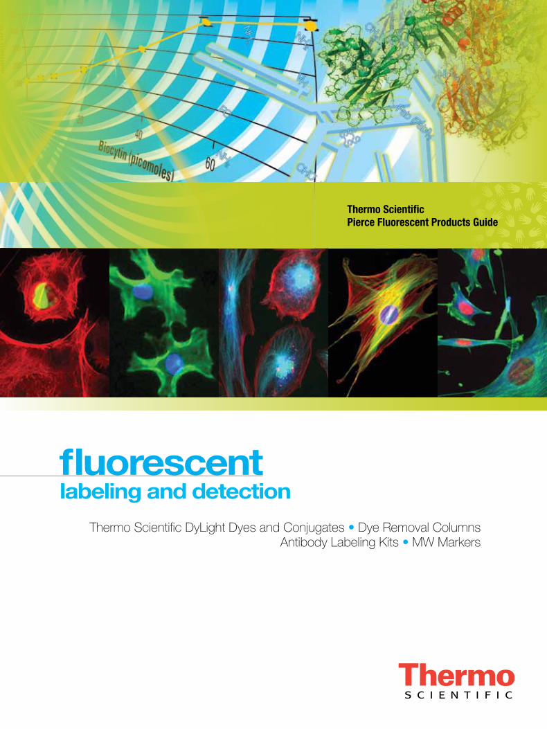
Thermo Scientific Pierce Fluorescent Products Guide
fluorescent
Thermo Scientific DyLight Dyes and Conjugates • Dye Removal Columns Antibody Labeling Kits • MW Markers
labeling and detection
For more information, or to download product instructions, visit www.thermoscientific.com/pierce
Fluorescent Products Guide
of contentstable
Fluorescent Labeling 1–17
Introduction 1
ThermoScientificDyLightAmine-ReactiveandSulfhydryl-ReactiveDyes 2–3
ThermoScientificDyLightSpecialtyDyes 4–5
ThermoScientificDyLightLongStokeShift(LS)Dyes 6
ThermoScientificDyLightFluorescentQuenchers 7
ThermoScientificDyLightAntibodyLabelingKits 8–9
ThermoScientificDyLightFluorPhosphineReagents 10
ThermoScientificFluoresceinDyes 11–12
ThermoScientificRhodamineDyes 13
ThermoScientificPierceImmunostainEnhancer 14
ThermoScientificDAPIStain 15
ThermoScientificHoechst33342Stain 16–17
Thermo Scientific DyLight Conjugates 18–19
LI-COR Odyssey Instrument Products 20–21
Fluorescence Biotin Quantitation Kit 22–23
Fluorescent Protein Gel Stains 24
1To order, call 800.874.3723 or 815.968.0747. Outside the U.S., contact your local branch office or distributor.
Bright new alternatives to other fluorescent dyes
Thermo Scientific DyLight Amine-Reactive and Sulfhydryl-Reactive Fluorescent Dyes
DyLight® Dyes have absorption spectra ranging from 353nm to 770nm (Table 1) and match the principal output wavelengths of common fluorescence instrumentation. The DyLight Dyes exhibit higher fluorescence intensity and photostability than Alexa Fluor®, CyDye® and LI-COR® Dyes in many applications and remain highly fluorescent over a broad pH range (pH 4-9). Additionally, the water solubility of the DyLight Dyes allows a high dye-to-protein ratio without precipitation during conjugation.
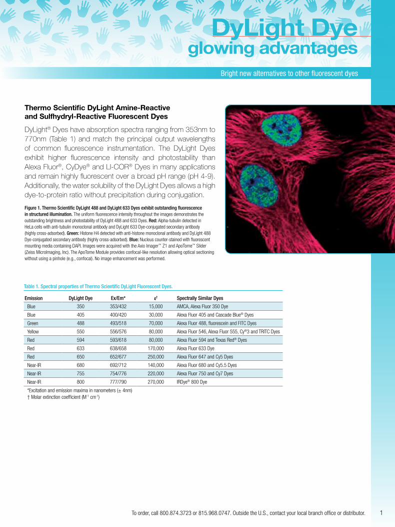
Figure 1. Thermo Scientific DyLight 488 and DyLight 633 Dyes exhibit outstanding fluorescence in structured illumination. The uniform fluorescence intensity throughout the images demonstrates the outstanding brightness and photostability of DyLight 488 and 633 Dyes. Red: Alpha-tubulin detected in HeLa cells with anti-tubulin monoclonal antibody and DyLight 633 Dye-conjugated secondary antibody (highly cross-adsorbed). Green: Histone H4 detected with anti-histone monoclonal antibody and DyLight 488 Dye-conjugated secondary antibody (highly cross-adsorbed). Blue: Nucleus counter-stained with fluorescent mounting media containing DAPI. Images were acquired with the Axio Imager™ Z1 and ApoTome™ Slider (Zeiss MicroImaging, Inc). The ApoTome Module provides confocal-like resolution allowing optical sectioning without using a pinhole (e.g., confocal). No image enhancement was performed.
DyLight Dyeglowing advantages
Table 1. Spectral properties of Thermo Scientific DyLight Fluorescent Dyes.
Emission DyLight Dye Ex/Em* ε† Spectrally Similar Dyes
Blue 350 353/432 15,000 AMCA, Alexa Fluor 350 Dye
Blue 405 400/420 30,000 Alexa Fluor 405 and Cascade Blue® Dyes
Green 488 493/518 70,000 Alexa Fluor 488, fluorescein and FITC Dyes
Yellow 550 556/576 80,000 Alexa Fluor 546, Alexa Fluor 555, Cy®3 and TRITC Dyes
Red 594 593/618 80,000 Alexa Fluor 594 and Texas Red® Dyes
Red 633 638/658 170,000 Alexa Fluor 633 Dye
Red 650 652/677 250,000 Alexa Fluor 647 and Cy5 Dyes
Near-IR 680 692/712 140,000 Alexa Fluor 680 and Cy5.5 Dyes
Near-IR 755 754/776 220,000 Alexa Fluor 750 and Cy7 Dyes
Near-IR 800 777/790 270,000 IRDye® 800 Dye
*Excitation and emission maxima in nanometers (± 4nm)† Molar extinction coefficient (M-1 cm-1)
fluorescent labelingExcellent photostability make these dyes the clear alternative
2 For more information, or to download product instructions, visit www.thermoscientific.com/pierce
Thermo Scientific DyLight Amine-Reactive and Sulfhydryl-Reactive Dyes
Highlights:• Available in both amine- and sulfhydryl-reactive chemistries for fast
and efficient labeling of IgG or other proteins
• High water solubility
• Excellent photostability
• Compatible with common fluorescence instrumentation
Applications: • Fluorescence microscopy
• Western blot detection
• Protein arrays
• Flow cytometry
• ELISA
• FRET-based technology
• And many more
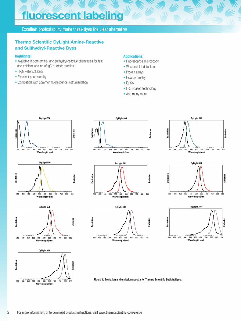
350 400 450 500 550 600 650Wavelength (nm)
700 750 800 850
Exci
tatio
n
Emis
sion
DyLight 350
350 400 450 500 550 600 650 700 750 800 850Wavelength (nm)
Exci
tatio
n
Emis
sion
DyLight 405
350 400 450 500 550 600 650 700 750 800 850Wavelength (nm)
Exci
tatio
n
Emis
sion
DyLight 488
350 400 450 500 550 600 650 700 750 800 850Wavelength (nm)
Exci
tatio
n
Emis
sion
DyLight 550
350 400 450 500 550 600 650 700 750 800 850Wavelength (nm)
Exci
tatio
n
Emis
sion
DyLight 594
350 400 450 500 550 600 650 700 750 800 850Wavelength (nm)
Exci
tatio
n
Emis
sion
DyLight 633
350 400 450 500 550 600 650 700 750 800 850Wavelength (nm)
Exci
tatio
n
Emis
sion
DyLight 650
Wavelength (nm)
Exci
tatio
n
Emis
sion
DyLight 680
350 400 450 500 550 600 650 700 750 800 850 350 400 450 500 550 600 650 700 750 800 850Wavelength (nm)
Exci
tatio
n
Emis
sion
DyLight 755
350 400 450 500 550 600 650 700 750 800 850Wavelength (nm)
Exci
tatio
n
Emis
sion
DyLight 800
Figure 1. Excitation and emission spectra for Thermo Scientific DyLight Dyes.
3To order, call 800.874.3723 or 815.968.0747. Outside the U.S., contact your local branch office or distributor.
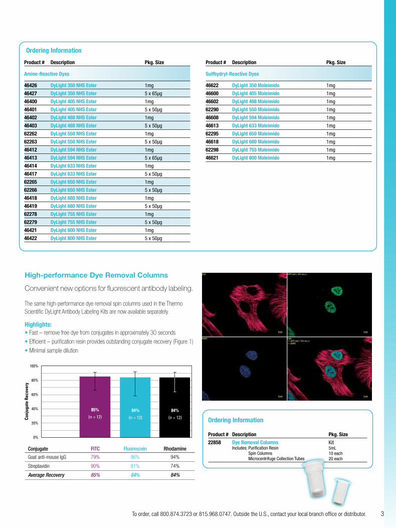
High-performance Dye Removal Columns
Convenient new options for fluorescent antibody labeling.
The same high-performance dye removal spin columns used in the Thermo Scientific DyLight Antibody Labeling Kits are now available separately.
Highlights:• Fast – remove free dye from conjugates in approximately 30 seconds
• Efficient – purification resin provides outstanding conjugate recovery (Figure 1)
• Minimal sample dilution
0.0%
20.0%
40.0%
60.0%
80.0%
100.0%
1
FITC-SA FITC-GAM Fluorescein-SA Fluorescein-GAM Rhodamine-SA Rhodamine-GAM
70.0%
75.0%
80.0%
85.0%
90.0%
95.0%
100.0%
1
FITC Fluorescein Rhodamine
70.0%
75.0%
80.0%
85.0%
90.0%
95.0%
100.0%
1
100%
80%
60%
40%
20%
0%
85%
(n = 12) 84%
(n = 12)
84%
(n = 12) Conj
ugat
e Re
cove
ry
Conjugate FITC Fluorescein Rhodamine
Goat anti-mouse IgG 79% 86% 94%
Streptavidin 90% 81% 74%
Average Recovery 85% 84% 84%
Ordering Information
Product # Description Pkg. Size
Amine-Reactive Dyes
46426 DyLight 350 NHS Ester 1mg
46427 DyLight 350 NHS Ester 5 x 65µg
46400 DyLight 405 NHS Ester 1mg
46401 DyLight 405 NHS Ester 5 x 50µg
46402 DyLight 488 NHS Ester 1mg
46403 DyLight 488 NHS Ester 5 x 50µg
62262 DyLight 550 NHS Ester 1mg
62263 DyLight 550 NHS Ester 5 x 50µg
46412 DyLight 594 NHS Ester 1mg
46413 DyLight 594 NHS Ester 5 x 65µg
46414 DyLight 633 NHS Ester 1mg
46417 DyLight 633 NHS Ester 5 x 50µg
62265 DyLight 650 NHS Ester 1mg
62266 DyLight 650 NHS Ester 5 x 50µg
46418 DyLight 680 NHS Ester 1mg
46419 DyLight 680 NHS Ester 5 x 50µg
62278 DyLight 755 NHS Ester 1mg
62279 DyLight 755 NHS Ester 5 x 50µg
46421 DyLight 800 NHS Ester 1mg
46422 DyLight 800 NHS Ester 5 x 50µg
Product # Description Pkg. Size
Sulfhydryl-Reactive Dyes
46622 DyLight 350 Maleimide 1mg
46600 DyLight 405 Maleimide 1mg
46602 DyLight 488 Maleimide 1mg
62290 DyLight 550 Maleimide 1mg
46608 DyLight 594 Maleimide 1mg
46613 DyLight 633 Maleimide 1mg
62295 DyLight 650 Maleimide 1mg
46618 DyLight 680 Maleimide 1mg
62298 DyLight 755 Maleimide 1mg
46621 DyLight 800 Maleimide 1mg
Ordering Information
Product # Description Pkg. Size
22858 Dye Removal ColumnsIncludes: Purification Resin
Spin Columns Microcentrifuge Collection Tubes
Kit5mL 10 each 20 each
fluorescent labelingExcellent photostability make these dyes the clear alternative
4 For more information, or to download product instructions, visit www.thermoscientific.com/pierce
Thermo Scientific DyLight Specialty Dyes
DyLight Specialty Dyes consist of fluorophores which vary in spectral characteristics, degree of sulfonation, charge and hydrophobicity. These different characteristics allow the dyes to be used in wide range of applications, including protein coupling, imaging, FRET, microarrays and PCR.
The large selection of near infrared (NIR) and infrared (IR) dyes may be used in variety of in vivo and near IR fluorescence (NIRF) applications. The DyLight Specialty Dyes are offered as amine-reactive NHS esters, which can form covalent bonds with primary amines on proteins. DyLight 690-B1 and DyLight 747 near IR dyes, for use in in vivo applications, are also offered as non-reactive free acid dyes for use as experimental control.
Applications
Blue• Molecular imaging
• Microscopy
• Flow cytometry
Green • Imaging
• Antibody labeling
• Direct immunofluorescence staining
• Flow cytometry
• Fluorescence correlation spectroscopy
• ELISA
• Western blotting
• Protein microarrays
• Polymer labeling
• Peptide labeling
• Phalloidin labeling for actin staining
• Fluorescence imaging
• Fluorescence correlation spectroscopy
• Flow cytometry
Yellow• Fluorescence imaging
• Fluorescence correlation spectroscopy
• Flow cytometry
• Imaging
• Antibody labeling
• Direct immunofluorescence staining
• ELISA
• Western blotting
• Protein microarrays
• Polymer labeling
• Peptide labeling
• Phalloidin labeling for actin staining
Orange• Imaging
• Antibody labeling
• Direct immunofluorescence staining
• Flow cytometry
• Fluorescence correlation spectroscopy
• ELISA
• Western blotting
• Protein microarrays
• Polymer labeling
• Peptide labeling
• Phalloidin labeling for actin staining
• Staining in acidic media
• Biofilm microorganism staining
Red• Fluorescence imaging
• Confocal microscopy
• Flow cytometry
• Spectral fluorescence imaging
• In vivo imaging
• Fluorescent western blotting
• Protein microarrays
• Antibody labeling
• Peptide labeling
• Fluorescence correlation spectroscopy
• Protein arrays
• Single molecule detection
• Nanoparticle conjugation
• Biotin/streptavidin conjugation
Far Red• Fluorescence imaging
• Confocal microscopy
• Flow cytometry
• Spectral fluorescence imaging
• In vivo imaging
• Fluorescent western blotting
• Protein microarrays
• Antibody labeling
• Peptide labeling
• Fluorescence correlation spectroscopy
• Protein arrays
• Single molecule detection
• Nanoparticle conjugation
• Biotin/streptavidin conjugation
Near IR• In vivo or ex vivo imaging
• Tumor imaging with labeled peptides
• NIR fluorescence (NIRF) imaging of labeled silica nanoparticles
• NIR in vitro imaging and characterization
• Determination of thermal stability
• Cytotoxicity assays
• Molecular imaging
• UV-VIS-NIR spectroscopy
• Fluorescence correlation spectroscopy
• MRI applications
• DNA sequencing
• Primer labeling for PCR
• 2-D gel electrophoresis
• Flow cytometry/fluorescence-activated cell sorting (FACS)
• Laser scanning confocal microscopy
5To order, call 800.874.3723 or 815.968.0747. Outside the U.S., contact your local branch office or distributor.
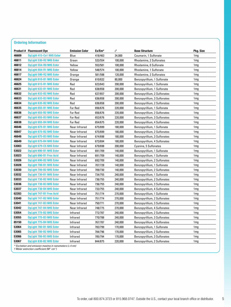
Ordering Information
Product # Fluorescent Dye Emission Color Ex/Em* ε† Base Structure Pkg. Size
46609 DyLight 415-Co1 NHS Ester Blue 418/463 34,000 Coumarin, 1 Sulfonate 1mg
46611 DyLight 530-R2 NHS Ester Green 533/554 100,000 Rhodamine, 2 Sulfonates 1mg
46612 DyLight 554-R0 NHS Ester Yellow 553/581 100,000 Rhodamine, 0 Sulfonate 1mg
46614 DyLight 554-R1 NHS Ester Yellow 548/574 100,000 Rhodamine, 1 Sulfonate 1mg
46617 DyLight 590-R2 NHS Ester Orange 581/598 120,000 Rhodamine, 2 Sulfonates 1mg
46624 DyLight 610-B1 NHS Ester Orange 610/632 80,000 Benzopyrillium, 1 Sulfonate 1mg
46625 DyLight 615-B1 NHS Ester Red 623/643 200,000 Benzopyrillium, 1 Sulfonate 1mg
46631 DyLight 633-B1 NHS Ester Red 638/658 200,000 Benzopyrillium, 1 Sulfonate 1mg
46632 DyLight 633-B2 NHS Ester Red 637/657 200,000 Benzopyrillium, 2 Sulfonates 1mg
46633 DyLight 633-B3 NHS Ester Red 636/658 200,000 Benzopyrillium, 3 Sulfonates 1mg
46634 DyLight 635-B2 NHS Ester Red 636/658 200,000 Benzopyrillium, 2 Sulfonates 1mg
46635 DyLight 655-B1 NHS Ester Far Red 656/676 220,000 Benzopyrillium, 1 Sulfonate 1mg
46636 DyLight 655-B2 NHS Ester Far Red 656/676 220,000 Benzopyrillium, 2 Sulfonates 1mg
46637 DyLight 655-B3 NHS Ester Far Red 653/676 220,000 Benzopyrillium, 3 Sulfonates 1mg
46638 DyLight 655-B4 NHS Ester Far Red 654/675 220,000 Benzopyrillium, 4 Sulfonates 1mg
46646 DyLight 675-B1 NHS Ester Near Infrared 675/699 180,000 Benzopyrillium, 1 Sulfonate 1mg
46647 DyLight 675-B2 NHS Ester Near Infrared 675/699 180,000 Benzopyrillium, 2 Sulfonates 1mg
46648 DyLight 675-B3 NHS Ester Near Infrared 674/698 180,000 Benzopyrillium, 3 Sulfonates 1mg
46649 DyLight 675-B4 NHS Ester Near Infrared 673/694 180,000 Benzopyrillium, 4 Sulfonates 1mg
53003 DyLight 679-C5 NHS Ester Near Infrared 679/698 200,000 Cyanine, 5 Sulfonates 1mg
53022 DyLight 690-B1 NHS Ester Near Infrared 691/709 140,000 Benzopyrillium, 1 Sulfonate 1mg
53023 DyLight 690-B1 Free Acid Near Infrared 691/709 140,000 Benzopyrillium, 1 Sulfonate 1mg
53026 DyLight 690-B2 NHS Ester Near Infrared 692/709 140,000 Benzopyrillium, 2 Sulfonates 1mg
53028 DyLight 700-B1 NHS Ester Near Infrared 707/728 140,000 Benzopyrillium, 1 Sulfonate 1mg
53030 DyLight 700-B2 NHS Ester Near Infrared 709/730 140,000 Benzopyrillium, 2 Sulfonates 1mg
53032 DyLight 730-B1 NHS Ester Near Infrared 734/755 240,000 Benzopyrillium, 1 Sulfonate 1mg
53033 DyLight 730-B2 NHS Ester Near Infrared 736/755 240,000 Benzopyrillium, 2 Sulfonates 1mg
53036 DyLight 730-B3 NHS Ester Near Infrared 736/755 240,000 Benzopyrillium, 3 Sulfonates 1mg
53037 DyLight 730-B4 NHS Ester Near Infrared 733/755 240,000 Benzopyrillium, 4 Sulfonates 1mg
62283 DyLight 747-B1 Free Acid Near Infrared 751/774 270,000 Benzopyrillium, 1 Sulfonate 1mg
53040 DyLight 747-B2 NHS Ester Near Infrared 751/774 270,000 Benzopyrillium, 2 Sulfonates 1mg
53041 DyLight 747-B3 NHS Ester Near Infrared 750/771 270,000 Benzopyrillium, 3 Sulfonates 1mg
53042 DyLight 747-B4 NHS Ester Near Infrared 748/775 270,000 Benzopyrillium, 4 Sulfonates 1mg
53054 DyLight 775-B2 NHS Ester Infrared 772/787 240,000 Benzopyrillium, 2 Sulfonates 1mg
53055 DyLight 775-B3 NHS Ester Infrared 770/788 240,000 Benzopyrillium, 3 Sulfonates 1mg
85150 DyLight 775-B4 NHS Ester Infrared 767/787 240,000 Benzopyrillium, 4 Sulfonates 1mg
53064 DyLight 780-B1 NHS Ester Infrared 783/799 170,000 Benzopyrillium, 1 Sulfonate 1mg
53065 DyLight 780-B2 NHS Ester Infrared 784/796 170,000 Benzopyrillium, 2 Sulfonates 1mg
53066 DyLight 780-B3 NHS Ester Infrared 785/794 170,000 Benzopyrillium, 3 Sulfonates 1mg
53067 DyLight 830-B2 NHS Ester Infrared 844/875 220,000 Benzopyrillium, 2 Sulfonates 1mg
*Excitationandemissionmaximainnanometers(±4nm)†Molarextinctioncoefficient(M-1cm-1)
Excellent photostability make these dyes the clear alternative
6 For more information, or to download product instructions, visit www.thermoscientific.com/pierce
fluorescent labeling
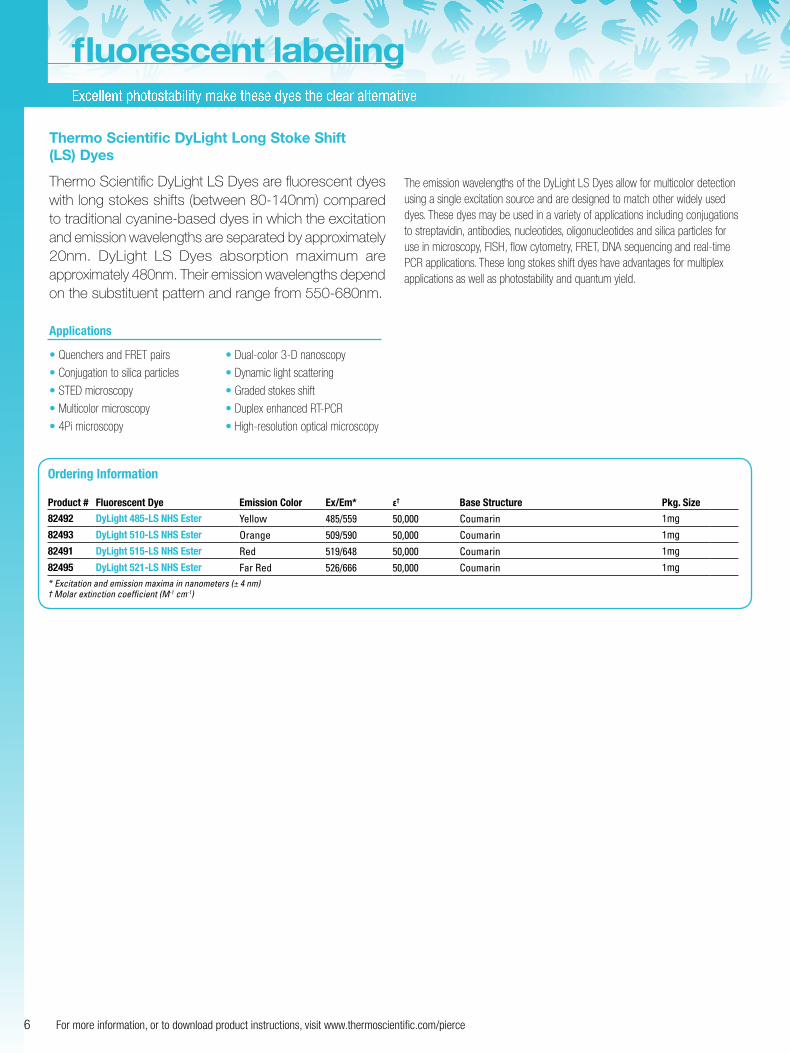
Thermo Scientific DyLight Long Stoke Shift (LS) Dyes
Thermo Scientific DyLight LS Dyes are fluorescent dyes with long stokes shifts (between 80-140nm) compared to traditional cyanine-based dyes in which the excitation and emission wavelengths are separated by approximately 20nm. DyLight LS Dyes absorption maximum are approximately 480nm. Their emission wavelengths depend on the substituent pattern and range from 550-680nm.
The emission wavelengths of the DyLight LS Dyes allow for multicolor detection using a single excitation source and are designed to match other widely used dyes. These dyes may be used in a variety of applications including conjugations to streptavidin, antibodies, nucleotides, oligonucleotides and silica particles for use in microscopy, FISH, flow cytometry, FRET, DNA sequencing and real-time PCR applications. These long stokes shift dyes have advantages for multiplex applications as well as photostability and quantum yield.
Applications
• Quenchers and FRET pairs
• Conjugation to silica particles
• STED microscopy
• Multicolor microscopy
• 4Pi microscopy
• Dual-color 3-D nanoscopy
• Dynamic light scattering
• Graded stokes shift
• Duplex enhanced RT-PCR
• High-resolution optical microscopy
Ordering Information
Product # Fluorescent Dye Emission Color Ex/Em* ε† Base Structure Pkg. Size
82492 DyLight 485-LS NHS Ester Yellow 485/559 50,000 Coumarin 1mg
82493 DyLight 510-LS NHS Ester Orange 509/590 50,000 Coumarin 1mg
82491 DyLight 515-LS NHS Ester Red 519/648 50,000 Coumarin 1mg
82495 DyLight 521-LS NHS Ester Far Red 526/666 50,000 Coumarin 1mg
*Excitationandemissionmaximainnanometers(±4nm)†Molarextinctioncoefficient(M-1cm-1)
7To order, call 800.874.3723 or 815.968.0747. Outside the U.S., contact your local branch office or distributor.
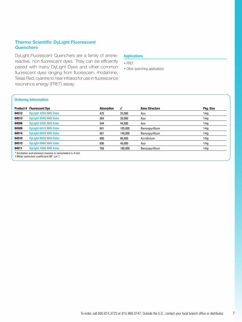
Thermo Scientific DyLight Fluorescent Quenchers
DyLight Fluorescent Quenchers are a family of amine-reactive, non-fluorescent dyes. They can be efficiently paired with many DyLight Dyes and other common fluorescent dyes ranging from fluorescein, rhodamine, Texas Red, cyanine to near infrared for use in fluorescence resonance energy (FRET) assay.
Applications
• FRET
• Other quenching applications
Ordering Information
Product # Fluorescent Dye Absorption ε† Base Structure Pkg. Size
84512 DyLight 425Q NHS Ester 425 25,000 Azo 1mg
84513 DyLight 504Q NHS Ester 504 39,000 Azo 1mg
84508 DyLight 543Q NHS Ester 544 44,000 Azo 1mg
84509 DyLight 641Q NHS Ester 641 105,000 Benzopyrillium 1mg
84514 DyLight 662Q NHS Ester 661 140,000 Benzopyrillium 1mg
84510 DyLight 683Q NHS Ester 680 80,000 Acridinium 1mg
84515 DyLight 696Q NHS Ester 696 49,000 Azo 1mg
84511 DyLight 766Q NHS Ester 766 180,000 Benzopyrillium 1mg
*Excitationandemissionmaximainnanometers(±4nm)†Molarextinctioncoefficient(M-1cm-1)
Label and purify antibodies in one hour
8 For more information, or to download product instructions, visit www.thermoscientific.com/pierce
fluorescent labeling
Thermo Scientific DyLight Antibody Labeling Kits
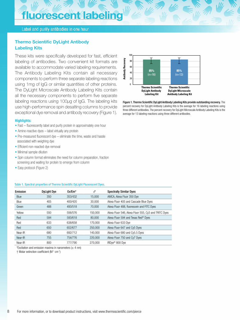
These kits were specifically developed for fast, efficient labeling of antibodies. Two convenient kit formats are available to accommodate varied labeling requirements. The Antibody Labeling Kits contain all necessary components to perform three separate labeling reactions using 1mg of IgG or similar quantities of other proteins. The DyLight Microscale Antibody Labeling Kits contain all the necessary components to perform five separate labeling reactions using 100µg of IgG. The labeling kits use high-performance spin desalting columns to provide exceptional dye removal and antibody recovery (Figure 1).
Highlights:• Fast – fluorescently label and purify protein in approximately one hour
• Amine-reactive dyes – label virtually any protein
• Pre-measured fluorescent dye – eliminate the time, waste and hassle associated with weighing dye
• Efficient non-reacted dye removal
• Minimal sample dilution
• Spin column format eliminates the need for column preparation, fraction screening and waiting for protein to emerge from column
• Easy protocol (Figure 2)
0
20
40
60
80
100
Thermo ScientificDyLight Antibody
Labeling Kit
85%(n=16)
85%(n=13)
Thermo ScientificDyLight Microscale
Antibody Labeling Kit
Ant
ibod
y Re
cove
ry
Figure 1. Thermo Scientific DyLight Antibody Labeling Kits provide outstanding recovery. The percent recovery for DyLight Antibody Labeling Kits is the average for 16 labeling reactions using three different antibodies. The percent recovery for DyLight Microscale Antibody Labeling Kits is the average for 13 labeling reactions using three different antibodies.
Table 1. Spectral properties of Thermo Scientific DyLight Fluorescent Dyes.
Emission DyLight Dye Ex/Em* ε† Spectrally Similar Dyes
Blue 350 353/432 15,000 AMCA, Alexa Fluor 350 Dye
Blue 405 400/420 30,000 Alexa Fluor 405 and Cascade Blue Dyes
Green 488 493/518 70,000 Alexa Fluor 488, fluorescein and FITC Dyes
Yellow 550 556/576 150,000 Alexa Fluor 546, Alexa Fluor 555, Cy3 and TRITC Dyes
Red 594 593/618 80,000 Alexa Fluor 594 and Texas Red® Dyes
Red 633 638/658 170,000 Alexa Fluor 633 Dye
Red 650 652/677 250,000 Alexa Fluor 647 and Cy5 Dyes
Near-IR 680 692/712 140,000 Alexa Fluor 680 and Cy5.5 Dyes
Near-IR 755 754/776 220,000 Alexa Fluor 750 and Cy7 Dyes
Near-IR 800 777/790 270,000 IRDye® 800 Dye
*Excitation and emission maxima in nanometers (± 4 nm)† Molar extinction coefficient (M-1 cm-1)
9To order, call 800.874.3723 or 815.968.0747. Outside the U.S., contact your local branch office or distributor.
Microscale Kits
Contains sufficient reagent to label and purify 5 x100µg of IgG.
All Microscale Kits include:
• Appropriate DyLight NHS Ester, 5 vials
• Reaction Buffer, 1mL
• Spin Columns, 5 each
• Microcentrifuge Collection Tubes, 10 each
• Purification Resin, 5mL
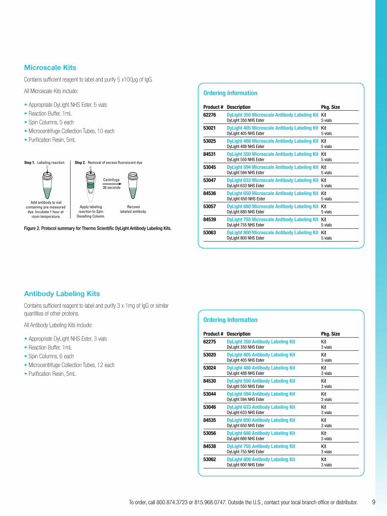
Step 1. Labeling reaction
Add antibody to vialcontaining pre-measured
dye. Incubate 1 hour at room temperature.
Apply labelingreaction to Spin
Desalting Column.
Recoverlabeled antibody.
Centrifuge
30 seconds
Step 2. Removal of excess fluorescent dye
Figure 2. Protocol summary for Thermo Scientific DyLight Antibody Labeling Kits.
Antibody Labeling Kits
Contains sufficient reagent to label and purify 3 x 1mg of IgG or similar quantities of other proteins.
All Antibody Labeling Kits include:
• Appropriate DyLight NHS Ester, 3 vials
• Reaction Buffer, 1mL
• Spin Columns, 6 each
• Microcentrifuge Collection Tubes, 12 each
• Purification Resin, 5mL
Ordering Information
Product # Description Pkg. Size
62276 DyLight 350 Microscale Antibody Labeling KitDyLight 350 NHS Ester
Kit5 vials
53021 DyLight 405 Microscale Antibody Labeling KitDyLight 405 NHS Ester
Kit5 vials
53025 DyLight 488 Microscale Antibody Labeling KitDyLight 488 NHS Ester
Kit5 vials
84531 DyLight 550 Microscale Antibody Labeling KitDyLight 550 NHS Ester
Kit5 vials
53045 DyLight 594 Microscale Antibody Labeling KitDyLight 594 NHS Ester
Kit5 vials
53047 DyLight 633 Microscale Antibody Labeling KitDyLight 633 NHS Ester
Kit5 vials
84536 DyLight 650 Microscale Antibody Labeling KitDyLight 650 NHS Ester
Kit 5 vials
53057 DyLight 680 Microscale Antibody Labeling KitDyLight 680 NHS Ester
Kit5 vials
84539 DyLight 755 Microscale Antibody Labeling KitDyLight 755 NHS Ester
Kit5 vials
53063 DyLight 800 Microscale Antibody Labeling KitDyLight 800 NHS Ester
Kit 5 vials
Ordering Information
Product # Description Pkg. Size
62275 DyLight 350 Antibody Labeling KitDyLight 350 NHS Ester
Kit 3 vials
53020 DyLight 405 Antibody Labeling KitDyLight 405 NHS Ester
Kit 3 vials
53024 DyLight 488 Antibody Labeling KitDyLight 488 NHS Ester
Kit 3 vials
84530 DyLight 550 Antibody Labeling KitDyLight 550 NHS Ester
Kit 3 vials
53044 DyLight 594 Antibody Labeling KitDyLight 594 NHS Ester
Kit 3 vials
53046 DyLight 633 Antibody Labeling KitDyLight 633 NHS Ester
Kit 3 vials
84535 DyLight 650 Antibody Labeling KitDyLight 650 NHS Ester
Kit 3 vials
53056 DyLight 680 Antibody Labeling KitDyLight 680 NHS Ester
Kit 3 vials
84538 DyLight 755 Antibody Labeling KitDyLight 755 NHS Ester
Kit 3 vials
53062 DyLight 800 Antibody Labeling KitDyLight 800 NHS Ester
Kit 3 vials
Fluorescent probes for Staudinger ligation and detection of azide-labeled cellular targets
10 For more information, or to download product instructions, visit www.thermoscientific.com/pierce
fluorescent labeling
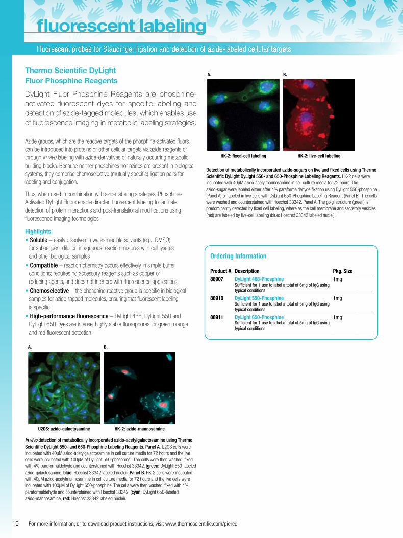
Thermo Scientific DyLight Fluor Phosphine Reagents
DyLight Fluor Phosphine Reagents are phosphine-activated fluorescent dyes for specific labeling and detection of azide-tagged molecules, which enables use of fluorescence imaging in metabolic labeling strategies.
Azide groups, which are the reactive targets of the phosphine-activated fluors, can be introduced into proteins or other cellular targets via azide reagents or through in vivo labeling with azide-derivatives of naturally occurring metabolic building blocks. Because neither phosphines nor azides are present in biological systems, they comprise chemoselective (mutually specific) ligation pairs for labeling and conjugation.
Thus, when used in combination with azide labeling strategies, Phosphine-Activated DyLight Fluors enable directed fluorescent labeling to facilitate detection of protein interactions and post-translational modifications using fluorescence imaging technologies.
Highlights:• Soluble – easily dissolves in water-miscible solvents (e.g., DMSO)
for subsequent dilution in aqueous reaction mixtures with cell lysates and other biological samples
• Compatible – reaction chemistry occurs effectively in simple buffer conditions; requires no accessory reagents such as copper or reducing agents, and does not interfere with fluorescence applications
• Chemoselective – the phosphine reactive group is specific in biological samples for azide-tagged molecules, ensuring that fluorescent labeling is specific
• High-performance fluorescence – DyLight 488, DyLight 550 and DyLight 650 Dyes are intense, highly stable fluorophores for green, orange and red fluorescent detection.
A.
U2OS: azido-galactosamine HK-2: azido-mannosamine
B.
In vivo detection of metabolically incorporated azido-acetylgalactosamine using Thermo Scientific DyLight 550- and 650-Phosphine Labeling Reagents. Panel A. U2OS cells were incubated with 40µM azido-acetylgalactosamine in cell culture media for 72 hours and the live cells were incubated with 100µM of DyLight 550-phosphine . The cells were then washed, fixed with 4% paraformaldehyde and counterstained with Hoechst 33342. (green: DyLight 550-labeled azido-galactosamine, blue: Hoechst 33342 labeled nuclei). Panel B. HK-2 cells were incubated with 40µM azido-acetylmannosamine in cell culture media for 72 hours and the live cells were incubated with 100µM of DyLight 650-phosphine. The cells were then washed, fixed with 4% paraformaldehyde and counterstained with Hoechst 33342. (cyan: DyLight 650-labeled azido-mannosamine, red: Hoechst 33342 labeled nuclei).
A.
HK-2: fixed-cell labeling HK-2: live-cell labeling
B.
Detection of metabolically incorporated azido-sugars on live and fixed cells using Thermo Scientific DyLight DyLight 550- and 650-Phosphine Labeling Reagents. HK-2 cells were incubated with 40µM azido-acetylmannosamine in cell culture media for 72 hours. The azido-sugar were labeled either after 4% paraformaldehyde fixation using DyLight 550-phosphine (Panel A) or labeled in live cells with DyLight 650-Phosphine Labeling Reagent (Panel B). The cells were washed and counterstained with Hoechst 33342. Panel A. The golgi structure (green) is predominantly detected by fixed cell labeling, where as the cell membrane and secretory vesicles (red) are labeled by live-cell labeling (blue: Hoechst 33342 labeled nuclei).
Ordering Information
Product # Description Pkg. Size
88907 DyLight 488-PhosphineSufficient for 1 use to label a total of 6mg of IgG using typical conditions
1mg
88910 DyLight 550-PhosphineSufficient for 1 use to label a total of 5mg of IgG using typical conditions
1mg
88911 DyLight 650-Phosphine Sufficient for 1 use to label a total of 5mg of IgG using typical conditions
1mg
11To order, call 800.874.3723 or 815.968.0747. Outside the U.S., contact your local branch office or distributor.
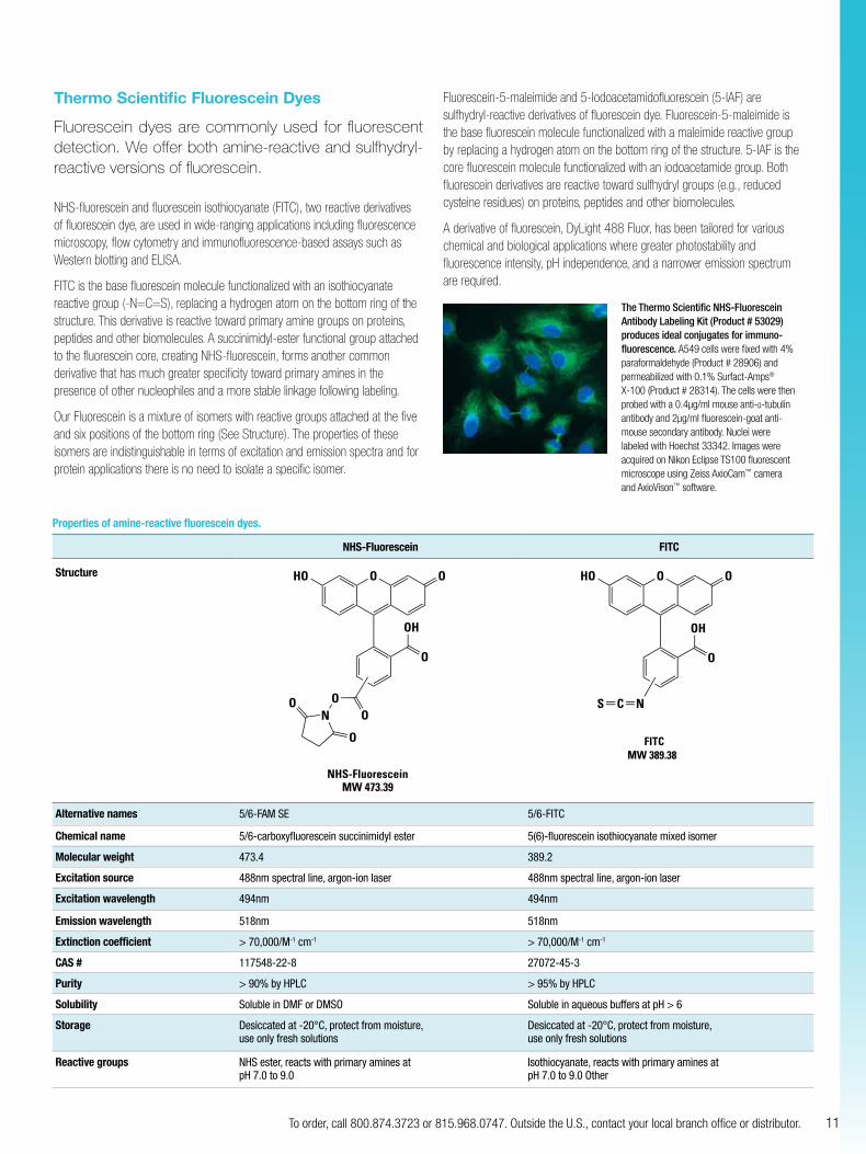
Thermo Scientific Fluorescein Dyes
Fluorescein dyes are commonly used for fluorescent detection. We offer both amine-reactive and sulfhydryl-reactive versions of fluorescein.
NHS-fluorescein and fluorescein isothiocyanate (FITC), two reactive derivatives of fluorescein dye, are used in wide-ranging applications including fluorescence microscopy, flow cytometry and immunofluorescence-based assays such as Western blotting and ELISA.
FITC is the base fluorescein molecule functionalized with an isothiocyanate reactive group (-N=C=S), replacing a hydrogen atom on the bottom ring of the structure. This derivative is reactive toward primary amine groups on proteins, peptides and other biomolecules. A succinimidyl-ester functional group attached to the fluorescein core, creating NHS-fluorescein, forms another common derivative that has much greater specificity toward primary amines in the presence of other nucleophiles and a more stable linkage following labeling.
Our Fluorescein is a mixture of isomers with reactive groups attached at the five and six positions of the bottom ring (See Structure). The properties of these isomers are indistinguishable in terms of excitation and emission spectra and for protein applications there is no need to isolate a specific isomer.
Fluorescein-5-maleimide and 5-Iodoacetamidofluorescein (5-IAF) are sulfhydryl-reactive derivatives of fluorescein dye. Fluorescein-5-maleimide is the base fluorescein molecule functionalized with a maleimide reactive group by replacing a hydrogen atom on the bottom ring of the structure. 5-IAF is the core fluorescein molecule functionalized with an iodoacetamide group. Both fluorescein derivatives are reactive toward sulfhydryl groups (e.g., reduced cysteine residues) on proteins, peptides and other biomolecules.
A derivative of fluorescein, DyLight 488 Fluor, has been tailored for various chemical and biological applications where greater photostability and fluorescence intensity, pH independence, and a narrower emission spectrum are required.
Properties of amine-reactive fluorescein dyes.
NHS-Fluorescein FITC
Structure
NHS-FluoresceinMW 473.39
O
ON
O
OH
O
OOHO
O
FITCMW 389.38
Ex/Em 494/518
OHO O
O
OH
NCS
Alternative names 5/6-FAM SE 5/6-FITC
Chemical name 5/6-carboxyfluorescein succinimidyl ester 5(6)-fluorescein isothiocyanate mixed isomer
Molecular weight 473.4 389.2
Excitation source 488nm spectral line, argon-ion laser 488nm spectral line, argon-ion laser
Excitation wavelength 494nm 494nm
Emission wavelength 518nm 518nm
Extinction coefficient > 70,000/M-1 cm-1 > 70,000/M-1 cm-1
CAS # 117548-22-8 27072-45-3
Purity > 90% by HPLC > 95% by HPLC
Solubility Soluble in DMF or DMSO Soluble in aqueous buffers at pH > 6
Storage Desiccated at -20°C, protect from moisture, use only fresh solutions
Desiccated at -20°C, protect from moisture, use only fresh solutions
Reactive groups NHS ester, reacts with primary amines at pH 7.0 to 9.0
Isothiocyanate, reacts with primary amines at pH 7.0 to 9.0 Other
The Thermo Scientific NHS-Fluorescein Antibody Labeling Kit (Product # 53029) produces ideal conjugates for immuno- fluorescence. A549 cells were fixed with 4% paraformaldehyde (Product # 28906) and permeabilized with 0.1% Surfact-Amps® X-100 (Product # 28314). The cells were then probed with a 0.4µg/ml mouse anti-α-tubulin antibody and 2µg/ml fluorescein-goat anti- mouse secondary antibody. Nuclei were labeled with Hoechst 33342. Images were acquired on Nikon Eclipse TS100 fluorescent microscope using Zeiss AxioCam™ camera and AxioVison™ software.
Other reactive derivatives of fluorescein and rhodamine dyes (cont.)
12 For more information, or to download product instructions, visit www.thermoscientific.com/pierce
fluorescent labeling
Properties of sulfhydryl-reactive dyes.
Fluorescein-5-maleimide 5-Iodoacetamido-fluorescein
Structure
Fluorescein-5-MaleimideMW 427.36
O N O
OH
O
OOHO
5-IAF5-Iodoacetamido-fluorescein
MW 515.25
OH
O
OOHO
NH
O
I
Alternative names 5-MF, 5-maleimido-fluorescein 5-IAF, 5-iodoacetamidofluorescein
Chemical name 1H-Pyrrole-2,5-dione, 1-(3’,6’-dihydroxy-3-oxospiro(isobenzofuran-1(3H),9’-(9H)xanthen-5-yl)-
Acetamide, N-(3’,6’-dihydroxy-3-oxospiro(isobenzofuran-1(3H), 9’-(9H)xanthen)-5-yl)-2-iodo
Molecular weight 427.36 ±3 515.26 ±3
Excitation source 488nm spectral line, argon-ion laser 488nm spectral line, argon-ion laser
Excitation wavelength 494nm 494nm
Emission wavelength 518nm 518nm
Extinction coefficient ~ 68,000/M-1 cm-1 > 80,000/M-1 cm-1
CAS # 75350-46-8 63368-54-7
Solubility Soluble in DMF or DMSO Soluble in DMF; aqueous buffers at pH > 6
Storage Desiccated at -20°C, protect from moisture, use only fresh solutions Desiccated at -20°C, protect from moisture, use only fresh solutions
Reactive groups Maleimide, reacts with sulfhydryls at pH 6.5 to 7.5 Iodoacetamide, reacts with sulfhydryls at pH 7.0 to 7.5
Ordering Information
Product # Description Pkg. Size
46424 FITC (Fluorescein Isothiocyanate) 1g
46425 FITC (Fluorescein Isothiocyanate) 100mg
46409 NHS-Fluorescein 1g
46410 NHS-Fluorescein 100mg
53027 FITC Antibody Labeling KitEfficiently labels and purifies 3 x 1mg of IgG or other protein in about 1 hour.Includes: FITC
Borate Buffer Spin Columns Microcentrifuge Collection Tubes Purification Resin
Kit
3 vials1mL6 each12 each5mL
Product # Description Pkg. Size
53029 Fluorescein Antibody Labeling KitEfficiently labels and purifies 3 x 1mg of IgG or other protein in about 1 hour.Includes: NHS Fluorescein
Borate Buffer Spin Columns Microcentrifuge Collection Tubes Purification Resin
Kit
3 vials 1mL6 each12 each5mL
62245 Fluorescein-5-Maleimide 25mg
62246 5-Iodoacetamido-fluorescein (5-IAF) 25mg
13To order, call 800.874.3723 or 815.968.0747. Outside the U.S., contact your local branch office or distributor.
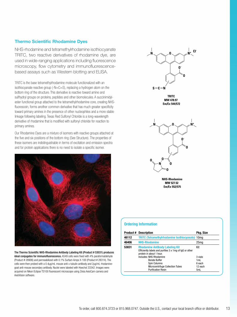
Thermo Scientific Rhodamine Dyes
NHS-rhodamine and tetramethylrhodamine isothiocyanate TRITC, two reactive derivatives of rhodamine dye, are used in wide-ranging applications including fluorescence microscopy, flow cytometry and immunofluorescence-based assays such as Western blotting and ELISA.
TRITC is the base tetramethylrhodamine molecule functionalized with an isothiocyanate reactive group (-N=C=S), replacing a hydrogen atom on the bottom ring of the structure. This derivative is reactive toward amine and sulfhydryl groups on proteins, peptides and other biomolecules. A succinimidyl-ester functional group attached to the tetramethylrhodamine core, creating NHS-fluorescein, forms another common derivative that has much greater specificity toward primary amines in the presence of other nucleophiles and a more stable linkage following labeling. Texas Red Sulfonyl Chloride is a long-wavelength derivative of rhodamine that is modified with sulfonyl chloride for reaction to primary amines.
Our Rhodamine Dyes are a mixture of isomers with reactive groups attached at the five and six positions of the bottom ring (See Structure). The properties of these isomers are indistinguishable in terms of excitation and emission spectra and for protein applications there is no need to isolate a specific isomer.
TRITCMW 478.97
Em/Ex 544/572
ON N+
O
O-
NCS
Cl-
NHS-RhodamineMW 527.52
Em/Ex 552/575
O N+N
O
O-
OO
N
O
O
Ordering Information
Product # Description Pkg. Size
46112 TRITC (Tetramethylrhodamine Isothiocyanate) 10mg
46406 NHS-Rhodamine 25mg
53031 Rhodamine Antibody Labeling KitEfficiently labels and purifies 3 x 1mg of IgG or other protein in about 1 hour.Includes: NHS Rhodamine
Borate Buffer Spin Columns Microcentrifuge Collection Tubes Purification Resin
Kit
3 vials1mL6 each12 each5mL
The Thermo Scientific NHS-Rhodamine Antibody Labeling Kit (Product # 53031) produces ideal conjugates for immunofluorescence. A549 cells were fixed with 4% paraformaldehyde (Product # 28906) and permeabilized with 0.1% Surfact-Amps X-100 (Product # 28314). The cells were then probed with a 0.4µg/mL mouse anti-α-tubulin antibody and 2µg/mL rhodamine-goat anti-mouse secondary antibody. Nuclei were labeled with Hoechst 33342. Images were acquired on Nikon Eclipse TS100 fluorescent microscope using Zeiss AxioCam camera and AxioVision software.
14 For more information, or to download product instructions, visit www.thermoscientific.com/pierce
Alleviates common immunostaining problems such as low signal and poor sensitivity
fluorescent labeling
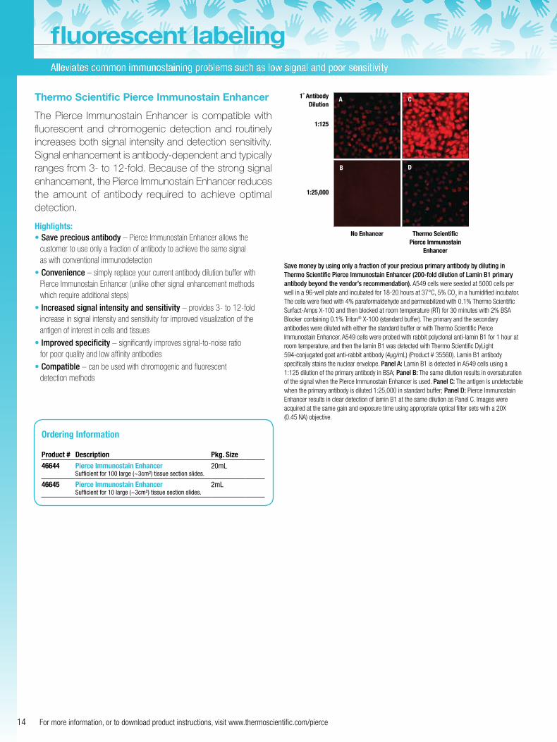
Thermo Scientific Pierce Immunostain Enhancer
The Pierce Immunostain Enhancer is compatible with fluorescent and chromogenic detection and routinely increases both signal intensity and detection sensitivity. Signal enhancement is antibody-dependent and typically ranges from 3- to 12-fold. Because of the strong signal enhancement, the Pierce Immunostain Enhancer reduces the amount of antibody required to achieve optimal detection.
Highlights:• Save precious antibody – Pierce Immunostain Enhancer allows the
customer to use only a fraction of antibody to achieve the same signal as with conventional immunodetection
• Convenience – simply replace your current antibody dilution buffer with Pierce Immunostain Enhancer (unlike other signal enhancement methods which require additional steps)
• Increased signal intensity and sensitivity – provides 3- to 12-fold increase in signal intensity and sensitivity for improved visualization of the antigen of interest in cells and tissues
• Improved specificity – significantly improves signal-to-noise ratiofor poor quality and low affinity antibodies
• Compatible – can be used with chromogenic and fluorescentdetection methods
1˚ Antibody Dilution
1:125
1:25,000
No Enhancer Thermo Scientific Pierce Immunostain
Enhancer
A C
B D
Ordering Information
Product # Description Pkg. Size
46644 Pierce Immunostain EnhancerSufficient for 100 large (~3cm²) tissue section slides.
20mL
46645 Pierce Immunostain EnhancerSufficient for 10 large (~3cm²) tissue section slides.
2mL
Save money by using only a fraction of your precious primary antibody by diluting in Thermo Scientific Pierce Immunostain Enhancer (200-fold dilution of Lamin B1 primary antibody beyond the vendor’s recommendation). A549 cells were seeded at 5000 cells per well in a 96-well plate and incubated for 18-20 hours at 37°C, 5% CO
2 in a humidified incubator.
The cells were fixed with 4% paraformaldehyde and permeabilized with 0.1% Thermo Scientific Surfact-Amps X-100 and then blocked at room temperature (RT) for 30 minutes with 2% BSA Blocker containing 0.1% Triton® X-100 (standard buffer). The primary and the secondary antibodies were diluted with either the standard buffer or with Thermo Scientific Pierce Immunostain Enhancer. A549 cells were probed with rabbit polyclonal anti-lamin B1 for 1 hour at room temperature, and then the lamin B1 was detected with Thermo Scientific DyLight 594-conjugated goat anti-rabbit antibody (4µg/mL) (Product # 35560). Lamin B1 antibody specifically stains the nuclear envelope. Panel A: Lamin B1 is detected in A549 cells using a 1:125 dilution of the primary antibody in BSA; Panel B: The same dilution results in oversaturation of the signal when the Pierce Immunostain Enhancer is used. Panel C: The antigen is undetectable when the primary antibody is diluted 1:25,000 in standard buffer; Panel D: Pierce Immunostain Enhancer results in clear detection of lamin B1 at the same dilution as Panel C. Images were acquired at the same gain and exposure time using appropriate optical filter sets with a 20X (0.45 NA) objective.
15To order, call 800.874.3723 or 815.968.0747. Outside the U.S., contact your local branch office or distributor.
fluorescent cellular stains
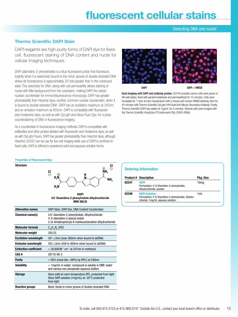
Thermo Scientific DAPI Stain
DAPI reagents are high-purity forms of DAPI dye for fixed-cell, fluorescent staining of DNA content and nuclei for cellular imaging techniques.
DAPI (diamidino-2-phenylindole) is a blue fluorescent probe that fluoresces brightly when it is selectively bound to the minor groove of double-stranded DNA where its fluorescence is approximately 20-fold greater than in the nonbound state. This selectivity for DNA, along with cell permeability allows staining of nuclei with little background from the cytoplasm, making DAPI the classic nuclear counterstain for immunofluorescence microscopy. DAPI has greater photostability than Hoechst dyes, another common nuclear counterstain, when it is bound to double-stranded DNA. DAPI has an excitation maximum at 345nm and an emission maximum at 455nm. DAPI is compatible with fluorescein and rhodamine dyes, as well as with DyLight and Alexa Fluor Dye, for nuclear counterstaining of DNA in fluorescence imaging.
As a counterstain in fluorescence imaging methods, DAPI is compatible with antibodies and other probes labeled with fluorescein and rhodamine dyes, as well as with DyLight Fluors. DAPI has greater photostability than Hoechst dyes, although Hoechst 33342 can be use for live cell imaging while use of DAPI is confined to fixed cells. DAPI is offered in powdered solid and aqueous solution forms.
DAPI + HMGBDAPI
Dual imaging with DAPI and antibody probes. DU145 prostate cancer cells were grown in 96-well plates, fixed with paraformaldehyde and permeabilized for 15 minutes. Cells were incubated for 1 hour at room temperature with a mouse anti-human HMGB antibody, then for 45 minutes with Thermo Scientific DyLight 549 Goat Anti-Mouse Secondary Antibody. Finally, Thermo Scientific DAPI was added at 1µg/mL for 5 minutes. Stained cells were imaged with the Thermo Scientific ArrayScan VTI Instrument (Obj. 20X/0.45NA).
Properties of Fluorescent Dye.
Structure
DAPI4,6’-Diamidino-2-phenylindole dihydrochloride
MW 350.25
H2N
H2N
NH2
NH2Cl– +
Cl
HN
–+
Alternative names DAPI Stain, DAPI Dye, DNA Content Counterstain
Chemical name(s) 4,6’-diamidino-2-phenylindole, dihydrochloride 4’,6-diamidine-2-phenyl indole 2- (4-Amidinophenyl)-6-indolecarbamidine dihydrochloride
Molecular formula C16H15N5 2HCl
Molecular weight 350.25
Excitation wavelength 341 ±3nm (near 360nm when bound to dsDNA)
Emission wavelength 452 ±3nm (456 to 460nm when bound to dsDNA)
Extinction coefficient > 30,600/M-1 cm-1 at 347nm in methanol
CAS # 28718-90-3
Purity > 95% (most lots >98%) by HPLC at 240nm
Solubility > 1mg/mL in water; compound is soluble in DMF, water and various non-phosphate aqueous buffers
Storage Store solid at room temperature (RT), protected from light Store DAPI solution (1mg/mL) at -20°C protected from light
Reactive groups None; binds to minor groove of double-stranded DNA
Detecting DNA and nuclei
Ordering Information
Product # Description Pkg. Size
62247 DAPIFormulation: 4’,6-Diamidino-2-phenylindole, dihydrochloride; powder.
10mg
62248 DAPI SolutionFormulation: 4’,6-Diamidino-2-phenylindole, dihydro-chloride; 1mg/mL aqueous solution.
1mL
Detecting DNA and nuclei (cont.)
16 For more information, or to download product instructions, visit www.thermoscientific.com/pierce
fluorescent cellular stains
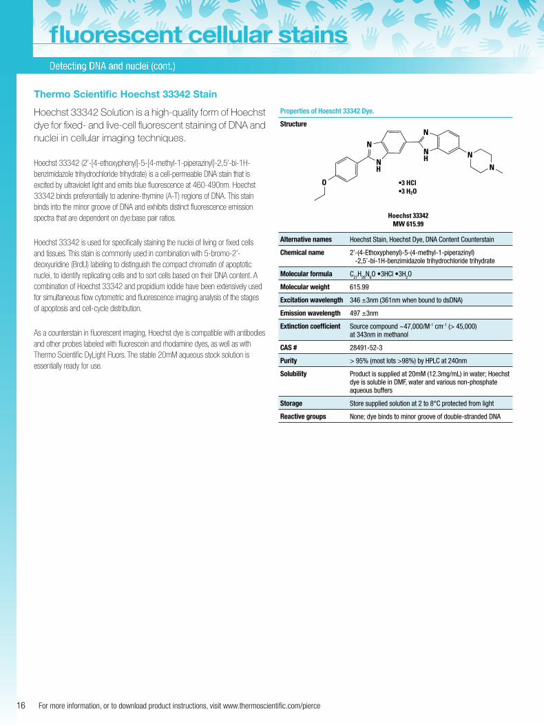
Thermo Scientific Hoechst 33342 Stain
Hoechst 33342 Solution is a high-quality form of Hoechst dye for fixed- and live-cell fluorescent staining of DNA and nuclei in cellular imaging techniques.
Hoechst 33342 (2’-[4-ethoxyphenyl]-5-[4-methyl-1-piperazinyl]-2,5’-bi-1H-benzimidazole trihydrochloride trihydrate) is a cell-permeable DNA stain that is excited by ultraviolet light and emits blue fluorescence at 460-490nm. Hoechst 33342 binds preferentially to adenine-thymine (A-T) regions of DNA. This stain binds into the minor groove of DNA and exhibits distinct fluorescence emission spectra that are dependent on dye:base pair ratios.
Hoechst 33342 is used for specifically staining the nuclei of living or fixed cells and tissues. This stain is commonly used in combination with 5-bromo-2’-deoxyuridine (BrdU) labeling to distinguish the compact chromatin of apoptotic nuclei, to identify replicating cells and to sort cells based on their DNA content. A combination of Hoechst 33342 and propidium iodide have been extensively used for simultaneous flow cytometric and fluorescence imaging analysis of the stages of apoptosis and cell-cycle distribution.
As a counterstain in fluorescent imaging, Hoechst dye is compatible with antibodies and other probes labeled with fluorescein and rhodamine dyes, as well as with Thermo Scientific DyLight Fluors. The stable 20mM aqueous stock solution is essentially ready for use.
Properties of Hoescht 33342 Dye.
Structure
Alternative names Hoechst Stain, Hoechst Dye, DNA Content Counterstain
Chemical name 2’ -(4-Ethoxyphenyl)-5-(4-methyl-1-piperazinyl) -2,5’-bi-1H-benzimidazole trihydrochloride trihydrate
Molecular formula C27H28N6O •3HCl •3H2O
Molecular weight 615.99
Excitation wavelength 346 ±3nm (361nm when bound to dsDNA)
Emission wavelength 497 ±3nm
Extinction coefficient Source compound ~47,000/M-1 cm-1 (> 45,000) at 343nm in methanol
CAS # 28491-52-3
Purity > 95% (most lots >98%) by HPLC at 240nm
Solubility Product is supplied at 20mM (12.3mg/mL) in water; Hoechst dye is soluble in DMF, water and various non-phosphate aqueous buffers
Storage Store supplied solution at 2 to 8°C protected from light
Reactive groups None; dye binds to minor groove of double-stranded DNA
Hoechst 33342MW 615.99
•3 HCl•3 H2O
N
O
NH
N
NH N
N
17To order, call 800.874.3723 or 815.968.0747. Outside the U.S., contact your local branch office or distributor.
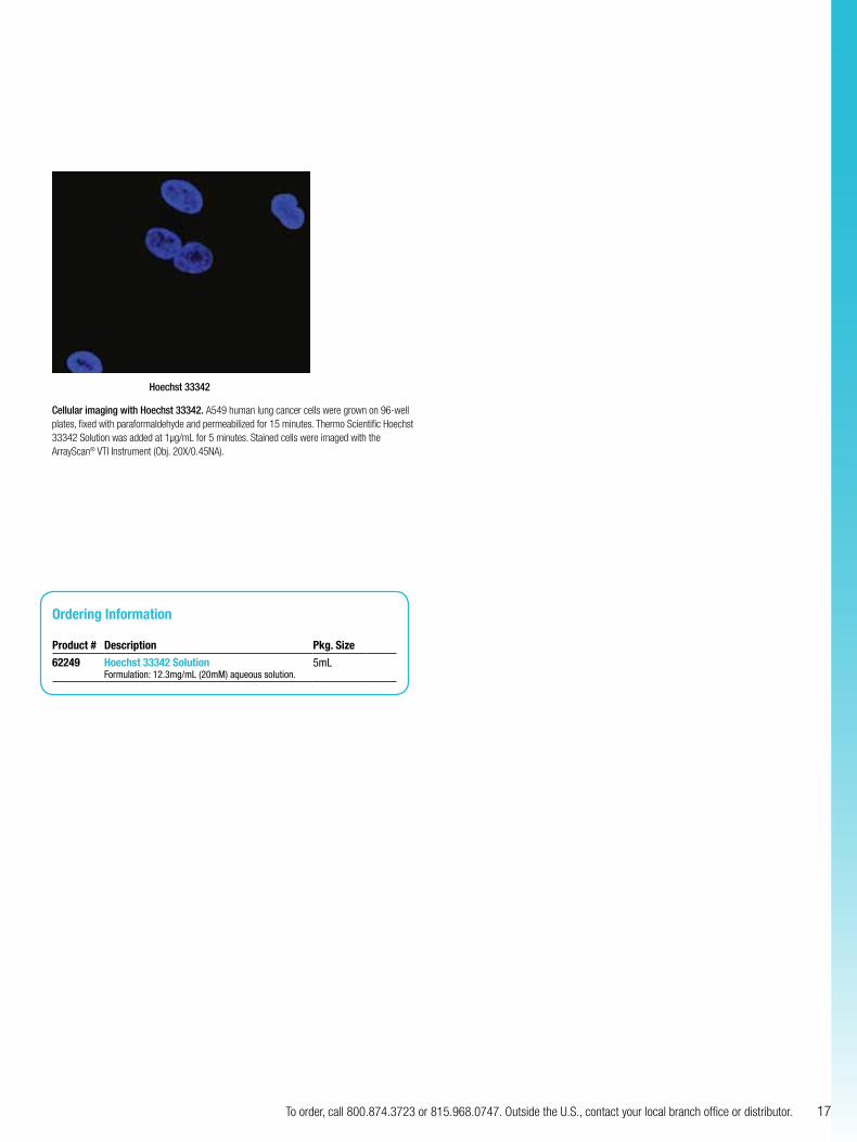
Hoechst 33342
Cellular imaging with Hoechst 33342. A549 human lung cancer cells were grown on 96-well plates, fixed with paraformaldehyde and permeabilized for 15 minutes. Thermo Scientific Hoechst 33342 Solution was added at 1µg/mL for 5 minutes. Stained cells were imaged with the ArrayScan® VTI Instrument (Obj. 20X/0.45NA).
Ordering Information
Product # Description Pkg. Size
62249 Hoechst 33342 SolutionFormulation: 12.3mg/mL (20mM) aqueous solution.
5mL
18 For more information, or to download product instructions, visit www.thermoscientific.com/pierce
Excellent brightness make these conjugates a clear alternative
conjugatesDyLight
Thermo Scientific DyLight Conjugates
Pre-conjugated secondary antibodies and biotin-binding proteins of DyLight Fluors make excellent secondary detection reagents for fluorescent assays. Our DyLight Fluor Conjugates are optimally labeled with the highest dye-to-protein ratio (F/P ratio) and does not cause quenching of the fluorescent signal or problems with conjugate solubility.
Highlights:• Available conjugated to commonly used secondary antibodies, Streptavidin and
NeutrAvidin Biotin-Binding Protein
• Molar ratio (dye:protein) optimized to provide excellent fluorescent intensity
• Stable for 1 year at 4°C
• Antibody conjugates are affinity-purified to minimize cross-reactivity
1 2 3 4 5
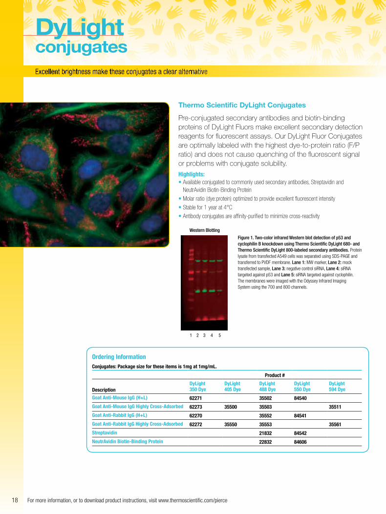
Figure 1. Two-color infrared Western blot detection of p53 and cyclophilin B knockdown using Thermo Scientific DyLight 680- and Thermo Scientific DyLight 800-labeled secondary antibodies. Protein lysate from transfected A549 cells was separated using SDS-PAGE and transferred to PVDF membrane. Lane 1: MW marker, Lane 2: mock transfected sample, Lane 3: negative control siRNA, Lane 4: siRNA targeted against p53 and Lane 5: siRNA targeted against cyclophilin. The membranes were imaged with the Odyssey Infrared Imaging System using the 700 and 800 channels.
Western Blotting
Ordering InformationConjugates: Package size for these items is 1mg at 1mg/mL.
Product #
DescriptionDyLight 350 Dye
DyLight 405 Dye
DyLight 488 Dye
DyLight 550 Dye
DyLight 594 Dye
Goat Anti-Mouse IgG (H+L) 62271 35502 84540
Goat Anti-Mouse IgG Highly Cross-Adsorbed 62273 35500 35503 35511
Goat Anti-Rabbit IgG (H+L) 62270 35552 84541
Goat Anti-Rabbit IgG Highly Cross-Adsorbed 62272 35550 35553 35561
Streptavidin 21832 84542
NeutrAvidin Biotin-Binding Protein 22832 84606
19To order, call 800.874.3723 or 815.968.0747. Outside the U.S., contact your local branch office or distributor.
Rela
tive
Fluo
resc
ent I
nten
sity
0
10,000
20,000
30,000
40,000
50,000
60,000
0 50 100 200150 250Biotinylated BSA (ng/well)
DyLight 649 StreptavidinDyLight 649 NeutrAvidinAlexa Fluor 647 Streptavidin
Rela
tive
Fluo
resc
ent I
nten
sity
0
5,000
10,000
15,000
20,000
25,000
30,000
35,000
40,000
45,000
50,000
0 200 400 800600 1,2001,000Mouse IgG (ng/well)
DyLight 549 Goat anti-mouse IgG
Alexa Fluor 555 Goat anti-mouse IgG
DyLight 800 StreptavidinIRDye 800 Streptavidin
DyLight 680 StreptavidinAlexa Fluor 680 Streptavidin
Rela
tive
Fluo
resc
ent I
nten
sity
4,000,000
8,000,000
12,000,000
16,000,000
0 2 4 86 1210Biotinylated BSA (ng per well)
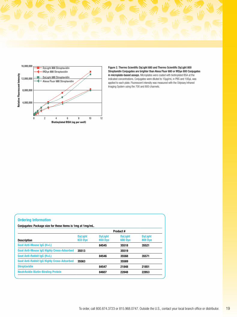
Figure 2. Thermo Scientific DyLight 680 and Thermo Scientific DyLight 800 Streptavidin Conjugates are brighter than Alexa Fluor 680 or IRDye 800 Conjugates in microplate-based assays. Microplates were coated with biotinylated BSA at the indicated concentrations. Conjugates were diluted to 10µg/mL in PBS and 100µL was applied to each plate. Fluorescent intensity was measured with the Odyssey Infrared Imaging System using the 700 and 800 channels.
Ordering InformationConjugates: Package size for these items is 1mg at 1mg/mL.
Product #
DescriptionDyLight 633 Dye
DyLight 650 Dye
DyLight 680 Dye
DyLight 800 Dye
Goat Anti-Mouse IgG (H+L) 84545 35518 35521
Goat Anti-Mouse IgG Highly Cross-Adsorbed 35513 35519
Goat Anti-Rabbit IgG (H+L) 84546 35568 35571
Goat Anti-Rabbit IgG Highly Cross-Adsorbed 35563 35569
Streptavidin 84547 21848 21851
NeutrAvidin Biotin-Binding Protein 84607 22848 22853
20 For more information, or to download product instructions, visit www.thermoscientific.com/pierce
Infrared dyes with the LI-COR Odyssey instrument for easy, sensitive detection
for high-sensitivity applicationsNear-IR dyes
Thermo Scientific DyLight 680 and 800 Near Infrared Dyes for use with the LI-COR Odyssey Instrument
Near-IR dyes are becoming more widely used in a variety of cellular assays and imaging applications. The dyes fluoresce in the 670-1,000nm range, at which biomolecules exhibit low background fluorescence. Combined with a high extinction coefficient, this low autofluorescence property allows high- sensitivity immunoassays, blotting procedures and in vivo imaging applications. In immunoblotting, for example, detection sensitivity with near-IR dye-conjugated antibodies are comparable with direct radioactive detection or indirect detection using enzyme-based assays. Near-IR dyes allow for a wider dynamic detection range and more reliable quantitation.
The LI-COR Odyssey Infrared Imaging System is useful for detecting infrared dyes. It is a two-channel laser-based infrared detection system made by LI-COR Biosciences. It
provides high sensitivity, multiplexing capabilities and accurate quantitation. We offer a variety of products based on near-IR dyes that are compatible with the LI-COR Odyssey Imager (Figure 1), allowing the full utilization of this instrument’s features.
1 1A. B.
2 23 34 45 56 67 78 8
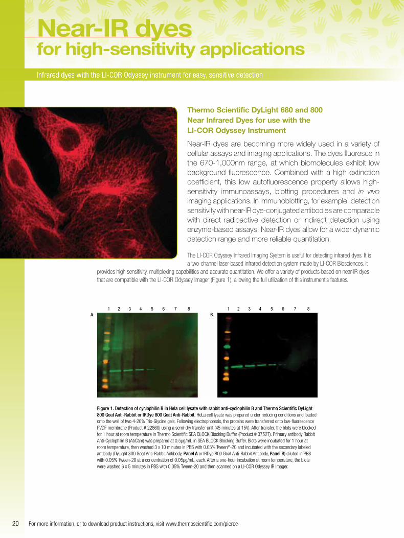
Figure 1. Detection of cyclophilin B in Hela cell lysate with rabbit anti-cyclophilin B and Thermo Scientific DyLight 800 Goat Anti-Rabbit or IRDye 800 Goat Anti-Rabbit. HeLa cell lysate was prepared under reducing conditions and loaded onto the well of two 4-20% Tris-Glycine gels. Following electrophoresis, the proteins were transferred onto low-fluorescence PVDF membrane (Product # 22860) using a semi-dry transfer unit (45 minutes at 15V). After transfer, the blots were blocked for 1 hour at room temperature in Thermo Scientific SEA BLOCK Blocking Buffer (Product # 37527). Primary antibody Rabbit Anti-Cyclophilin B (AbCam) was prepared at 0.5µg/mL in SEA BLOCK Blocking Buffer. Blots were incubated for 1 hour at room temperature, then washed 3 x 10 minutes in PBS with 0.05% Tween®-20 and incubated with the secondary labeled antibody (DyLight 800 Goat Anti-Rabbit Antibody, Panel A or IRDye 800 Goat Anti-Rabbit Antibody, Panel B) diluted in PBS with 0.05% Tween-20 at a concentration of 0.05µg/mL, each. After a one-hour incubation at room temperature, the blots were washed 6 x 5 minutes in PBS with 0.05% Tween-20 and then scanned on a LI-COR Odyssey IR Imager.
21To order, call 800.874.3723 or 815.968.0747. Outside the U.S., contact your local branch office or distributor.
Infrared dye-based products currently offered: • Amine- and sulfhydryl-reactive dyes comparable to IRDye 800
and Alexa Fluor 680 Dye
• Labeling kits for fluorescently labeling various amounts of primary or secondary antibodies
• Streptavidin and NeutrAvidin® Protein-conjugated DyLight 680 and DyLight 800 Dyes
• Goat Anti-Mouse- and Goat Anti-Rabbit-conjugated DyLight 680 and DyLight 800 Dyes
• Thermo Scientific Krypton Infrared Protein Stain for total protein detection in polyacrylamide gels – the only infrared total protein stain available (see page 24)
DyLight IR Fluors offer many benefits for labeling and imaging applications. The dyes are extremely photostable compared to other available dyes that excite and emit at the same wavelengths. In addition, DyLight Dyes exhibit high solubility, making them extremely useful for protein labeling. We offer a choice of two fluors, DyLight 680 and 800 Dyes, that are compatible with the LI-COR Odyssey System (Figure 2). We also offer the Krypton Infrared Protein Stain, which is the only infrared total protein stain available. It offers low nanogram level sensitivity and multiplexing capabilities when used with the LI-COR Odyssey.
Low-autofluorescence properties combined with high extinction coefficients allow DyLight Infrared Dyes to be used in a variety of applications, including:
• Western blotting • In-gel detection • Immunoassays • In vivo imaging • In-cell Western assays
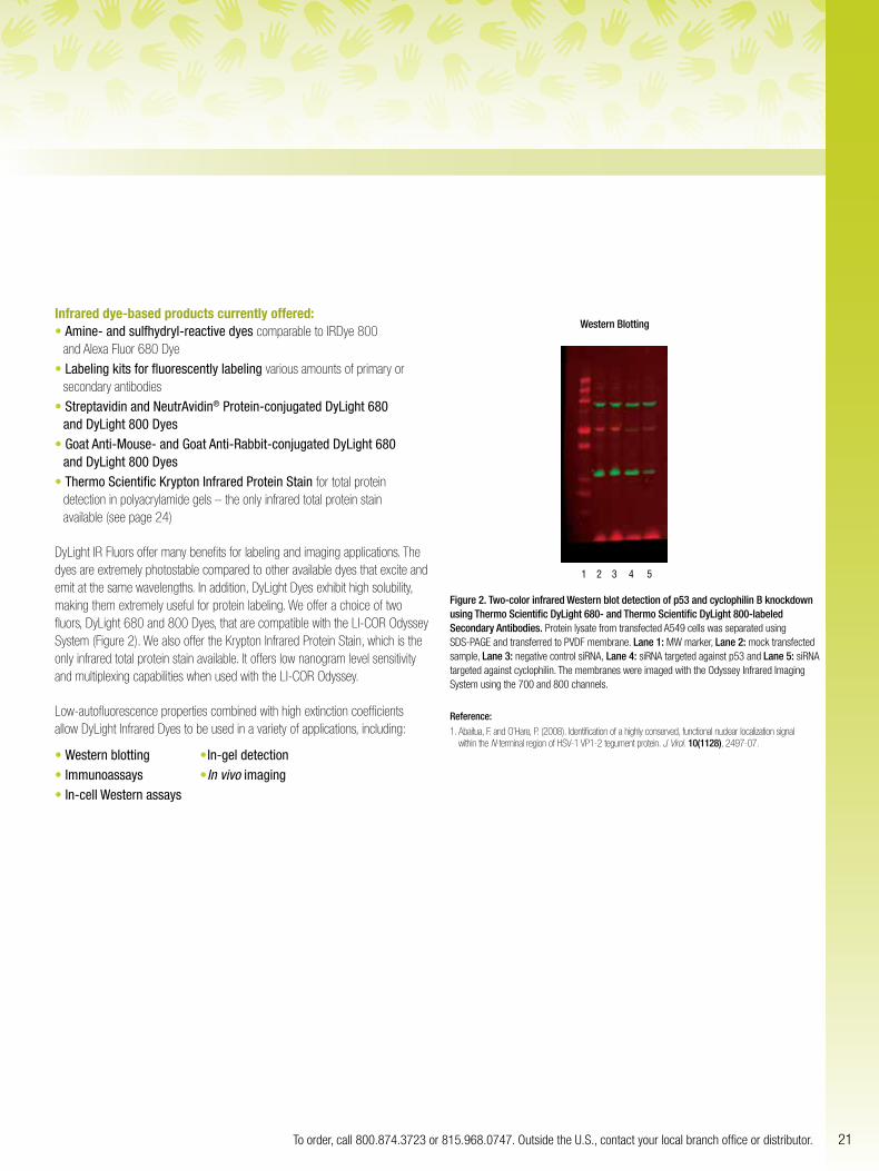
1 2 3 4 5
Figure 2. Two-color infrared Western blot detection of p53 and cyclophilin B knockdown using Thermo Scientific DyLight 680- and Thermo Scientific DyLight 800-labeled Secondary Antibodies. Protein lysate from transfected A549 cells was separated using SDS-PAGE and transferred to PVDF membrane. Lane 1: MW marker, Lane 2: mock transfected sample, Lane 3: negative control siRNA, Lane 4: siRNA targeted against p53 and Lane 5: siRNA targeted against cyclophilin. The membranes were imaged with the Odyssey Infrared Imaging System using the 700 and 800 channels.
Reference:1. Abaitua, F. and O’Hare, P. (2008). Identification of a highly conserved, functional nuclear localization signal
within the N-terminal region of HSV-1 VP1-2 tegument protein. J. Virol. 10(1128), 2497-07.
Western Blotting
22 For more information, or to download product instructions, visit www.thermoscientific.com/pierce
Detect down to 750ng of biotinylated IgG
quantitationfluorescent
Thermo Scientific Fluorescence Biotin Quantitation Kit
Accurately measure biotinylation level with a new highly sensitive fluorescent assay.
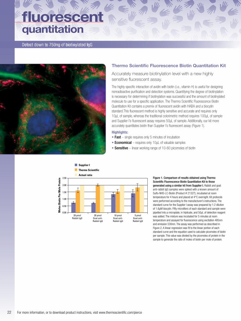
The highly specific interaction of avidin with biotin (i.e., vitamin H) is useful for designing nonradioactive purification and detection systems. Quantifying the degree of biotinylation is necessary for determining if biotinylation was successful and the amount of biotinylated molecule to use for a specific application. The Thermo Scientific Fluorescence Biotin Quantitation Kit contains a premix of fluorescent avidin with HABA and a biocytin standard.This fluorescent method is highly sensitive and accurate and requires only 10µL of sample, whereas the traditional colorimetric method requires 100µL of sample and Supplier I’s fluorescent assay requires 50µL of sample. Additionally, our kit more accurately quantitates biotin than Supplier I’s fluorescent assay (Figure 1).
Highlights:• Fast – single requires only 5 minutes of incubation
• Economical – requires only 10µL of valuable samples
• Sensitive – linear working range of 10-60 picomoles of biotin
Mol
es B
iotin
Per
Mol
e Pr
otei
n 2.50
Supplier I
Thermo Scientific
Actual ratio
20 pmolRabbit IgG
20 pmolGoat anti-Rabbit IgG
10 pmolGoat anti-Rabbit IgG
5 pmolGoat anti-Rabbit IgG
2.00
1.50
1.00
0.50
0.00
Figure 1. Comparison of results obtained using Thermo Scientific Fluorescence Biotin Quantitation Kit to those generated using a similar kit from Supplier I. Rabbit and goat anti-rabbit IgG samples were spiked with a known amount of Sulfo-NHS-LC-Biotin (Product # 21327), incubated at room temperature for 4 hours and placed at 4°C overnight. Kit protocols were performed according to the manufacturer’s instructions. The standard curve for the Supplier I assay was prepared by 1:2 dilution of 1.6µM biocytin. Fifty microliters of each standard and sample were pipetted into a microplate, in triplicate, and 50µL of detection reagent was added. The mixture was incubated for 5 minutes at room temperature and assayed for fluorescence using excitation 485nm and emission 530nm. The assay was performed as described in Figure 2. A linear regression was fit to the linear portion of each standard curve and the equation used to calculate picomoles of biotin per sample. This value was divided by the picomoles of protein in the sample to generate the ratio of moles of biotin per mole of protein.
23To order, call 800.874.3723 or 815.968.0747. Outside the U.S., contact your local branch office or distributor.
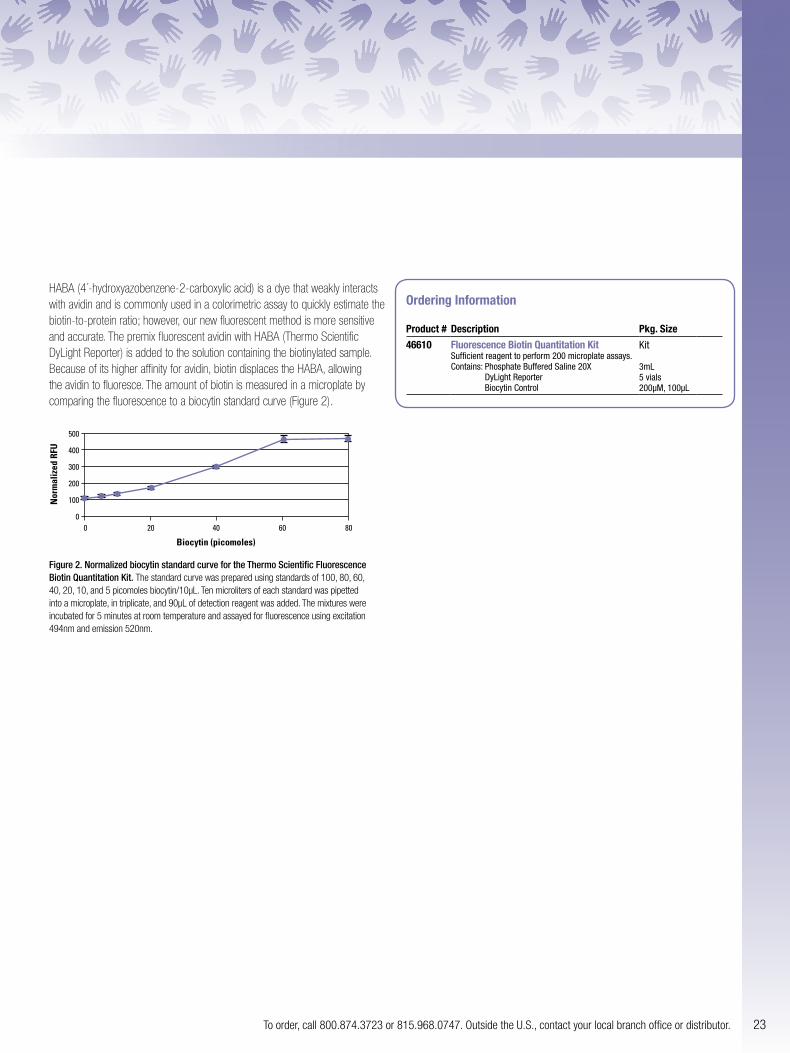
HABA (4´-hydroxyazobenzene-2-carboxylic acid) is a dye that weakly interacts with avidin and is commonly used in a colorimetric assay to quickly estimate the biotin-to-protein ratio; however, our new fluorescent method is more sensitive and accurate. The premix fluorescent avidin with HABA (Thermo Scientific DyLight Reporter) is added to the solution containing the biotinylated sample. Because of its higher affinity for avidin, biotin displaces the HABA, allowing the avidin to fluoresce. The amount of biotin is measured in a microplate by comparing the fluorescence to a biocytin standard curve (Figure 2).
00 20
Biocytin (picomoles)
Nor
mal
ized
RFU
40 60 80
100
200
300
400
500
Figure 2. Normalized biocytin standard curve for the Thermo Scientific Fluorescence Biotin Quantitation Kit. The standard curve was prepared using standards of 100, 80, 60, 40, 20, 10, and 5 picomoles biocytin/10µL. Ten microliters of each standard was pipetted into a microplate, in triplicate, and 90µL of detection reagent was added. The mixtures were incubated for 5 minutes at room temperature and assayed for fluorescence using excitation 494nm and emission 520nm.
Ordering Information
Product # Description Pkg. Size
46610 Fluorescence Biotin Quantitation KitSufficient reagent to perform 200 microplate assays. Contains: Phosphate Buffered Saline 20X
DyLight Reporter Biocytin Control
Kit 3mL 5 vials 200µM, 100µL
24 For more information, or to download product instructions, visit www.thermoscientific.com/pierce
A faster, affordable fluorescent stain that provides excellent performance
protein gel stainsfluorescent
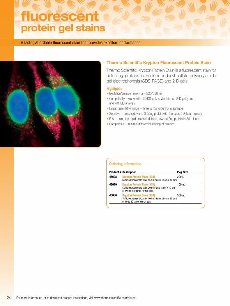
Thermo Scientific Krypton Fluorescent Protein Stain
Thermo Scientific Krypton Protein Stain is a fluorescent stain for detecting proteins in sodium dodecyl sulfate-polyacrylamide gel electrophoresis (SDS-PAGE) and 2-D gels.
Highlights:• Excitation/emission maxima – 520/580nm
• Compatibility – works with all SDS-polyacrylamide and 2-D gel types and with MS analysis
• Linear quantitative range – three to four orders of magnitude
• Sensitive – detects down to 0.25ng protein with the basic 2.3-hour protocol
• Fast – using the rapid protocol, detects down to 2ng protein in 30 minutes
• Comparative – minimal differential staining of proteins
Ordering Information
Product # Description Pkg. Size
46628 Krypton Protein Stain (10X)Sufficient reagent to stain four mini gels (8 cm x 10 cm)
20mL
46629 Krypton Protein Stain (10X)Sufficient reagent to stain 20 mini gels (8 cm x 10 cm) or two to four large-format gels
100mL
46630 Krypton Protein Stain (10X) Sufficient reagent to stain 100 mini gels (8 cm x 10 cm) or 10 to 20 large-format gels
500mL

25To order, call 800.874.3723 or 815.968.0747. Outside the U.S., contact your local branch office or distributor.
Thermo Scientific Pierce Antibody Immunostaining Guide
Immunofluorescence (IF) and immunohistochemistry (IHC) are two methods commonly used to detect proteins in a cellular context. This guide contains technical information, dyes, stains and antibodies to help you in your research of many cellular pathways, structures, organelles and processes.
Thermo Scientific Pierce Antibody Production and Purification Technical Handbook
The updated Antibody Production and Purification Technical Handbook is an essential resource for any laboratory working with antibodies. The handbook provides an overview of antibody structure and types, as well as technical information on the procedures, reagents and tools used to produce, purify, fragment and label antibodies.
www.thermoscientific.com/pierce
1602361 01/12 Printed in the U.S.
Life Science Research
Africa /Belgium/Europe/Middle East +32 53 85 71 84France +0 800 50 82 15Germany +0228 9125650Netherlands +076 50 31 880Switzerland +0800 56 31 40 UK +0800 252 185
Email: [email protected] www.thermoscientific.com/perbio
For other regions, visit www.thermoscientific.com/piercedistributors
USA +815-968-0747 or +800-874-3723Customer Assistance E-mail:[email protected] www.thermoscientific.com/pierce
www.thermoscientific.com/pierce© 2012 Thermo Fisher Scientific Inc. All rights reserved. These products are supplied for laboratory or manufacturing applications only. Prices listed herein were accurate at the time of printing. Visit our website for up-to-date prices. Facebook is a registered trademark of Facebook, Inc. Alexa Fluor® and Texas Red® are trademarks of Molecular Probes, Inc. CyDye®, Cy® and Typhoon® are trademarks of GE Healthcare. Odyssey® and IRDye® are trademarks of LI-COR Biosciences. All other trademarks are the property of Thermo Fisher Scientific Inc. and its subsidiaries.