Embed Size (px)
Citation preview
Review
Fluorescence imaging of synapse dynamics in normal circuit maturationand in developmental disorders
By Shigeo OKABE*1,†
(Communicated by Nobutaka HIROKAWA, M.J.A.)
Abstract: One of the most fundamental questions in neurobiology is how proper synapticconnections are established in the developing brain. Live-cell imaging of the synaptic structure andfunctional molecules can reveal the time course of synapse formation, molecular dynamics, andfunctional maturation. Using postsynaptic scaffolding proteins as a marker of synapse development,fluorescence time-lapse imaging revealed rapid formation of individual synapses that occurredwithin hours and their remodeling in culture preparations. In vivo two-photon excitationmicroscopy development enabled us to directly measure synapse turnover in living animals. In vivosynapse dynamics were suppressed in the adult rodent brain, but were maintained at a high levelduring the early postnatal period. This transition in synapse dynamics is biologically important andcan be linked to the pathology of juvenile-onset psychiatric diseases. Indeed, the upregulation ofsynapse dynamics was observed in multiple mouse models of autism spectrum disorders.Fluorescence imaging of synapses provides new information regarding the physiology and pathologyof neural circuit construction.
Keywords: live-cell imaging, fluorescent proteins, postsynaptic density, two-photonmicroscopy, in vivo imaging, autism
1. Introduction
In the vertebrate brain, postmitotic neurons aregenerated from neuroepithelial cells and they start toextend long axonal processes and relatively short,branched dendrites after their migration to appro-priate brain regions. Axonal processes recognize their
target postsynaptic dendrites using multiple molecu-lar keys and start to differentiate synaptic struc-tures.1) In the mammalian forebrain, synaptogenesisstarts before birth, but the number of synapsessignificantly increases in the early postnatal period.2)
Early studies on synapse formation utilized severaltypes of model synapses in the peripheral nervoussystem (PNS). One popular preparation is that ofneuromuscular junctions (NMJs), which have simpleconnectivity between presynaptic motor neurons andpostsynaptic muscles.3) Another advantage of NMJsis their large size, which facilitates experimentalmanipulation and structural analyses. Finally, NMJsare accessible by relatively simple operative proce-dures and can be imaged by conventional fluores-cence imaging systems.4) By capitalizing on theseadvantages, cell and molecular biological analyses ofNMJs have greatly advanced our understanding ofsynaptogenesis in PNS.
In contrast, studies on synapse formation inthe central nervous system (CNS) were technicallydifficult for multiple reasons. First, connectivity ofthe neurons in CNS is complex, and reliable
*1 Department of Cellular Neurobiology, Graduate School ofMedicine, The University of Tokyo, Tokyo, Japan.
† Correspondence should be addressed: S. Okabe, Depart-ment of Cellular Neurobiology, Graduate School of Medicine, TheUniversity of Tokyo, 7-3-1 Hongo, Bunkyo-ku, Tokyo 113-0033,Japan (e-mail: [email protected]).
Abbreviations: PNS: peripheral nervous system; NMJ:neuromuscular junction; CNS: central nervous system; EM:electron microscopy; FIB-SEM: focused ion beam/scanning elec-tron microscopy; GFP: green fluorescent protein; PSD: postsy-naptic density; NMDA: N-methyl-D-aspartic acid; AMPA:,-amino-3-hydroxy-5-methyl-4-isoxazolepropionic acid; TARP:transmembrane AMPA receptor regulatory protein; ASD: autismspectrum disorder; CNV: copy number variation; IEG: immedi-ately early gene; FRAP: fluorescence recovery after photobleach-ing; GKAP: guanylate kinase-associated protein; LRRTM: leucine-rich repeat transmembrane protein; SALM: synaptic adhesion-likemolecule; YFP: yellow fluorescent protein; CFP: cyan fluorescentprotein.
Proc. Jpn. Acad., Ser. B 93 (2017)No. 7] 483
doi: 10.2183/pjab.93.029©2017 The Japan Academy

identification of presynaptic neurons connecting totheir target is sometimes impossible because of thelack of appropriate markers.5) Therefore, studiesexamining CNS synapse formation were mainlyconducted in a few specific types of synapses, namely,those in which presynaptic and postsynaptic compo-nents are well-defined and both divergence andconvergence of axonal projections are low, such asclimbing fiber synapses on cerebellar Purkinje cells.6)
Second, the synaptic structure in CNS is small, witha typical size of a few micrometers in diameter(Fig. 1).7) Details of the synaptic structure cannot beresolved by conventional light microscopy, and theirshape and size could only be measured using electron
microscopy (EM). However, three-dimensional re-construction of EM images requires a large amount oftime and labor.7) Recently this situation dramaticallychanged because of the introduction of new tech-nologies in light and electron microscopy, suchas super-resolution microscopy8),9) and focused ionbeam/scanning electron microscopy (FIB-SEM).10)
Finally, synapses in CNS are difficult to accessexperimentally.11) In the 1980s, there were noavailable technologies that would allow access tothe small rodent cranium-covered brain withoutdamaging the CNS neurons, which are highlyvulnerable to lower oxygen concentrations, increasesin intracranial pressure, and inflammation.
A B
C D
Fig. 1. Morphology and molecular assembly of the postsynaptic component. (A) EM images of synapses formed in the hippocampal sliceculture (left) and dissociated culture of hippocampal neurons. Synaptic vesicles, presynaptic active zone, and PSDs can be recognized.Bars, 200 nm. (B) A scheme of presynaptic and postsynaptic structures. Most of the excitatory glutamatergic synapses on theforebrain pyramidal neurons are formed onto dendritic spines. (C) Glutamate receptors (AMPA receptor, NMDA receptor, andmGluR), postsynaptic scaffolding molecules (PSD-95, guanylate kinase-associated protein (GKAP), Shank, and homer), andpostsynaptic adhesion molecules (neuroligin, leucine-rich repeat transmembrane protein (LRRTM), synaptic adhesion-like molecule(SALM), and synCAM) and their organization in PSD. Presynaptic cell adhesion molecules, neurexins, bind to both neuroligins andleucine-rich repeat transmembrane protein LRRTMs. (D) Organization of polymerized actin and actin regulators (Arp2/3 andWAVE complex, formin family proteins, and VASP) in the spine.
S. OKABE [Vol. 93,484

2. Imaging synapses in vitro
To circumvent the difficulty in analyzing CNSsynapses, isolated preparations of the nervous system,such as dissociated neuronal cultures, were exten-sively utilized to identify the nascent synapses formedbetween CNS neurons.12) The identification of syn-apses was further facilitated by the development ofa culture system of pure hippocampal or corticalneurons without contamination by glial cells.12),13)
A turning point in our ability to detect synapseformation in live samples was reliable markerdevelopment for the detection of presynaptic orpostsynaptic structures (Fig. 1B). Integral mem-brane proteins in synaptic vesicles, such as synapto-physin, or proteins associated with synaptic vesicles,such as synapsin I, are good candidates for presy-naptic markers. These presynaptic molecules taggedwith green fluorescent protein (GFP) and otherfluorescent proteins are widely used for the detectionof presynaptic components.14),15) A characteristicstructure present in the postsynaptic compartmentis the postsynaptic density (PSD), a disk-like mo-lecular assembly with a diameter of less than 1 µmthat consists of both membrane and cytoplasmicproteins.16),17) Biochemical and proteomic studiesidentified several hundred protein species present inPSD (Fig. 1C). PSD scaffolding proteins are a smallsubset of PSD proteins that are proposed to forma framework for the PSD structure.18) They areabundant in the PSD fraction and comprise multipledomains for binding with other postsynaptic mole-cules.19) A predominant PSD scaffolding proteinPSD-95 was originally identified by the biochemicalcharacterization of the purified PSD fraction.20) PSD-95 comprises three PDZ domains, one GK domain,and one SH3 domain. These domains have specificbinding partners, including N-methyl-D-aspartic acid(NMDA)-type glutamate receptors,21) synaptic celladhesion molecules neuroligins,22) and transmem-brane ,-amino-3-hydroxy-5-methyl-4-isoxazolepro-pionic acid (AMPA) receptor regulatory proteins(TARPs).23) Several research groups, including ours,found that PSD-95 tagged with GFP can reliablyreport the position of PSDs in the dendrites of livingcortical and hippocampal neurons.24)–26) This findinggreatly facilitated studies on synapse formation incell biology. By expressing PSD-95-GFP in culturedneurons, the fate of single postsynaptic sites can befollowed over several weeks. Live imaging of PSD-95-GFP provided quantitative information aboutthe basic behavior and turnover of the synaptic
structure, its regulation by neuronal activity, and itsresponses to pathological conditions.27)
3. Basic principles of synapse turnover in vitro
In the initial studies on CNS synapse develop-ment, researchers collected static images of neuronsstained with antibodies specific for presynaptic orpostsynaptic molecular markers at different timepoints. These immunocytochemical studies reportedgradual increases in the density of presynapticboutons and postsynaptic PSDs throughout thecourse of neuronal circuit development. Consistentwith data obtained in vitro, immunohistochemicalanalyses of cortical and hippocampal tissue sectionsalso revealed a gradual increase in synaptic densityduring postnatal development. These results weretaken as evidence for the stability of synapses afterinitial formation and for their slow differentiationinto mature synapses.
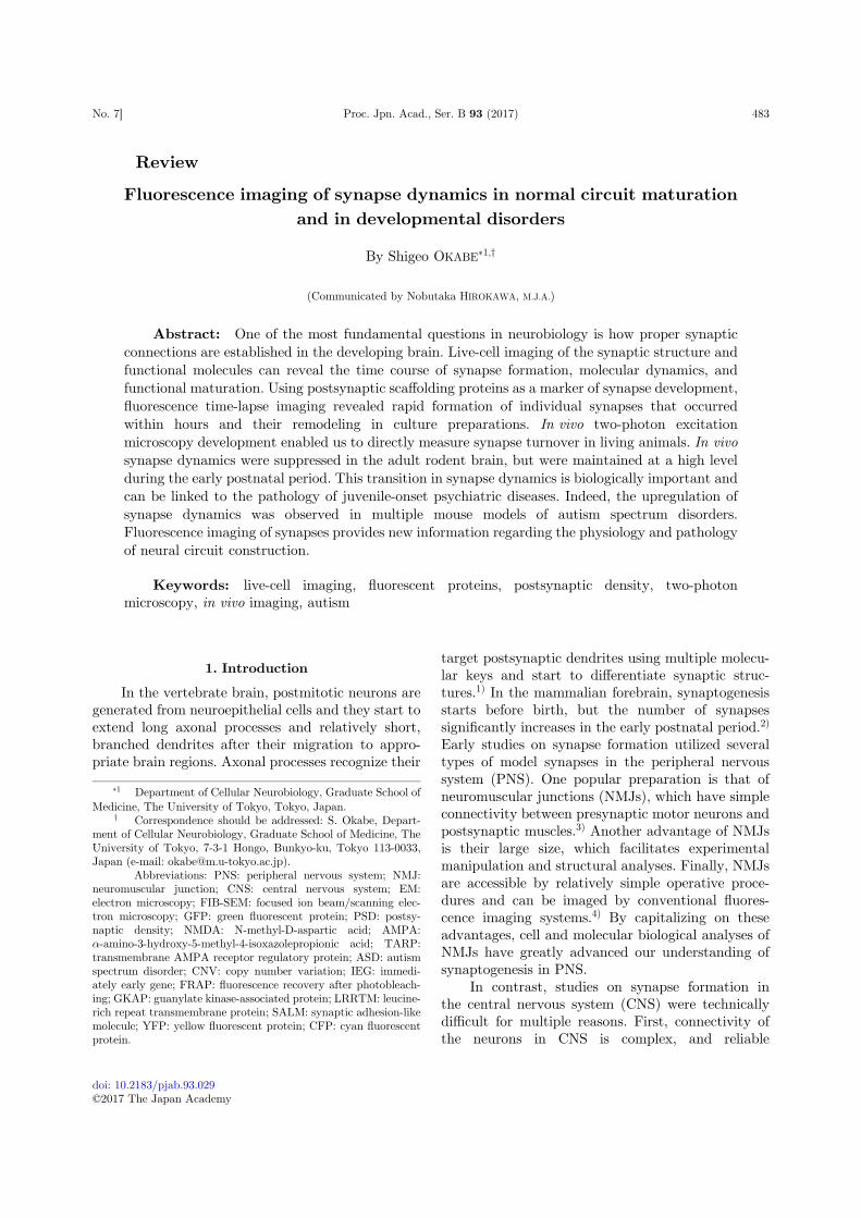
Based on the model of immediate stabilizationand monotonous accumulation of differentiatingsynapses, we initially hypothesized that imaging ofsynapses in live neurons with an interval of severaldays could detect very few changes in synapticconnections, with only a minor fraction of gained orlost synapses. To our surprise, distribution of PSD-95puncta at two time points was markedly different,even if the time interval was set to be 24 h(Fig. 2A).26) This observation clearly indicates thatformation of individual synapses is a rapid processthat takes place over hours. A large proportion ofnewly formed PSD-95 clusters were already associ-ated with the synaptophysin-positive presynapticpuncta (Fig. 2C). Formation of PSD-95 clusterswas synchronized with the formation of dendriticspines (Fig. 2B).15) These observations are consistentwith the idea that presynaptic and postsynapticcomponents are assembled within a narrow timewindow, and their differentiation is coordinated withone another.28)
Another surprising observation in the synapselive imaging was a high fraction of lost synapseswithin a short period. Synapse elimination was high,not only in the early phase of differentiation but alsoafter complete maturation of neurons that had beenmaintained for more than 3 weeks in culture. Thenumber of gained synapses is balanced with that oflost synapses after the maturation of culture prepa-rations, indicating that the balance between gainand loss of synapses determines the overall trendof synapse density increase. Our initial protocolof expression of fluorescent probes was based on
Live-cell imaging of synapse formationNo. 7] 485

adenovirus-mediated gene transduction.26),29) Thistechnique can achieve high efficiency of gene trans-duction, but cell toxicity associated with adenovirusinfection limited the observation period to less thanseveral days. It is difficult to trace the fate ofindividual synapses from images taken at shortintervals. To achieve long-term observation of thesynapse population on the same dendrites, it isnecessary to generate a long-term, stable expression
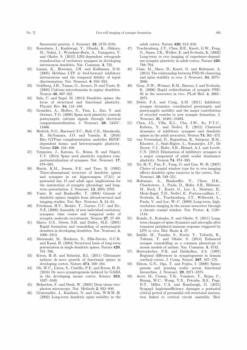
system of GFP-tagged synaptic proteins. We gen-erated transgenic mouse lines expressing PSD-95-GFP or another abundant postsynaptic scaffoldingprotein, Homer1c, tagged with GFP and performedfluorescence time-lapse imaging of cultured neuronstaken from these mouse lines.30) Observation ofsynapse turnover along the same dendrites overweeks provided further information about basicprinciples of synapse turnover (Fig. 3). The patterns
A
B C
Fig. 2. Formation of synapses detected by time-lapse imaging in culture preparations.15),26) (A) Time-lapse fluorescence imaging of PSD-95-GFP in a culture of hippocampal neurons. Formation of new PSD-95 clusters can be detected within 12h (arrows). (B) Formationof PSD-95 clusters (arrows in row b) in newly formed dendritic spines filled with cyan fluorescent protein (CFP, arrows in row a).Merged color images are shown in row c (green: CFP, red: PSD-95- yellow fluorescent protein (YFP)). Times are shown in the rightupper corners in minutes. (C) Simultaneous imaging of synaptophysin-CFP (column a) and PSD-95-YFP (column b). Arrowsindicate the formation of presynaptic synaptophysin and postsynaptic PSD-95 clusters. Scale bars, 3 µm.
S. OKABE [Vol. 93,486

of increase in synaptic density within dendriticsegments were variable; half of the dendritic seg-ments showed a monotonous increase in synapticdensity (Fig. 3A). Namely, majority of the synapsespersist after formation. Interestingly, the other halfof dendritic segments showed a complex pattern inchanges of synaptic density, with higher chance ofsynapse elimination within several days after for-mation (Fig. 3B). The newly formed dendritesrapidly acquired new PSD structures and theirdensity became comparable with that of parentaldendrites within a few days. When the fate of singlePSD structures was monitored over several days,elimination of newly formed synapses was frequentlyobserved. These data confirmed the importance ofthe balance between the gain and loss of synapsesduring neural circuit development and that synapseloss is not confined to a specific subgroup in the totalsynapse population.
4. Relationship between synaptogenesisand spinogenesis
Dendritic spines are small protrusions from thesurface of dendritic shafts. Although dendritic spinesare taken as a structural marker of excitatorysynapses, a subset of neurons, such as forebraininhibitory neurons, develop dendrites with few spinesand receive most of the excitatory synapses directlyonto dendritic shafts.31) This observation clearlyindicates that dendritic spines are not essential inbasic excitatory synaptic transmission.32),33) Theaccumulating evidences indicate that spines havemodulatory functions in postsynaptic signal trans-duction and synaptic plasticity.34) Dendritic spinesare proposed to be important in multiple aspects ofplasticity-related mechanisms, such as confinement ofsignaling molecules, such as calcium35) and activatedsmall GTPases,36) and attenuation of local membrane
A B
Fig. 3. Long-term observation of the same dendrites for turnover of postsynaptic densities.30) (A) Time-lapse images of the dendriticsegment showing a monotonous increase in PSD density. The right column shows binarized images of PSDs. (B) Dendritic segmentsshowing a complex pattern in PSD density. DIV; days in vitro. PSDs were detected by Homer1c-GFP. Scale bars, 5 µm.
Live-cell imaging of synapse formationNo. 7] 487

depolarization induced by neurotransmitter recep-tors and voltage-dependent channels within spineheads.37)
Relationship between excitatory postsynapticsites and dendritic spines is not fixed but regulatedalong development of pyramidal neurons. In themature brain, pyramidal neuron dendrites arecovered by spines. However, immature pyramidalneurons express highly motile filopodia and lackspines.38) It is likely that a fraction of the dendriticfilopodia in an immature stage is converted to spines.However, it has not yet been clarified whether mostof spine synapses are indeed generated from filopodia,working as precursors of spines. The filopodia modelof spine formation proposes that interactions ofmotile filopodia with nearby axons initiate synapticcontact development.39) This model is based on lightmicroscopic observations of synapse formation incultured neurons.15),40) In these experiments, theappearance of dendritic filopodia generally precededthe presynaptic contacts and formation of the PSDstructure (Fig. 2A and B). On the other hand, EM-based observations of developing cortical tissueindicated an abundance of shaft synapses in theearly stage of cortical development.38) The “Miller-Peters model” was proposed based on this observa-tion and states that direct contact between presy-naptic axons and dendritic shafts leads to theformation of synaptic junctions, and this eventtriggers the subsequent formation of dendriticspines.39) In vitro slice culture data support thismodel by showing an initial formation of PSDclusters on dendritic shafts and subsequent gener-ation of spines at the same dendritic positions.41)
In spite of many previous efforts to identify realprecursors of mature dendritic spines, the debatebetween the filopodia model and Miller-Peters modelhas not yet been settled.
Recent technical developments in uncaging ofneurotransmitters by two-photon excitation lasershave enabled researchers to stimulate cell surfacereceptors with a high spatial resolution.42) Uncagingof glutamate or GABA on dendritic shafts can inducethe formation of new dendritic protrusions, whichsubsequently acquire synaptic contacts.43),44) Thedistinction between filopodia in immature dendritesand the dendritic protrusions induced by localneurotransmitter application has not been clarified.Nevertheless, these studies suggest that dendriticprotrusive activity triggered by neurotransmittersreleased from nearby axons contributes to theformation of new synaptic connections.
5. Imaging synapses in vivo
As described in previous chapters, the turnoverof single synapses was mainly investigated in isolatedpreparations, such as dissociated cultures and orga-notypic slices, until late 1990s. This situation wasdramatically changed by two-photon excitation laserscanning microscopy development and its applicationin the detection of single synapses in vivo.45) Usingthis technique, the behavior and fate of singlesynapses can be followed for hours, days, and monthsin living animals. Nowadays, in vivo two-photonimaging has become an indispensable imagingtechnique for investigating synapse developmentand plasticity; it has greatly contributed to theidentification of new mechanisms that underlieneural network development, learning-related forma-tion of new synapses, and synapse pathology relatedto neurological and psychiatric disorders.
Initial studies on in vivo synapse imaging reliedon dendritic spines for the identification of synap-ses.46),47) As discussed in the previous chapter, reliabledetection of multiple types of synapses, in thedeveloping and mature brain, requires other detectionmethods. Application of PSD-95-GFP as a secondmarker of postsynaptic specialization was alreadyproven to be useful in culture preparations, andrecent studies have demonstrated reliable detection ofsynapse-containing spines by double imaging of thespine structure and PSD-95 clusters (Fig. 4).48),49) Alarge fraction of inhibitory synapses are also presenton dendritic shafts, and their detection requiresspecific molecular markers.31) Gephyrin is a majorscaffolding molecule of inhibitory postsynaptic spe-cialization. Gephyrin tagged with GFP has beenshown to be useful in the detection of inhibitory syn-apses in live neurons, both in vitro and in vivo.50)–52)
Application of two-photon microscopy for thevisualization of dendritic spines in vivo has beenrestricted to adult rodents until recently. Earlyin vivo imaging studies reported discrepancies inthe spine turnover rate for transcranial imagingdepending on the surgical procedures.46),47) Thiscontroversy led to the finding of excess activationof microglial cells due to inappropriate open-skullsurgery and microglia-dependent mechanisms ofspine pruning.53) Further studies confirmed similarturnover rates of spines obtained by carefullycontrolled open-skull surgery and by thinning thecranial bone to be transparent enough for spineimaging, indicating that appropriate surgical proce-dures are necessary for the proper measurement of
S. OKABE [Vol. 93,488

spine turnover.54) The consensus of researchersworking on in vivo two-photon imaging of spines inadult animals is that spine dynamics in the adultcortex is highly suppressed, with a remainingdynamic spine fraction of less than 5% of the totalnumber of spines (Fig. 5).55) As mentioned inprevious sections, synapse dynamics in culturedneurons was maintained at a high level, even morethan 3 weeks after plating.26) Thus, synapse turnoverin vivo is highly suppressed by factors that are notpresent in reduced preparations of cultured neurons.
There have been few quantitative studies onsynapse dynamics in vivo during the early postnatal
period. This scarcity of data mainly originates fromthe difficulty of imaging synapses in the immaturebrain tissues without activating glial cells. Thecranial bone in young pups is thin and less opaquebut more vulnerable to invasive techniques. There-fore, the thin skull method cannot be used on miceyounger than 2 weeks of age. Our group performedthin skull window-based in vivo two-photon imagingof spines at postnatal 2 and 3 weeks and foundextensive gain and loss of both spines and PSD-95clusters (Fig. 6).56) By comparing images separatedby an interval of 2 days, fractions of spine gain andloss were found to be close to 20%. Notably, the
PSD-95-GFP + DsRed2 plasmids
In utero electroporationIn vivo two-photon imaging
at postnatal 2-8 weeks
DsRed2/PSD-95-GFP
A B
CDE
Fig. 4. Procedures for in vivo two-photon imaging. (A) Electroporation of plasmids for the expression of PSD-95-GFP and DsRed2 (redfluorescent protein) into the mouse embryos. (B) In vivo two-photon imaging of mice which underwent in utero electroporation ofexpression plasmids. (C) A single two-photon excitation scanning image taken from the anesthetized mouse. (D) Three-dimensionalreconstruction of an image stuck with two fluorescence signals. (E) Enlargement of a dendritic segment marked by a white rectangle in(D). Individual synapses can be reliably detected by the morphology of spines and presence of PSD-95-GFP clusters. In vivo two-photon images are shown as pseudocolor pictures with PSD-95-GFP in green and volume filler DsRed2 in red. Scale bars, 10 µm (C),3 µm (E).
Live-cell imaging of synapse formationNo. 7] 489

turnover rate in vivo in the early postnatal periodwas comparable with that measured in the culturedhippocampal neurons. This comparison suggeststhat during early stages of synapse development,spine turnover in in vitro and in vivo preparationsshare similar mechanisms and that the balancebetween the gain and loss of synapses determinesthe profile of synapse density increases duringdevelopment.
6. Imaging synapse pathology in vivo
Interactions between genetic and environmentalfactors are critical for the expression of physiologicalfunctions of the neural circuits. Construction andrefinement of the neural circuits in the neocortex
mainly takes place in the postnatal period, andsensory information plays a key role in this process.The developmental profile of spine density in specificbrain regions can be used as a readout of the neuralcircuit construction. In the human neocortex, thepeak of spine density is at the age of 2–6 years.57) Therate of spine increase was high before reaching thepeak, but the subsequent decline in spine densitytoward adolescence was very slow. The initial rapidincrease of spines and its gradual decrease afterthe peak is preserved in both monkey58) and mouseneocortex.59) Our quantitative analysis confirmedthat spines in mouse layer II/III pyramidal neuronsrapidly increase until postnatal week 3.56) The spinepopulation shifts to the phase of gradual decrease
Fig. 5. Stability of dendritic spines in the adult mouse neocortex. The somatosensory cortex of Thy1-GFP mouse lines were imagedthrough the thinned skull window with two-photon scanning microscopy.56) The same dendritic segments were imaged at intervals of1, 14, and 33 days. Most of the spines can be detected on the second imaging session. Scale bar, 3 µm.
S. OKABE [Vol. 93,490

thereafter. These observations collectively indicatethat an initial phase of rapid construction of neuronalcircuits with excess connectivity followed by a phaseof slow pruning of unnecessary connections is acommon strategy across mammalian species forcircuit development and maturation in the neocortex.
The onset of psychiatric diseases is variable, andpatients with childhood psychiatric diseases experi-ence symptom onset and diagnosis early in life.Autism spectrum disorders (ASDs) is one of thechildhood psychiatric diseases with a high preva-lence.60) Patients with ASD show their initialsymptoms and can be diagnosed at the ages of 2–6years. This postnatal period matches the peak ofspine density in the human neocortex, suggestingthat postnatal neural circuit development is linkedto functional changes in the brain of children with
ASD.61) Another line of evidence suggests thatdysregulated synaptic connectivity has an importantrole in ASD pathophysiology. Recent genetic studiesidentified a large number of candidate rare genevariants associated with non-syndromic ASDs.62)
Rare variants in genes of postsynaptic scaffoldingmolecules (Shank2 and Shank3), postsynaptic celladhesion molecules (neurogilin-3 and -4), and theirpresynaptic binding partners (neurexin-1) are linkedto ASDs.63) These synaptic scaffolding proteins andcell adhesion molecules are critical for the regulationof synaptic connectivity. Therefore, it is likely thatdysregulated synapse construction due to geneticchanges in key synaptic molecules leads to the accu-mulation of mismatches in neuronal connectivity,which subsequently triggers symptoms associatedwith ASDs.
A
B
C
Fig. 6. Downregulation of spine synapse dynamics in the postnatal period.56) (A) In vivo imaging of dendrites with the interval of 1 day.In vivo two-photon images are shown as pseudocolor pictures with PSD-95-GFP in green and volume filler DsRed2 in magenta. Arrowindicates a new spine and arrowheads indicate lost spines. Scale bar, 3 µm. (B) Spine turnover rates at different postnatal days. At 8weeks after birth, spine gain and loss are highly suppressed. (C) Trend of PSD turnover rates during the postnatal period. Note similarsuppression of dynamics in both spines (B) and PSDs (C). One-way analysis of variance, Tukey’s test. ***p < 0.001. Number ofanimals analyzed (2 weeks; n F 6, 3 weeks; n F 6, 8 weeks; n F 3).
Live-cell imaging of synapse formationNo. 7] 491

To test this hypothesis of circuit dysfunction inthe neocortex of patients with ASD, we applied thetechnique of in vivo two-photon imaging to multipleASD mouse models.56) These mouse models includepatDp/D,64) NLG R451C,65) and BTBR mice.66)
patDp/D mice mimic the most frequently reportedcopy number variation in ASD, the duplication of15q11-13. NLG R451C mice are genetically engi-neered to contain a rare missense mutation of theNlgn3 gene, a mutation also found in some individ-uals with ASDs. The BTBR inbred mouse has beenextensively studied and reflect major behavioralfeatures of ASD: deficits in social interactions,unusual vocalization behaviors, and increase inrepetitive activities. Because these three ASD mousemodels are heterogeneous in their genetic properties,identification of circuit-level impairments shared bythese models will lead to the establishment of theworking hypothesis for the core pathophysiology ofASDs. Indeed, these three mouse models showconsistent upregulation in the turnover of spinespositively associated with PSD-95 clusters (Fig. 7).This alteration was specific to the spine synapsesreceiving intracortical projections; the dynamics ofspines receiving inputs from the thalamus wereunaffected. Similar upregulation of spine turnover in
the neocortex was reported in Fmr1 knockout mice,a fragile X syndrome and syndromic autistic mousemodel, supporting the hypothesis of shared synapse-level phenotypes across ASD mouse models withheterogeneous genetic mutations.67),68) patDp/D andNLG R451C mice further showed impairment in theactivity-dependent remodeling of spine synapses inthe somatosensory cortex after manipulation of theirwhiskers and also demonstrated reduced immediatelyearly gene (IEG) responses of layer II/III neurons tothe upregulation of whisker-dependent sensory in-puts.56) The sensory stimulus-dependent upregula-tion of IEG was comparable in layer IV neuronsbetween ASD model and control mice, suggestingintact transmission of sensory information from thethalamus to the postsynaptic layer IV neurons. Thesedata indicate that upregulation in spine synapseturnover during the early postnatal period results inweaker functional connectivity within the layer II/IIIneuron population and/or between layer II/III andlayer IV neurons. This dysfunction in informationtransfer may not be specific to the sensory cortex andmay be observed in other neocortical areas includingthe prefrontal cortex, where circuit impairmentscan be associated with behavioral deficits in socialinteractions in patients with ASD.
patDp/+ NLG R451C BTBR
Fig. 7. Upregulation of spine synapse dynamics in three ASD mouse models.56) Both formation and elimination rates of spines areupregulated in patDp/D, NLG R451C, and BTBR mouse lines. Arrows and arrowheads indicate spines formed and eliminated,respectively. In vivo two-photon images are shown as pseudocolor pictures with PSD-95-GFP in green and volume filler DsRed2 inmagenta. Scale bar, 2 µm. Student’s t-test, *p < 0.05, ***p < 0.001. Number of animals analyzed (patDp/D; control F 5, mutant F 6,NLG R451C; control F 4, mutant F 6, BTBR; C57Bl/6 F 3, BTBR F 3).
S. OKABE [Vol. 93,492

7. Synapse stability and instability
An initial phase characterized by the rapidconstruction of neuronal circuits with excess con-nectivity followed by a slow synapse pruning phase isa common strategy for neocortical circuit develop-ment. In the mature neocortex, turnover of spinesynapses is highly suppressed and these synapses canbe maintained for several months or even throughoutthe life of an animal.69)–71) The analysis of synapsestability is as important as that of synapse pruningand plasticity. Without the establishment of stableneuronal connections, long-term storage of informa-tion in the brain may be very difficult to achieve.However, there are few studies aiming to clarifyunderlying principles that support long-term synapsestability. It is possible to propose that unidentified,very stable proteins form the core of PSD and othersynaptic molecular interactions are based on thepresence of these stable core proteins. Studiesinvestigating molecular mobility in synapses throughfluorescence recovery after photobleaching orthrough photoactivation have provided us with richinformation regarding the rate of protein replacementat individual postsynaptic sites. These studies onmolecular mobility have failed to find any postsy-naptic molecules that stay in the same synapse fora prolonged period.49),72),73) A large fraction of PSDscaffolding molecules are replaced if we monitorFRAP recovery for more than a few hours. Theturnover of glutamate receptors, such as AMPA-typeand NMDA-type receptors, is in the same range asthat of PSD scaffolding proteins.74),75) Therefore, theidea of a stable PSD core complex presently lacksexperimental support. Another possibility is theexistence of a feedback mechanism for the homeo-stasis of synaptic molecular assembly and spinevolume. Previous studies provided evidence that theactin cytoskeleton supports spine stability.76),77) If areduction in the actin content in spines activates afeedback loop that triggers the increase of anotherfactor, which then in turn facilitates the recruitmentof actin back into the spines, spine actin contents canbe maintained automatically. Obvious candidates foractin recruitment factors are regulators of actinpolymerization, such as the Arp2/3 and WAVEcomplexes (Fig. 1D). Indeed, recent super-resolutionimaging revealed that PSD can function as arecruitment platform for the WAVE complex.78),79)
Previous modeling studies on neutrophil motilityproposed the presence of a feedback mechanismbetween actin assembly and WAVE complex activa-
tion.80) In this feedback model, actin is both anoutput and input to the membrane-tethered WAVEcomplex, and the spatiotemporal cycling of actinassembly is driven by an active feedback process withthe WAVE complex. If a similar feedback mechanismexists in the spine actin meshwork and PSD-tetheredWAVE complex, long-term maintenance of spinestructure and spine actin organization can beexplained. To confirm the presence of this feedbackpathway, new technologies that combine mathemat-ical modeling with actin and WAVE complexmanipulation in a higher temporal resolution maybe required.
8. Future research
Both reduced preparations of neuronal circuitsin vitro and recently developed monitoring technol-ogy of in vivo synapses are required for an in-depthunderstanding of the underlying mechanisms ofsynapse formation, remodeling, and stabilization.The measurement of multiple parameters withinsingle synapses across a large volume of the braintissue will become important for studies that requirecomprehensive information regarding the local circuitfunction associated with specific cortical functions.These imaging experiments will require high speedimaging and an improvement in the spatial resolutionof two-photon microscopy.81),82) New technologiesthat will enable the acquisition of images from widerfields of view that encompass multiple neocorticalareas while still maintaining the resolution of singlesynapses will be required.83) To achieve this goal, itwill be necessary to design new microscopic objectiveswith higher numerical apertures and a wider field ofview; additionally, a redesign of optical systems andscanning devices should follow thereafter. Acquisitionof data from large volume samples increases thedata size and necessitates automatic pipeline devel-opment for data analysis, which may require collab-oration with data scientists. Finally, the applicationof synapse imaging techniques to mouse models ofpsychiatric disorders facilitates the identification ofthe core pathology in the dysfunctions of neuralcircuits. This information will be useful for newbiomarker development of psychiatric disorders andfor the evaluation of new drug candidates and othertherapeutic approaches.
Acknowledgments
The author expresses his gratitude to thecollaborators on the study described in this review.This study was supported by grants-in-aid for
Live-cell imaging of synapse formationNo. 7] 493

Scientific Research (26250014 and 25117006),Core Research for Evolutional Science and Technol-ogy from the Japanese Science and TechnologyAgency (JPMJCR14W2), and the Uehara MemorialFoundation.
References
1) Munno, D.W. and Syed, N.I. (2003) Synaptogenesisin the CNS: an odyssey from wiring together tofiring together. J. Physiol. 552, 1–11.
2) Elston, G.N. and Fujita, I. (2014) Pyramidal celldevelopment: postnatal spinogenesis, dendriticgrowth, axon growth, and electrophysiology.Front. Neuroanat. 10.3389/fnana.2014.00078.
3) Sanes, J.R. and Lichtman, J.W. (1999) Developmentof the vertebrate neuromascular junction. Annu.Rev. Neurosci. 22, 389–442.
4) Turney, S.G., Walsh, M.K. and Lichtman, J.W.(2012) In vivo imaging of the developing neuro-muscular junction in neonatal mice. Cold SpringHarb. Protoc. 7, 1166–1176.
5) Livet, J., Weissman, T.A., Kang, H., Draft, R.W.,Lu, J., Bennis, R.A., Sanes, J.R. and Lichtman,J.W. (2007) Transgenic strategies for combinato-rial expression of fluorescent proteins in thenervous system. Nature 450, 56–62.
6) Uesaka, N., Uchigashima, M., Mikuni, T., Hirai, H.,Watanabe, M. and Kano, M. (2015) Retrogradesignaling for climbing fiber synapse elimination.Cerebellum 14, 4–7.
7) Sorra, K.E. and Harris, K.M. (2000) Overview on thestructure, composition, function, development, andplasticity of hippocampal dendritic spines. Hippo-campus 10, 501–511.
8) Frost, N.A., Shroff, H., Kong, H., Betzig, E. andBlanpied, T.A. (2010) Single-molecule discrimina-tion of discrete perisynaptic and distributed sitesof actin filament assembly within dendritic spines.Neuron 67, 86–99.
9) Dani, A., Huang, B., Bergan, J., Dulac, C. andZhuang, X. (2010) Superresolution imaging ofchemical synapses in the brain. Neuron 68, 843–856.
10) Knott, G., Marchman, H., Wall, D. and Lich, B.(2008) Serial section scanning electron microscopyof adult brain tissue using focused ion beammilling. J. Neurosci. 28, 2959–2964.
11) Yang, G., Pan, F., Parkhurst, C.N., Grutzendler, J.and Gan, W.-B. (2010) Thinned-skull cranialwindow technique for long-term imaging of thecortex in live mice. Nat. Protoc. 5, 201–208.
12) Banker, G.A. and Cowan, W.M. (1977) Rat hippo-campal neurons in dispersed cell culture. BrainRes. 126, 342–397.
13) Brewer, G.J., Torricelli, J.R., Evege, E.K. and Price,P.J. (1993) Optimized survival of hippocampal-neurons in B27-supplemented Neurobasal(Tm), anew serum-free medium combination. J. Neurosci.Res. 35, 567–576.
14) Ahmari, S.E., Buchanan, J. and Smith, S.J. (2000)
Assembly of presynaptic active zones from cyto-plasmic transport packets. Nat. Neurosci. 3, 445–451.
15) Okabe, S., Miwa, A. and Okado, H. (2001) Spineformation and correlated assembly of presynapticand postsynaptic molecules. J. Neurosci. 21, 6105–6114.
16) Ziff, E.B. (1997) Enlightening the postsynapticdensity. Neuron 19, 1163–1174.
17) Okabe, S. (2007) Molecular anatomy of the post-synaptic density. Mol. Cell. Neurosci. 34, 503–518.
18) Kim, E. and Sheng, M. (2004) PDZ domain proteinsof synapses. Nat. Rev. Neurosci. 5, 771–781.
19) Sugiyama, Y., Kawabata, I., Sobue, K. and Okabe, S.(2005) Determination of absolute protein numbersin single synapses by a GFP-based calibrationtechnique. Nat. Methods 2, 677–684.
20) Cho, K.O., Hunt, C.A. and Kennedy, M.B. (1992)The rat brain postsynaptic density fraction con-tains a homolog of the drosophila discs-large tumorsuppressor protein. Neuron 9, 929–942.
21) Kornau, H.-C., Schenker, L.T., Kennedy, M.B. andSeeburg, P.H. (1995) Domain interaction betweenNMDA receptor subunits and the postsynapticdensity protein PSD-95. Source Sci. New Ser. 269,1737–1740.
22) Irie, M., Hata, Y., Takeuchi, M., Ichtchenko, K.,Toyoda, A., Hirao, K., Takai, Y., Rosahl, T.W.and Südhof, T.C. (1997) Binding of neuroligins toPSD-95. Science 277, 1511–1515.
23) Dakoji, S., Tomita, S., Karimzadegan, S., Nicoll,R.A. and Bredt, D.S. (2003) Interaction of trans-membrane AMPA receptor regulatory proteinswith multiple membrane associated guanylatekinases. Neuropharmacology 45, 849–856.
24) Arnold, D.B. and Clapham, D.E. (1999) Moleculardeterminants for subcellular localization of PSD-95with an interacting KD channel. Neuron 23, 149–157.
25) Craven, S.E., El-Husseini, A.E. and Bredt, D.S.(1999) Synaptic targeting of the postsynapticdensity protein PSD-95 dediated by lipid andprotein motifs. Neuron 22, 497–509.
26) Okabe, S., Kim, H.D., Miwa, A., Kuriu, T. andOkado, H. (1999) Continual remodeling of post-synaptic density and its regulation by synapticactivity. Nat. Neurosci. 2, 804–811.
27) Okabe, S. (2013) Fluorescence imaging of synapseformation and remodeling. J. Electron Microsc. 62,51–62.
28) Ahmari, S.E. and Smith, S.J. (2002) Knowing anascent synapse when you see it. Neuron 34, 333–336.
29) Okabe, S., Miwa, A. and Okado, H. (1999) Alter-native splicing of the C-terminal domain regulatescell surface expression of the NMDA receptor NR1subunit. J. Neurosci. 19, 7781–7792.
30) Ebihara, T., Kawabata, I., Usui, S., Sobue, K. andOkabe, S. (2003) Synchronized formation andremodeling of postsynaptic densities: long-termvisualization of hippocampal neurons expressingpostsynaptic density proteins tagged with green
S. OKABE [Vol. 93,494
fluorescent protein. J. Neurosci. 23, 2170–2181.31) Kawabata, I., Kashiwagi, Y., Obashi, K., Ohkura,
M., Nakai, J., Wynshaw-Boris, A., Yanagawa, Y.and Okabe, S. (2012) LIS1-dependent retrogradetranslocation of excitatory synapses in developinginterneuron dendrites. Nat. Commun. 3, 722.
32) Lamsa, K., Heeroma, J.H. and Kullmann, D.M.(2005) Hebbian LTP in feed-forward inhibitoryinterneurons and the temporal fidelity of inputdiscrimination. Nat. Neurosci. 8, 916–924.
33) Goldberg, J.H., Tamas, G., Aronov, D. and Yuste, R.(2003) Calcium microdomains in aspiny dendrites.Neuron 40, 807–821.
34) Sala, C. and Segal, M. (2014) Dendritic spines: thelocus of structural and functional plasticity.Physiol. Rev. 94, 141–188.
35) Grunditz, A., Holbro, N., Tian, L., Zuo, Y. andOertner, T.G. (2008) Spine neck plasticity controlspostsynaptic calcium signals through electricalcompartmentalization. J. Neurosci. 28, 13457–13466.
36) Hedrick, N.G., Harward, S.C., Hall, C.E., Murakoshi,H., McNamara, J.O. and Yasuda, R. (2016)Rho GTPase complementation underlies BDNF-dependent homo- and heterosynaptic plasticity.Nature 538, 104–108.
37) Tønnesen, J., Katona, G., Rózsa, B. and Nägerl,U.V. (2014) Spine neck plasticity regulates com-partmentalization of synapses. Nat. Neurosci. 17,678–685.
38) Harris, K.M., Jensen, F.E. and Tsao, B. (1992)Three-dimensional structure of dendritic spinesand synapses in rat hippocampus (CA1) atpostnatal day 15 and adult ages: implications forthe maturation of synaptic physiology and long-term potentiation. J. Neurosci. 12, 2685–2705.
39) Yuste, R. and Bonhoeffer, T. (2004) Genesis ofdendritic spines: insights from ultrastructural andimaging studies. Nat. Rev. Neurosci. 5, 24–34.
40) Friedman, H.V., Bresler, T., Garner, C.C. and Ziv,N.E. (2000) Assembly of new individual excitatorysynapses: time course and temporal order ofsynaptic molecule recruitment. Neuron 27, 57–69.
41) Marrs, G.S., Green, S.H. and Dailey, M.E. (2001)Rapid formation and remodeling of postsynapticdensities in developing dendrites. Nat. Neurosci. 4,1006–1013.
42) Matsuzaki, M., Honkura, N., Ellis-Davies, G.C.R.and Kasai, H. (2004) Structural basis of long-termpotentiation in single dendritic spines. Nature 429,761–766.
43) Kwon, H.-B. and Sabatini, B.L. (2011) Glutamateinduces de novo growth of functional spines indeveloping cortex. Nature 474, 100–104.
44) Oh, W.C., Lutzu, S., Castillo, P.E. and Kwon, H.-B.(2016) De novo synaptogenesis induced by GABAin the developing mouse cortex. Science 353,1037–1040.
45) Helmchen, F. and Denk, W. (2005) Deep tissue two-photon microscopy. Nat. Methods 2, 932–940.
46) Grutzendler, J., Kasthuri, N. and Gan, W.B.W.-B.(2002) Long-term dendritic spine stability in the
adult cortex. Nature 420, 812–816.47) Trachtenberg, J.T., Chen, B.E., Knott, G.W., Feng,
G., Sanes, J.R., Welker, E. and Svoboda, K. (2002)Long-term in vivo imaging of experience-depend-ent synaptic plasticity in adult cortex. Nature 420,788–794.
48) Cane, M., Maco, B., Knott, G. and Holtmaat, A.(2014) The relationship between PSD-95 clusteringand spine stability in vivo. J. Neurosci. 34, 2075–2086.
49) Gray, N.W., Weimer, R.M., Bureau, I. and Svoboda,K. (2006) Rapid redistribution of synaptic PSD-95 in the neocortex in vivo. PLoS Biol. 4, 2065–2075.
50) Dobie, F.A. and Craig, A.M. (2011) Inhibitorysynapse dynamics: coordinated presynaptic andpostsynaptic mobility and the major contributionof recycled vesicles to new synapse formation. J.Neurosci. 31, 10481–10493.
51) Chen, J.L., Villa, K.L., Cha, J.W., So, P.T.C.,Kubota, Y. and Nedivi, E. (2012) Clustereddynamics of inhibitory synapses and dendriticspines in the adult neocortex. Neuron 74, 361–373.
52) van Versendaal, D., Rajendran, R., Saiepour, H.M.,Klooster, J., Smit-Rigter, L., Sommeijer, J.P., DeZeeuw, C.I., Hofer, S.B., Heimel, A.J. and Levelt,C.N. (2012) Elimination of inhibitory synapses isa major component of adult ocular dominanceplasticity. Neuron 74, 374–383.
53) Xu, H.-T., Pan, F., Yang, G. and Gan, W.-B. (2007)Choice of cranial window type for in vivo imagingaffects dendritic spine turnover in the cortex. Nat.Neurosci. 10, 549–551.
54) Holtmaat, A., Bonhoeffer, T., Chow, D.K.,Chuckowree, J., Paola, D., Hofer, S.B., Hübener,M., Keck, T., Knott, G., Lee, A., Mostany, R.,Mrsic-flogel, T.D., Nedivi, E., Portera-cailliau, C.,Svoboda, K., Trachtenberg, J.T., Wilbrecht, L.,Paola, V. and Lee, W.-C. (2009) Long-term, high-resolution imaging in the mouse neocortex througha chronic cranial window. Nat. Protoc. 4, 1128–1144.
55) Kondo, S., Kohsaka, S. and Okabe, S. (2011) Long-term changes of spine dynamics and microglia aftertransient peripheral immune response triggered byLPS in vivo. Mol. Brain 4, 27.
56) Isshiki, M., Tanaka, S., Kuriu, T., Tabuchi, K.,Takumi, T. and Okabe, S. (2014) Enhancedsynapse remodelling as a common phenotype inmouse models of autism. Nat. Commun. 5, 4742.
57) Huttenlocher, P.R. and Dabholkar, A.S. (1997)Regional differences in synaptogenesis in humancerebral cortex. J. Comp. Neurol. 387, 167–178.
58) Elston, G.N., Oga, T. and Fujita, I. (2009) Spino-genesis and pruning scales across functionalhierarchies. J. Neurosci. 29, 3271–3275.
59) Aceti, M., Creson, T.K., Vaissiere, T., Rojas, C.,Huang, W.C., Wang, Y.X., Petralia, R.S., Page,D.T., Miller, C.A. and Rumbaugh, G. (2015)Syngap1 haploinsufficiency damages a postnatalcritical period of pyramidal cell structural matura-tion linked to cortical circuit assembly. Biol.
Live-cell imaging of synapse formationNo. 7] 495

Psychiatry 77, 805–815.60) Geschwind, D.H. (2008) Autism: many genes,
common pathways? Cell 135, 391–395.61) Penzes, P., Cahill, M.E., Jones, K.A., VanLeeuwen,
J.-E. and Woolfrey, K.M. (2011) Dendritic spinepathology in neuropsychiatric disorders. Nat.Neurosci. 14, 285–293.
62) Huguet, G., Ey, E. and Bourgeron, T. (2013) Thegenetic landscapes of autism spectrum disorders.Annu. Rev. Genomics Hum. Genet. 14, 191–213.
63) Bourgeron, T. (2015) From the genetic architectureto synaptic plasticity in autism spectrum disorder.Nat. Rev. Neurosci. 16, 551–563.
64) Nakatani, J., Tamada, K., Hatanaka, F., Ise, S.,Ohta, H., Inoue, K., Tomonaga, S., Watanabe, Y.,Chung, Y.J., Banerjee, R., Iwamoto, K., Kato, T.,Okazawa, M., Yamauchi, K., Tanda, K., Takao,K., Miyakawa, T., Bradley, A. and Takumi, T.(2009) Abnormal behavior in a chromosome-engineered mouse model for human 15q11-13duplication seen in autism. Cell 137, 1235–1246.
65) Tabuchi, K., Blundell, J., Etherton, M.R., Hammer,R.E., Liu, X., Powell, C.M. and Südhof, T.C.(2007) A neuroligin-3 mutation implicated inautism increases inhibitory synaptic transmissionin mice. Science 318, 71–76.
66) McFarlane, H.G., Kusek, G.K., Yang, M., Phoenix,J.L., Bolivar, V.J. and Crawley, J.N. (2008)Autism-like behavioral phenotypes in BTBRTDtf/J mice. Genes Brain Behav. 7, 152–163.
67) Pan, F., Aldridge, G.M., Greenough, W.T. and Gan,W.-B. (2010) Dendritic spine instability andinsensitivity to modulation by sensory experiencein a mouse model of fragile X syndrome. Proc.Natl. Acad. Sci. U.S.A. 107, 17768–17773.
68) Cruz-Martín, A., Crespo, M. and Portera-Cailliau, C.(2010) Delayed stabilization of dendritic spines infragile X mice. J. Neurosci. 30, 7793–7803.
69) Zuo, Y., Lin, A., Chang, P. and Gan, W.B. (2005)Development of long-term dendritic spine stabilityin diverse regions of cerebral cortex. Neuron 46,181–189.
70) Xu, T., Yu, X., Perlik, A.J., Tobin, W.F., Zweig,J.A., Tennant, K., Jones, T. and Zuo, Y. (2009)Rapid formation and selective stabilization ofsynapses for enduring motor memories. Nature462, 915–919.
71) Yang, G., Pan, F. and Gan, W.B. (2009) Stablymaintained dendritic spines are associated withlifelong memories. Nature 462, 920–924.
72) Okabe, S., Urushido, T., Konno, D., Okado, H. andSobue, K. (2001) Rapid redistribution of thepostsynaptic density protein PSD-Zip45 (Homer1c) and its differential regulation by NMDAreceptors and calcium channels. J. Neurosci. 21,9561–9571.
73) Kuriu, T., Inoue, A., Bito, H., Sobue, K. and Okabe,S. (2006) Differential control of postsynapticdensity scaffolds via actin-dependent and-independent mechanisms. J. Neurosci. 26, 7693–
7706.74) Makino, H. and Malinow, R. (2009) AMPA receptor
incorporation into synapses during LTP: the roleof lateral movement and exocytosis. Neuron 64,381–390.
75) Constals, A., Penn, A.C., Compans, B., Toulme, E.,Phillipat, A., Marais, S., Retailleau, N., Hafner,A.S., Coussen, F., Hosy, E. and Choquet, D. (2015)Glutamate-induced AMPA receptor desensitiza-tion increases their mobility and modulates short-term plasticity through unbinding from stargazin.Neuron 85, 787–803.
76) Bencsik, N., Sziber, Z., Liliom, H., Tarnok, K.,Borbely, S., Gulyas, M., Ratkai, A., Szucs, A.,Hazai-Novak, D., Ellwanger, K., Racz, B.,Pfizenmaier, K., Hausser, A. and Schlett, K.(2015) Protein kinase D promotes plasticity-induced F-actin stabilization in dendritic spinesand regulates memory formation. J. Cell Biol. 210,771–783.
77) Hotulainen, P. and Hoogenraad, C.C. (2010) Actinin dendritic spines: Connecting dynamics tofunction. J. Cell Biol. 189, 619–629.
78) Chen, B., Brinkmann, K., Chen, Z., Pak, C.W., Liao,Y., Shi, S., Henry, L., Grishin, N.V., Bogdan, S.and Rosen, M.K. (2014) The WAVE regulatorycomplex links diverse receptors to the actincytoskeleton. Cell 156, 195–207.
79) Chazeau, A., Mehidi, A., Nair, D., Gautier, J.J.,Leduc, C., Chamma, I., Kage, F., Kechkar, A.,Thoumine, O., Rottner, K., Choquet, D.,Gautreau, A., Sibarita, J.-B. and Giannone, G.(2014) Nanoscale segregation of actin nucleationand elongation factors determines dendritic spineprotrusion. EMBO J. 33, 2745–2764.
80) Weiner, O.D., Marganski, W.A., Wu, L.F.,Altschuler, S.J. and Kirschner, M.W. (2007) Anactin-based wave generator organizes cell motility.PLoS Biol. 5, 2053–2063.
81) Nadella, K.M.N.S., Roš, H., Baragli, C., Griffiths,V.A., Konstantinou, G., Koimtzis, T., Evans, G.J.,Kirkby, P.A. and Silver, R.A. (2016) Randomaccess scanning microscopy for 3D imaging inawake behaving animals. Nat. Methods 13, 1001–1004.
82) Ingaramo, M., York, A.G., Wawrzusin, P., Milberg,O., Hong, A., Weigert, R., Shroff, H. andPatterson, G.H. (2014) Two-photon excitationimproves multifocal structured illumination mi-croscopy in thick scattering tissue. Proc. Natl.Acad. Sci. U.S.A. 111, 5254–5259.
83) Sofroniew, N.J., Flickinger, D., King, J. andSvoboda, K. (2016) A large field of view two-photon mesoscope with subcellular resolution forin vivo imaging. eLife 14, el4472. doi: 10.7554/eLife.14472.
(Received Jan. 23, 2017; accepted May 26, 2017)
S. OKABE [Vol. 93,496

Profile
Shigeo Okabe was born in Tokyo in 1960 and graduated from The University ofTokyo, School of Medicine in 1986. He was appointed as a research assistant in theDepartment of Anatomy and Cell Biology, The University of Tokyo in 1988 andobtained Ph.D. degree in 1993 from the Graduate School of Medicine, The University ofTokyo. The main research topic as a graduate student was dynamics of neuronalcytoskeleton. From 1993 to 1996, he worked as a Visiting Associate in National Instituteof Neurological Disorders and Stroke, National Institutes of Health and studied neuralstem cell biology. In 1996, he came back to Japan and appointed as a principalinvestigator in National Institute of Bioscience and Human-Technology in Tsukuba. Inthis new laboratory, he started imaging research in synapse formation and remodeling. In1999, he moved to Department of Cell Biology, Tokyo Medical and Dental University as a professor. In 2007, hewas appointed as a full professor in Department of Cellular Neurobiology, Graduate School of Medicine, TheUniversity of Tokyo. His main research interest is dynamic behaviour of synaptic molecules in the process of neuralcircuit formation. He discovered that dynamic balance between gain and loss of synapses is essential in properformation and function of cortical neuronal circuits. This finding is important in pathophysiology ofneurodevelopmental disorders, such as autism spectrum disorders. He received Young Investigator Award(Japanese Association of Anatomists) in 1996, Tsukahara Nakaakira Award (Japan Neuroscience Society) in 2004,and Seto Award (Japanese Society of Microscopy) in 2010.
Live-cell imaging of synapse formationNo. 7] 497