Embed Size (px)
Citation preview
216 J. Layboum-Parr t t al.
Hamburger. K.: Respiratory rate through the growth-di ision cycle of Acanthatizoeha sp, C. RLab. Carlsberg 40. 175-185 (1975)Hamilton. R. D.. and J. E. Preslan: Cultural characteristics of a pelagic marine hymenostomeüronetna sp.). J. Exp. Mar. Biol. Ecol. 4, 90-99119691
9. Hamilton. R. D., and J. E. Preslan: Observations on the continuous culture of a planktonic phk,(protozoan. J. Exp. Mar. Biol. Ecol. 5, 94-1114(19701
I0. Harding. J. P.: Quantitative studies on the ciliate Glaucoma. 1. The regulation of body size and fissi,by the bacterial food supply. J. Exp. Biol. 14. 422-430 (1937)
II. Holter. H. and E. Zeuthen: Metabolism and reduced sr eight in starving Chaos chaos. C. R. Tr, .Carlsberg 26. 277-296(1948)James. T. W. and C. P. Read: The effect of incubation temperature on the cell size of Tetrah .. yapvriformis. Exp. Cell Res. 13.51(1-516 (19571Klebowski, R. Z.: Cartesian diver microrespirometry for aquatic animals. Pol. Arch. Hydrobi, : 18.93-114(1971)Laybourri. J.: An investigation of the factors influencin g mean cell volume in populations of the ateColpidium campylum. J. Zool. 177. 17 I -177 ( 1975)Laybourn, 1.: Energy budgets for Stentor coeruleus Ehrenberg (Ciliophora). Oecologia (Bed 22.431-437(19761Layboum. J. and B. J. Finlay: Resiratory energy losses related to cell weight and temperature inprotozoa. Oecologia (Berl.) 24, 349-355 (19761Lee, C. C.. and T. Fenchel: Studies on ciliates assoc.iated with sea ice from Antartica. II. Temperz.are
responses and tolerances in ciliates from antartic. temperate and tropical habitats. Arch. Protistenk Bd.
114. 2371 244 (1972)Pace. D. M.. and W. H. Belda: The effect of food content and temperature on respiration in Pelf ttl
carolinensis Wilson. Biol. Bull. Woods Hole. S6. 146-153 (19441Pace, D. M., and K. K. Kimura: The effect of temperature on the respiration of P. eau P.aurelia. J. Cell Comp. Physiol. 24. 173-183 (1944)Page. F. C.: Taxonomic criteria for Infix( amoebae. with descriptions of three new spe.i . ofHartmannella and three of Vahlkampfia. J. Protozool. 14. 499-521 (1967)Page. F. C.: An illustrated key to the freshwater and soil amoebae, with notes on cultivation and ece!,2.y.Freshwater Biol. Assoc. publication No. 34. (1976)Reich. K.: Studies on the respiration rate of an amoeba Mayorella palestinensis. Physiol. Zool. 21,
390-412)1948)Rogerson. A.: The energetics of Amoeba proteus Leidy . Ph.D. thesis. University of Stirling. (197,Sarojini. R.. and R. Nagabushanam: A comparative study of the respiration of some free-livin g:ated
protozoa. J. An im. Monph. Physiol. 14, 158-107 (1967)Stewart. J. M.: Aging in Paramecium aurelia: the rate of change of oxygen consumption. Ph.D. thesis.University of Aberdeen. (1966)Taylor. W. D.: Maximum growth rate, size and commonness in a community of hacterivorous cli:Oecologia (Berl.) 36. 263-272 (1978)Wieser. W.: Temperature relations of ectotherms. A speculative review. In W. Weiser (ed.): Eft_,'( ofTemperature on Ectothermic Organisms. Springer-Verlag. Berlin (1973)
Fluorescein Diacetate Hydrolysis as an Estimatorof Microbial Biomass on Coniferous Needle Surfaces
Ronald Swisher' and George C. Carroll'
I Department of Science. Oregon Institute of Technology. Klamath Falls. Oregon 97601and 2 Department of Biology, University of Oregon, Eugene, Oregon 97403
Abstract. Estimating microbial standing crops and microbial production in naturalhabitats has been difficult for microbial ecologists. The present paper describes asimple spectrophotometric assay based on the hydrolysis of fluorescein diacetatewhich estimates well the standing crops of microbial cells on coniferous needles andtwigs. A technique is also presented for correlating optical density readings withactual dry weights of microbial cells epiphytic on needles, and thus forstandardizing the assay. The assay shows promise of broad applicability to othermicrobial habitats.
Introduction
Estimating microbial standin g crops and microbial production in natural habitats hasproved one of the most intractable problems microbial ecologists have confronted.Techniques for microbial biomass estimation have relied on visual measurements. ondeterminations of various chemical components of cells, or on measurements of certaincellular activities. All of these approaches have been used in the recent literature. All ofthem involve significant inherent difficulties; in addition, several entail considerabletechnical sophistication and expense.
Visual measurements of microbial cell volumes must be made under the microscope:they are extremely laborious and often yield uncertain data in a form unsuitable for easystatistical analysis (4, 5, 9, 17). The resultant cell-volume estimates must then beconverted to dry weight biomass estimates on the basis of values for cell density andwater content, which are frequently inadequately known. The proportion of dead toliving cells should also enter into such computations (14).
Chemical estimators of microbial biomass have included ATP (1. 7, 10). hexosamine(3, 16), and ergosterol (II, 12). The latter two determinations have been applied chieflyto the fungi. All such approaches assume a relatively constant ratio between theestimated chemical component and the total cell biomass from which the component istaken. Where that assumption is not met, the values obtained can be used only as an
Microb. Ecol. 6:217-226 (1980)
NOTICETIM MATERIAL MP.
BE PROTECTED BY
(TICT°L.PEY1:371GUHT US CODE)
MICROBIAL ECOLOGY
0095-3628/80 0006-0217 S02.00r 1°8 01
218 R. Swisher and G. C. Carroll
index of biomass, not as an absolute measure of it. For many applications, cells grown inpure culture—not those from natural habitats—must be used for standardizing theassays. Both ATP and ergosterol determinations require expensive pieces of equipmentand experienced laboratory personnel.
Respirometry (8), uptake of I4C-labeled g lucose (18), and measurement of de-hydrogenase activity (13) exemplify a third approach, in which microbial metabolicactivity has been used to estimate biomass. Such techniques may be blind to the presenceof livin g but inactive cells such as spores or quiescent vegetative cells, and thus o;seriously underestimate total living biomass.
The need then, still exists for a simple. inexpensive, and accurate methodolo gy thatwill permit easy estimation of microbial standin g crops in or on natural substrates. Herewe describe a method, based on the hydrolysis of fluorescein diacetate (FDA). whichshows promise of fulfillin g these requirements. FDA has been widely adopted as a vitalfluorescent stain for soil fungi (14, 15); esterases associated with living fungal cellshydrolyze FDA, releasing fluorescein dye, which causes active cells to fluoresce abrilliant yellow-green in ultraviolet light.
In the present study we have determined rates of FDA hydrolysisspectrophotometrically and have shown them to be proportional to the amounts ofmicrobial cell mass present in populations of microepiphytes on Dou g las fir[PseUdotsuga menziesii (M irb. ) Franco) needles. Previous studies (2, 4, 5) have shownthis habitat to be dominated by fun g i, but also to contain algae and bacteria.Observations under the fluorescence microscope of twig and needle microepiphytesstained with FDA show that all categories of microbial cells may fluoresce and thus showFDA hydrolytic activity. The method described below has thus been applied to acomposite microbial community, not to a restricted group of microorganisms.
We further describe a procedure for standardizing the assay by weighin g microbialcells dislodged from the needles in solution with a stirring bar. Although this methodsuffers from some of the same difficulties noted above for other methods (e.g., lack ofconstancy in the actual activity/biomass ratio), it can be performed quickly and cheaply.Thus multiple determinations and a large sample size can in part compensate for theimprecision of single determinations.
Methods
Foliage was taken from the lower canopy of two large old- g rowth Douglas fir trees located in the H. J. Andr'.%Experimental Forest 1443 10' N latitude. 122° 20' W longitude) in the western Cascade Mountainsapproximately 70 km east of Eugene. Oregon. The trees occur in a stand corresponding to the Ts(;,(aheterophylla—Rhododendron macrophyllum—Berheris nervosa community of the Tsuga heterophylla zone i hSamples were brought back to the laboratory in large plastic bags and kept in the dark at 4°C. They k1/4 ere
normally used within 2 weeks of collection. in no case were they stored for longer than 3 weeks. Needles wereremoved manually and separated by age class.
NI icroparticulate matter from Douglas fir needles was removed by puttin g a sample of needles (normallyin 25 ml of 60 mM sodium phosphate buffer (pH 7.6) and stirring with a Star Head stirrin g bar (Nalge Co.). Thestirring time varied from 1.5 to 6 h. The microparticulate maner suspended in solution was then separated fromthe needles by screening through a 30 gauge stainless steel screen. FDA (Aldrich Chemical Co.,Wisconsin) was dissolved in acetone at a concentration of 2 mg/ml and stored at — I0°C. Next, 100 plot thisstock solution was added to the microparticulate matter suspended in solution to make a final concentration ofpg/ml. The samples were then shaken on a shaker bath for 1.5 h at 20°C. The microparticulate matter v• ascollected on a 0.2 pot Nuclepore filter by vacuum filtration. The filters were dried by heatin g at 85°C for "t
FDA Hydrolysis as Estimator of Microbial Biomass 219
and stored in a desiccator until weight detemiinations were made. The filtrate was collected in a test tube. puton ice, and the optical density at 490 nm was measured immediately. Thalli of Atichia glomerulosa (Ach. exMann) Stein were detached by hand with a razor blade from needles of an Abies concolor ( Gord. et G lend.)Lind tree located on the University of Oregon campus. FDA hydrolytic activity and dry weights weredetermined as for needle microepiphytes on Douglas fir.
Background absorhance was corrected with a control run under identical conditions but without the additionof FDA. The background absorhance was normally quite small, 0.015 or less. Occasional values higher thanthis were due to contamination of the glassware with residual FDA from previous experiments. Spuriously highcontrol values were readily identified by the sharp absorption peak at 490 nm. which is characteristic of thefluorescein absorhance spectrum. This problem was avoided by soaking glassware and stirrin g bars overnightin deter gent solution, followed by thorough scrubbing and rinsin g to remove trace amounts of FDA adsorbedon the glass.
To determine the stirring time necessary for effective removal of microparticulate matter, stirring was donefor 1.5 h. microparticulate matter suspended in solution was separated from the needles as above, fresh bufferwas added to the needles, and stirring was continued for additional I .5-h periods followed by separations ofnewly removed microparticulate matter and resuspension of the needles in fresh buffer. FDA was addeddirectly to the stirring suspension to measure the amount of FDA hydrolysis activity still anached to theneedles.
Results
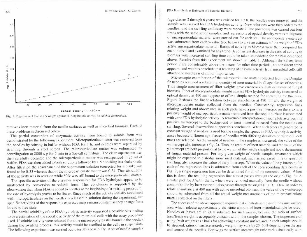
Several lines of evidence indicate that FDA hydrolytic activity is associated withmicrobial epiphytes rather than the needles: (a) first-year needles on which microbialepiphytes are almost entirely absent (2, 5) showed no significant FDA hydrolyticactivity; (b) older age class needles showed FDA hydrolytic activity increasing with age,a pattern consistent with the previous results of Bernstein and Carroll (2) and Carroll(5); (c) needles which had been swirled thoroughly (6-8 h) with buffer to removemicrobial epiphytes showed no significant FDA hydrolytic activity; (d) high levels ofFDA hydrolytic activity were associated with Atichia glomerulosa, the anamorph of acommon epiphytic loculoascomycete on coniferous needles. Atichia thalli could bepicked manually from locally collected white fir (Abies concolor) needles withoutsignificant contamination from the needles themselves; the amount of activity wasproportional to the weight of Atichia present, as shown in Fig. 1; (e) finally, as discussedin more detail below, the amount of FDA hydrolytic activity was proportional to theamount of total microbial biomass removed from the Douglas fir needles by swirling.with coefficients of determination for the regressions generally ranging from 0.85 to0.90.
Observation of FDA hydrolysis in the same samples over long periods (3-6 h)revealed that the rate of hydrolysis was linear with respect to time until an optical densityof 1 is reached, after which hydrolysis slowed and eventually stopped, an effect whichcan probably be ascribed to exhaustion of the FDA substrate. Incubation ofmicroparticulate matter with FDA for 1.5 h gave optimal optical density values (0.2-0.6) for needle samples weighing 1 g with average microbial populations. Either time ofshaking with FDA or the weight of needles could be easily adjusted to suit higher orlower FDA hydrolytic activities.
In order to use FDA hydrolysis as a measure of microbial biomass, it is necessary tocorrelate accurately a given amount of microbial biomass with a given activity. Thiscorrelation is made difficult by two factors: the enzyme (or enzymes) responsible forFDA hydrolysis are rendered partially soluble by swirling, and the swirling technique
220
7 0
6 0
50
40
3.0
0
Z 2.0
1.0
R. Swisher and G. C. Carroll
El
10 .20 30 40 50 60 70 80 90 1.00
optical density 1 .1 490nm,
Fig. 1. Regression of thallus dry weight a gainst FDA hydrolytic activity for Atichia glomerulosa.
removes inert material from the needle surfaces as well as microbial biomass. Each ofthese problems is discussed below.
The partial conversion of enzymatic activity from bound to soluble form wasdemonstrated by the following experiment. Microparticulate matter was removed fromthe needles by stirring in buffer without FDA for 1 h, and needles were separated bystraining through a steel screen. The microparticulate matter was sedimented bycentrifugation at 8000 x g for 5 min in a Sorvall centrifuge. The clear supernatant wasthen carefully decanted and the microparticulate matter was resuspended in 25 ml ofbuffer. FDA was then added to both solutions followed by 1.5 h shaking in a shaker bath.After filtration the absorbance of the supernatant solution (corrected for a blank ) wasfound to be 0.33 whereas that of the microparticulate matter was 0.34. Thus about 50%of the activity was in solution while 50% was still bound to the microparticulate matter.
The specific activities of the enzymes responsible for FDA hydrolysis appear to heunaffected by conversion to soluble form. This conclusion is supported by theobservation that when FDA is added to needles at the beginnin g of a swirling procedure.the rate of hydrolysis proves to be linear. Since much of the activity initially associatedwith microparticulates on the needles is released in solution during the experiment, thespecific activities of the responsible esterases must remain constant as they change frombound to free state.
The partial solubility of the FDA hydrolytic activity suggested a possible bias leadingto overestimation of the specific activity of the microbial cells with the assay procedureused. If soluble activity were released from the microepiphytes still bound to the needlesduring the swirling process, this activity would be ascribed to the cells in suspension.The followin g experiment was carried out to test this possibility. A set of needle samples
FDA Hydrolysis as Estimator of Microbial Biomass 221
E
(age-classes 2 through 6 years) was swirled for 1.5 h. the needles were removed, and thesample was assayed for FDA hydrolytic activity. New solutions were then added to theneedles, and the swirling and assay were repeated. The procedure was carried out fourtimes with the same set of samples, and re gressions of optical density versus milligramsof microparticulate material were carried out for each set. The appropriate y-interceptwas subtracted from each y-value (see below) to give an estimate of the weight of FDAactive microparticulate material. Ratios of activity to biomass were then computed foreach interval and examined for any trend. A consistent decrease in the ratio of activit y tobiomass with increased swirling time could be taken as evidence for the bias describedabove. Results from this experiment are shown in Table 1. Although the values fromperiod 2 are considerably above the means for other time periods. no consistent trendappears, and we thus conclude that leaching of enzyme activity from microbial cells stillattached to needles is of minor importance.
Microscopic examination of the microparticulate matter collected from the Douglasfir needles revealed a substantial quantity of inert material in all age classes of needles.Thus simple measurement of filter weights gave erroneously hi g h estimates of fungalbiomass. Plots of microparticulate weight against FDA hydrol y tic activity (measured asoptical density at 490 nm) appear to offer a simple method for correctin g for this bias.Figure 2 shows the linear relation between absorbance at 490 nm and the wei g ht ofmicroparticulate matter collected from the needles. Consistently, regression linesrelating weight and absorbance in such plots have a positive intercept on the y axis: apositive wei ght of microparticulate matter removed from the needle surface is associatedwith zero FDA hydrolytic activity. A reasonable interpretation of such plots ascribes thispositive y-intercept to the background of inert material released from the needles onswirling. Several observations support this conclusion. When such plots are generated. aconstant weight of needles is used for the sample; the spread in FDA hydrolytic activityarises because different age-classes of needles with differing densities of microbial cellmass are selected. As the wei ght of needles in the samples is increased, the value of they-intercept also increases (Fig. 2). Thus the amount of inert material and the value of they-intercept are both proportional to the weight of the needle sample and not to the amountof fungal material present. For any given weight of needle sample, treatments whichmight be expected to dislod ge more inert material, such as increased time or speed ofswirling, also increase the value of the y-intercept. When the value of the y-intercept foreach of the regression lines is subtracted from each of the corresponding data points inFig. 2, a single regression line can be determined for all of the corrected values. Whenthis is done, the resulting regression line almost passes throu g h the ori g in (Fig. 3). Asimilar plot for Atichia thalli, which were removed manuall y from the needle withoutcontamination by inert material, also passes through the origin (Fig. 1). Thus, in order torelate absorbance at 490 nm with active microbial biomass, the value of the y-interceptshould be subtracted from all the actual weight measurements of the microparticulatematter collected on the filters.
The success of the above approach requires that substrate samples of the same surfacearea which release approximately the same amount of inert material/sample be used.Needles or leaves are an ideal substrate for such assays, because the ratio of surfacearea/fresh weight is acceptably constant within the samples chosen. The importance ofusing fresh weights as a basis for achieving a constant surface area in each sample shouldbe stressed; ratios of surface area/dry weight may vary by 25-50% depending on the ageand source of the needles. For twi g s the surface area/weight ratio varies drastica:ly with

R. Swisher and G. C. Carroll
1.3
c
3
CC
Cal
51,
7=-3C
C
0
0
51,E
0
OO
5.4
0O
0C
- -r
6 c
r‘ i r--
X
("1
6 6
r K r I .7
6 6
in occc.6 6 6 6
C6 6
-6 6 C :It:
in r", Inr- iC-6 6 6 -
rr.
-
C
517.
`g,C c71:,
O 6. 6 6
I)L 6 0
-■ r- r- r•-••ri -
-6 6 CiTC C^,
-"J
- -r X ("IE C." - 6
C
C-0
In 3C
O .7.% 6 6
- I r- -rCZ: 6
FDA Hydrolysis as Estimator of Microbial Biomass 223
7.0
6.0
••••• 0
,.••0.5 gram needles--- •1.0 gram needles 1.5 gram needles
10 .20 .30 .40 .50 .60 .70 .80 .90 1.00
optical density (.t 490nm)
Fig. 2. Regressions of dry weight against FDA hydrolytic activity for microepiphytes on Douglas fir needles.Each line represents a separate regression for a different sample weight, uncorrected for inert material. Needleswere swirled for 3 h to dislod ge microbial cells. r2 for regression with 0.5 g sample = 0.86; for 1.0 2 sample =0.75; for 1.5 g sample = 0.86. Differences in slopes of the line are not significant at the 1% level.
0
7.0
6.0
E 5.0
03
- .2 4.0
0 0
2-2
co
3.0
a)
2.00
.,
0
0.5 gram needles-
1.0 gram needies--.:,1.5 gram needles-t,Y
E 10
50
E
O 4 0
0a 3.0
E
2.0
IN X rr,,C 0, C
1 rr,
,.....5e
.2 E- Iru .6 12
.10 .20 .30 .40 50 .60 .70 80 .90 1.00
optical density l A= 490nm)
Fig. 3. Single regression of all data points from Fig. 2, with intercepts subtracted to correct for inert material.r2 = 0.90.
CC O
R. Swisher and G. C. Carroll
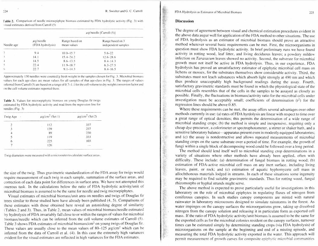
Fable 2. Comparison of needle microepiphyte biomass estimated by FDA hydrolytic activity (Fig. 3) withvisual estimates derived from Carroll (5)
kg/needle ICarroll (5)1
Needle agepg/needle(FDA hydrolysis)
Range based onmean values
Range based on 3independent samples
4
9.414.114.522.422.7
10.0-15.715.4-24.28.6-13.5
11.9-18.79.1-14.3
5.6-2212.6-28.68.4-14.36.3-27.58.4-16.5
Approximately 130 needles were counted/g fresh weight in the samples chosen for Fig. 3. Microbial biomassvalues for each age-class are mean values for all samples of that age-class in Fig. 3. The ranges of valuesinferred from Carroll (5) are based on a range of 0.7-1. I for the cell volume to dry weight conversion factor andon the cell volume estimates reported there.
Table 3. Values for microepiphytic biomass on young Douglas fir twigsestimated by FDA hydrolytic activity and read from the regression line forneedles (Fig. 3)
Twig Age Pgiem 2 (Set I) pg/cm2 (Set 2)
112 3373 139 2374 89 2305 119 3336 225 1037 126
Twig diameters were measured with a micrometer to calculate surface areas.
the size of the twig. Thus gravimetric standardization of the FDA assay for twigs wouldrequire measurement of each twig in each sample. summation of the surface areas, andcorrection of each data point for differences in surface area. We have not attempted thisonerous task. In the calculations below the ratio of FDA hydrolytic activity/unit ofmicrobial biomass is assumed to be the same for needle and twig microepiphytes.
Visual estimates of microbial biomass/unit area on needles and twigs of Douglas firtrees similar to those studied here have already been published (4, 5). Comparisons ofthese estimates with those obtained here reveal an astonishing degree of similarity(Tables 2 and 3). Thus, in Table 2, mean values of microbial biomass/needle estimatedby hydrolysis of FDA invariably fall close to or within the ranges of values for microbialbiomass/needle which can be inferred from the cell volume estimates of Carroll (5).Table 3 shows FDA estimates of microbial biomass/cm = on young twigs of Douglas fir.These values are usually close to the mean values of 80-125 big/cm = which can beinferred from the data of Carroll et al. (4). In this case the extremely high variancesevident for the visual estimates are reflected in hi g h variances for the FDA estimates.
FDA Hydrolysis as Estimator of Microbial Biomass 275
Discussion
The degree of agreement between visual and chemical estimation procedures evident inthe above data augur well for application of the FDA method to other situations. The useof FDA hydrolysis as an estimator of microbial biomass should prove an attractivemethod wherever several basic requirements can be met. First, the microoroanisms inquestion must show FDA hydrolytic activity. In brief preliminary runs we have foundactivity in rotting wood, leaf litter, and living deciduous leaves; a powdery mildewinfection on Taraxactim leaves showed no activity. Second, the substrate for microbialgrowth must not itself be active in FDA hydrolysis. Thus, in our experience, FDAhydrolysis has proved an unsatisfactory estimator of epiphytic microbial cell mass onlichens or mosses, for the substrates themselves show considerable activity. Third, thesubstrates must not leach substances which absorb light strongly at 490 nm and whichthus produce unacceptably high background readings during the assay. Fourth.satisfactory gravimetric standards must be found in which the physiological state of themicrobial cells resembles that of the cells in the samples to be assayed as closely aspossible. Finally, the fluctuations in biomass/activity ratio for the microbial cells underinvestigation must be acceptably small: coefficients of determination (r 2 ) for there g ression lines should be above 0.85.
Where these requirements can be met, the assay offers several advantages over othermethods currently in use: (a) rates of FDA hyrdolysis are linear with respect to time overa great range of optical densities; this permits the determination of a wide range ofmicrobial standing crops; (b) the method is simple and inexpensive, requirin g only acheap dye-precursor, a colorimeter or spectrophotometer, a stirrer or shaker bath, and asensitive laboratory balance—apparatus present even in modestly equipped laboratories;and (c) the assay is nondestructive and allows repeated measurements of microbialstanding crops on the same substrate over a period of time. For example, the growth offungi within a single block of decomposing wood could be followed over a lon g period.
The method should lend itself well to microbial standing crop determinations in avariety of situations where other methods have already been applied, often withdifficulty. These include: (a) determination of fungal biomass in rotting wood; (b)estimation of FDA-active microbial cell mass on any inert surface, includin g livingleaves, paint, or rock; and (c) estimation of aquatic hyphomycete cell mass inallochthonous materials lodged in streams. In each of these situations some ingenuitymay be required to find suitable gravimetric standards. Thus for rottin g wood. fungalrhizomorphs or hyphal strands might serve.
The above method is expected to prove particularly useful for investi gations in thislaboratory on the role of microbial epiphytes in regulating fluxes of nitro gen fromconiferous canopies. In such studies canopy components are misted with naturalrainwater in laboratory microcosms designed to simulate rainstorms in the forest. Aswater impinges on the canopy surfaces the microorganisms grow, takin g up dissolvednitrogen from the canopy solution and releasing it in particulate form as exported cellmass. If the ratio of FDA hydrolytic activity/unit biomass is assumed to be the same forthe exported cells as for the microbial colonies resident on the canopy surfaces, turnovertimes can be estimated simply by determining standing crops (via FDA hydrolysis) ofmicroorganisms on the sample at the beginning and end of a misting episode, andmeasuring the total FDA hydrolytic activity exported in the water. This approach willpermit measurement of growth curves for composite epiph y tic microbial communities
22( R. Swisher and G. C. Carroll
Microb. Ecol. 6:227-240 (1980)
MICROBIAL ECOLOGY
on their natural substrates and will allow determination of various microbial growthparameters. The use of computer simulation models to extrapolate from microcosms toreal canopies with regard to patterns of nitrogen flux is a long-term research goal in ourlaboratory. The discovery of a method to actually measure parameters for the modelwhich previously could only be guessed at makes realization of this goal far more likely.
Acknowledgement. The authors wish to acknowledge support for this work from NSF Grant DEB 78-03583.
References
Ausmus, B. S.: The use of ATP assay in terrestrial decomposition studies. Bull. Ecol. Res. Commit.(Stockholm) 17, 223-234 (1973)Bernstein, M. E.. and G. Carroll: Microbial production on Douglas fir needle surfaces. Microb. Ecol. 4.41-52(1977)Blanchette, R. A., and C. G. Shaw: Associations amon g bacteria, yeasts, and basidiomycetes dub%wood decay. Phytopathol. 68, 631-637 (1978)Carroll, G. C., L. H. Pike, J. R. Perkins, and M. A. Sherwood: Biomass and distribution patterns ofconifer twi g microepiphytes in a Douglas fir forest. Can. J. Bot. 58. 624-630(19SO)Carroll, G. C.: Needle microepiphytes in a Douglas fir canopy: biomass and distribution patterns. Can. J.Bot. 57, 1000-1007 (1979)Franklin. J. F., and C. T. Dyrness: Natural Vegetation of Oregon and Washington. U.S. Dept. AgriculFor. Serv. Gen. Tech. Rept. PNW-8. Portland (1973)Holm-Hansen. 0.: The use of ATP determinations in ecological studies. Bull. Ecol. Res. Commit(Stockh. ) 17. 215-222(1973)Hubbard, J. S.: Radiorespirometric methods in measurement of metabolic activities in soil. Bull. Ecol.Res. Commit. (Stockh.) 17, 199-206(1973)Jones, P. C. T.. and J. E. Mollison: A technique for the quantitative estimation of soil microorganisms. J.Gen. Microbiol. 2. 54-68 (1948)Oades, J. M., and D. S. Jenkinson: The ATP content of the soil microbial biomass. Soil Biol. Biochem11, 201-204 (1979)Seitz, L. M., H. E. Mohr, R. Burroughs, and D. B. Sauer: Ergosterol as an indicator of fungal invasion ingrains. Cereal Chem. 54, 1207-1217 (1978)Seitz. L. M., D. B. Sauer, R. Burroughs, H. E. Mohr, and J. D. Hubbard: Ergosterol as a measure offungal growth. Phytopathol. 69. 1202-1203 (1979)Skujins, J.: Dehydrogenase: an indicator of biological activities in arid soils. Bull. Ecol. Res. Commit.(Stockh.) 17, 235-241 (1973)SOderstrOm, B. E.: Vital staining of fungi in pure cultures and in soil with fluorescein diacetate. Soil Biol.Biochem. 9, 59-63 (1977)SiiderstrOm, B. E.: Some problems in assessing the fluorescein diacetate active fun gal biomass in thesoil. Soil Biol. Biochem. 11, 147-148 (1979)Swift, M. J.: The estimation of mycelial biomass by determination of the hexosamine content of woodtissue decayed by fungi. Soil Biol. Biochem. 5, 321-332 (1973)Visser, S., and D. Parkinson: Fungal succession on aspen poplar leaf litter. Can. J. Bot. 53, 1640-I6f'l(1975)Waid, J. S., K. J. Preston, and P. J. Harris: A method to detect metabolically-active microorganisms inleaf litter habitats. Soil Biol. Biochem. 3, 235-241 (1971)
Influence of Zinc, Lead, and Cadmium Pollutants on the Microfloraof Hawthorn Leaves
Richard J. F. Bewley and Richard Campbell
Department of Botany, University of Bristol, Bristol, BS8 1UG, England
Abstract. Transect studies were conducted to determine the relative effects of zinc.lead, and cadmium pollution on microorganisms occurring on ham. thorn leaves atvarying distances from a smelting complex. Sporobolomyces roseus was absentfrom the most heavily contaminated leaves but, although lead was inhibitory, otherenvironmental factors were also important in determining its overall populationlevel. Conversely, Aureobasidium pullulans and nonpigmented yeasts showed asignificant partial positive correlation with lead but were inhibited by zinc and/orcadmium. Numbers of bacterial colonies were only slightly reduced by thecombined effect of all three metals, but total numbers of bacteria were highlynegatively correlated with lead. Filamentous fungi, isolated by leaf washin g , wereonly sli g htly inhibited by all three metals, and the degree of mycelial proliferationon senescent leaves was little affected by heavy metal pollution. Computer-generated maps were produced of the distribution of A. pullulans in relation to zincand lead fallout.
Introduction
The effects of heavy metal pollution on microbial ecology are poorly understood (2. 3).Many areas of environmental pollution involve simultaneous contamination by a varietyof elements, and it is particularly difficult to determine the relative importance ofindividual effects and synergistic interactions. In the vicinity of a smelting complex atAvonmouth near Bristol, En g land, there is heavy contamination of aerial plant surfacesby zinc, lead, and cadmium (12, 13). Many leaf surface microor g anisms appear toflourish under such conditions although there are species differences in tolerance (5, 6).
Preliminary studies conducted by the authors showed that Aureobasidium pullulans(de Bary) Arnaud is particularly tolerant to heavy metals, bacteria are somewhat lesstolerant, and Sporobolomyces roseus KI. and van Niel is a particularly sensitive species.The aim of the present study was to investigate the relative impact of zinc, lead, and
I Present address: Department of Biology, New York University, Washington Square, Nev. York. NY 10003.
0095-3628/80/0006-0227 52.801980 Sprin ger-Verla g Ne York Inc