Embed Size (px)
Citation preview

UNESCO-EOLS
S
SAMPLE C
HAPTERS
BIOMECHANICS - Fluid Biomechanics And Circulation - Natacha DePaola and Michael P. Burns
©Encyclopedia of Life Support Systems (EOLSS)
FLUID BIOMECHANICS AND CIRCULATION Natacha DePaola and Michael P. Burns Illinois Institute of Technology, USA Keywords: shear stress, pressure, hemodynamics, equations of fluid motion, venous flow, arterial flow, microcirculation, inflammation, thrombosis, atherosclerosis Contents 1. Introduction 2. Fluid mechanics of blood flow 3. Fluid dynamics of the vascular beds 4. Bio-fluid dynamics in pathophysiology Glossary Bibliography Biographical Sketches
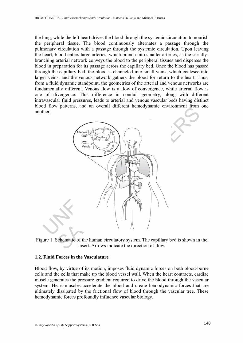
Summary This chapter discusses the fluid mechanics of the human circulatory system, illustrating the importance of the local hemodynamic (blood flow) environment in the regulation of cardiovascular function and maintenance of health. Basic concepts of fluid mechanics are introduced, and the general equations of fluid motion are derived in a summarized form. The complexity of fully characterizing the dynamics of blood flow throughout the vascular system is acknowledged, and the key characteristics of blood flow in each of the vascular beds (arterial, venous and capillary) are discussed. The role of fluid forces on vascular tissue biology is presented in the context of selected pathophysiological examples. This chapter is not intended to provide a rigorous mathematical analysis of fluid flow in the human circulation, but rather to describe the distinctive physical flow environment found throughout the vascular tree and its relationship to biological function. 1. Introduction 1.1. Circulation Blood circulation is essential for maintaining the stability of the body’s normal physiological condition (i.e., homeostasis). The circulatory system coordinates a loop of blood flow that delivers oxygen and nutrients to the body’s tissues and organs while waste products are removed and transported to the liver and kidneys for excretion. The circulation also aids in other vital processes including temperature regulation, transport of hormones, and delivery of cells controlling the body’s immune response. Figure 1 is a cartoon representation of the human circulatory system. The heart is a four-chambered muscular pump that functions as two distinct pumps working side-by-side. The right heart drives the blood through the pulmonary circulation for gas exchange in
147
UNESCO-EOLS
S
SAMPLE C
HAPTERS
BIOMECHANICS - Fluid Biomechanics And Circulation - Natacha DePaola and Michael P. Burns
©Encyclopedia of Life Support Systems (EOLSS)
the lung, while the left heart drives the blood through the systemic circulation to nourish the peripheral tissue. The blood continuously alternates a passage through the pulmonary circulation with a passage through the systemic circulation. Upon leaving the heart, blood enters large arteries, which branch into smaller arteries, as the serially-branching arterial network conveys the blood to the peripheral tissues and disperses the blood in preparation for its passage across the capillary bed. Once the blood has passed through the capillary bed, the blood is channeled into small veins, which coalesce into larger veins, and the venous network gathers the blood for return to the heart. Thus, from a fluid dynamic standpoint, the geometries of the arterial and venous networks are fundamentally different. Venous flow is a flow of convergence, while arterial flow is one of divergence. This difference in conduit geometry, along with different intravascular fluid pressures, leads to arterial and venous vascular beds having distinct blood flow patterns, and an overall different hemodynamic environment from one another.
Figure 1. Schematic of the human circulatory system. The capillary bed is shown in the insert. Arrows indicate the direction of flow.
1.2. Fluid Forces in the Vasculature Blood flow, by virtue of its motion, imposes fluid dynamic forces on both blood-borne cells and the cells that make up the blood vessel wall. When the heart contracts, cardiac muscle generates the pressure gradient required to drive the blood through the vascular system. Heart muscles accelerate the blood and create hemodynamic forces that are ultimately dissipated by the frictional flow of blood through the vascular tree. These hemodynamic forces profoundly influence vascular biology.
148

UNESCO-EOLS
S
SAMPLE C
HAPTERS
BIOMECHANICS - Fluid Biomechanics And Circulation - Natacha DePaola and Michael P. Burns
©Encyclopedia of Life Support Systems (EOLSS)
Since blood flow rate and blood vessel geometry vary considerably throughout the circulation, the hemodynamic forces in the various vascular beds vary considerably as well. Arterial blood flow is characterized by high-pressure, pulsatile flow through vessels with branching geometry; capillary flow is characterized by steadier flow through a vessel whose diameter approaches the size of the red blood cell; venous flow is characterized by low-pressure flow through collapsible vessels with a converging geometry. The fluid dynamic forces that characterize each of these particular hemodynamic environments are highly dependent on the vascular bed being considered. The fluid dynamics of vascular beds is discussed in Section 3 of this chapter. Biological events in the circulation are regulated by the local blood flow and its interaction with the vascular tissue. The lumenal surface of blood vessels is lined by the endothelium, a single-cell layer tissue with key functions known to be regulated by fluid forces. Endothelial cells are in direct contact with the flowing blood and, as a result, are exposed to fluid shear stress (the force exerted by a moving fluid in the direction of the flow). Blood shear forces alter endothelial cell gene expression and regulate endothelial cell release of vasoactive compounds. In fact, blood shear forces elicit endothelial cell feedback responses that help regulate the shear stress imposed on the vascular wall. For example, high wall shear stresses cause endothelial cells to release nitric oxide, a vasodilator, which relaxes vascular smooth muscle cells, thereby increasing the vessel bore and lowering the fluid shear stress imposed on the vessel wall. Conversely, when endothelial cells experience low shear stress, they release endothelin-1, a vasoconstrictor. Thus, the endothelium’s response to blood shear forces helps keep arterial wall shear stress relatively constant throughout the arterial tree. Regulation of blood shear stress levels is important because high wall shear stress equates to high vascular resistance and, consequently, a large workload on the heart. Although physiological biological events are regulated by the local blood flow and its interaction with the vascular tissue, blood forces can also be detrimental. High shear environments can activate platelets and damage red blood cells. In addition, elevated levels of wall shear stress often have a role in the culminating clinical episodes of vascular disease. For example, in atherosclerosis, vessel narrowing due to plaque growth produces high wall shear stresses that ultimately cause plaque rupture. In discussing fluid biomechanics in the context of the human circulatory system, this chapter describes the physical forces associated with blood flow in the different vascular beds (Section 3) and establishes functional correlations between fluid forces and cardiovascular pathophysiology (Section 4). 1.3. Flow Characterization It has been long recognized that hemodynamics (or blood fluid dynamics) has a regulatory role in biological events of daily incidence and life-long lasting impact in the vascular system. To better understand this relationship, a detailed characterization of the fluid biomechanics of the human circulation is required. Fluid dynamic analysis of blood flow describes the hemodynamic environment and quantifies the forces within the vasculature. A fluid dynamic environment is completely
149

UNESCO-EOLS
S
SAMPLE C
HAPTERS
BIOMECHANICS - Fluid Biomechanics And Circulation - Natacha DePaola and Michael P. Burns
©Encyclopedia of Life Support Systems (EOLSS)
defined if the fluid pressure and velocity are known for each location, at any given time, within the flow field. The equations of motion that govern blood flow are complex, involving multiple parameters that interact with one another in a non-linear fashion, therefore (with exception of certain simple flow environments), there is often no analytical solution to the full governing flow equations that describe the dynamics of blood flow in the human circulation. An overall challenge in the field of fluid biomechanics is to accurately characterize the flow field within the limitations associated with the ability to solve the resultant governing equations. The basic strategy is to modify the equations of motion by imposing simplifying assumptions that allow the elimination of smaller-order terms that are insignificant within the particular vascular bed being considered (vein, artery, or capillary). Alternatively, numerical solutions of the full equations can be obtained using computational fluid dynamic techniques. Section 2 of this chapter illustrates how the methods of fluid dynamic analysis are applied to the circulation. Modern medicine increasingly uses fluid dynamic analysis of the hemodynamic environment to support clinical decision making. Advances in computing and medical imaging now allow patient-specific data to be incorporated into simulation of the hemodynamic environment. These individually-tailored simulations help in patient assessment, prediction of outcomes, and design of treatment strategies. Detailed hemodynamic analysis also aids the development of vascular implants that are efficient and minimally thrombogenic. 2. Fluid Mechanics of Blood Flow This section describes basic concepts of fluid mechanics and derives the Navier-Stokes (N-S) equations, which are the general equations of motion that govern fluid flow. A derivation will also be presented for the Hagen-Poiseuille equation—the equation that governs the special case of steady laminar flow through a cylindrical pipe. 2.1. Basic Concepts of Fluid Mechanics 2.1.1. Forces in Blood Flow Blood flow, like all fluid flows, is governed by a balance of forces. Fluid forces can act either at a fluid surface or at the center of mass of the fluid. Accordingly, fluid forces may be classified as normal forces, shearing forces, or body forces. Normal forces act perpendicular to a fluid surface, and shearing forces act parallel to a fluid surface. Body forces are forces that act at the center of mass of the fluid. For blood flow, the relevant forces are pressure forces, viscous forces, and inertial forces. Pressure forces are normal forces—they act perpendicular to a fluid surface. Viscous forces, by contrast, are shearing forces—they act parallel to a fluid surface. Inertial forces are body forces—they act at the fluid’s center of mass and arise when fluid accelerates. Fluid may accelerate by changing its velocity with respect to time (i.e., flow rate) or space (i.e., flow direction).
150

UNESCO-EOLS
S
SAMPLE C
HAPTERS
BIOMECHANICS - Fluid Biomechanics And Circulation - Natacha DePaola and Michael P. Burns
©Encyclopedia of Life Support Systems (EOLSS)
Viscous forces are frictional; they always resist fluid motion. Pressure forces, on the other hand, usually drive flow. However, it is possible for fluid to move against an adverse pressure gradient, but in doing so, the fluid will decelerate. Inertial forces descend from fluid acceleration; thus inertial forces reflect the comparative strengths of the pressure and viscous forces. When pressure forces exceed the resistive viscous forces, the fluid accelerates and generates inertia, which may be consumed later to overcome viscous forces or adverse pressure gradients. Blood forces create stress, where stress is defined as force per unit area. For example, shear stress is calculated by dividing the shear force by the area upon which the force is applied. Fluids are materials which continually deform under the application of a shear stress. The rate at which the fluid deforms is called the shear rate, and this is the rate at which the fluid particles are displaced relative to one another. In a flowing fluid, the shear rate is equal to the fluid’s velocity gradient normal to the surface being considered. 2.1.2. Blood Viscosity: Newtonian Vs Non-Newtonian Fluid The relationship between the applied shear stress and the resultant shear rate depends on the fluid viscosity. For some fluids, the relationship between shear stress and shear rate is linear and independent of the shear rate—this type of fluid is called Newtonian. Thus, for a Newtonian fluid, the relationship between shear stress (τ ), shear rate (γ ), and viscosity (μ ) is given by the following equation: τ μγ= (1) In general, blood may be considered Newtonian, but when the shear rate of blood is small (i.e. < 100 s-1), the non-Newtonian aspects of blood become evident. Since shear rates below 100 s-1 are found in certain regions of the circulation (e.g., capillaries and postcapillary venules), the non-Newtonian nature of blood warrants discussion. Blood’s non-Newtonian nature is due to red blood cell (RBC) aggregation. When low blood shear rates are coupled with the presence of the plasma proteins fibrinogen and globulin, human RBCs form linear stacks called rouleaux. Rouleaux formation becomes more extensive as blood shear rate decreases; both the number and size of the rouleaux increase as the shear rate declines. Inside the vessel, blood velocity is non-uniform, with the blood near the vessel wall moving slower than the blood near the center of the vessel. The non-uniform velocity profile of the blood imparts a moment on the rouleaux, causing them to tumble. Rouleaux tumbling disturbs the flow field, which consumes energy, and increases blood viscosity. As shear rate increases, the rouleaux are broken up and blood viscosity decreases. Thus, at low shear rates blood displays shear-thinning characteristics since blood’s viscosity decreases with increasing shear rate. At shear rates above 100 s-1, rouleaux formation is abolished, and the blood behaves as a Newtonian fluid. The non-linear nature of blood viscosity has been extensively investigated, and it has been found that blood is best described by Casson’s
151

UNESCO-EOLS
S
SAMPLE C
HAPTERS
BIOMECHANICS - Fluid Biomechanics And Circulation - Natacha DePaola and Michael P. Burns
©Encyclopedia of Life Support Systems (EOLSS)
relationship, which is given by the equation: y ckτ τ γ= + . In this equation, yτ
denotes the yield stress and ck is a constant. In addition to shear-rate effects, blood viscosity is also influenced by hematocrit. Blood viscosity increases with increasing hematocrit, and RBC flexibility helps alleviate this effect. Somewhat surprisingly, RBCs are more flexible than oil droplets. Blood is amazing in its ability to flow with hematocrits as high as ninety percent. Oil emulsions, by contrast, cease to flow once the suspension volume nears fifty percent. Blood rheology is discussed extensively by Fung in his book Biomechanics: Mechanical Properties of Living Tissues, which is cited in the Bibliography of this chapter.
2.2. The Navier-Stokes Equations The Navier-Stokes equations are the general equations of motion that govern fluid flow. These equations are derived from fluid force analysis. Figure 2 depicts a conceptual control volume of fluid, which is fixed in space and completely enclosed by an outer surface—the control volume surface—through which the fluid can freely pass. The fluid density is represented by ρ and the x-, y-, and z-components of the fluid velocity are represented by u, v, and w, respectively.
Figure 2. Fluid control volume. Conservation of mass, applied to the control volume, requires that the net flow of mass across the control volume surface must equal the change in mass within the control volume. This is expressed mathematically by the continuity equation:
152

UNESCO-EOLS
S
SAMPLE C
HAPTERS
BIOMECHANICS - Fluid Biomechanics And Circulation - Natacha DePaola and Michael P. Burns
©Encyclopedia of Life Support Systems (EOLSS)
( ) ( ) ( ) 0u v wx y z t
ρρ ρ ρ∂ ∂ ∂ ∂+ + + =
∂ ∂ ∂ ∂ (2)
Since the bulk modulus of blood is similar to that of water (~2 GPa) and is large compared to the pressure within the circulatory system (~13 kPa), blood may be considered an incompressible fluid. The density of an incompressible fluid is constant. Thus, the continuity equation for an incompressible fluid reduces to 0V∇⋅ = . Conservation of momentum requires that the sum of the external forces acting on the control volume must equal the net efflux of linear momentum from the control volume plus the time rate of change of linear momentum within the control volume. As mentioned earlier, the forces that act on the control volume can be classified as either body forces or surface forces. Body forces act at the control volume’s center of mass, while surface forces act (somewhat obviously) on the control volume surface. Surface forces may be further classified as either normal forces or shearing forces. With these classifications in mind, it is clear that gravity is a body force, pressure is a normal force, and viscous forces are shearing forces. Enforcing conservation of mass and conservation of momentum on the control volume gives the following set of equations:
yxxx zxx
u u u ug u v wx y z t x y z
τσ τρ ρ∂ ⎛ ⎞∂ ∂ ∂ ∂ ∂ ∂
+ + + = + + +⎜ ⎟∂ ∂ ∂ ∂ ∂ ∂ ∂⎝ ⎠ (3)
xy yy zyy
v v v vg u v wx y z t x y zτ σ τ
ρ ρ∂ ∂ ∂ ⎛ ⎞∂ ∂ ∂ ∂
+ + + = + + +⎜ ⎟∂ ∂ ∂ ∂ ∂ ∂ ∂⎝ ⎠ (4)
yzxz zzz
w w w wg u v wx y z t x y z
ττ σρ ρ∂ ⎛ ⎞∂ ∂ ∂ ∂ ∂ ∂
+ + + = + + +⎜ ⎟∂ ∂ ∂ ∂ ∂ ∂ ∂⎝ ⎠ (5)
The above equations contain fluid stress terms and fluid velocity terms. However, fluid velocity will be related to fluid stress through the intrinsic properties of the fluid (e.g., viscosity). In other words, fluid stress may be expressed as a function of the velocity field and the viscosity. As mentioned earlier, blood can always be considered incompressible and can usually be considered a Newtonian fluid. For an incompressible, Newtonian fluid the relationship between fluid stress and fluid motion is given by Eqs. (6-8). The derivation of these equations is extensive and may be found in the references cited at the end of this chapter.
2xx xy yxu u vpx y x
σ μ τ τ μ⎛ ⎞∂ ∂ ∂
= − + = = +⎜ ⎟∂ ∂ ∂⎝ ⎠ (6)
153
UNESCO-EOLS
S
SAMPLE C
HAPTERS
BIOMECHANICS - Fluid Biomechanics And Circulation - Natacha DePaola and Michael P. Burns
©Encyclopedia of Life Support Systems (EOLSS)
2yy yz zyv w vpy y z
σ μ τ τ μ⎛ ⎞∂ ∂ ∂
= − + = = +⎜ ⎟∂ ∂ ∂⎝ ⎠ (7)
2zz zx xzw u wpz z x
σ μ τ τ μ∂ ∂ ∂⎛ ⎞= − + = = +⎜ ⎟∂ ∂ ∂⎝ ⎠ (8)
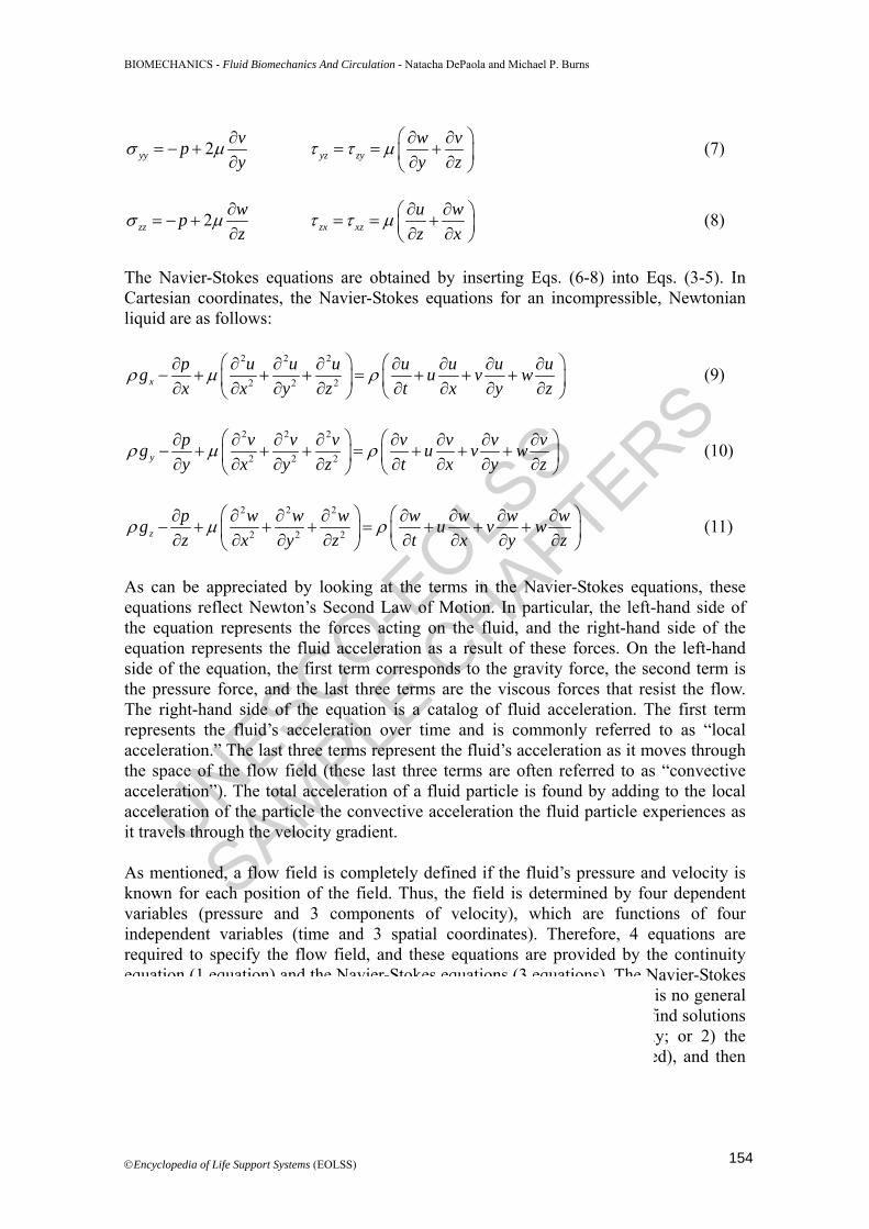
The Navier-Stokes equations are obtained by inserting Eqs. (6-8) into Eqs. (3-5). In Cartesian coordinates, the Navier-Stokes equations for an incompressible, Newtonian liquid are as follows:
2 2 2
2 2 2xp u u u u u u ug u v wx x y z t x y z
ρ μ ρ⎛ ⎞ ⎛ ⎞∂ ∂ ∂ ∂ ∂ ∂ ∂ ∂
− + + + = + + +⎜ ⎟ ⎜ ⎟∂ ∂ ∂ ∂ ∂ ∂ ∂ ∂⎝ ⎠⎝ ⎠ (9)
2 2 2
2 2 2yp v v v v v v vg u v wy x y z t x y z
ρ μ ρ⎛ ⎞ ⎛ ⎞∂ ∂ ∂ ∂ ∂ ∂ ∂ ∂
− + + + = + + +⎜ ⎟ ⎜ ⎟∂ ∂ ∂ ∂ ∂ ∂ ∂ ∂⎝ ⎠⎝ ⎠ (10)
2 2 2
2 2 2zp w w w w w w wg u v wz x y z t x y z
ρ μ ρ⎛ ⎞ ⎛ ⎞∂ ∂ ∂ ∂ ∂ ∂ ∂ ∂
− + + + = + + +⎜ ⎟ ⎜ ⎟∂ ∂ ∂ ∂ ∂ ∂ ∂ ∂⎝ ⎠⎝ ⎠ (11)
As can be appreciated by looking at the terms in the Navier-Stokes equations, these equations reflect Newton’s Second Law of Motion. In particular, the left-hand side of the equation represents the forces acting on the fluid, and the right-hand side of the equation represents the fluid acceleration as a result of these forces. On the left-hand side of the equation, the first term corresponds to the gravity force, the second term is the pressure force, and the last three terms are the viscous forces that resist the flow. The right-hand side of the equation is a catalog of fluid acceleration. The first term represents the fluid’s acceleration over time and is commonly referred to as “local acceleration.” The last three terms represent the fluid’s acceleration as it moves through the space of the flow field (these last three terms are often referred to as “convective acceleration”). The total acceleration of a fluid particle is found by adding to the local acceleration of the particle the convective acceleration the fluid particle experiences as it travels through the velocity gradient. As mentioned, a flow field is completely defined if the fluid’s pressure and velocity is known for each position of the field. Thus, the field is determined by four dependent variables (pressure and 3 components of velocity), which are functions of four independent variables (time and 3 spatial coordinates). Therefore, 4 equations are required to specify the flow field, and these equations are provided by the continuity equation (1 equation) and the Navier-Stokes equations (3 equations). The Navier-Stokes equations are second order, non-linear, partial differential equations; there is no general analytical solution for equations of this form. Two approaches are used to find solutions to the Navier-Stokes equations: 1) the equations are solved numerically; or 2) the equations are modified (if certain simplifying assumptions may be applied), and then the modified equations are solved analytically.
154

UNESCO-EOLS
S
SAMPLE C
HAPTERS
BIOMECHANICS - Fluid Biomechanics And Circulation - Natacha DePaola and Michael P. Burns
©Encyclopedia of Life Support Systems (EOLSS)
Bibliography Barbee J.H., Cokelet G.R. (1971). The Fahraeus effect. Microvascular Research 3(1), 6-16. [A landmark study that identified the apparent viscosity of blood flowing through small tubes is reduced due to red blood cell margination].
Damiano E.R., Long D.S., and Smith M.L. (2004). Estimation of viscosity profiles using velocimetry data from parallel flows of linearly viscous fluids: application to microvascular haemodynamics. Journal of Fluid Mechanics 512, 1-19. [An approach that uses velocimetry data to estimate a spatial distribution of viscosity. This is an in vitro study that supports the treatment of heterogeneous RBC distribution in capillary vessels as a Newtonian fluid with a spatially varying kinematic viscosity].
Fahraeus R. (1929). The suspension stability of the blood. Physiological Review 9:241-274. [The first report of increased discharge hematocrit for blood flowing through small diameter tubes.]
Fahraeus R., Lindqvist T. (1931). Viscosity of blood in narrow capillary tubes. American Journal of Physiology 96:562-568. [The seminal report that apparent viscosity of blood flowing through tubes decreases with decreasing tube diameter.]
Fung Y.C. (1993). Biomechanics: Mechanical Properties of Living Tissue. New York: Springer. [A comprehensive discussion of the rheological properties of blood].
Goldsmith H.L. (1971). Red cell motions and wall interactions in tube flow. Federation Proceedings 30:1578-1590. [The first demonstration that cell deformability is required for margination.]
Lurie F., Kistner R.L., Eklof B., Kessler D.. (2002). Mechanism of venous valve closure and role of the valve in circulation: a new concept. Journal of Vascular Surgery 38, 955-961. [A study that investigates the self-sustained mechanism of vein valve closure].
Malone P.C., Agutter P.S. (2006). The aetiology of deep venous thrombosis. The Quarterly Journal of Medicine 99, 581-593. [A compelling presentation of an alternative hypothesis to the consensus model of deep vein thrombosis].
Pearson M.J., Lipowsky H.H. (2004). Effect of fibrinogen on leukocyte margination and adhesion in postcapillary venules. Microcirculation 11:295-306. [A demonstration that rouleaux formation enhances transport of leukocytes to the postcapillary vessel wall.]
Schmid-Schobein G.W., Usami S., Skalak R., Chien S. (1980). The interaction of leukocytes and erythrocytes in capillary and postcapillary vessels. Microvascular Research 19:45-70. [An early study proposing that red blood cells force leukocytes to the postcapillary wall.]
Sun C.H., Migliorini C., Munn L.L. (2003). Red blood cells initiate leukocyte rolling in postcapillary expansions: a lattice Boltzmann analysis. Biophysics Journal 85:208-222. [A numerical study that demonstrates postcapillary expansion ratios and red blood cell shape are critical determinants for causing leukocyte transport to the vessel wall.]
Biographical Sketches Natacha DePaola received her Ph.D. in Medical Engineering/Medical Physics from the Harvard Medical School – Massachusetts Institute of Technology Division of Health Science and Technology (1991). She completed her postdoctoral training at Columbia University (1992), where she also taught as an adjunct assistant professor of chemical engineering. Dr. DePaola held faculty positions in Biomedical Engineering at Northwestern University (1993) and at Rensselaer Polytechnic Institute (1994-2009), where she served as department head from 2004-2009 and held an adjunct faculty position at the Center for Cardiovascular Sciences at the Albany Medical College. Since 2009, Dr. DePaola serves as Dean of the Armour College of Engineering at the Illinois Institute of Technology and is a member of its Biomedical Engineering faculty. Her research investigates the role of physical mechanisms on cellular behavior, emphasizing its importance in the understanding of human disease, the development of new therapies, and the engineering of functional tissues. Dr. DePaola is a Fellow of the American Institute for Medical and Biological Engineering (AIMBE) and a member of the Biomedical Engineering
175

UNESCO-EOLS
S
SAMPLE C
HAPTERS
BIOMECHANICS - Fluid Biomechanics And Circulation - Natacha DePaola and Michael P. Burns
©Encyclopedia of Life Support Systems (EOLSS)
Society (BMES), the American Society of Mechanical Engineers (ASME), and the American Society for Engineering Education (ASEE). Michael Burns was born in Schenectady, New York. He earned his B.S. in biomedical engineering in 1994 from Case Western Reserve University, Cleveland, Ohio. In 1996, he received his M.S. in biomedical engineering from Rensselaer Polytechnic Institute, Troy, New York. He received his doctorate in biomedical engineering from Rensselaer in 2003. His doctoral thesis was on fluid force regulation of leukocyte adhesion to endothelial layers with regard to early-stage atherosclerosis. From 2004 to 2006, he was a post-doctoral researcher at the Davis Heart and Lung Research Institute at The Ohio State University in Columbus, Ohio. There, his research was directed to fluid shear regimes that reduced endothelial cell damage in ischemia-reperfusion injury. From 2006 to 2008, he was awarded the Ruth L. Kirschstein National Research Service Award and was a post-doctoral fellow at the University of Chicago, where he studied angiogenesis in lung endothelial cells subjected to hypoxia. In 2010, he was a senior research associate at Illinois Institute of Technology. Currently, he is pursuing a career in intellectual property law and specializes in biotechnology patent law.
176