Embed Size (px)
DESCRIPTION
Flow Cytometry at Boston University Medical Campus. Introduction to some methods that we offer. Yan Deng (X4-5225), [email protected]. Gerald Denis (X4-1371), [email protected]. Definitions. Flow cytometry simultaneously measures and analyzes - PowerPoint PPT Presentation
Citation preview

Flow Cytometry at
Boston University Medical Campus
Introduction to some methods that we offer
Yan Deng (X4-5225), [email protected]
Gerald Denis (X4-1371), [email protected]
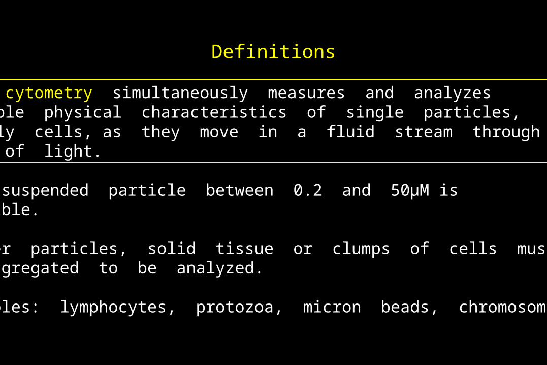
Flow cytometry simultaneously measures and analyzes multiple physical characteristics of single particles, usually cells, as they move in a fluid stream through a beam of light.
Any suspended particle between 0.2 and 50μM issuitable.
Larger particles, solid tissue or clumps of cells must bedisaggregated to be analyzed.
Examples: lymphocytes, protozoa, micron beads, chromosomes
Definitions
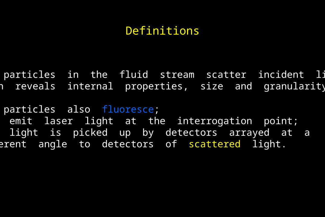
The particles in the fluid stream scatter incident light,which reveals internal properties, size and granularity.
The particles also fluoresce; they emit laser light at the interrogation point; this light is picked up by detectors arrayed at a different angle to detectors of scattered light.
Definitions
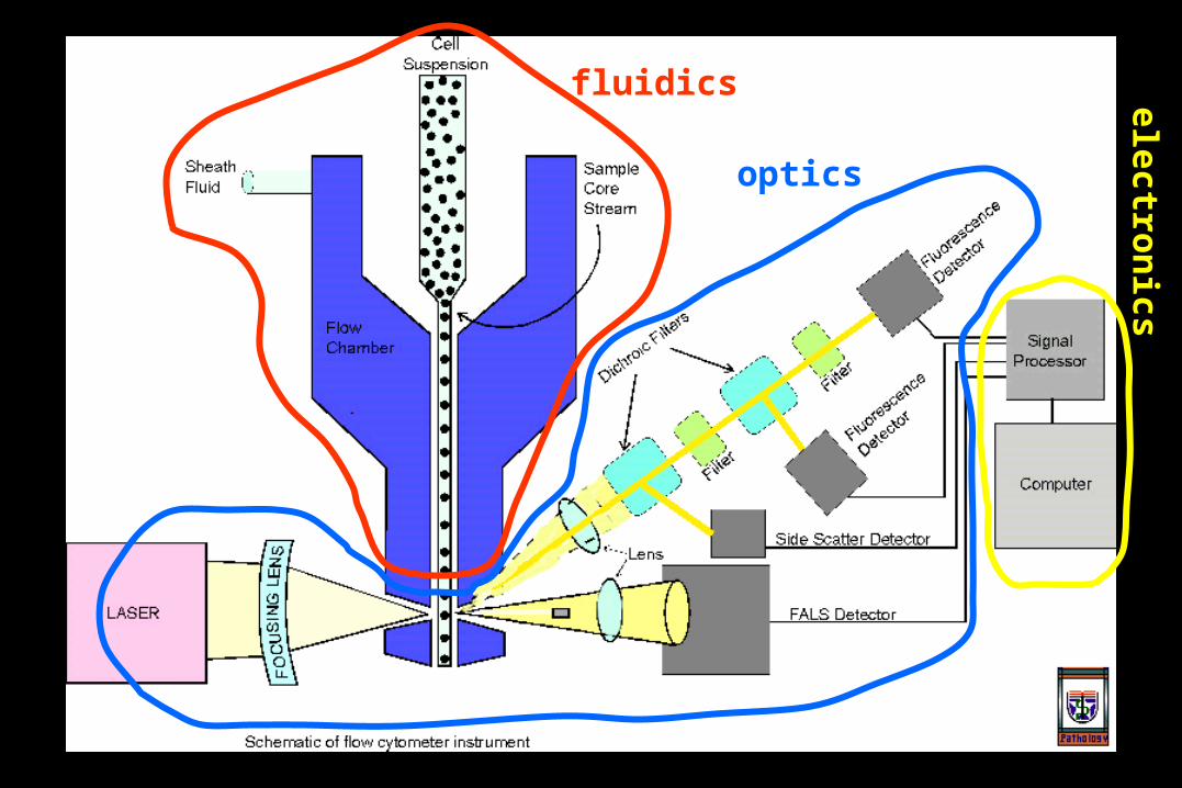
fluidics
optics
electro
nics
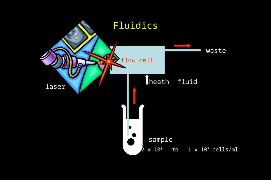
Fluidics
sample
flow cell
laser
waste
~2 x 10⁵ to 1 x 10⁷ cells/ml
sheath fluid
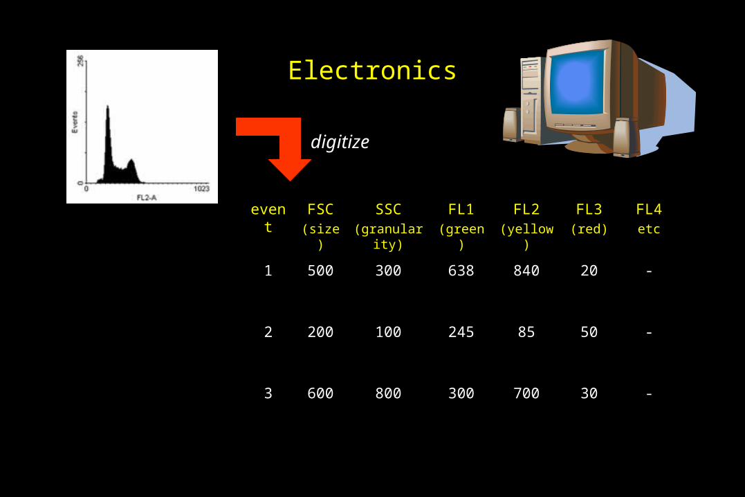
Electronics
event FSC(size)
SSC(granularity)
FL1(green)
FL2(yellow)
FL3(red)
FL4etc
1 500 300 638 840 20 -
2 200 100 245 85 50 -
3 600 800 300 700 30 -
digitize

FluidicsPurpose of the fluidics system:
1. Transport particles in a fluid stream to the laser beam to be interrogated
2. Position the sample core in the center of the laser beam
sheath fluid
samplefluid
‘hydrodynamic focusing’
single file particles
● low flow rate
● narrow sample core
● high resolution
● high flow rate
● wide sample core
● low resolution
Fluidics
Concerns
1. Shear rates for cells: check after you complete a run to ensure that the cells are intact.
2. Larger tips are needed for cell sorting.
Optics
Excitation is accomplished by lasers that emit light at specified wavelengths. The atomic properties of the excitation media define this wavelength for a particular laser.
The laser beam is focused on the sample core; lasers must be fixed in place.
This is the most common and versatile wavelength for excitation of fluorochromes.
Argon blue lasers emit light at 488 nm.
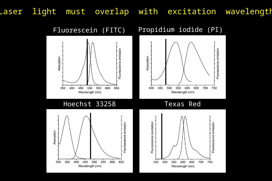
Fluorescein (FITC)
Hoechst 33258 Texas Red
Propidium iodide (PI)
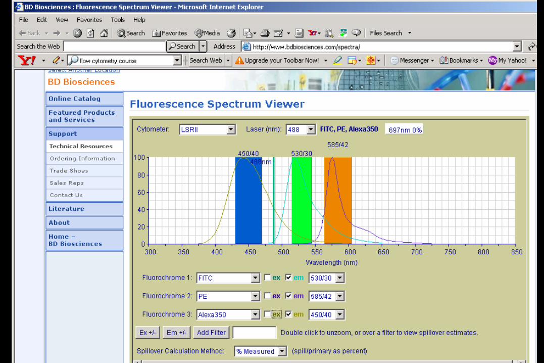
Laser light must overlap with excitation wavelength

Optics
Filters resolve overlapping wavelengths of emitted light
Longpass filter: transmits light of longer than or equal toa specific wavelength
Shortpass filter: transmits light of shorter than or equal toa specific wavelength
Bandpass filter: transmits light only within a narrow rangeof wavelengths
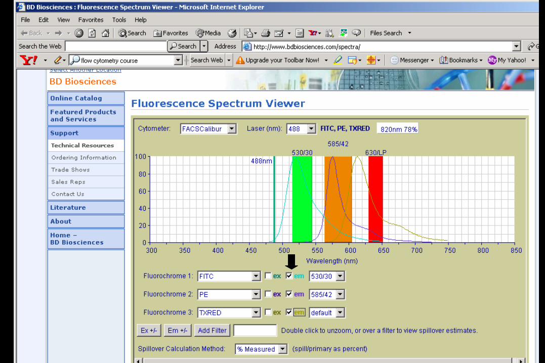

Excitation ofTexas Red is notoptimal with 488 nmlaser
http://probes.invitrogen.com/resources/spectraviewer/
http://www.bdbiosciences.com/spectra/
For more information
Electronics
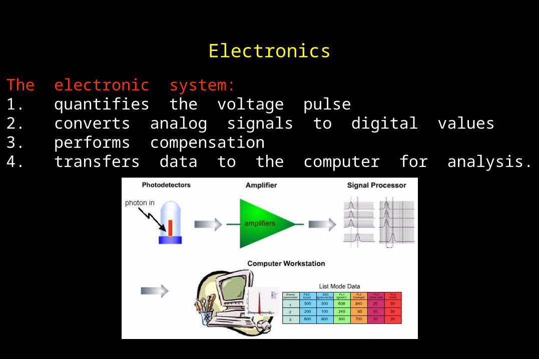
The electronic system: 1. quantifies the voltage pulse2. converts analog signals to digital values3. performs compensation4. transfers data to the computer for analysis.
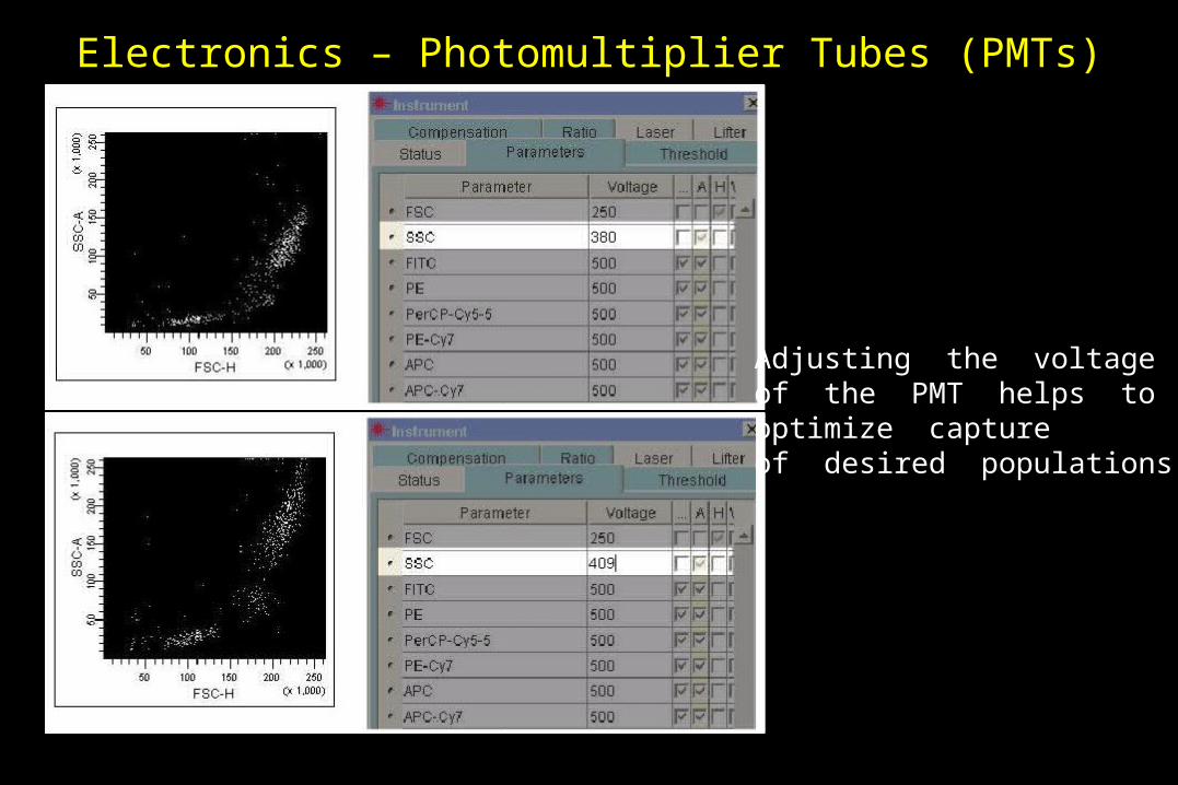
Adjusting the voltageof the PMT helps tooptimize captureof desired populations
Electronics – Photomultiplier Tubes (PMTs)
Electronics - Amplifiers
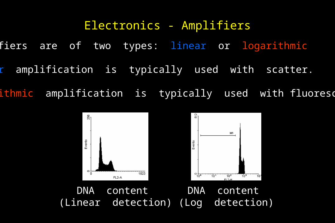
Amplifiers are of two types: linear or logarithmic
Linear amplification is typically used with scatter.
Logarithmic amplification is typically used with fluorescence.
DNA content (Linear detection)
DNA content (Log detection)
Electronics
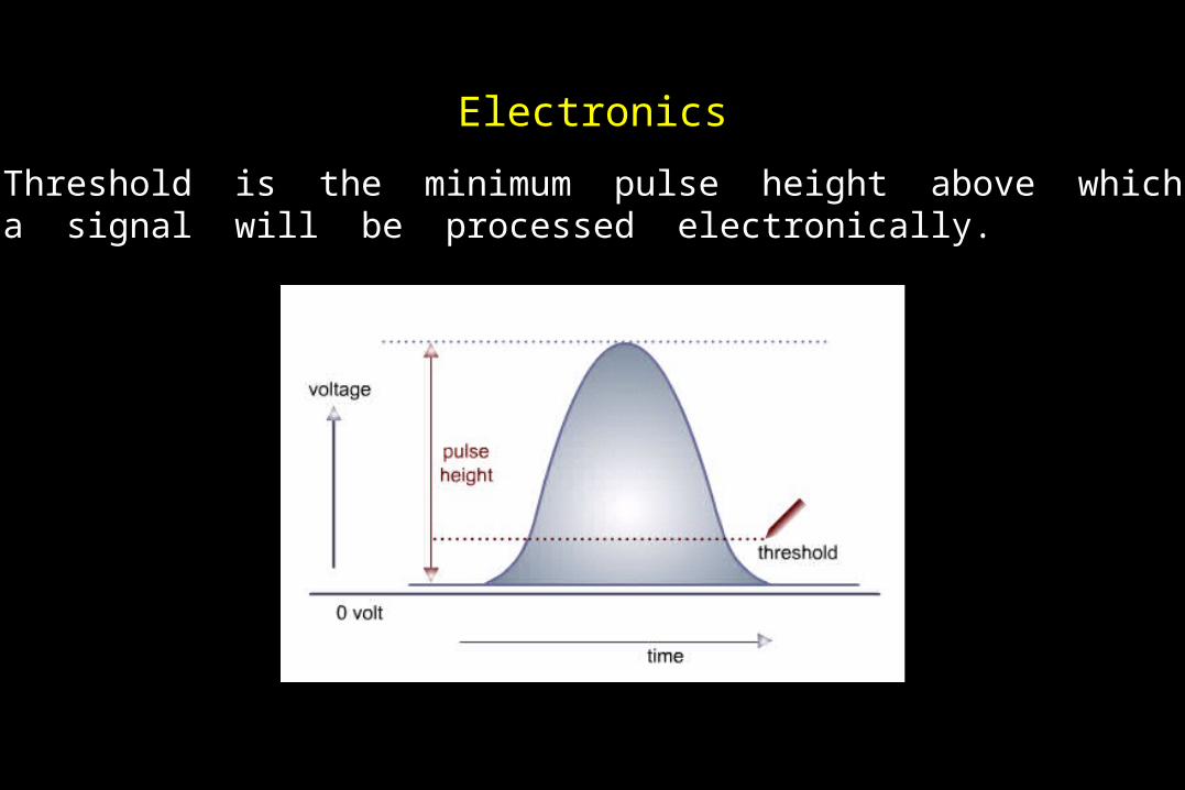
Threshold is the minimum pulse height above whicha signal will be processed electronically.
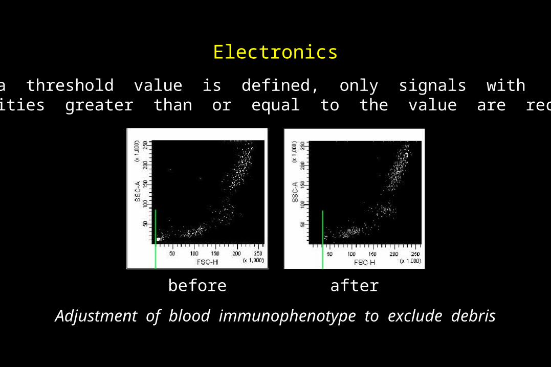
Electronics
When a threshold value is defined, only signals with intensities greater than or equal to the value are recorded.
Adjustment of blood immunophenotype to exclude debris
before after

Fluorescence
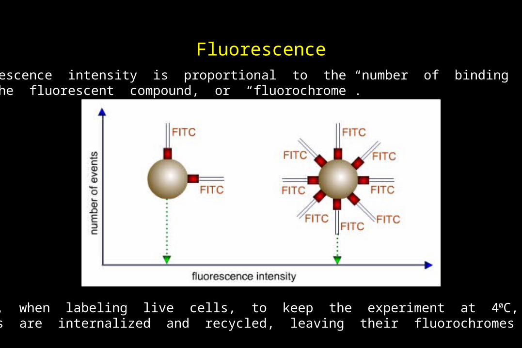
FluorescenceFluorescence intensity is proportional to the number of binding sitesof the fluorescent compound, or “fluorochrome”.
Remember, when labeling live cells, to keep the experiment at 40C, becausereceptors are internalized and recycled, leaving their fluorochromes behind.
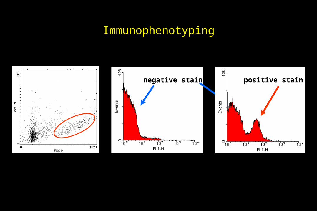
Immunophenotyping
negative stain positive stain
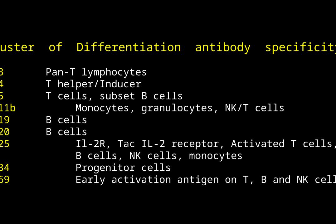
Cluster of Differentiation antibody specificity
CD3 Pan-T lymphocytes CD4 T helper/Inducer CD5 T cells, subset B cells CD11b Monocytes, granulocytes, NK/T cells CD19 B cellsCD20 B cells CD25 Il-2R, Tac IL-2 receptor, Activated T cells,
B cells, NK cells, monocytes CD34 Progenitor cells CD69 Early activation antigen on T, B and NK cells
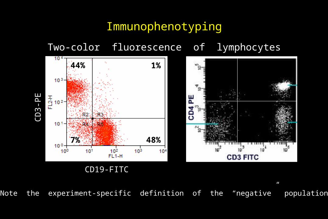
Immunophenotyping
Two-color fluorescence of lymphocytes
48%
44%
7%
1%
CD19-FITC
CD
3-P
E
Note the experiment-specific definition of the “negative” population

Fluorescence
Multicolor analysis
Spectral overlap
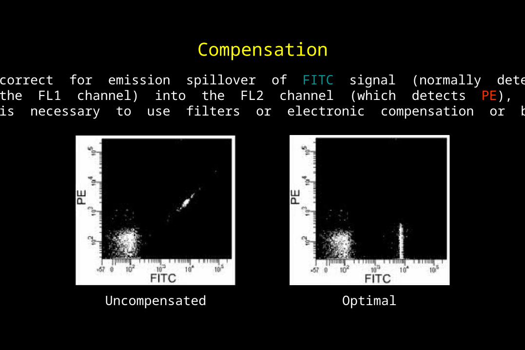
Compensation
To correct for emission spillover of FITC signal (normally detected in the FL1 channel) into the FL2 channel (which detects PE), it is necessary to use filters or electronic compensation or both.
Uncompensated Optimal
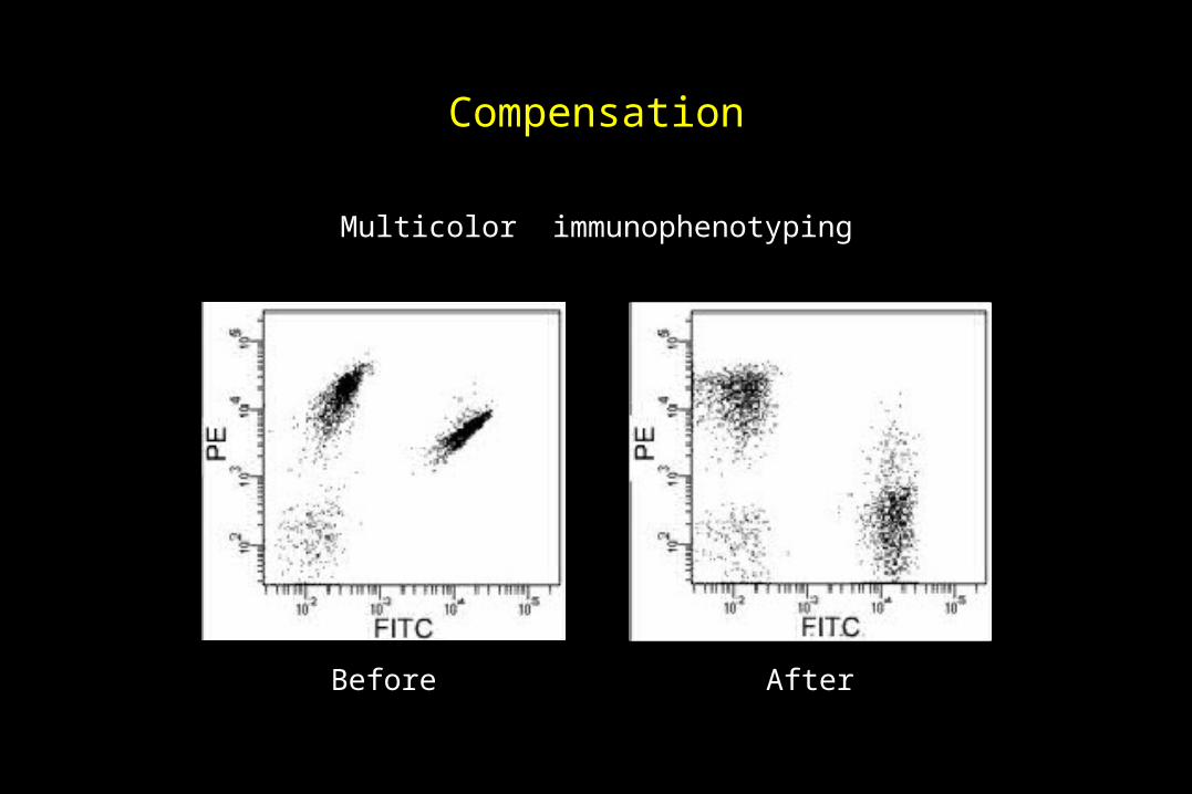
Compensation
Before After
Multicolor immunophenotyping
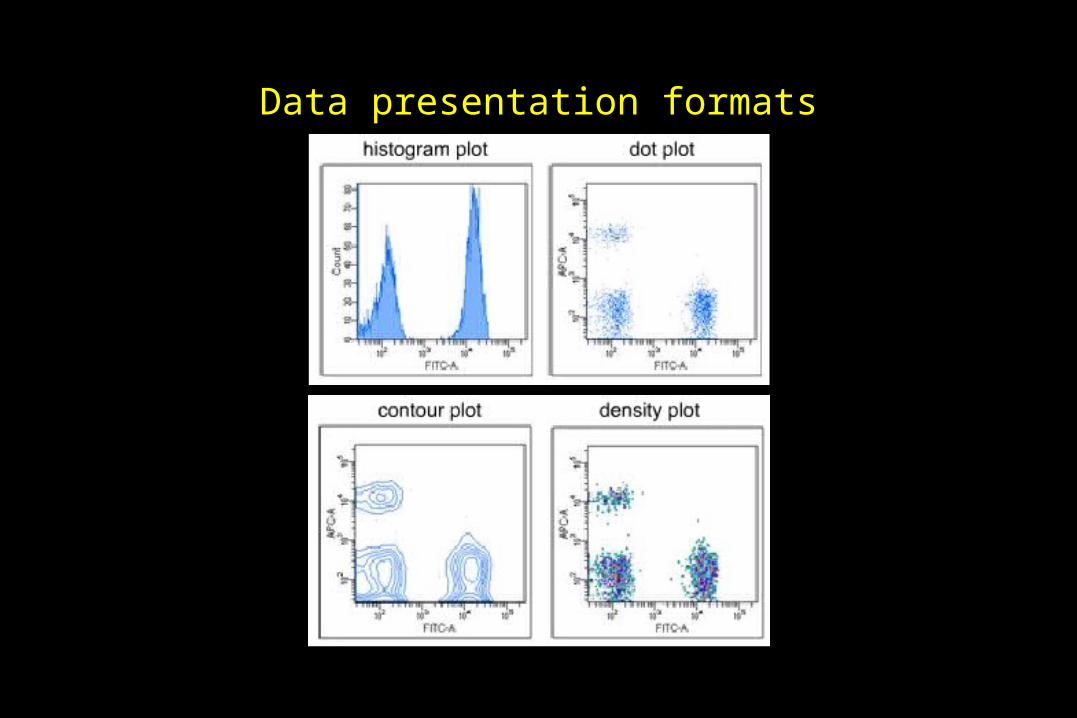
Data presentation formats
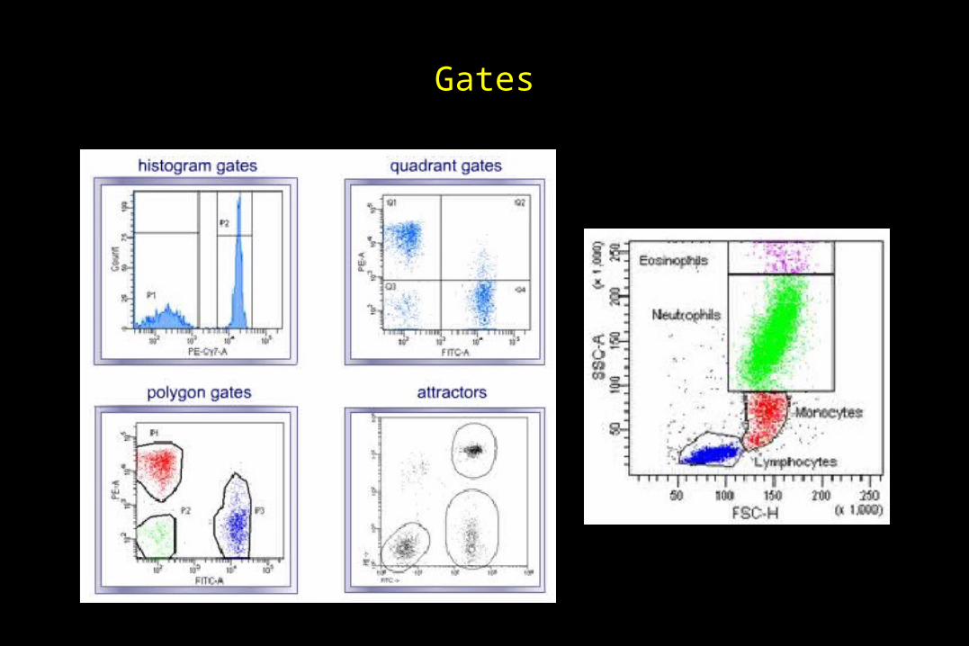
Gates

Gates
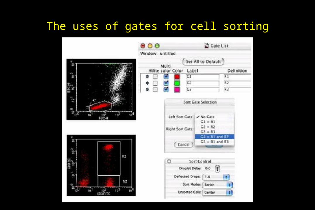
The uses of gates for cell sorting
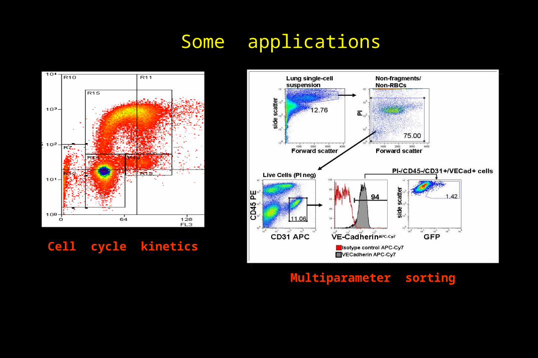
Some applications
Multiparameter sorting
Cell cycle kinetics

Some applications
DNA content for cell cycle / apoptosis
Immune system activation