Embed Size (px)
Citation preview

Flow Cytometric Analysis of Endothelial Progenitor Cells
Authors: Dorota Sadowicz, Vasilis Toxavidis, John Tigges
Affiliation: Beth Israel Deaconess Medical Center Harvard Stem Cell Institute
a B
eckm
an C
oulte
r Life
Sci
ence
s: W
hite
Pap
er

B E C K M A N C O U L T E R • W H I T E P A P E R 2
Flow Cytometric Analysis of Endothelial Progenitor Cells
PRINCIPLE OF THE TECHNIQUE
Background:
Endothelial Progenitor Cells (EPCs), first described by Asahara et al. in 1997, have become an important tool in the field of cardiovascular research (1). It is believed that they serve an important role in mitigating dysfunction by maintaining vascular homeostasis through repair and/or growth of blood vessels (1, 3, 4). The difficulty with studying EPCs has been in the lack of a clearly defined antigenic profile. Most of the markers used to isolate and study EPCs, such as the VEGF receptor-2 and CD34, have come from deductions and assumptions based on profiles of related cells, such as Hematopoietic Stem Cells (HSCs) and endothelial cells. Many years and publications later, there is still disagreement about which markers are more accurate and reliable, as well as ideal conditions for maintaining EPCs in culture (3, 5). Things are further hampered by the idea that there is a heterogeneous population of EPCs. This has made it difficult to discern which cells contribute to which “EPC” capabilities and traits. Therefore, most of the published data has come from the study of various types of cells that co-mingle, instead of a single-cell population.
The following protocol focuses on the use of a bacteriophage, found using a Phage Display library screen, which expresses a small peptide shown to recognize cells from the bone marrow (and peripheral circulation) that resemble characteristics of EPCs (2). Using Flow Cytometry, we analyze cells harvested from peripheral blood and bone marrow for phage positive and negative cells, co-stain with various EPC markers and analyze their co-expression, and sort these populations for further cellular and biochemical examination. The aim of this protocol is to develop a novel, biology-based method of identification of EPCs so they may be more accurately and uniformly studied.
B E C K M A N C O U L T E R • W H I T E P A P E R 3
RESEARCH APPLICATIONS
Flow Cytometry is a very useful tool for rare event analysis (cells occurring in very low frequency). It also enables easy and reproducible comparison of multiple sample types under varying conditions, as well as rapid positive and negative cell counts. Prep and analysis can take mere hours versus days/weeks with tissue culture..
PROTOCOL
Standard Procedure:
1. Collect fresh blood in EDTA-coated tubes, mix by gently inverting to prevent coagulation.
2. Dilute blood 1:1 with Dulbecco’s Phosphate Buffered Saline (DPBS). In a 15 mL conical tube layer 9.5 mL of blood/DPBS mixture atop 5 mL Ficoll-Paque Plus. To use larger volumes, refer to Ficoll-Paque manual. Instructions 71-7167-00 AG Ficoll-Paque Plus.pdf
3. Centrifuge tubes for 15 minutes at 2000 rpm, 10°C, with medium acceleration and no brake.
4. Carefully harvest the “buffy coat” (thin white band seen) above the red blood cells and between the Ficoll and DPBS/serum layers. Pool in 15 mL conical tube, fill to 15 mL with DPBS. Spin for 5 minutes at 1700 rpm, 10°C, with full brake and acceleration.
5. Aspirate supernatant. Dilute 10X IOTest 3 Lysing Solution 1:10 in de-ionized water. Resuspend pellet(s) in 3 mL IOTest3 reagent (cells should be less than (104/mL) and incubate 10 minutes at room temperature. Add 9 mL DPBS and centrifuge (same conditions as step 4).
6. Pool pellets together and resuspend to a final volume of 10 mL DBPS.
7. Count the cells using Trypan Blue and a Hemacytometer or a commercially available cell counter.
8. Adjust final cell concentration to 10x106 cells/mL using DPBS. Then aliquot the appropriate amount of cells into 14 polycarbonate tubes of 5 mL each.

B E C K M A N C O U L T E R • W H I T E P A P E R 4
The following sample tubes will be necessary:
Tube Antibody Volume to add
FcR Blocking reagent
1 Unstained control - -2 Isotype controls for FITC 5 mL 20 mL3 Isotype controls for PE 20 mL 20 mL4 Isotype controls for APC 5 mL 20 mL5 Isotype controls for PerCP-Cy5.5L 10 mL 20 mL6 KDR-PE single stain 20 mL(1) 20 mL7 CD31-PE single stain 20 mL(1) 5 mL8 CD34-PerCP-Cy5.5 single stain 20 mL(1) 20 mL9 CD133-APC single stain 10 mL(2) 20 mL
10 CD45-APC single stain 10 mL(1) 5 mL11 Phage-FITC single stain - -12 CD34/KDR/CD45/Phage sample stain - 20 mL13 CD34/KDR/CD133/Phage sample stain - 20 mL14 CD45/CD31/Phage sample stain - 5 mL
(1) Volume per 5x10⁵ cells (2) Volume per 5x10⁶ cells
9. Add FcR Blocking Reagent to all tubes receiving antibodies, except for the phage single stain in the volumes listed above. Incubate for 15 minutes on ice, in the dark.
10. Add antibodies to tubes using the volumes noted above. Incubate for 45 minutes on ice, in the dark.
11. Add 2 mL of DPBS and spin for 7 minutes, at 1700 rpm, 10°C.
12. Aspirate the supernatant, then either resuspend in 0.5 mL DPBS (if analyzing right away or sorting), or fix in 1.6% PFA/PBS (if not analyzing right away) and store, in the dark, at 4°C. Paraformaldehyde (PFA) fixed cells can be analyzed up to 5 days after fixation.
13. Analyze samples on Beckman Coulter Gallios* flow cytometer. Determine and set voltages so scatter properties and fluorescence (set to negative) are optimized.
14. Compensation was done using the QuickCOMP function of the Beckman Coulter Gallios CXP software.
15. Once all compensation has been set, analyze and record 10,000 events for the unstained and single stain controls, then 1 million events for the triple and dual stain samples. Set gates first based on the FSC vs SSC, and then focus on CD34-positive populations to determine how many are also positive for the other markers.
* Gallios is for research use only. Not for use in diagnostic procedures.
B E C K M A N C O U L T E R • W H I T E P A P E R 5
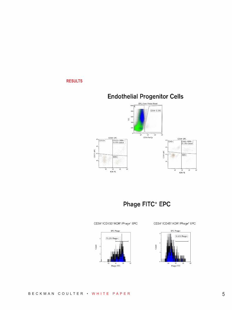
RESULTS

B E C K M A N C O U L T E R • W H I T E P A P E R 6
Our intention was to show that the phage we isolated could indeed be used as a novel marker or stain for EPC’s, and using the Beckman Coulter Gallios flow cytometer we were able to effectively show that phage-staining of cells isolated from peripheral blood greatly overlaps with the expression of accepted EPC markers, such as CD34 and VEGF-R2 (KDR). Additionally, this phage-staining was for the most part restricted to CD45-negative cells, as would be expected if the phage was indeed a marker of EPCs. The Beckman Coulter Gallios flow cytometer rapidly ran through samples and allowed us to clearly distinguish populations that support our belief that the phage is a marker for EPCs and would allow us to focus on isolating a specific population of cells for further biochemical analysis.
Tips: • Increased plot size and resolution can be essential in CD34+ gating. • Voltages should be set according to Unstained Control and adjustments made from use of isotype controls
only if necessary.• Whole Blood samples can be spiked with CD34+ cells in order to define the CD34 population more clearly.• It is best to set Phage FITC gate according to CD45+/KDR+ gate. This gate can be duplicated and applied
to the CD133+/KDR+ Phage FITC+ histogram.• For Voltage and Compensation guidelines please refer to Appendix. These are guidelines and
adjustments should be made accordingly.• Compensation was determined during acquisition, but can be determined (AutoComp with single stained
controls) or adjusted using the features of Kaluza* Flow Cytometry Analysis Software (Beckman Coulter).
*Gallios and Kaluza are for research use only. Not for use in diagnostics procedures.

B E C K M A N C O U L T E R • W H I T E P A P E R 7
REFERENCES1. Asahara T, Murohara T, Sullivan A, Silver M, Van der Zee R, Li T, Witzenbichler B, Schatteman G, and
Isner JM. Isolation of Putative Progenitor Endothelial Cells for Angiogenesis. Science. 1997 275: 964-967.
2. Willats WGT. Phage Display: Practicalities and Prospects. Plant Molecular Biology. 2002 50: 837.
3. Rosenzweig A. Endothelial Progenitor Cells. New England Journal of Medicine. 2003 348: 581-582.
4. Hill JM, Zalos G, Halcox JP, Schenke WH, Waclawiw MA, Quyyumi AA, and Finkle T. Circulating Endothe-lial Progenitor Cells, Vascular Function, and Cardiovascular Risk. New England Journal of Medicine. 2003 348: 593-600.
5. Schmeisser A, Strasser RH. Phenotypic Overlap Between Hematopoietic Cells with Suggested Angioblas-tic Potential and Vascular Endothelial Cells. Journal of Hematotherapy & Stem Cell Research. 2002 11(1): 69-79.
REAGENT DETAILSReagent Supplier Order DetailsAnti-Human CD34-PerCP-Cy5.5 Beckman Coulter IM2648UAnti-Human CD34-FITC Beckman Coulter IM1870UAnti-Human CD133-APC Miltenyi Biotec 130-090-826Anti-Human VEGF-R2-PE (KDR-PE) Beckman Coulter A64615 Anti-Human CD45-APC Beckman Coulter IM2473UAnti-Human CD31-PE Beckman Coulter IM2409FcR Blocking Reagent Miltenyi Biotec 130-059-901Ficoll-Paque PLUS GE Healthcare 17-1440-031X DPBS (-Ca2+, -Mg2+) Invitrogen (or any) 14190136IOTest 3 Lysing Solution, 10X Beckman Coulter A07799Trypan Blue (0.4%) Sigma (or any) T8154 (or any)PerCP-Cy5.5 Isotype Beckman Coulter IM2663UHuman IgG APC BD Bioscience 550931Mouse IgG PE Beckman Coulter IM0670UHuman IgG FITC BD Bioscience 555786Phage-FITC Antibody, made in our lab, patent pending.
NOTES
The results demonstrated in this application sheet represent those generated on the Beckman Coulter Gallios Flow Cytometer. As differences exist in the performance between analyzers, the author cannot guarantee a similar appearance with the use of other flow cytometers. Quality control and assurance were performed to meet manufacturer’s specifications using Flow-Check Pro (Beckman Coulter) and 8-peak Rainbow Beads (Spherotech).

B E C K M A N C O U L T E R • W H I T E P A P E R 8
APPENDIX
Instrument Laser Set-up
Compensation Matrix
BR-18062A B2103-14112