Embed Size (px)
Citation preview

Ⅰ Introduction
Arrhythmogenic right ventricular cardiomyopathy (ARVC), a heart muscle disease of unknown etiology, is characterized by replacement of the right ventricular (RV) myocardium by fibrofatty tissue and electrical instability associated with ventricular tachycardia (VT)1)2). The regional or diffuse fibrofatty atrophy of the RV free wall causes wall thinning and chamber dilatation, leading to right heart failure1). It has been reported that one-lung ventilation (OLV) increases RV preload3) as well as afterload3)-6), possibly resulting in dysfunction of the right heart for ARVC patients. We report a case of ARVC in which OLV caused flattening of the interventricular septum (IVS), which is a sign of the development of RV dysfunction due to RV overload, revealed by transesophageal echocardiography (TEE).
Ⅱ Case Report
A 70-year-old male (height, 165 cm ; weight, 66 kg) had been suffering from sudden attacks of unconsciousness for 13 years. An echocardiogram showed RV dilatation (RV end-diastolic dimension of 43.2 mm (>28 mm)), an endomyocardial biopsy showed fibrofatty replacement of the myocardium, and an electrocardiogram showed VT exhibiting left bundle-branch block morphology. ARVC was diagnosed by these findings according to the criteria for diagnosis of ARVC/dysplasia7). He had been treated with amiodarone (100 mg/day) and had been implanted with an implantable cardioverter-defibrillator (ICD). He had no familial history of premature sudden death due to suspected ARVC. He underwent abdominoperineal resection of the rectum for treatment of rectal cancer three years ago. Follow-up blood chemistry studies showed elevation of tumor makers such as carbohydrate antigen 19-9 and carcinoembryonic antigen. Computed tomography and positron emission tomography revealed lung cancer in the left upper lobe. The patient
Flattening of the Interventricular Septum during One-lung Ventilation in a Patient with Arrhythmogenic Right
Ventricular Cardiomyopathy
Satoshi Fuseya*, Takashi Ishida, Takashi Ichino and Mikito KawamataDepartment of Anesthesiology and Resuscitology, Shinshu University School of Medicine
Arrhythmogenic right ventricular cardiomyopathy (ARVC) is characterized by ventricular arrhythmia and right heart failure due to right ventricular (RV) fibrofatty atrophy. A 70-year-old male with ARVC underwent elective endoscopic left upper lobectomy for treatment of lung cancer. We found that one-lung ventilation (OLV) caused flattening of the interventricular septum (IVS) during diastole and changed the left ventricular (LV) configuration from a circle to a “D-shape” in the patient, which was imaged with transesophageal echocardiography (TEE). The LV “D-shape” returned to a normal circular configuration immediately after conversion from OLV to two-lung ventilation. Since flattening of the IVS is a sign of RV overload and since continuous RV overload may result in the development of RV dysfunction, early diagnosis is important. We recommend monitoring of IVS movement by TEE during OLV in patients with ARVC. Shinshu Med J 66 : 443―449, 2018
(Received for publication June 5, 2018 ; accepted in revised form July 25, 2018)
Key words : flattening of the interventricular septum, one-lung ventilation, arrhythmogenic right ventricular cardiomyopathy
* Corresponding author : Satoshi Fuseya Department of Anesthesiology and Resuscitology, Shinshu University School of Medicine, 3-1-1 Asahi, Matsumoto, Nagano 390-8621, Japan E-mail : [email protected]
443No. 6, 2018
Shinshu Med J, 66⑹:443~449, 2018

was thus scheduled for elective endoscopic left upper lobectomy for treatment of lung cancer. Preoperative physical examination did not reveal any symptoms of heart failure such as dyspnea during daily activities, orthopnea, or pitting edema. Results of hematologic and blood chemistry studies and results of spirometry were in normal ranges
except for creatinine of 1.23 mg/dl and forced expiratory volume % in one second of 67.4 %. An electrocardiogram showed typical findings2) as the one minor sign and the two major signs of the criteria for diagnosis of ARVC/dysplasia (Fig. 1)7). Preoperative transthoracic echocardiography showed remarkable RV dilation (Fig. 2), tricuspid valve regurgita
Fig . 1 Preoperative electrocardiogram. An electrocardiogram showed low voltage in extremity leads, right axis deviation and T-wave inversions in leads V1-6 without right bundle-branch block as the minor sign of the criteria for diagnosis of ARVC/dysplasia7); the electrocardiogram also revealed QRS complex prolongation (>110 msec) in leads V1-3 and epsilon waves as the two major signs of the criteria for diagnosis of ARVC/dysplasia7). These findings of the electrocardiogram are typical for ARVC2).
Fig . 2 Preoperative transthoracic echocardiography. (A) and (B) are a short axis cross-sectional view and a four-chamber view at end diastole, respectively. Note the remarkable dilation of the right ventricle so that the volume of the right ventricle is larger than that of the left ventricle (white arrowhead) (A, B) and apex consisting of the right ventricle (white arrow) (B). Right ventricular trabeculae carneae were also well developed. RV : right ventricle, LV : left ventricle.
444 Shinshu Med J Vol. 66
Fuseya・Ishida・Ichino et al.
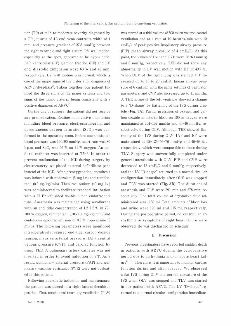
tion (TR) of mild to moderate severity diagnosed by a TR jet area of 4.2 cm2, vena contracta width of 4 mm, and pressure gradient of 27.8 mmHg between the right ventricle and right atrium. RV wall motion, especially at the apex, appeared to be hypokinetic. Left ventricular (LV) ejection fraction (EF) and LV end-diastolic dimension were 63 % and 43 mm, respectively. LV wall motion was normal, which is one of the major signs of the criteria for diagnosis of ARVC/dysplasia7). Taken together, our patient fulfilled the three signs of the major criteria and two signs of the minor criteria, being consistent with a positive diagnosis of ARVC7). On the day of surgery, the patient did not receive any premedication. Routine noninvasive monitoring including blood pressure, electrocardiogram, and percutaneous oxygen saturation (SpO2) was performed in the operating room. Before anesthesia, his blood pressure was 140/90 mmHg, heart rate was 80 b.p.m, and SpO2 was 96 % on 21 % oxygen. An epidural catheter was inserted at T5-6. In order to prevent malfunction of the ICD during surgery by electrocautery, we placed external defibrillator pads instead of the ICD. After preoxygenation, anesthesia was induced with midazolam (5 mg i.v.) and remifentanil (0.2 μg/kg/min). Then rocuronium (40 mg i.v.) was administered to facilitate tracheal intubation with a 37 Fr left-sided double-lumen endotracheal tube. Anesthesia was maintained using sevoflurane with an end-tidal concentration of 1.2-1.5 % in 72-100 % oxygen, remifentanil (0.05-0.1 μg/kg/min), and continuous epidural infusion of 0.2 % ropivacaine (3 ml/h). The following parameters were monitored intraoperatively : expired end-tidal carbon dioxide tension, invasive arterial pressure (IAP), central venous pressure (CVP), and cardiac function by using TEE. A pulmonary artery catheter was not inserted in order to avoid induction of VT. As a result, pulmonary arterial pressure (PAP) and pulmonary vascular resistance (PVR) were not evaluated in this patient. Following anesthetic induction and maintenance, the patient was placed in a right lateral decubitus position. First, mechanical two-lung ventilation (TLV)
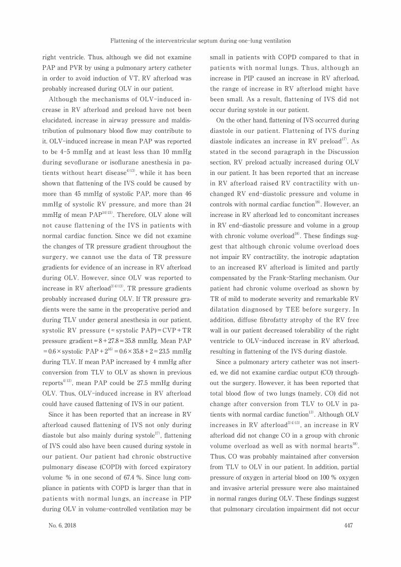
was started at a tidal volume of 500 ml on volume-control ventilation and at a rate of 10 breaths/min with 12 cmH2O of peak positive inspiratory airway pressure (PIP) (mean airway pressure of 4 cmH2O). At this point, the values of IAP and CVP were 98/60 mmHg and 8 mmHg, respectively. TEE did not show any abnormality in LV wall motion with EF of 49.7 %. When OLV of the right lung was started, PIP increased up to 18 to 20 cmH2O (mean airway pressure of 6 cmH2O) with the same settings of ventilator parameters, and CVP also increased up to 11 mmHg. A TEE image of the left ventricle showed a change to a “D-shape” by flattening of the IVS during diastole (Fig. 3A). Partial pressures of oxygen and carbon dioxide in arterial blood on 100 % oxygen were maintained at 101-157 mmHg and 45-46 mmHg, respectively, during OLV. Although TEE showed flattening of the IVS during OLV, IAP and EF were maintained at 92-125/56-76 mmHg and 46-63 % , respectively, which were comparable to those during TLV. Surgery was uneventfully completed under general anesthesia with OLV. PIP and CVP were decreased to 13 cmH2O and 9 mmHg, respectively, and the LV “D-shape” returned to a normal circular configuration immediately after OLV was stopped and TLV was started (Fig. 3B). The durations of anesthesia and OLV were 391 min and 278 min, respectively. The total volume of crystalloid fluid administered was 1150 ml. Total amounts of blood loss and urine were 120 ml and 215 ml, respectively. During the postoperative period, no ventricular arrhythmia or symptoms of right heart failure were observed. He was discharged on schedule.
Ⅲ Discussion
Previous investigators have reported sudden death in patients with ARVC during the perioperative period due to arrhythmia and/or acute heart failure8)-11). Therefore, it is important to monitor cardi ac function during and after surgery. We observed a flat IVS during OLV and normal curvature of the IVS when OLV was stopped and TLV was started in our patient with ARVC. The LV “D-shape” returned to a normal circular configuration immediate
445
Flattening of the interventricular septum during one-lung ventilation
No. 6, 2018

ly after conversion from OLV to TLV. These results strongly suggest that pre- and/or after-overload of the right ventricle due to OLV caused flattening of the IVS in this patient. Indeed, in our patient, CVP during OLV was 3 mmHg higher than that during TLV, being consistent with the results of a previous study showing that CVP during OVL is significantly higher than that during TLV under the condition of the same settings of ventilator parameters12). These findings indicate that preload of the right ventricle in
creases after conversion from TLV to OLV. In addition, Abe et al.4) and Carlsson et al.13) reported that mean PAP of 4-5 mmHg and PVR were significantly increased after conversion from TLV to OLV at volume-controlled ventilation with the same settings of ventilator parameters under sevoflurane or isoflurane anesthesia. Wilkinson et al.3) also reported that both RV end-systolic area and RV end-diastolic area were significantly increased during OLV compared to those during TLV. These findings indicate that OLV increases afterload as well as preload of the
Fig . 3 Transesophageal echocardiographic short axis cross-sectional view at end diastole. (A) and (B) are a view during one-lung ventilation and a view during two-lung ventilation, respectively. (A1) and (B1) are original photographs, and (A2) and (B2) are shown to emphasize the biventricular wall shape by tracing the outline. Note that left ventricular wall shape was changed to a “D-shape” by flattening of the interventricular septum (IVS) in (A1) and (A2) (black arrow). Note that the left ventricular wall “D-shape” completely recovered to the normal circular configuration (black arrow) and that left chamber size was enlarged to the original size in (B1) and (B2).
446 Shinshu Med J Vol. 66
Fuseya・Ishida・Ichino et al.
right ventricle. Thus, although we did not examine PAP and PVR by using a pulmonary artery catheter in order to avoid induction of VT, RV afterload was probably increased during OLV in our patient. Although the mechanisms of OLV-induced increase in RV afterload and preload have not been elucidated, increase in airway pressure and maldistribution of pulmonary blood flow may contribute to it. OLV-induced increase in mean PAP was reported to be 4-5 mmHg and at least less than 10 mmHg during sevoflurane or isoflurane anesthesia in patients without heart disease4)13), while it has been shown that flattening of the IVS could be caused by more than 45 mmHg of systolic PAP, more than 46 mmHg of systolic RV pressure, and more than 24 mmHg of mean PAP14)15). Therefore, OLV alone will not cause flattening of the IVS in patients with normal cardiac function. Since we did not examine the changes of TR pressure gradient throughout the surgery, we cannot use the data of TR pressure gradients for evidence of an increase in RV afterload during OLV. However, since OLV was reported to increase in RV afterload3)4)13), TR pressure gradients probably increased during OLV. If TR pressure gradients were the same in the preoperative period and during TLV under general anesthesia in our patient, systolic RV pressure (≈systolic PAP)=CVP+TR pressure gradient=8+27.8=35.8 mmHg. Mean PAP=0.6×systolic PAP+216)=0.6×35.8+2=23.5 mmHg during TLV. If mean PAP increased by 4 mmHg after conversion from TLV to OLV as shown in previous reports4)13), mean PAP could be 27.5 mmHg during OLV. Thus, OLV-induced increase in RV afterload could have caused flattening of IVS in our patient. Since it has been reported that an increase in RV afterload caused flattening of IVS not only during diastole but also mainly during systole17), flattening of IVS could also have been caused during systole in our patient. Our patient had chronic obstructive pulmonary disease (COPD) with forced expiratory volume % in one second of 67.4 %. Since lung compliance in patients with COPD is larger than that in patients with normal lungs, an increase in PIP during OLV in volume-controlled ventilation may be
small in patients with COPD compared to that in patients with normal lungs. Thus, although an increase in PIP caused an increase in RV afterload, the range of increase in RV afterload might have been small. As a result, flattening of IVS did not occur during systole in our patient. On the other hand, flattening of IVS occurred during diastole in our patient. Flattening of IVS during diastole indicates an increase in RV preload17). As stated in the second paragraph in the Discussion section, RV preload actually increased during OLV in our patient. It has been reported that an increase in RV afterload raised RV contractility with unchanged RV end-diastolic pressure and volume in controls with normal cardiac function18). However, an increase in RV afterload led to concomitant increases in RV end-diastolic pressure and volume in a group with chronic volume overload18). These findings suggest that although chronic volume overload does not impair RV contractility, the inotropic adaptation to an increased RV afterload is limited and partly compensated by the Frank-Starling mechanism. Our patient had chronic volume overload as shown by TR of mild to moderate severity and remarkable RV dilatation diagnosed by TEE before surgery. In addition, diffuse fibrofatty atrophy of the RV free wall in our patient decreased tolerability of the right ventricle to OLV-induced increase in RV afterload, resulting in flattening of the IVS during diastole. Since a pulmonary artery catheter was not inserted, we did not examine cardiac output (CO) throughout the surgery. However, it has been reported that total blood flow of two lungs (namely, CO) did not change after conversion from TLV to OLV in patients with normal cardiac function13). Although OLV increases in RV afterload3)4)13), an increase in RV afterload did not change CO in a group with chronic volume overload as well as with normal hearts18). Thus, CO was probably maintained after conversion from TLV to OLV in our patient. In addition, partial pressure of oxygen in arterial blood on 100 % oxygen and invasive arterial pressure were also maintained in normal ranges during OLV. These findings suggest that pulmonary circulation impairment did not occur
447
Flattening of the interventricular septum during one-lung ventilation
No. 6, 2018

during OLV in our patient. Since flattening of the IVS is a sign of development of RV dysfunction, early diagnosis is important. TEE enables real-time and noninvasive monitoring of IVS movement during surgery. Therefore, we recommend monitoring of IVS movement by TEE during OLV in patients with ARVC. While positive end-expiratory pressure (PEEP) is commonly used to improve hypoxia during OLV, PEEP can increase RV afterload. It has been shown that PEEP of more than 20 cmH2O causes flattening of the IVS in patients without heart disease during TLV19). Since diffuse fibrofatty atrophy of the RV free wall as observed in our patient decreases tolerability of the right ventricle to OLV-induced increase in RV afterload, when PEEP is applied in patients with ARVC during OLV, PEEP should be set up with observation of IVS movement by TEE. In our patient, since blood pressure and EF were not changed when flattening of the IVS occurred, we carefully monitored hemodynamic status without treatment. If hemodynamics deteriorate with flattening of the IVS after starting OLV, administration of inotropic agents such as dobutamine, vasopressors, pulmonary vasodilators such as phosphodiesterase-3 inhibitors, or nitroglycerin or inhalation of nitric oxide in addition to cessation of OLV should be con
sidered20). Since hypoxia and hypercapnia can increase PVR, arterial blood gas should also be checked. One of the weaknesses of the present study is that evidence for an increase in RV afterload including systolic PAP calculated by TR pressure gradients or mean PAP calculated by early-diastolic pulmonary regurgitation velocity was not obtained. These parameters and CO calculated by velocity-time integral and diameter of the RV outflow tract could be useful for estimating PVR. If these parameters had been investigated after conversion from TLV to OLV and from OLV to TLV, we could have directly shown that OLV-induced increase in RV afterload caused flattening of IVS. In addition, we were not able to use 3D echocardiography in our patient because the equipment was outdated. If we had examined the configuration of the heart by 3D echocardiography, we could have shown evidence that OLV changed the configuration of the tricuspid valve complex, resulting in impairment of tricuspid valve coaptation, which deteriorates TR and leads to an increase in RV preload. In conclusion, we experienced OLV-induced pathologic IVS movement (flattening) in a patient with ARVC. We recommend monitoring of IVS movement by TEE during OLV in patients with ARVC.
References
1) Kiès P, Bootsma M, Bax J, Schalij MJ, Wall EE : Arrhythmogenic right ventricular dysplasia/cardiomyopa
thy : screening, diagnosis, and treatment. Heart Rhythm 3 : 225-234, 2006
2) McKenna WJ, Thiene G, Nava A, Fontaliran F, Blomstorm-Lundqvist C, Fontaine G, Camerini F : Diagnosis of
arrhythmogenic right ventricular dysplasia/ cardiomyopathy. Task Force of the Working Group Myocardial and
Pericardial Diseases of the European Society of Cardiology and the Scientific Council on Cardiomyopathies of the
International Society and Federation of Cardiology. Br Heart J 71 : 215-218, 1994
3) Wilkinson JN, Scanlan M, Skinner H, Malik M : Right heart function during one-lung ventilation-observations using
transoesophageal echocardiography. Anaesthesia 64 : 1387-1388, 2009
4) Abe K, Mashimo T, Yoshida I : Arterial oxygenation and shunt fraction during one-lung ventilation : a comparison
of isoflurane and sevoflurane. Anesth Analg 86 : 1266-1270, 1998
5) Mcmahon CC, Irvine T, Conacher ID : Transoesophageal echocardiography in the management of whole lung
lavage. Br J Anaesth 81 : 262-264, 1998
6) Pagel PS, Fu JL, Damask MC, Davis RF, Samuelson PN, Howie MB, Warltier DC : Desflurane and isoflurane produce
similar alterations in systemic and pulmonary hemodynamics and arterial oxygenation in patients undergoing one-
lung ventilation during thoracotomy. Anesth Analg 87 : 800-807, 1998
448 Shinshu Med J Vol. 66
Fuseya・Ishida・Ichino et al.

7) Thiene G, Corrado D, Basso C : Arrhythmogenic right ventricular cardiomyopathy/dysplasia. Orphanet J Rare Dis
2 : 45, 2007
8) Bonnet F, Samain E, Bocquet R, Corre FL, Marty J : Perioperative management of severe head injury in a patient
with arrhythmogenic right ventricular dysplasia. Anesthesiology 95 : 255-256, 2001
9) Houfani B, Meyer P, Merckx J, Roure P, Padovani JP, Fontaine G, Carli P : Postoperative sudden death in two
adolescents with myelomeningocele and unrecognized arrhythmogenic right ventricular dysplasia. Anesthesiology
95 : 257-259, 2001
10) Toh KW, Nadesan K, Sie MY, Vijeyasingam R, Tan PSK : Postoperative death in a patient with unrecognized
arrhythmogenic right ventricular dysplasia syndrome. Anesth Analg 99 : 350-352, 2004
11) Bastien O, Guerin JM, Artru F, Lehot JJ : Arrhythmogenic right ventricular dysplasia : an underestimated cause of
perioperative death? J Cardiothorac Vasc Anesth 16 : 357-358, 2002
12) Lee SH, Kim N, Kim HI, Oh YJ : Echocardiographic evaluation of pulmonary venous blood flow and cardiac function
changes during one-lung ventilation. Int J Clin Exp Med 8 : 13099-13108, 2015
13) Carlsson AJ, Bindslev L, Hedenstierna G : Hypoxia-induced pulmonary vasoconstriction in the human lung. The
effect of isoflurane anesthesia. Anesthesiology 66 : 312-316, 1987
14) Ryan T, Petrovic O, Dillon JC, Feigenbaum H, Conley MJ, Armstrong WF : An echocardiographic index for
separation of right ventricular volume and pressure overload. J Am Coll Cardiol 5 : 918-924, 1985
15) Shimada R, Takeshita A, Nakamura M : Noninvasive assessment of right ventricular systolic pressure in atrial
septal defect : analysis of the end-systolic configuration of the ventricular septum by two-dimensional echo
cardiography. Am J Cardiol 53 : 1117-1123, 1984
16) Chemla D, Castelain V, Humbert M, Hébert JL, Simonneau G, Lecarpentier Y, Hervé P : New formula for predicting
mean pulmonary artery pressure using systolic pulmonary artery pressure. Chest 126 : 1313-1317, 2004
17) Rudski LG, Lai WW, Afilalo J, Hua L, Handschumacher MD, Chandrasekaran K, Solomon SD, Louie EK, Schiller
NB : Guidelines for the echocardiographic assessment of the right heart in adults : a report from the American
Society of Echocardiography endorsed by the European Association of Echocardiography, a registered branch of
the European Society of Cardiology, and the Canadian Society of Echocardiography. J Am Soc Echocardiogr
23 : 685-713, 2010
18) Szabó G, Soós P, Bährle S, Radovits T, Weigang E, Kékesi V, Merkely B, Hagl S : Adaptation of the right ventricle to
an increased afterload in the chronically volume overloaded heart. Ann Thorac Surg 82 : 989-995, 2006
19) Jardin F, Farcot JC, Boisante L, Curien N, Margairaz A, Bourdarais JP : Influence of positive end-expiratory
pressure on left ventricular performance. N Engl J Med 304 : 387-392, 1981
20) Lahm T, McCaslin CA, Wozniak TC, Ghumman W, Fadl YY, Obeidat OS, Schwab K, Meldrum DR : Medical and
surgical treatment of acute right ventricular failure. J Am Coll Cardiol 56 : 1435-1446, 2010
(2018. 6. 5 received;2018. 7. 25 accepted)
449
Flattening of the interventricular septum during one-lung ventilation
No. 6, 2018


![Multiscale Computational Analysis of Right Ventricular ... contraction and relaxation in the RV wall, LV wall, and the interventricular septum [29]. Biventricular contraction and relaxa-tion](https://img.pdfslide.us/doc/110x75/5ea917417ce82a350563d687/multiscale-computational-analysis-of-right-ventricular-contraction-and-relaxation.jpg)