Embed Size (px)
Citation preview

RESEARCH ARTICLE
Flagellar pocket restructuring through the Leishmania life cycleinvolves a discrete flagellum attachment zoneRichard J. Wheeler1,2,*,‡, Jack D. Sunter2,*,‡ and Keith Gull2
ABSTRACTLeishmania promastigote parasites have a flagellum, which protrudesfrom the flagellar pocket at the cell anterior, yet, surprisingly, havehomologs of many flagellum attachment zone (FAZ) proteins –
proteins used in the related Trypanosoma species to laterally attachthe flagellum to the cell body from the flagellar pocket to the cellposterior. Here, we use seven Leishmania mexicana cell lines thatexpressed eYFP fusions of FAZ protein homologs to show that theLeishmania flagellar pocket includes a FAZ structure. Electrontomography revealed a precisely defined 3D organisation for boththe flagellar pocket and FAZ, with striking similarities to those ofTrypanosoma brucei. Expression of two T. brucei FAZ proteins inL. mexicana showed that T. brucei FAZ proteins can assemble intothe Leishmania FAZ structure. Leishmania therefore have apreviously unrecognised FAZ structure, which we show undergoesmajor structural reorganisation in the transition from the promastigote(sandfly vector) to amastigote (in mammalian macrophages).Morphogenesis of the Leishmania flagellar pocket, a structureimportant for pathogenicity, is therefore intimately associated with aFAZ; a finding with implications for understanding shape changesinvolving component modules during evolution.
KEY WORDS: Leishmania, Trypanosoma brucei, Amastigote,Electron tomography, Flagellum attachment zone, Life cycledifferentiation
INTRODUCTIONTrypanosoma and Leishmania are two related genera that includemany major human and livestock pathogens, such as Leishmaniamexicana, which causes New World cutaneous leishmaniasis, andTrypanosoma brucei, which causes African sleeping sickness. Likemany unicellular eukaryotes, these cells have a highly definedinternal organisation (Lacomble et al., 2009, 2010; Sherwin and Gull,1989), and undergo precise morphogenesis during division (Ambitet al., 2011; Robinson et al., 1995; Sheriff et al., 2014; Sherwin andGull, 1989; Wheeler et al., 2011, 2013a) and precise morphogenesisduring adaptation of cell shape to different environments (Gadelhaet al., 2013; Rotureau et al., 2011, 2012; Sharma et al., 2008;Wheeleret al., 2015). How they generate their shape is of interest because of itsapparent co-evolution with different pathogenic life cycles and the
capacity of parasites to persist in different niches within their hosts(Maslov et al., 2013; Wheeler et al., 2013b).
Trypanosomes and Leishmania most likely arose from anancestor with a promastigote Leishmania-like morphology – anovoid cell with a single flagellum protruding from the flagellarpocket at the anterior (Flegontov et al., 2013). The flagellar pocket iscentral to the morphogenesis of trypanosomes and is associatedwith many single copy organelles – the basal body and pro-basalbody pair, the flagellum, the Golgi and the mitochondrial DNA (thekinetoplast) (Gheiratmand et al., 2013; Gull, 2003; Lacomble et al.,2009, 2010). The pocket is also the site of all endo- and exocytosisand hence has a crucial role in the pathogenicity of the parasite(Engstler et al., 2007; Field and Carrington, 2009; Gadelha et al.,2009). Correct pocket formation is vital for cell morphogenesis,viability and infectivity, as evidenced in T. brucei (Absalon et al.,2008; Bonhivers et al., 2008).
Trypanosomes, uniquely among genera of the Trypanosomatidaefamily, have developed a life cycle that includes a free-swimmingstage in the vertebrate bloodstream. This genus has the innovation oftrypomastigote morphology with the flagellar pocket towards thecell posterior and a flagellum that is laterally attached to the cellsurface and runs towards the anterior. The lateral attachment of theflagellum is mediated by a large complex cytoskeletal structurecalled the flagellum attachment zone (FAZ), which connects theflagellar skeleton to the cell body cytoskeleton through both theflagellum and cell body membranes (Hayes et al., 2014; Höög et al.,2012; Robinson et al., 1995; Sherwin and Gull, 1989; Vaughanet al., 2008; Vickerman, 1969). The morphological innovation ofthe FAZ seems to be associated with adaptation to the hostenvironment of the bloodstream (Wheeler et al., 2013b). Flagellarpocket and FAZ formation are linked; the proximal end of the FAZ(in the flagellar pocket) is the site of FAZ assembly (Sunter et al.,2015; Zhou et al., 2015), and the FAZ is physically connected withthe flagellar pocket cytoskeleton through microtubules (Lacombleet al., 2009, 2010) and the bi-lobe structure (Esson et al., 2012;Gheiratmand et al., 2013). In T. brucei, correct FAZ formation isvital for cell morphogenesis and viability (Hayes et al., 2014;LaCount et al., 2002; Rotureau et al., 2014; Sun et al., 2013;Vaughan et al., 2008; Zhou et al., 2011, 2015).
The flagellar pocket is likely to be important to a similardegree in Leishmania species. Evidence for a complexLeishmania pocket structure incorporating cytoskeletalcomponents exists from many years of electron microscopyanalysis – Leishmania have a microtubule quartet next to theflagellar pocket (Molyneux et al., 1975; Weise et al., 2000), atleast one specialised cytoplasmic microtubule starting near thepocket (Weise et al., 2000) and electron-dense structures linkingthe cell body and flagellum (Aleman, 1969; Gadelha et al., 2013;Molyneux et al., 1975). However, little is known about the three-dimensional (3D) organisation of the pocket. Leishmaniagenomes encode homologs of most FAZ proteins (Sunter et al.,Received 9 November 2015; Accepted 29 December 2015
1Max Planck Institute of Molecular Cell Biology and Genetics, Pfotenhauerstraße108, Dresden 01307, Germany. 2Sir William Dunn School of Pathology, South ParksRoad, Oxford OX1 3RE, UK.*These authors contributed equally to this work
‡Authors for correspondence ([email protected];[email protected])
This is an Open Access article distributed under the terms of the Creative Commons AttributionLicense (http://creativecommons.org/licenses/by/3.0), which permits unrestricted use,distribution and reproduction in any medium provided that the original work is properly attributed.
854
© 2016. Published by The Company of Biologists Ltd | Journal of Cell Science (2016) 129, 854-867 doi:10.1242/jcs.183152
Journal
ofCe
llScience

2015) but do not have the laterally attached flagellumcharacteristic of the trypanosome trypomastigote morphology,leaving their function completely unknown. It has beensuggested that structures in the Leishmania flagellar pocketregion correspond to a FAZ-related structure (Gadelha et al.,2013), although this has not been proven. Furthermore, unliketrypanosomes, Leishmania undergo a large change in flagellumstructure through the life cycle, from a 9+2 axoneme to acollapsed 9+0 (9v) axoneme (Alexander, 1978; Gluenz et al.,2010; Wheeler et al., 2015). How a complex pocket organisationlike that of T. brucei (Lacomble et al., 2009, 2010) canaccommodate this flagellum restructuring is also unknown.We used a combination of electron tomography analysis and
eYFP tagging of L. mexicana FAZ protein homologs to determinethe 3D structure and molecular composition of the L. mexicanaflagellar pocket region, which showed that it includes a previouslyunrecognised structure homologous to the T. brucei FAZ. Thisallowed us to determine how the T. brucei FAZ structure evolvedfrom the ancestral morphology, infer potential functional groupingsof FAZ proteins and analyse how Leishmania use FAZ proteins toadapt their flagellar pocket structure through their life cycle. Theseresults highlight the importance of the expression and regulation ofthe assembly of cohorts of proteins (which we term componentmodules) in determining different cell shapes and cell form in boththe life cycle and evolution.
RESULTSL. mexicana FAZ protein homologs localise to structuresadjacent to the pocketThere are 34 known FAZ-protein-coding genes in T. brucei(Hayes et al., 2014; Hu et al., 2015; LaCount et al., 2002;McAllaster et al., 2015; Morriswood et al., 2013; Nozakiet al., 1996; Oberholzer et al., 2011; Rotureau et al., 2014; Sunet al., 2013; Sunter et al., 2015; Vaughan et al., 2008; Woodset al., 2013; Zhou et al., 2011, 2015), and the majority of theseproteins have at least one clear homolog in L. mexicana speciesbased on sequence similarity, retention of domains and synteny(Table 1). As Leishmania species do not have a flagellum laterallyattached to the cell body, the function of this FAZ protein cohortin Leishmania is unclear. We therefore generated L. mexicana celllines expressing eYFP fusions of a subset of FAZ homologs todetermine their localisation, using the Leishmania endogenoustagging plasmid pLEnTv2-YB (Dean et al., 2015). We choseto localise FAZ1 (LmxM.36.5970), FAZ2 (LmxM.12.1130),FAZ5 (LmxM.36.5970), FAZ8 (LmxM33.2570), ClpGM6(LmxM.27.0490), FLA1BP (LmxM.10.0620) and FAZ10(LmxM.22.1320) as these proteins are found along the majorityof the length of the FAZ in T. brucei (accession numbers aregiven in brackets). All cell lines also expressed mCherry-taggedSMP1 (SMP1–mCh) as a fluorescent marker for the flagellarmembrane (Tull et al., 2004; Wheeler et al., 2015), and weconfirmed correct integration of the eYFP-tagging constructs andfusion protein expression by using PCR and western blotting forGFP (Fig. S1). All seven fusion proteins localised to the flagellarpocket region in L. mexicana promastigotes (Fig. 1A–G), so tofurther characterise this region of the cell, we also generated celllines expressing an eYFP fusion of the L. mexicana homolog ofthe bilobe protein LRRP1 (LmxM.28.1990) using pLEnTv2-YBand a Myc-epitope-tag fusion of the flagellar pocket collar proteinBILBO1 (LmxM.09.0100) using pPOTv2 and fusion PCRtagging (Dean et al., 2015) as reference structures (Fig. 1H–I).We also attempted immunofluorescence analysis with the L3B2
and L6B3 antibodies against T. brucei FAZ1 (Kohl et al., 1999)but saw no signal (data not shown).
The flagellar pocket region is identifiable in phase contrast andSMP1–mCh fluorescence images of Leishmania cells, where theSMP1–mCh flagellar membrane signal penetrates 1.5–2 μm intothe cell anterior. Myc–BILBO1 localised to a line or ring acrossthe flagellum mid-way through the flagellar pocket region,approximately 1 μm from the base of the flagellum (Fig. 1I). Allseven eYFP–FAZ protein fusions localised to structures in thepocket region, at either a similar or more distal distance from thebase of the flagellum as BILBO1. Localisation of these proteinsindicates a complex asymmetric structure because not all proteinshad identical localisation patterns and often lay to one side of theflagellum. The localisation patterns could be separated into fourclasses: a short linear structure either in the flagellum or in veryclose proximity to the flagellar membrane (ClpGM6, FLA1BP;Fig. 1A,B), a short linear structure to one side of the flagellum(FAZ1, FAZ2, FAZ5, FAZ8; Fig. 1C–F), a ring structure around theflagellum mid-way through the flagellar pocket (FAZ1, FAZ8;Fig. 1C,D), or a horseshoe or ring structure around the exit point ofthe flagellum from the flagellar pocket (FAZ10; Fig. 1G). A ringstructure was inferred from the FAZ1 and FAZ8 localisation fromthe spur of fluorescence signal that crossed the flagellum andappeared to be bifurcated or ring-like. This suggested that L.mexicana possesses a complex flagellar pocket organisation,including a short linear structure (with additional elaborations)positioned to one side of the flagellum that is homologous to theT. brucei FAZ. This region also appeared to include a structure thatis homologous to the bilobe (Fig. 1H), similar to the proximal end ofthe FAZ filament domain densities in T. brucei (Esson et al., 2012).
L. mexicana flagellar pocket organisation has manysimilarities but also key differences to that of T. bruceiWe characterised the structure of the L. mexicana flagellar pocketusing electron tomography to generate a 3D reconstruction of theorganisation of the basal and pro-basal body, the flagellar pocket,the pocket collar, the flagellum exit from the cell and any structuresthat could be orthologous to the T. brucei FAZ. Analysis was basedon tomograms from longitudinal sections of the entire pocketvolume of three cells, supported by tomograms from transversesections through parts of the pocket of a further three cells (toavoid misinterpretation from the lower z-resolution in tomograms),at 1–2 nm/voxel (Table S1, Movies 1–5).
The L. mexicana flagellar pocket was similar overall to that ofT. bruceiwith two distinct domains separated by the flagellar pocketcollar, which was visible as a double line of electron-dense material(Fig. 2A,B). The flagellum extended through the pocket with theparaflagellar rod (PFR) present only past the pocket collar. Thepocket had an overall curved shape. In the proximal region (beforethe collar), the pocket was bulbous and surrounded by many smallvesicles. In the distal region (after the collar), the pocket narrowed toa cylindrical neck connecting the bulbous proximal pocket to thecell anterior and was surrounded by complex structures with highelectron density. A quartet of four microtubules (MtQ) ran as a tightparallel array along the pocket surface. They followed a left-handedhelical path, starting near to the basal body, passing through a gap inthe collar, then terminating irregularly in the neck region. Thisirregular termination of the MtQ is unlike that in T. brucei. In onetomogram (PL2), it could be seen that the pro-basal body had begunextending into a very short new axoneme, whereas in the remainder,the pro-basal body had not extended. However, in all tomogramsanalysed, the pro-basal body also had an MtQ, which was much
855
RESEARCH ARTICLE Journal of Cell Science (2016) 129, 854-867 doi:10.1242/jcs.183152
Journal
ofCe
llScience

shorter and only extended around half-way to the pocket collar.Note that as L. mexicana has few discrete morphological markers ofthe cell cycle stage in the early cell cycle (Wheeler et al., 2011), wecould not determine precisely which cell cycle stage these cells werein, so at which cell cycle stage newMtQ nucleation occurred. Threeor four additional microtubules were also always present in twogroups – one singlet or pair running along the surface of the pocketand one singlet or pair extending into the cytoplasm. Structuresconsistent with this organisation were seen in all six promastigotetomograms.The axoneme central pair of L. mexicana has a fixed orientation
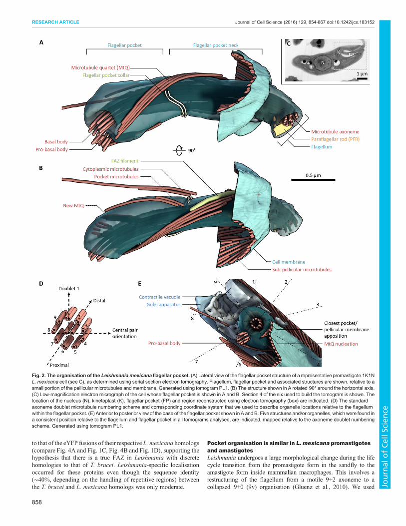
and can be used to unambiguously number the outer doublets. Thisprovides a polar coordinate system for describing the location ofstructures in and near the flagellum and pocket using the proximal–distal axis of the axoneme and the angle around the proximal–distalaxis, described by using the doublet number (steps of 40° perdoublet, clockwise looking proximal to distal) (Fig. 2D).The flagellar pocket itself was asymmetric and had a consistently
asymmetric positioning within the anterior of the cell. Otherorganelles and structures with a consistently asymmetric position(Fig. 2E) were the pro-basal body (adjacent to doublets 6 and 7 ofthe axoneme, 200–240°), the nucleation of the MtQ (doublets 4 and
5, 120–160°), the closest apposition of the pocket with one side ofthe cell pellicle (doublets 3 and 4, 80–120°), the contractile vacuole(doublets 9 and 1, 320–360°) and the Golgi (doublets 7 and 8, 240–280°). These structure and organelle positions were seen in thetomograms of all six promastigote cells where the structure ororganelle fell within the tomogram volume.
FAZ-like structural features are found around the pocket inL. mexicanaThe flagellar pocket neck also had a consistent chiral organisation,clearly seen in transverse views of the neck structure (Fig. 3A–D).At the proximal end of the neck, theMtQ passed through an openingin the collar (adjacent to doublets 2 and 3 of the axoneme, 40–80°)with the pocket microtubules starting adjacent to doublets 9 and 1(320–360°) and the cytoplasmic microtubules starting adjacent todoublet 9 (∼320°) (Fig. 3D). By the mid-section of the neck, thehelical organisation of the MtQ had rotated the MtQ position so thatit was adjacent to doublets 9 and 1 (320–360°), and the PFR waspresent next to doublets 5–7 (160–200°) (Fig. 3C). In this region, aregular organisation of the electron density around the neck wasvisible; there was an electron-dense filament, similar to the T. bruceiFAZ filament, running parallel to the path of the MtQ (doublets 8
Table 1. The known FAZ proteins in T. brucei and the L. mexicana homologs, as defined by sequence similarity, synteny and retention of domain.
T. brucei accession number T. brucei name ReferenceL. mexicanaaccession number Localised
Cross-expressed
Tb927.8.4010 FLA1 Nozaki et al., 1996 LmxM.10.0630Tb927.8.4060 FLA2 LaCount et al., 2002 LmxM.10.0630Tb927.8.4110 FLA3 Sun et al., 2013 LmxM.10.0630Tb927.8.4050 FLA1BP Sun et al., 2013 LmxM.10.0620 localisedTb927.8.4100 FLA1BP Sun et al., 2013 LmxM.10.0620Tb927.5.4570 FLA3 Woods et al., 2013 LmxM.10.0620Tb927.5.4580 FLA3 Woods et al., 2013 LmxM.10.0620Tb927.10.2880 FAZ Ca2+ channel Oberholzer et al., 2011 LmxM.33.0480Tb927.10.8830 FAZ5 Sunter et al., 2015 LmxM.36.5970 localisedTb927.10.14320 FAZ9 Sunter et al., 2015 LmxM.31.0140Tb927.11.1090 ClpGM6 Hayes et al., 2014 LmxM.27.0490 localised
LmxM.27.0500LmxM.27.0510
Tb927.8.4780 FLAM3 Rotureau et al., 2014 LmxM.16.1660Tb927.1.4310 FAZ2 Sunter et al., 2015 LmxM.12.1120 localisedTb927.11.12530 FAZ3 Sunter et al., 2015 LmxM.09.0520Tb927.4.3740 FAZ1 Vaugan et al., 2008 LmxM.33.0680 cross-expressed
LmxM.33.0690 localisedLmxM.33.2530
Tb927.9.10530 FAZ4 Sunter et al., 2015 LmxM.33.2570 localisedTb927.4.2060 FAZ8 Sunter et al., 2015 LmxM.33.2570 cross-expressedTb927.7.3330 FAZ10 Morriswood et al., 2012 LmxM.22.1320 localisedTb927.4.2080 CC2D Zhou et al., 2011 LmxM.33.2540Tb927.10.840 FAZ6 Sunter et al., 2015 LmxM.21.1240Tb927.4.5340 FAZ11 Morriswood et al., 2012 LmxM.30.3110Tb927.10.15390 FAZ7 Sunter et al., 2015 LmxM.19.0680
LmxM.19.0690Tb927.11.13230 TbVAP Lacomble et al., 2012 LmxM.09.1050Tb927.9.13820 KMP11 Zhou et al., 2015 LmxM.34.2210Tb927.9.13880 KMP11 Zhou et al., 2015 LmxM.34.2220Tb927.9.13920 KMP11 Zhou et al., 2015 LmxM.34.2221Tb927.11.2590 FAZ12 Hu et al., 2015 LmxM.32.2460Tb927.3.1020 FAZ13 Hu et al., 2015 No reciprocal best BLASTTb927.8.6980 FAZ14 Hu et al., 2015 LmxM.30.3110Tb927.11.3300 TbSAS4 Hu et al., 2015 LmxM.13.1590Tb927.8.7070 FAZ15 McAllaster et al., 2015 LmxM.30.3020Tb927.5.3460 FAZ16 McAllaster et al., 2015 LmxM.16.0700Tb927.10.7210 FAZ17 McAllaster et al., 2015 LmxM.36.2770Tb927.11.15800 TOEFAZ1 McAllaster et al., 2015 LmxM.31.2610
856
RESEARCH ARTICLE Journal of Cell Science (2016) 129, 854-867 doi:10.1242/jcs.183152
Journal
ofCe
llScience

and 9, 280–320°) and there was an area where attachment of theflagellar membrane to the pocket membrane was visible (doublets1–3, 0–80°) (Fig. 3C). The similarity of the filament and attachmentregions to those of T. brucei suggests that they are homologous tothe structures in the T. brucei FAZ. At the distal end of theLeishmania neck, the MtQ and FAZ filament had terminated; in thisregion, the attachment area had extended from spanning adjacentdoublets to spanning doublets 8 to 2 (280 to 40°) (Fig. 3B). The cellbody protruded further on the side to which the flagellum wasattached, giving an asymmetric pocket neck opening (Fig. 3B).These structural features have distinct appearances in longitudinal
sections through the neck region – sections tangential to the pocketmembrane at the collar revealed the collar as a double line ofelectron density, with the more proximal line appearing thicker(Fig. 3E). Sections perpendicular to the pocket membrane in theneck showed the regions of attachment as tight membrane junctionsmediated by pseudo-regularly arranged junctional complexes(Fig. 3F,G). Sections at around 40° relative to the axonemeshowed junctional complexes along the length of the pocket neck(Fig. 3F), whereas at other orientations, junctional complexes wereonly ever at the distal limit of the pocket neck (Fig. 3G). Thejunctional complexes were approximately evenly spaced but were
not in a regular array. Sections through the FAZ filament, tangentialto the pocket membrane, showed an electron-dense filament thatwas ∼5 nm wide, which was positioned between a broad region ofelectron density on the left (looking from the outside of the pocket,with distal oriented up) and the MtQ on the right (Fig. 3H). In distalregions, junctional complexes were present between the FAZfilament and the MtQ (Fig. 3H).
T. brucei FAZ proteins can localise to the L. mexicana FAZThe structures in the L. mexicana neck have several similarities tothose in the T. brucei FAZ – the junctional complexes and the FAZfilament appear similar to the T. brucei junctional complexes andFAZ filament, and moreover, both of these structures are associatedwith the MtQ as in T. brucei. Given these similarities, we expressedeYFP fusions of two T. brucei FAZ proteins (FAZ1 and FAZ8) inL. mexicana to determine whether they could localise to the pocketneck region (Fig. 4). FAZ1 and FAZ8 were selected as theL. mexicana homologs of these proteins and showed a localisationpattern (a line with a ring; Fig. 1C,D) that is not observed inT. brucei, and the genes were sufficiently small to be readily cloned.Both T. brucei FAZ1 and T. brucei FAZ8 localised to the distalpocket region of L. mexicana and had a similar localisation pattern
Fig. 1. L. mexicana FAZ protein homologs localise to the distal flagellar pocket. Fluorescence micrographs of L. mexicana promastigotes expressingthe indicated fusion proteins of FAZ and pocket-associated genes from their endogenous loci. SMP1–mCh is a flagellar membrane marker. Detail of the pocketregion is shown on the right in each panel. (A–G) Native fluorescence from cells expressing eYFP fusions (eYFP–) of L. mexicana homologs of T. brucei FAZproteins. (H) Native fluorescence from a cell expressing an eYFP fusion of the L. mexicana homolog of LRRP1, a T. brucei bilobe protein. (I) Anti-Mycimmunofluorescence of cells expressing the L. mexicana homolog of BILBO1, a T. brucei pocket collar protein, tagged with the Myc-epitope tag.
857
RESEARCH ARTICLE Journal of Cell Science (2016) 129, 854-867 doi:10.1242/jcs.183152
Journal
ofCe
llScience
to that of the eYFP fusions of their respective L. mexicana homologs(compare Fig. 4A and Fig. 1C, Fig. 4B and Fig. 1D), supporting thehypothesis that there is a true FAZ in Leishmania with discretehomologies to that of T. brucei. Leishmania-specific localisationoccurred for these proteins even though the sequence identity(∼40%, depending on the handling of repetitive regions) betweenthe T. brucei and L. mexicana homologs was only moderate.
Pocket organisation is similar in L. mexicana promastigotesand amastigotesLeishmania undergoes a large morphological change during the lifecycle transition from the promastigote form in the sandfly to theamastigote form inside mammalian macrophages. This involves arestructuring of the flagellum from a motile 9+2 axoneme to acollapsed 9+0 (9v) organisation (Gluenz et al., 2010). We used
Fig. 2. The organisation of the Leishmania mexicana flagellar pocket. (A) Lateral view of the flagellar pocket structure of a representative promastigote 1K1NL. mexicana cell (see C), as determined using serial section electron tomography. Flagellum, flagellar pocket and associated structures are shown, relative to asmall portion of the pellicular microtubules and membrane. Generated using tomogram PL1. (B) The structure shown in A rotated 90° around the horizontal axis.(C) Low-magnification electron micrograph of the cell whose flagellar pocket is shown in A and B. Section 4 of the six used to build the tomogram is shown. Thelocation of the nucleus (N), kinetoplast (K), flagellar pocket (FP) and region reconstructed using electron tomography (box) are indicated. (D) The standardaxoneme doublet microtubule numbering scheme and corresponding coordinate system that we used to describe organelle locations relative to the flagellumwithin the flagellar pocket. (E) Anterior to posterior view of the base of the flagellar pocket shown in A and B. Five structures and/or organelles, which were found ina consistent position relative to the flagellum and flagellar pocket in all tomograms analysed, are indicated, mapped relative to the axoneme doublet numberingscheme. Generated using tomogram PL1.
858
RESEARCH ARTICLE Journal of Cell Science (2016) 129, 854-867 doi:10.1242/jcs.183152
Journal
ofCe
llScience

electron tomography to assess how the flagellar pocket andLeishmania FAZ structures are modified in this different life cyclestage.Analysiswas based on tomograms from longitudinal sections ofthe pocket region of three amastigote cells 96 h after infection of J774macrophages at 1–3 nm/voxel (Table S1, Movies 6–8).
The pocket organisation in the amastigote was similar overall tothat in the promastigote (Fig. 5), with the exception of the narrowerflagellum neck region (as the flagellum axoneme has a 9v structurewith no PFR) and a larger pocket width (Fig. 5A,B). The flagellarpocket collar, MtQ, pocket and cytoplasmic microtubules, and FAZ
Fig. 3. FAZ-like structures in the L. mexicana flagellar pocket neck. (A) Overview of the key non-membrane structures in the L. mexicana flagellar pocketneck. Dashed lines indicate regions of particular interest, illustrated in B–D. Generated using tomogram PL1. (B–D) Transverse views (looking proximal to distal)of the structure of the flagellar pocket neck at three key points along its length, and 10-nm virtual transverse sections (generated from tomogram volumes),showing the electron density corresponding to the structures segmented. (B) The exit of the flagellum from the flagellar pocket. Tomogram PT2. (C) The centralportion of the flagellar pocket neck. Tomogram PT1b. (D) The flagellar pocket collar. Tomogram PT1a. Dashed lines indicate the location of longitudinal sectionsof particular interest, with example images shown in E to H, as indicated. (E-H) 10-nm virtual longitudinal sections (generated from tomogram volumes), illustratingelectron densities corresponding to key structures in the flagellar pocket neck region. E andH are tangential to the flagellar membrane, F andG are perpendicular.Oriented with distal upwards. (E) The double line (arrows) of the flagellar pocket collar. Tomogram PL2. (F) The primary flagellum attachment area. Digitallystraightened from tomogram PL3. (G) Example of additional flagellum attachment areas (Att. area) at the flagellar pocket neck lip. Tomogram PL1. (H) Theelectron-dense FAZ filament and neighbouring electron densities, next to the MtQ. Digitally straightened from Tomogram PL2.
859
RESEARCH ARTICLE Journal of Cell Science (2016) 129, 854-867 doi:10.1242/jcs.183152
Journal
ofCe
llScience

filament were all present (Fig. 5A,B), although the microtubuleorganisation was more variable than in the promastigote. Two cellslacked one of the microtubules of the MtQ for much of the MtQlength, one cell lacked any cytoplasmic microtubules and one hadan extension of a pocket microtubule in the distal direction. All cellsalso lacked a MtQ associated with the pro-basal body that had beenseen in promastigotes.In the absence of a central pair, axonemeorientationwas determined
assuming that the pro-basal body was positioned between doublets 6and 7 (200–240°), as in the promastigote (Fig. 2E). This indicated thatthe position of MtQ nucleation, closest flagellar pocket and pellicularmembrane apposition, transit of theMtQ through thepocket collar, andnucleation of the pocket and cytoplasmic microtubules occurred at thesame location as in the promastigote. In the distal pocket neck, theirregular inward collapse of the outer doublets makes axonemeorientation a meaningless measure; however, the FAZ filament waspositionedparallel to the trajectoryof theMtQ (Fig.5A,B,G).Owing toa lackof a PFR, the tip of the flagellum at the exit of the flagellar pocketwas more symmetrical than the promastigote. The amastigote flagellarpocket exit also did not have such an asymmetric extension of the cellbody (Fig. 5A,B).
Flagellum attachment area organisation has adaptations inL. mexicana amastigotesUnlike in the promastigote, clearly separated junctional complexeswere not visible, instead, large areas showed tight flagellar andpocket membrane links with close apposition of the membranebilayers in many areas (Fig. 5E,F). As a result, the space between theflagellar and pocket membranes for access to the flagellar pocketappears to be greatly reduced. We quantified the cross-sectional areaof the flagellum, the flagellar pocket and the resulting opening at10-nm steps proximal to distal, from 100 nm before the flagellarpocket collar to beyond the distal end of the pocket, for the mostcomplete promastigote and amastigote tomograms (PL1 and AL2)from the segmented membrane models (Fig. 5H,I). Thepromastigote and amastigote had comparable flagellum andpocket cross-sectional areas at the collar (around 0.05 and0.12 μm2, respectively); however, the promastigote pocket neckwas narrowest at the collar – the pocket, flagellum and cross-sectional area of the access space all increasedwith distance from thecollar. In contrast, the amastigote pocket, flagellum and access spaceall had a constriction in the cross-sectional area distal of the collar by0.4 μm,with the cross-section of the area throughwhichmaterial canaccess the flagellar pocket dropping to under 0.01 μm2 (assuming noblocking of access by the adhesion structures themselves).The differences in structure of the promastigote and amastigote
pocket and flagellum attachment areas suggest that the localisationof L. mexicana FAZ protein homologs should show changes in theamastigote. We analysed this by triggering differentiation in our cell
lines that expressed eYFP fusions of FAZ1, FAZ2, FAZ5, FAZ8,FAZ10, ClpGM6 and FLA1BP and the SMP1–mCh flagellummembrane marker into axenic amastigotes (Fig. 6). The constrictionin flagellum width in the pocket neck was clearly visible in theSMP1–mCh signal. The FAZ proteins could be separated into threeclasses based on the localisation patterns – a line or ring in thecytoplasm across the flagellar neck constriction (FAZ1, FAZ2,FAZ5, FAZ10, FAZ8), in the flagellum in the neck constrictionfollowing the shape of the flagellum (FLA1BP) or in the flagellumproximal and distal of the neck constriction (ClpGM6). This isconsistent with the restructuring of the extended attachment regionin the promastigote pocket neck, with distinct neck and lipattachment regions, into a compacted structure focused on theneck constriction in the amastigote, as we saw using electrontomography (Figs 2, 3 and 5). It also suggests a function of ClpGM6outside of the primary attachment areas and serves to furtherhighlight the diversity of the overall FAZ structures that these FAZproteins can form.
DISCUSSIONHere, we provide an integrated view of the Leishmania flagellarpocket region in both the promastigote and amastigote usingelectron tomography analysis and eYFP tagging. Our data suggestthat the Leishmania flagellar pocket and associated FAZ structure(which includes a short FAZ filament) have crucial roles inmorphogenesis, signalling and motility, like their T. bruceicounterparts. The flagellar pocket in T. brucei is essential forpathogenicity; it is the only site for exo- and endocytosis and alsocontains numerous receptors that are crucial for immune evasionand survival (Engstler et al., 2007; Field and Carrington, 2009). InLeishmania, the flagellar pocket is likely to be equally important,especially in the intracellular amastigote form, as molecules thatinfluence the behaviour of both the parasite and the macrophagemight transit through it.
Flagellar pocket and FAZ structures in L. mexicana andT. bruceiOverall, the flagellar pocket structure in L. mexicana is strikinglysimilar to that previously described for T. brucei (Lacomble et al.,2009, 2010). Both share the same core structures (MtQ and collar) ina similar asymmetric organisation, with this similarity including thearrangement of the pro-basal body and Golgi around the pocket.Surprisingly, the similarities also extend into the flagellar pocketneck with the anchoring of the L. mexicana flagellum in the neckregion, showing similarity to the proximal end of the T. brucei FAZ,particularly in the promastigote. Furthermore, L. mexicana FAZprotein homologs localised to this neck region, and T. brucei FAZproteins expressed in L. mexicana had the same asymmetry oflocalisation as their L. mexicana homologs. The attachments we and
Fig. 4. T. brucei FAZ proteins localise to theL. mexicana flagellar pocket neck. Fluorescencemicrographs of L. mexicana promastigotesexpressing eYFP fusions (eYFP–) of the indicatedT. brucei FAZ proteins. SMP1–mCh is a flagellarmembrane marker. Detail of the pocket region isshown on the right in each panel. Native eYFPfluorescence shows a localisation similar to that ofeYFP fusions of the L. mexicana homologs (Fig. 1).
860
RESEARCH ARTICLE Journal of Cell Science (2016) 129, 854-867 doi:10.1242/jcs.183152
Journal
ofCe
llScience

others have described in the L. mexicana pocket neck regiontherefore appear to be truly homologous to the T. brucei FAZ –Leishmania have a FAZ, despite not having a trypomastigotemorphology with a laterally attached flagellum.A naïve assumption from the overall promastigote cell shape is
that Leishmania are bilaterally or radially symmetric. The chiral
organisation of the pocket and presence of an asymmetric FAZshows neither is the case, with the flagellar pocket consistentlypositioned asymmetrically within the cell anterior and the plane ofthe flagellar beat (perpendicular to the axoneme central pair) notaligned with the FAZ filament and attachment zone. This suggeststhat a possible role of the Leishmania FAZ is in defining radial cell
Fig. 5. See next page for legend.
861
RESEARCH ARTICLE Journal of Cell Science (2016) 129, 854-867 doi:10.1242/jcs.183152
Journal
ofCe
llScience

polarity, with the orientation of the cell and cytoplasmic structuresrigidly defined by the orientation of the axoneme and flagellar beatplane through the FAZ, or vice versa. This might have important
consequences for cell swimming behaviours through subtleasymmetries in cell shape.
There were some small differences in pocket organisation. Thepocket collar in L. mexicanawas a clear double filament around thepocket, whereas in T. brucei, it was a single filament, perhapsindicating an additional collar protein and/or structure inL. mexicana. In promastigotes, we always saw the pro-basal bodywith an associated short MtQ. In contrast, we did not see a pro-basalbody-associated MtQ in any of the amastigote tomograms, perhapssuggestive of a more quiescent state or longer cell cycle. Finally,L. mexicana had additional pocket and cytoplasmic microtubulesnucleated near the collar, of which one is likely to be the lysosome-associated microtubule previously described (Weise et al., 2000).
The differences in FAZ organisation are more extensive.In T. brucei the FAZ structures are arranged in an extendedlinear structure, with a filament and a line of regularly spacedjunctional complexes (Höög et al., 2012; Sherwin and Gull, 1989;Vickerman, 1969). Filamentous structures were seen in bothpromastigote and amastigote L. mexicana, yet junctionalcomplexes did not follow the regular linear organisation seen in T.brucei, especially in the amastigote. In T. brucei, the junctionalcomplexes consistently link to doublet 7 of the axoneme(Vickerman, 1969); however, in L. mexicana promastigotes, themost commonly used doublets were 1, 2 and 3, and in amastigotes,attachment extended around the entire flagellum. Therefore,although the L. mexicana FAZ structure is clearly homologous tothe T. brucei FAZ and is made up of homologous proteins, there aresignificant structural adaptations.
Fig. 6. L. mexicana FAZ protein localisations in theamastigote. Fluorescence micrographs of axenicL. mexicana amastigotes expressing eYFP fusions(eYFP–) of the indicated L. mexicana homologs ofT. brucei FAZ proteins. SMP1–mCh is a flagellarmembrane marker. Detail of the pocket region isshown on the right in each panel. Native eYFPfluorescence shows a change in structure from that inthe promastigote.
Fig. 5. Adaptation of the L. mexicana flagellar pocket and FAZ structuresin the amastigote. (A) Lateral view of the flagellar pocket structure of arepresentative amastigote 1K1N L. mexicana cell (see C), as determined usingserial section electron tomography. Flagellum, flagellar pocket and associatedstructures are shown, relative to a small portion of the pellicular microtubulesand membrane. The inferred path of the MtQ out of the tomogram volume isindicated with dashed structures. Generated from Tomogram AL2. (B) Thestructure shown in A rotated 90° around the horizontal axis. (C) Low-magnification electronmicrograph of the cell whose flagellar pocket is shown inA and B. Section 1 of the three used to build the tomogram is shown. Thelocation of the nucleus (N), kinetoplast (K), flagellar pocket (FP) and regionreconstructed by using electron tomography (box) are indicated. (D–G) 10-nmvirtual longitudinal sections (generated from tomogram volumes), illustratingelectron densities corresponding to key structures in the flagellar pocket neckregion. D and G are tangential to the flagellar membrane, E and F areperpendicular. Oriented with distal upwards. (D) The double line of the flagellarpocket collar (arrows). Tomogram AL2. (E) The flagellum embedded in theattachment area. Tomogram PL2. (F) Detail of the typical flagellum andflagellar pocket membrane spacing in the attachment area, and quantitation ofthe corresponding electron density profile. Both lipid bilayers can be seen,separated by ∼10 nm. Generated using tomogram PL1. (G) The electron-dense FAZ filament and neighbouring electron densities. Digitally straightenedfrom tomogram AL3. (H,I) Cross-sectional area of the flagellum and flagellarpocket, and remaining space allowing entry to the flagellar pocket, for arepresentative amastigote and promastigote. The small cartoon cross-sectionsillustrate the areas measured. Generated using tomograms PL1 and AL2,respectively.
862
RESEARCH ARTICLE Journal of Cell Science (2016) 129, 854-867 doi:10.1242/jcs.183152
Journal
ofCe
llScience
Despite the differences in FAZ organisation between T. bruceiand L. mexicana, the structures share a key organisational similarity– the FAZ runs from the point in the cell at which the divisioncleavage furrow starts to the flagellar pocket collar (Robinson et al.,1995; Wheeler et al., 2011, 2013a). This suggests a possible role ofthe Leishmania FAZ in the propagation of cell structure informationfor division, as in T. brucei, ensuring that single-copy organellesorientated relative to the MtQ and axoneme are positioned correctlyfor segregation into daughter cells by the spatial constraintsconferred by the FAZ and its links to the sub-pellicularmicrotubules. If the ancestral morphology of the trypanosomatidswas the promastigote, then this plausibly could have been theancestral FAZ function.
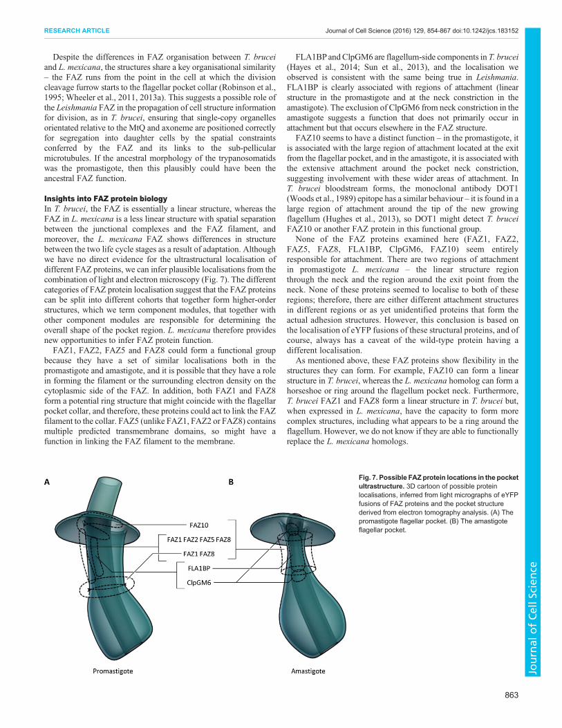
Insights into FAZ protein biologyIn T. brucei, the FAZ is essentially a linear structure, whereas theFAZ in L. mexicana is a less linear structure with spatial separationbetween the junctional complexes and the FAZ filament, andmoreover, the L. mexicana FAZ shows differences in structurebetween the two life cycle stages as a result of adaptation. Althoughwe have no direct evidence for the ultrastructural localisation ofdifferent FAZ proteins, we can infer plausible localisations from thecombination of light and electron microscopy (Fig. 7). The differentcategories of FAZ protein localisation suggest that the FAZ proteinscan be split into different cohorts that together form higher-orderstructures, which we term component modules, that together withother component modules are responsible for determining theoverall shape of the pocket region. L. mexicana therefore providesnew opportunities to infer FAZ protein function.FAZ1, FAZ2, FAZ5 and FAZ8 could form a functional group
because they have a set of similar localisations both in thepromastigote and amastigote, and it is possible that they have a rolein forming the filament or the surrounding electron density on thecytoplasmic side of the FAZ. In addition, both FAZ1 and FAZ8form a potential ring structure that might coincide with the flagellarpocket collar, and therefore, these proteins could act to link the FAZfilament to the collar. FAZ5 (unlike FAZ1, FAZ2 or FAZ8) containsmultiple predicted transmembrane domains, so might have afunction in linking the FAZ filament to the membrane.
FLA1BPandClpGM6 are flagellum-side components in T. brucei(Hayes et al., 2014; Sun et al., 2013), and the localisation weobserved is consistent with the same being true in Leishmania.FLA1BP is clearly associated with regions of attachment (linearstructure in the promastigote and at the neck constriction in theamastigote). The exclusion of ClpGM6 from neck constriction in theamastigote suggests a function that does not primarily occur inattachment but that occurs elsewhere in the FAZ structure.
FAZ10 seems to have a distinct function – in the promastigote, itis associated with the large region of attachment located at the exitfrom the flagellar pocket, and in the amastigote, it is associated withthe extensive attachment around the pocket neck constriction,suggesting involvement with these wider areas of attachment. InT. brucei bloodstream forms, the monoclonal antibody DOT1(Woods et al., 1989) epitope has a similar behaviour – it is found in alarge region of attachment around the tip of the new growingflagellum (Hughes et al., 2013), so DOT1 might detect T. bruceiFAZ10 or another FAZ protein in this functional group.
None of the FAZ proteins examined here (FAZ1, FAZ2,FAZ5, FAZ8, FLA1BP, ClpGM6, FAZ10) seem entirelyresponsible for attachment. There are two regions of attachmentin promastigote L. mexicana – the linear structure regionthrough the neck and the region around the exit point from theneck. None of these proteins seemed to localise to both of theseregions; therefore, there are either different attachment structuresin different regions or as yet unidentified proteins that form theactual adhesion structures. However, this conclusion is based onthe localisation of eYFP fusions of these structural proteins, and ofcourse, always has a caveat of the wild-type protein having adifferent localisation.
As mentioned above, these FAZ proteins show flexibility in thestructures they can form. For example, FAZ10 can form a linearstructure in T. brucei, whereas the L. mexicana homolog can form ahorseshoe or ring around the flagellum pocket neck. Furthermore,T. brucei FAZ1 and FAZ8 form a linear structure in T. brucei but,when expressed in L. mexicana, have the capacity to form morecomplex structures, including what appears to be a ring around theflagellum. However, we do not know if they are able to functionallyreplace the L. mexicana homologs.
Fig. 7. Possible FAZ protein locations in the pocketultrastructure. 3D cartoon of possible proteinlocalisations, inferred from light micrographs of eYFPfusions of FAZ proteins and the pocket structurederived from electron tomography analysis. (A) Thepromastigote flagellar pocket. (B) The amastigoteflagellar pocket.
863
RESEARCH ARTICLE Journal of Cell Science (2016) 129, 854-867 doi:10.1242/jcs.183152
Journal
ofCe
llScience

Implications for mechanisms of trypanosomatid parasitemorphogenesisTrypanosomatids have a set of characteristic morphologies, in partdefined by the positioning of the flagellar pocket and whether or notthey have a laterally attached flagellum (Hoare and Wallace, 1966).Taking the promastigote morphology as the ancestral morphology(Flegontov et al., 2013), the structure of the L. mexicana FAZ can beused to predict what the ultrastructure of these morphologies islikely to be, what their molecular composition is and how they couldhave evolved. Firstly, the opisthomastigote appears to simply havean extension of the neck region, pushing the collar and pocketdeeper into the cell, based on the presence of the PFR in theextended pocket (Janovy et al., 1974; Rowton et al., 1981; Yoshidaet al., 1978). Secondly, the choanomastigote appears to have anelaboration of the attachment around the distal end of the flagellarpocket neck, the region characterised by the presence of FAZ10,into a larger attachment structure previously described as containingmany desmosome-like junctional complexes (Brooker, 1970, 1971;Brooks, 1978; Kusel et al., 1967; Soares et al., 1986). Thirdly, thelaterally attached flagellum of the trypomastigote or epimastigoteappears to require shifting of the flagellar pocket to anchor withinthe sub-pellicular array (rather than at an opening at the end of thearray), the extension of the MtQ and FAZ filament into the sub-pellicular array, and relocation of junctional complexes into thevicinity of the FAZ filament. It will be of interest to determine howthese morphological changes have co-evolved with themorphogenesis of cell shape through the cell cycle, which differsbetween Leishmania (Ambit et al., 2011; Wheeler et al., 2011) andtrypanosomes (Elias et al., 2007; Robinson et al., 1995; Sherwinand Gull, 1989; Wheeler et al., 2013a).The difference in organisation of the promastigote and amastigote
pocket neck and FAZ region implies that the amastigotemorphology represents an adaptation that minimises total surfacearea. The amastigote is small and near spherical, suggesting aminimisation of cell volume (metabolic load for growth andmaintenance) and/or surface area (in the potentially harmfulenvironment of the macrophage endocytic system). Access to theflagellar pocket in the promastigote is relatively easy, with a largecross-sectional area open to fluid access, whereas in the amastigote,the flagellum essentially plugs the pocket entrance, acting to furtherreduce the exposed surface area of the parasite. In the promastigote,the structure of the narrowest part of the neck appears to bemodulated by the flagellar pocket collar, whereas in the amastigote,it is a region dominated by the FAZ structures. The FAZ thereforeappears to be involved in regulating access to the flagellar pocket. InT. brucei, the MtQ has been implicated in keeping an access channelto the flagellar pocket open (Gadelha et al., 2009), and T. bruceiMORN1 has recently been identified as controlling access to theflagellar pocket (Morriswood and Schmidt, 2015). T. bruceiMORN1 is associated with the pocket neck and the proximal endof the T. brucei FAZ (Esson et al., 2012), and it seems likely thatFAZ structure could be modulating L. mexicanaMORN1 control ofpocket access in L. mexicana. Not only do Leishmania have a FAZdespite lacking a laterally attached flagellum, but this FAZ plausiblyhas a key function in adapting morphology throughout the life cyclein order to modify the pocket structure.This discovery and description of the Leishmania FAZ in the
context of the flagellar pocket architecture has relevance tofundamental issues of how possession, expression and theregulation of assembly of component modules change throughoutthe life cycle and evolution to orchestrate different cell shapes andforms.
MATERIALS AND METHODSAll reagents were purchased from Sigma-Aldrich unless stated.
L. mexicana cultureL. mexicana (World Health Organization strain MNYC/BZ/62/M397)promastigotes were grown in M199 medium with Earle’s salts andL-glutamine (Thermo Fisher), supplemented with 10% foetal calf serum(FCS) (Thermo Fisher), 5 mM HEPES·NaOH (pH 7.4), 26 mM NaHCO3
and 5 µg/ml haemin, at 28°C. Cells weremaintained in logarithmic growth atculture densities between 1×10⁵ cells/ml and 1×10⁷ cells/ml through regularsubculture (Wheeler et al., 2011). Culture densities were measured with aCASYmodel TT cell counter (RocheDiagnostics). Axenic amastigotes weregenerated by subculturing into Schneider’s Drosophila medium (ThermoFisher) supplementedwith 20%FCS (ThermoFisher) and 25 mMMES·HCl(pH 5.5) at 34°C under 5% CO2 (Bates, 1994), and growth for 72 h withoutsubculture. Amastigotes were generated by infection of J774 macrophageswith stationary-phase promastigotes and allowed to differentiate intoamastigotes for 72 h. J774 macrophages were grown in RPMI (ThermoFisher), supplemented with 10% FCS (Thermo Fisher), at 37°C (prior toinfection) or at 34°C (after infection) under 5% CO2.
Tagging of proteinsFor the eYFP tagging of the L. mexicana proteins, the corresponding ORFsand UTRs were cloned into the pLEnTv2-YB plasmid, as previouslydescribed (Dean et al., 2015). To tag FAZ1, FAZ2, ClpGM6, FAZ10 andLRRP1 at the N-terminus with eYFP, ∼500 bp of the 5′ end of the genedirectly after the start codon and∼500 bp of 5′UTR directly upstream of thestart codon was amplified from genomic DNA. An XbaI site was added tothe forward primer and a NotI site added to the reverse primer for theamplification of the ORF. A NotI site was added to the forward primer and aBamHI site added to the reverse primer for the amplification of the 5′ UTR.The resulting PCR fragments were digested with the appropriate restrictionenzymes and cloned into the pLEnTv2-YB plasmid that had been digestedwith XbaI and BamHI (New England Biolabs). To tag FLA1BP, FAZ8 andFAZ5 at the C-terminus with eYFP, ∼500 bp of the 3′ end of the genedirectly before the stop codon and ∼500 bp of 3′ UTR directly downstreamof the stop codon was amplified from genomic DNA. A NotI site was addedto the forward primer and a SpeI site added to the reverse primer for theamplification of the ORF. A HindIII site was added to the forward primerand a NotI site added to the reverse primer for the amplification of the3′ UTR. The resulting PCR fragments were digested with the appropriaterestriction enzymes and cloned into the pLEnTv2-YB plasmid that had beendigested with SpeI and HindIII (New England Biolabs). The resultingplasmids were linearised with NotI (NEB, Hitchin, UK) and then ethanolprecipitated before transfection.
For Myc-tagging of the L. mexicana BILBO1 homolog, a fusion PCRapproach was taken, as previously described (Dean et al., 2015). To tagBILBO1 at the N-terminus with aMyc tag,∼500 bp of the 5′ end of the genedirectly after the start codon and∼500 bp of 5′UTR directly upstream of thestart codon was amplified from genomic DNA. The forward primer for theamplification of the BILBO1 ORF had a 27-bp region of homology to the 3′end of the actin 5′ UTR at its 5′ end followed by a Myc-epitope tag that wasin-frame with the ORF. The reverse primer for the amplification of the 5′UTR had a 25-bp region of identity to the start of the blasticidin-resistancegene. Three pieces of DNA, the 500 bp UTR and ORF fragments amplifiedfrom genomic DNA, and the region from the pPOTv2 plasmid containingthe blasticidin-resistance gene followed by the aldolase 3′UTR and then theactin 5′ UTR, released using an EcoRI HindIII (New England Biolabs)digest were combined in a fusion PCR. The PCR required 30 amplificationcycles, five of which were without any primers, followed by 25 cycles usingnested primers that annealed 40 bp from the 5′ end of the UTR fragment and40 bp from the 3′ end of the gene fragment. After amplification, theconstruct was purified and then used for the transfection.
Expression of the T. brucei FAZ proteins in L. mexicanaFor the expression of the T. brucei FAZ proteins in L. mexicana, a newmodular constitutive expression plasmid was made. The plasmid integratesinto the β-tubulin array. From the 5′ end of the plasmid, there is the tubulin
864
RESEARCH ARTICLE Journal of Cell Science (2016) 129, 854-867 doi:10.1242/jcs.183152
Journal
ofCe
llScience

upstream targeting sequence followed by the Crithidia fasciculata PGKB 5′UTR, followed by a Ty–eYFP–Ty ORF (where Ty is an epitope from theSaccharomyces Cerevisiae Ty1 virus-like particle) and then theC. fasciculata PGKA 3′ UTR and PGKB 5′ UTR, next is the blasticidinORF and then the C. fasciculata GSS 3′ UTR and finally the tubulindownstream targeting sequence. Every ORF and UTR can be readilyexchanged as each component is flanked by unique restriction enzymes. Theplasmid can support the expression of both N- and C-terminally eYFP-tagged proteins. To express T. brucei FAZ1 with an N-terminal eYFP tag,the T. brucei FAZ1 ORF was cloned into the XbaI BamHI sites, and forexpression of T. brucei FAZ8 with a C-terminal eYFP tag, the T. bruceiFAZ8 ORF was cloned into the HindIII SpeI sites. The plasmids werelinearised with Acc65I and BglII (New England Biolabs) and ethanolprecipitated before transfection. The plasmids and fusion PCR constructwere electroporated, as previously described (Dean et al., 2015), usingprogram X-001 on a Nucleofector 2b instrument (Lonza).
Immunofluorescence analysis and western blottingAll the cell lines expressing eYFP tagged proteins were examinedby using live-cell microscopy. The cells were washed three timesin PBS, resuspended in PBS with Hoescht 33342 (1 µg/ml) and then10 μl placed on a poly-lysine slide. The cells were imaged using aDM5500B (Leica, Milton Keynes, UK) microscope controlled bythe Micromanager software with 100×/1.4 objective and Neo 5.5sCMOS (Andor, Belfast, UK) camera (Edelstein et al., 2010).Immunofluorescence analysis of the Myc–BILBO1 cell line wasperformed by washing the cells three times in PBS, resuspendingthe cells in PBS and allowing them to settle on a poly-lysine slide.The cells were fixed with 4% (v/v) formaldehyde for 10 min, and theexcess formaldehyde was quenched by washing the slides in PBS with1% (w/v) glycine for 5 min. The cells were blocked for 1 h withblocking buffer [PBS with 1% (w/v) BSA]. After blocking, the cellswere incubated with undiluted anti-Myc antibody clone 9E10 (Evanet al., 1985) for 1 h and then washed thoroughly with PBS beforeincubating with 1:200 diluted FITC-conjugated rabbit anti-mouse IgGantibody (catalogue number F0261, DAKO) in blocking buffer. Theslides were thoroughly washed before mounting with DABCOcontaining DAPI (100 ng/ml). The cells were imaged using a LeicaDM5500B microscope.
Expression of eYFP and Myc fusion proteins were confirmed by westernblotting. Cell lysates were resolved on SDS-PAGE gels, transferred tonitrocellulose membrane and probed with 1:2000 diluted rabbit anti-GFP(catalogue number A11122, Thermo Fisher), then 1:5000 dilutedhorseradish peroxidase (HRP)-conjugated goat anti-rabbit (cataloguenumber P0448, Dako, Ely, UK) or 1:10 diluted clone 9E10 then 1:20,000HRP-conjugated rabbit anti-mouse IgG (catalogue number A9044, Sigma-Aldrich), and the signals were detected by using enhancedchemiluminescence.
Electron tomography imagingPromastigotes and amastigotes in J774 macrophages were prepared formicroscopy as previously described (Gluenz et al., 2015; Höög et al.,2010). Briefly, cells were fixed in culture with 2.5% (v/v)glutaraldehyde (TAAB, Aldermaston, UK) for 5 min, harvested bycentrifugation (for promastigotes) or by scraping and thencentrifugation (for amastigotes in macrophages) then washed andresuspended in 200 mM phosphate buffer (pH 7.0) with 2.5%glutaraldehyde and 2.0% paraformaldehyde for 2 h. The pellet waswashed, post-fixed with 1% osmium tetroxide (Amsbio, Abingdon, UK)for 2 h and stained en bloc with 2% uranyl acetate (Amsbio, Abingdon,UK) for 2 h, then dehydrated in an ethanol series and embedded in Agar100 resin (Agar Scientific, Stansted, UK). Serial sections with anominal thickness of between 150 and 200 nm (for 100 kVmicroscopes) or between 250 nm and 400 nm (for 300 kVmicroscopes) were cut and collected on formvar-coated slot grids, andstained with Reynolds lead citrate (TAAB, Aldermaston, UK) for 2 min.
Tilt series of images were captured on a Tecnai 12 or Tecnai T30instrument with an Ultrascan 1000 CCD camera between −55° and 55°, and
between −64° and 64°, respectively, using SerialEM software(Mastronarde, 2003). The tilt series were aligned using fiducial-less patchtracking, and tomographic volumes were generated by back projection. Thetomograms of serial sections were then joined to generate the completetomogram volume. Tomogram building and joining was performed usingeTomo, part IMOD (Kremer et al., 1996; Mastronarde, 1997). Structures inthe tomograms were manually traced or segmented using 3DMOD, also partof IMOD (Kremer et al., 1996), then refined and rendered for display usingBlender (http://www.blender.org). Virtual sections through the tomogramvolumes were generated using ImageJ (Collins, 2007). Summary videosshowing the tomogram volume and segmentation (Supplementary Movies1-8) are hosted on FigShare (https://dx.doi.org/10.6084/m9.figshare.1595927.v1).
AcknowledgementsWe thank past and present members of our laboratories for discussions and accessto archive materials and electron microscopy data, especially Eva Gluenz(University of Oxford) for providing the amastigote-infected macrophage electronmicroscopy samples and Johanna Hoog (University of Gothenburg) for capturingone of the amastigote tomograms.
Competing interestsThe authors declare no competing or financial interests.
Author contributionsR.J.W. performed the electron tomography analyses. J.D.S. generated the FAZ celllines and performed the light microscopy. R.J.W., J.D.S. and K.G. all contributed tointerpreting the data and writing the paper.
FundingWork in the Gull lab is supported by the Wellcome Trust [grant numbers 066839/B/02/Z and 104627/Z/14/Z], including a Wellcome Trust Senior Investigator Award toK.G. R.J.W. holds a Wellcome Trust Sir Henry Wellcome Fellowship [grant number103261/Z/13/Z]. Deposited in PMC for immediate release.
Supplementary informationSupplementary information available online athttp://jcs.biologists.org/lookup/suppl/doi:10.1242/jcs.183152/-/DC1
ReferencesAbsalon, S., Blisnick, T., Bonhivers, M., Kohl, L., Cayet, N., Toutirais, G.,
Buisson, J., Robinson, D. and Bastin, P. (2008). Flagellum elongation isrequired for correct structure, orientation and function of the flagellar pocket inTrypanosoma brucei. J. Cell Sci. 121, 3704-3716.
Aleman, C. (1969). Finestructure of cultured Leishmania brasiliensis. Exp.Parasitol. 24, 259-264.
Alexander, J. (1978). Unusual axonemal doublet arrangements in the flagellum ofLeishmania amastigotes. Trans. R. Soc. Trop. Med. Hyg. 72, 345-347.
Ambit, A., Woods, K. L., Cull, B., Coombs, G. H. and Mottram, J. C. (2011).Morphological events during the cell cycle of Leishmania major. Eukaryot. Cell 10,1429-1438.
Bates, P. A. (1994). Complete developmental cycle of Leishmania mexicana inaxenic culture. Parasitology 108, 1-9.
Bonhivers, M., Nowacki, S., Landrein, N. andRobinson, D. R. (2008). Biogenesisof the trypanosome endo-exocytotic organelle is cytoskeleton mediated. PLoSBiol. 6, e105.
Brooker, B. E. (1970). Desmosomes and hemidesmosomes in the flagellateCrithidia fasciculata. Z. Fur Zellforsch. Mikrosk. Anat. 105, 155-166.
Brooker, B. E. (1971). The fine structure of Crithidia fasciculata with specialreference to the organelles involved in the ingestion and digestion of protein.Z. Fur Zellforsch. Mikrosk. Anat. 116, 532-563.
Brooks, A. S. (1978). Ultrastructure of the flagellar attachment site in three speciesof trypanosomatids. Trans. Am. Microsc. Soc. 97, 287-296.
Collins, T. J. (2007). ImageJ for microscopy. Biotechniques 43, S25-S30.Dean, S., Sunter, J.,Wheeler, R. J., Hodkinson, I., Gluenz, E. andGull, K. (2015).
A toolkit enabling efficient, scalable and reproducible gene tagging intrypanosomatids. Open Biol. 5, 140197.
Edelstein, A., Amodaj, N., Hoover, K., Vale, R. and Stuurman, N. (2010).Computer control of microscopes using µManager. Curr. Protoc. Mol. Biol. 92,14.20.1-14.20.17. Ed. Frederick M Ausubel Al Chapter 14, Unit14.20.
Elias, M. C., da Cunha, J. P. C., de Faria, F. P., Mortara, R. A., Freymuller, E. andSchenkman, S. (2007). Morphological events during the Trypanosoma cruzi cellcycle. Protist 158, 147-157.
865
RESEARCH ARTICLE Journal of Cell Science (2016) 129, 854-867 doi:10.1242/jcs.183152
Journal
ofCe
llScience

Engstler, M., Pfohl, T., Herminghaus, S., Boshart, M., Wiegertjes, G.,Heddergott, N. and Overath, P. (2007). Hydrodynamic flow-mediated proteinsorting on the cell surface of trypanosomes. Cell 131, 505-515.
Esson, H. J., Morriswood, B., Yavuz, S., Vidilaseris, K., Dong, G. andWarren, G.(2012). Morphology of the trypanosome bilobe, a novel cytoskeletal structure.Eukaryot. Cell 11, 761-772.
Evan, G. I., Lewis, G. K., Ramsay, G. and Bishop, J. M. (1985). Isolation ofmonoclonal antibodies specific for human c-myc proto-oncogene product. Mol.Cell. Biol. 5, 3610-3616.
Field, M. C. and Carrington, M. (2009). The trypanosome flagellar pocket. Nat.Rev. Microbiol. 7, 775-786.
Flegontov, P., Votýpka, J., Skalický, T., Logacheva, M. D., Penin, A. A., Tanifuji,G., Onodera, N. T., Kondrashov, A. S., Volf, P., Archibald, J. M. et al. (2013).Paratrypanosoma is a novel early-branching trypanosomatid. Curr. Biol. 23,1787-1793.
Gadelha, C., Rothery, S., Morphew, M., McIntosh, J. R., Severs, N. J. and Gull,K. (2009). Membrane domains and flagellar pocket boundaries are influenced bythe cytoskeleton in African trypanosomes. Proc. Natl. Acad. Sci. USA 106,17425-17430.
Gadelha, A. P. R., Cunha-e-Silva, N. L. and de Souza, W. (2013). Assembly of theLeishmania amazonensis flagellum during cell differentiation. J. Struct. Biol. 184,280-292.
Gheiratmand, L., Brasseur, A., Zhou, Q. and He, C. Y. (2013). Biochemicalcharacterization of the bi-lobe reveals a continuous structural network linking thebi-lobe to other single-copied organelles in Trypanosoma brucei. J. Biol. Chem.288, 3489-3499.
Gluenz, E., Hoog, J. L., Smith, A. E., Dawe, H. R., Shaw,M. K. andGull, K. (2010).Beyond 9+0: noncanonical axoneme structures characterize sensory cilia fromprotists to humans. FASEB J. 24, 3117-3121.
Gluenz, E., Wheeler, R. J., Hughes, L. and Vaughan, S. (2015). Scanning andthree-dimensional electron microscopy methods for the study of Trypanosomabrucei and Leishmania mexicana flagella. Methods Cell Biol. 127, 509-542.
Gull, K. (2003). Host–parasite interactions and trypanosome morphogenesis: aflagellar pocketful of goodies. Curr. Opin. Microbiol. 6, 365-370.
Hayes, P., Varga, V., Olego-Fernandez, S., Sunter, J., Ginger, M. L. and Gull, K.(2014). Modulation of a cytoskeletal calpain-like protein induces major transitionsin trypanosome morphology. J. Cell Biol. 206, 377-384.
Hoare, C. A. and Wallace, F. G. (1966). Developmental stages of trypanosomatidflagellates: a new terminology. Nature 212, 1385-1386.
Hoog, J. L., Gluenz, E., Vaughan, S. and Gull, K. (2010). Ultrastructuralinvestigation methods for Trypanosoma brucei. Methods Cell Biol. 96, 175-196.
Hoog, J. L., Bouchet-Marquis, C., McIntosh, J. R., Hoenger, A. and Gull, K.(2012). Cryo-electron tomography and 3-D analysis of the intact flagellum inTrypanosoma brucei. J. Struct. Biol. 178, 189-198.
Hu, H., Zhou, Q. and Li, Z. (2015). SAS-4 in protein in Trypanosoma brucei controlslife cycle transitions by modulating the length of the flagellum attachment zonefilament. J. Biol. Chem. 290, 30453-30463.
Hughes, L., Towers, K., Starborg, T., Gull, K. andVaughan, S. (2013). A cell-bodygroove housing the new flagellum tip suggests an adaptation of cellularmorphogenesis for parasitism in the bloodstream form of Trypanosoma brucei.J. Cell Sci. 126, 5748-5757.
Janovy, J., Lee, K. W. and Brumbaugh, J. A. (1974). The differentiation ofHerpetomonas megaseliae: ultrastructural observations. J. Protozool. 21, 53-59.
Kohl, L., Sherwin, T. and Gull, K. (1999). Assembly of the paraflagellar rod and theflagellum attachment zone complex during the Trypanosoma brucei cell cycle.J. Eukaryot. Microbiol. 46, 105-109.
Kremer, J. R., Mastronarde, D. N. and McIntosh, J. R. (1996). Computervisualization of three-Dimensional image data using IMOD. J. Struct. Biol. 116,71-76.
Kusel, J. P., Moore, K. E. and Weber, M. M. (1967). The ultrastructure of Crithidiafasciculata and morphological changes induced by growth in acriflavin.J. Protozool. 14, 283-296.
Lacomble, S., Vaughan, S., Gadelha, C., Morphew, M. K., Shaw, M. K.,McIntosh, J. R. and Gull, K. (2009). Three-dimensional cellular architecture ofthe flagellar pocket and associated cytoskeleton in trypanosomes revealed byelectron microscope tomography. J. Cell Sci. 122, 1081-1090.
Lacomble, S., Vaughan, S., Gadelha, C., Morphew, M. K., Shaw, M. K.,McIntosh, J. R. and Gull, K. (2010). Basal body movements orchestratemembrane organelle division and cell morphogenesis in Trypanosoma brucei.J. Cell Sci. 123, 2884-2891.
LaCount, D. J., Barrett, B. and Donelson, J. E. (2002). Trypanosoma brucei FLA1is required for flagellum attachment and cytokinesis. J. Biol. Chem. 277,17580-17588.
Maslov, D. A., Votýpka, J., Yurchenko, V. and Lukes, J. (2013). Diversity andphylogeny of insect trypanosomatids: all that is hidden shall be revealed. TrendsParasitol. 29, 43-52.
Mastronarde, D. N. (1997). Dual-axis tomography: an approach with alignmentmethods that preserve resolution. J. Struct. Biol. 120, 343-352.
Mastronarde, D. (2003). SerialEM A program for automated tilt series acquisition ontecnai microcopes using prediction of specimen position. Microsc. Microanal. 9,1182-1183.
McAllaster, M. R., Ikeda, K. N., Lozano-Nun ez, A., Anrather, D.,Unterwurzacher, V., Gossenreiter, T., Perry, J. A., Crickley, R., Mercadante,C. J., Vaughan, S. et al. (2015). Proteomic identification of novel cytoskeletalproteins associated with TbPLK, an essential regulator of cell morphogenesis inT. brucei. Mol. Biol. Cell 26, 3013-3029.
Molyneux, D. H., Killick-Kendrick, R. and Ashford, R. W. (1975). Leishmania inphlebotomid sandflies. III. The ultrastructure of Leishmania mexicanaamazonensis in the midgut and pharynx of Lutzomyia longipalpis. Proc. R. Soc.B Biol. Sci. 190, 341-357.
Morriswood, B. and Schmidt, K. (2015). A MORN repeat protein facilitates proteinentry into the flagellar pocket of Trypanosoma brucei. Eukaryot. Cell 14,1081-1093.
Morriswood, B., Havlicek, K., Demmel, L., Yavuz, S., Sealey-Cardona, M.,Vidilaseris, K., Anrather, D., Kostan, J., Djinovic-Carugo, K., Roux, K. J., et al.(2013). Novel bilobe components in Trypanosoma brucei identified usingproximity-dependent biotinylation. Eukaryot. Cell 12, 356-367.
Nozaki, T., Haynes, P. A. and Cross, G. A. M. (1996). Characterization of theTrypanosoma brucei homologue of a Trypanosoma cruzi flagellum-adhesionglycoprotein. Mol. Biochem. Parasitol. 82, 245-255.
Oberholzer, M., Langousis, G., Nguyen, H. T., Saada, E. A., Shimogawa, M. M.,Jonsson, Z. O., Nguyen, S. M., Wohlschlegel, J. A. and Hill, K. L. (2011).Independent analysis of the flagellum surface and matrix proteomes providesinsight into flagellum signaling in mammalian-infectious Trypanosoma brucei.Mol. Cell. Proteomics 10, M111.010538.
Robinson, D. R., Sherwin, T., Ploubidou, A., Byard, E. H. and Gull, K. (1995).Microtubule polarity and dynamics in the control of organelle positioning,segregation, and cytokinesis in the trypanosome cell cycle. J. Cell Biol. 128,1163-1172.
Rotureau, B., Subota, I. and Bastin, P. (2011). Molecular bases of cytoskeletonplasticity during the Trypanosoma brucei parasite cycle. Cell. Microbiol. 13,705-716.
Rotureau, B., Subota, I., Buisson, J. and Bastin, P. (2012). A new asymmetricdivision contributes to the continuous production of infective trypanosomes in thetsetse fly. Development 139, 1842-1850.
Rotureau, B., Blisnick, T., Subota, I., Julkowska, D., Cayet, N., Perrot, S. andBastin, P. (2014). Flagellar adhesion in Trypanosoma brucei relies on interactionsbetween different skeletal structures in the flagellum and cell body. J. Cell Sci.127, 204-215.
Rowton, E. D., Lushbaugh, W. B. and McGhee, R. B. (1981). Ultrastructure of theflagellar apparatus and attachment of Herpetomonas ampelophilae in the gut andmalpighian tubules of Drosophila melanogaster 1. J. Protozool. 28, 297-301.
Sharma, R., Peacock, L., Gluenz, E., Gull, K., Gibson, W. and Carrington, M.(2008). Asymmetric cell division as a route to reduction in cell length and changein cell morphology in trypanosomes. Protist 159, 137-151.
Sheriff, O., Lim, L.-F. and He, C. Y. (2014). Tracking the biogenesis and inheritanceof subpellicular microtubule in Trypanosoma brucei with inducible YFP-α-tubulin.Biomed. Res. Int. 2014, 893272.
Sherwin, T. and Gull, K. (1989). The cell division cycle of Trypanosoma bruceibrucei: timing of event markers and cytoskeletal modulations. Philos.Trans. R. Soc. B. Biol. Sci. 323, 573-588.
Soares, M. J., Brazil, R. P., Tanuri, A. and de Souza, W. (1986). Someultrastructural aspects of Crithidia guilhermei n.sp. isolated from Phaeniciacuprina (Diptera: Calliphoridae). Can. J. Zool. 64, 2837-2842.
Sun, S. Y., Wang, C., Yuan, Y. A. and He, C. Y. (2013). An intracellularmembrane junction consisting of flagellum adhesion glycoproteins linksflagellum biogenesis to cell morphogenesis in Trypanosoma brucei. J. CellSci. 126, 520-531.
Sunter, J. D., Varga, V., Dean, S. and Gull, K. (2015). A dynamic coordination offlagellum and cytoplasmic cytoskeleton assembly specifies cell morphogenesis intrypanosomes. J. Cell Sci. 128, 1580-1594.
Tull, D., Vince, J. E., Callaghan, J. M., Naderer, T., Spurck, T., McFadden, G. I.,Currie, G., Ferguson, K., Bacic, A. and McConville, M. J. (2004). SMP-1, aMember of a new family of small myristoylated proteins in kinetoplastid parasites,is targeted to the flagellummembrane in leishmania.Mol. Biol. Cell15, 4775-4786.
Vaughan, S., Kohl, L., Ngai, I., Wheeler, R. J. and Gull, K. (2008). A repetitiveprotein essential for the flagellum attachment zone filament structure and functionin Trypanosoma brucei. Protist 159, 127-136.
Vickerman, K. (1969). On the surface coat and flagellar adhesion in trypanosomes.J. Cell Sci. 5, 163-193.
Weise, F., Stierhof, Y. D., Kuhn, C., Wiese, M. and Overath, P. (2000). Distributionof GPI-anchored proteins in the protozoan parasite Leishmania, based on animproved ultrastructural description using high-pressure frozen cells. J. Cell Sci.113, 4587-4603.
Wheeler, R. J., Gluenz, E. and Gull, K. (2011). The cell cycle of Leishmania:morphogenetic events and their implications for parasite biology. Mol. Microbiol.79, 647-662.
866
RESEARCH ARTICLE Journal of Cell Science (2016) 129, 854-867 doi:10.1242/jcs.183152
Journal
ofCe
llScience

Wheeler, R. J., Scheumann, N.,Wickstead, B., Gull, K. and Vaughan, S. (2013a).Cytokinesis in Trypanosoma brucei differs between bloodstream and tsetsetrypomastigote forms: implications for microtubule-based morphogenesis andmutant analysis. Mol. Microbiol. 90, 1339-1355.
Wheeler, R. J., Gluenz, E. and Gull, K. (2013b). The limits on trypanosomatidmorphological diversity. PLoS ONE 8, e79581.
Wheeler, R. J., Gluenz, E. and Gull, K. (2015). Basal body multipotency andaxonemal remodelling are two pathways to a 9+0 flagellum. Nat. Commun. 6,8964.
Woods,A., Sherwin, T., Sasse,R., MacRae, T. H., Baines, A. J. andGull, K. (1989).Definition of individual components within the cytoskeleton of Trypanosoma bruceiby a library of monoclonal antibodies. J. Cell Sci. 93, 491-500.
Woods, K., Nic a’Bhaird, N., Dooley, C., Perez-Morga, D. andNolan, D. P. (2013).Identification and characterization of a stage specific membrane protein involvedin flagellar attachment in Trypanosoma brucei. PLoS ONE 8, e52846.
Yoshida, N., Freymuller, E. and Wallace, F. G. (1978). Herpetomonasmariadeanei sp. n. (Protozoa, Trypanosomatidae) from Muscina stabulans(Falleen, 1816) (Diptera, Muscidae). J. Eukaryot. Microbiol. 25, 421-425.
Zhou, Q., Liu, B., Sun, Y. and He, C. Y. (2011). A coiled-coil- and C2-domain-containing protein is required for FAZ assembly and cell morphology inTrypanosoma brucei. J. Cell Sci. 124, 3848-3858.
Zhou, Q., Hu, H., He, C. Y. and Li, Z. (2015). Assembly and maintenance of theflagellum attachment zone filament in Trypanosoma brucei. J. Cell Sci. 128,2361-2372.
867
RESEARCH ARTICLE Journal of Cell Science (2016) 129, 854-867 doi:10.1242/jcs.183152
Journal
ofCe
llScience
J. Cell Sci. 129: doi:10.1242/jcs.183152: Supplementary information
A
eY
FP–L
RR
P
FAZ8
–eY
FP
eY
FP–F
AZ2
eY
FP–F
AZ1
0
eY
FP–C
lpG
M6
eY
FP–F
AZ1
FLA
1B
P–e
YFP
FAZ5
–eY
FP
L. mexicana SMP1–mCh
250150
10075
50
37
kDa
250150
10075
50
37
* *
*
*
*
*
*
10
9
91
17
1
47
2
29
6
25
1
10
8
99
Expectedsize (kDa)
Cell line
anti-GFP(long exposure)
anti-GFP(short exposure)
anti-PFR
B
2.01.51.0
0.5
3.0kbp
eY
FP–L
RR
P
FAZ8
–eY
FP
eY
FP–F
AZ2
FLA
1B
P–e
YFP
− + − +− +− +
− SMP1–mCh (parental cell line) gDNA template+ SMP1–mCh with eYFP tagged ORF gDNA template
FAZ5
–eY
FP
eY
FP–F
AZ1
eY
FP–C
lpG
M6
eY
FP–F
AZ1
0
− + − +− +− +
2.01.51.0
0.5
3.0kbp
eYFP ORF
N-terminal (eYFP–)
C-terminal (–eYFP)
eYFPORF
67
myc–B
ILB
O1
250150
10075
50
kDa
Expectedsize (kDa)
Cell line
anti-myc(9E10)
anti-PFR
L. mexicanaSMP1–mCh
*
C
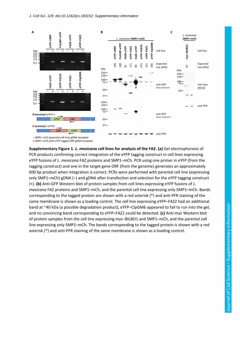
Supplementary Figure 1. L. mexicana cell lines for analysis of the FAZ. (a) Gel electrophoresis of
PCR products confirming correct integration of the eYFP tagging construct in cell lines expressing
eYFP fusions of L. mexicana FAZ proteins and SMP1–mCh. PCR using one primer in eYFP (from the
tagging construct) and one in the target gene ORF (from the genome) generates an approximately
600 bp product when integration is correct. PCRs were performed with parental cell line (expressing
only SMP1–mCh) gDNA (−) and gDNA after transfection and selection for the eYFP tagging construct
(+). (b) Anti-GFP Western blot of protein samples from cell lines expressing eYFP fusions of L.
mexicana FAZ proteins and SMP1–mCh, and the parental cell line expressing only SMP1–mCh. Bands
corresponding to the tagged protein are shown with a red asterisk (*) and anti-PFR staining of the
same membrane is shown as a loading control. The cell line expressing eYFP–FAZ2 had an additional
band at ~40 kDa (a possible degradation product), eYFP–ClpGM6 appeared to fail to run into the gel,
and no convincing band corresponding to eYFP–FAZ1 could be detected. (c) Anti-myc Western blot
of protein samples from the cell line expressing myc–BILBO1 and SMP1–mCh, and the parental cell
line expressing only SMP1–mCh. The bands corresponding to the tagged protein is shown with a red
asterisk (*) and anti-PFR staining of the same membrane is shown as a loading control.
Jour
nal o
f Cel
l Sci
ence
• S
uppl
emen
tary
info
rmat
ion

J. Cell Sci. 129: doi:10.1242/jcs.183152: Supplementary information
Supplementary Table 1. Summary of tomograms captured for pocket structure analysis.
Sample Name Resolution Serial
sections
Tomogram depth
Tomogram volume
Orientation (nm/px) (nm) (μm3)
Exponential phase promastigote PL1 1.534 6 896 8 Longitudinal Exponential phase promastigote PL2 1.101 5 719 3 Longitudinal Exponential phase promastigote PL3 1.534 4 626 6 Longitudinal Exponential phase promastigote PT1a 1.534 3 584 6 Transverse Exponential phase promastigote PT1b 1.534 3 419 4 Transverse Exponential phase promastigote PT2 1.101 4 713 3 Transverse Amastigote in J774 macrophage AL1 1.537 2 341 3 Longitudinal Amastigote in J774 macrophage AL2 2.320 3 324 7 Longitudinal Amastigote in J774 macrophage AL3 2.320 3 449 10 Longitudinal
Jour
nal o
f Cel
l Sci
ence
• S
uppl
emen
tary
info
rmat
ion

J. Cell Sci. 129: doi:10.1242/jcs.183152: Supplementary information
The eight supplemental movies are hosted at Figshare, and can be downloaded here:
https://figshare.com/articles/Leishmania_mexicana_promastigote_and_amastigote_flagellar_pocke
t_tomograms/1595927
Supplemental Movie 1: Tomogram PL1. The tomographic volume and segmented model of
tomogram PL1, generated from 6 serial longitudinal sections through the flagellar pocket and cell
anterior of a Leishmania mexicana promastigote.
Supplemental Movie 2: Tomogram PL2. The tomographic volume and segmented model of
tomogram PL2, generated from 5 serial longitudinal sections through the flagellar pocket and cell
anterior of a Leishmania mexicana promastigote.
Jour
nal o
f Cel
l Sci
ence
• S
uppl
emen
tary
info
rmat
ion

J. Cell Sci. 129: doi:10.1242/jcs.183152: Supplementary information
Supplemental Movie 3: Tomogram PL3. The tomographic volume and segmented model of
tomogram PL3, generated from 4 serial longitudinal sections through the flagellar pocket and cell
anterior of a Leishmania mexicana promastigote.
Supplemental Movie 4: Tomogram PT1. The tomographic volume and segmented model of
tomogram PT1, generated from 3 serial transverse sections through the flagellar pocket and cell
anterior of two Leishmania mexicana promastigotes.
Jour
nal o
f Cel
l Sci
ence
• S
uppl
emen
tary
info
rmat
ion

J. Cell Sci. 129: doi:10.1242/jcs.183152: Supplementary information
Supplemental Movie 5: Tomogram PT2. The tomographic volume and segmented model of
tomogram PT2, generated from 4 serial transverse sections through the flagellar pocket and cell
anterior of a Leishmania mexicana promastigote.
Supplemental Movie 6: Tomogram AL1. The tomographic volume and segmented model of
tomogram AL1, generated from 2 serial longitudinal sections through the flagellar pocket and cell
anterior of a Leishmania mexicana amastigote.
Jour
nal o
f Cel
l Sci
ence
• S
uppl
emen
tary
info
rmat
ion

J. Cell Sci. 129: doi:10.1242/jcs.183152: Supplementary information
Supplemental Movie 7: Tomogram AL2. The tomographic volume and segmented model of
tomogram AL2, generated from 3 serial longitudinal sections through the flagellar pocket and cell
anterior of a Leishmania mexicana amastigote.
Supplemental Movie 8: Tomogram AL3. The tomographic volume and segmented model of
tomogram AL3, generated from 3 serial longitudinal sections through the flagellar pocket and cell
anterior of a Leishmania mexicana amastigote.
Jour
nal o
f Cel
l Sci
ence
• S
uppl
emen
tary
info
rmat
ion