Embed Size (px)
Citation preview

CARDIOVASCULAR PHYSIOLOGY
LECTURE 1
The structure and function of the cardiovascular system.
Function and characteristics of the myocardium:
Excitability, Automatism and Conductivity
Assoc. Prof. Ana-Maria Zagrean
Physiology & Neuroscience Department
www.fiziologie.ro

The need for a competitive circulatory system
• Isolated/single cells vs. multicellular organisms
• Connection between external - internal milieu: from simple diffusion to complex, highly regulated, circulatory systems

Cardiovascular
System:
1)The heart, a dual pump,
that circulates a liquid
2) the blood,
3) through the vessels of
systemic and pulmonary
circulations
4) Integrated regulatory
system to adapt to the
changing circumstances

Circulatory System: an interplay of fast & complex
mechanisms to maintain body homeostasis and adaptive
capacity.
1. Functions of the circulatory system
The primary role of the circulatory system is the distribution
of dissolved gases and other molecules for nutrition,
growth, and repair (transport system function).
This function depends on maintaining a steep concentration gradient
from the blood to the intracellular medium for nutrients and in the
opposite direction for waste/metabolic products.
A fast blood circulation is needed between cells in the body and
circulatory areas in contact with the external milieu: lung, gut, and
kidney epithelia.

1. Circulatory system as a transport system :
- Nutrition, growth and repair
transport between all parts of the body: nutrients,
metabolic products, respiratory gases (O2, CO2):
relation CO2 pH homeostasis
- Maintenance of circulatory parameters:
cardiac output – 5L/min at rest, for an adult
! capacity to increase 5 fold during exercise
blood pressure
blood volume,
blood fluidity/viscosity

2. Functions of the circulatory system
Secondary roles:
(1) fast chemical signaling to cells by means of circulating
hormones, neurotransmitters, signaling molecules; secretory
function
(2) dissipation of heat by delivery of heat from the core to the
surface of the body
(3) mediation of inflammatory and host defense responses
against invading microorganisms:‘Channel’ for the immune response/defense mechanisms:
immune cells, antibodies, clotting proteins

3. Functions of the circulatory system
Secondary roles:
Secretory function of the circulatory system:
- Atrial Natriuretic Peptide (ANP):
released by atrial fibers as a response to heart overload
powerful vasodilator, reduce water & sodium on the circulatory
system, thereby reducing blood pressure
- Nitric oxide - NO (Endothelial Derived Relaxing Factor):
vasodilator, inhibits platelets adherence & aggregation
- Endothelial Derived Hyperpolarizing Factor
- Endothelin: vasoconstrictor on ETA, ETB2 receptors
- Prostaglandins PGE2, PGI2 (prostacyclin):
increase Na+ excretion by the kidneys, vasodilators

1. Components of the Cardiovascular System
and their function:
1. the heart consists in two pumps, operating in series, requiring
equalization of their outputs (5 L/min)
Left heart (LH) – main pump
Right heart (RH) – boost pump
2. the blood
3. a tubular system (two serial circuits):
-systemic / pulmonary circulation
-high-pressure (LV systemic capillaries)
low-pressure (systemic capillaries LA)
-vessels (arteries, veins, capillaries, lymphatics) respond with
changes in blood flow to changing metabolic demands
-angiogenesis: self-repairing / self-expanding capacity of endothelial
cells

4. an integrated regulation system to adapt to variable demands:
- intrinsic: automatism
- systemic: nervous system
endocrine system
- local/metabolic regulation
! level of activity – metabolic rate coupling
e.g., increase in muscular metabolic rate during physical activity
muscular blood perfusion - resting conditions – 4 ml/100g/min
- physical activity - 80 ml/100g/min
Components of the Cardiovascular System and
their function:

Distribution of blood flow (% Cardiac Output) at rest & during heavy exercise

Heart – morphological data:
- G = 250 - 340 g
- Longitudinal diam. =100 - 120 mm
- Transversal diam. = 80 - 100 mm
- Measurement methods:- clinical: aria of cardiac dullness- echocardiography- radiology

Morphological components of the heart
-Pericardium:thin-walled membranous cavity surrounding the heart;small volume of pericardial fluid in the space between itstwo surfaces.
- Myocardium, cardiac conduction system, peacemaker cells
- Fibrous skeleton
- 4 intracardiac valves:
AV valves: Right (tricuspid, 3-leaflet)
Left (mitral, 2-leaflet)
Semilunar valves: Aortic & Pulmonary
- chordae tendineae
- Endocardium
- Coronary circulation


The cardiac muscle: myocardium
• Peacemaker cells
• Conductive system
• Working myocardium
-involuntary contraction-striated in appearance (sarcomeres and myofilaments similar to skeletal muscle ones); small cells, electrical and mechanical cell-to-cell communication (syncitium); -mechanical performance complex and subtle.

Myocardial cell (MC) structure
Sarcolema - T tubules & terminal cistern: continuous with the cell
membrane, carry the AP; more developed in the ventricles
Sarcoplasmic reticulum (SR):
in close proximity to the contractile elements
site of storage and release of Ca
Sarcomere: contractile unit of the MC
between 2 Z lines
contains: thin filaments - actin, troponin, tropomyosin
thick filaments – myosin
Intercalated disks: paracellular connections
hold the cells (desmosomes)
connect them electrically (gap junctions),
heart behaves as an electrical syncytium(sync, synch: informal for synchronization)
Mitochondria (> than in skeletal muscle)


Myocardial properties
1. Excitability (bathmotropia)
2. Rhythmic activity/automaticity (chronotropia)
3. Conductibility (dromotropia)
4. Contractility (inotropia)
5. Relaxation (lusitropia)

Excitability
Myocardial capacity to respond to a stimulus that has a
minimum threshold intensity
Depends on ionic gradients across the cell membrane
(polarized membrane), through the action of the membrane
ionic transport system: ionic channels, pumps, exchangers
Membrane ionic transport system
Classification according to the mechanism that mediates
the transport:
• Pore proteins (water channels, connexons of gap junctions…)
• Transporters (carriers - exchangers, pumps…)
• Channels
Properties of the transport system:
• Functional state: opened or closed
• Rate of transport (109/sec - aquaporins, 106-108/sec. - gated
channels, 102-105/sec. - transporters)
• Transport mechanism: diffusion or binding of substrates
• Saturation
• Chemical specificity
• Regulation (e.g. by ATP, phosphorylation)

Membrane Ionic Transport System

Ionic Channels
− K+ channels − fast Na+ channels, slow Na+ and Ca2+ channels− Ca2+ channels− Cl- channels− nonspecific cation channels (Na+, K+, Li+), etc...

Membrane currents in the heart
-time-dependent
-voltage-dependant
Classification
1.Na Currents (INa)
2.Ca Currents (ICa)
3.K Currents (IK)
4.Pacemaker Currents (if)

Principal ionic currents & channels that generate AP in a cardiac cell

-Na inflow through fast voltage-dependent channels:a, b1 subunits: a subunit has a phosphorylation site
for cAMP-dependent PK
1.Na Currents (INa)
-opening/activation (m activation gate) in responseto local depolarization, at ~ -55 mV (0,1- 0,2 ms)…
-closing/inactivation (h inactivation gate) to a positivepotential (~1 ms); 99% of Na channels areinactivated at the peak of the AP
-channel reset to open again after the membranebecomes repolarized below -50 mV !
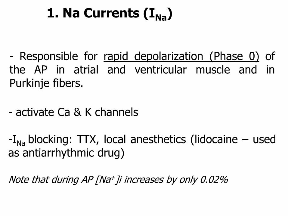
- Responsible for rapid depolarization (Phase 0) ofthe AP in atrial and ventricular muscle and inPurkinje fibers.
-INa blocking: TTX, local anesthetics (lidocaine – usedas antiarrhythmic drug)
Note that during AP [Na+]i increases by only 0.02%
1. Na Currents (INa)
- activate Ca & K channels

- Ca inflow through slow L-type voltage-dependent
channels/dihydropyridine receptors (L from long lived)
- Responsible for depolarization (Phase 0) of AP inSAN, AVN and their neighboring cells
2. Ca Current (ICa)
- activation ~ 1 ms
- inactivation ~ 10-20 ms
- participate in Phase 2 (plateau phase) of AP inatrial and ventricular muscle
long refractory period of the AP

2. Ca Current (ICa)
-activates Ca-dependent Ca release from SR;
-initiates excitation-contraction coupling in myocardium
-L-type Ca channels blockers:nifedipin, verapamil, diltiazem
- Other Ca channels: T-type (transient) -activated at a lower voltage threshold(< - 30 mV), inactivated at a lower voltage…
- initiation of the AP, automatism …N-type, P/Q-type

T (transverse) tubule
Ca2+ dependent Ca2+
releasing channel
(ryanodine receptor)
Sarcoplasmic
Reticulum
L-type Ca2+ channel
(Cav1.2. dihydropiridine
receptors)
Sarcoplasmic
Reticulum (SR)
Tetrad: 4 L-type Ca channels
faces a single Ca release
channel on SR
Triad: T tub + 2 SR cisternae

3. K Currents
IK-repolarization currents resulting from the slow opening
(20-200 ms) of KV channels
-responsible for Phase 3 of the AP
-deactivating at the diastolic membrane voltage
Ito- early transient outward K current (A-type)
- activated by depolarization; rapidly inactivates
- contributes to Phase 1 of the AP
IK 1 inward rectifying K channel - responsible for the resting membrane potential (Phase 4)

G-protein activated inwardly-rectifying K current-vagal nerv stim. of SAN & AVN Ach muscarinicreceptor G-protein (bg subunit) GIRK K channels:outward K current hyperpolarization slows pacemaker
rate & slows AP conduction through AVN
ATP-sensitive K channels:Low probability to open at normal [ATP]~ 5 mMK currents dependent on ATP/ADP ratio; hypoxia/ischemia
ATP & ADP K channels activation & K outflow
(early repolarisation, short plateau, weak beat; possible role inelectrical regulation of contractile behavior by coupling cellularmetabolism & membrane excitability; cardioprotection…)
Note that the [K+]i changes just by 0.001% during AP.
3. K Currents

4. Pacemaker Current If
- found in SAN & AVN cells and in Purkinje fibers
- slow activation (100 ms) by hyperpolarization at the end of
Phase 3 (“f” from funny)
-Produces an inward, depolarizing current of Na+
-If through a nonspecific cation channel (permeable for Na &
K) called HCN (hyperpolarization-activated cyclic nucleotide
(AMPc, GMPc)-gated channels)
-other pacemaker currents: Ica, IK

Ionic pumps
- Na+/K+ pump: action mechanism; blockers – ouabain
- Ca2+ pumps: function, distribution, action mechanism

sarcoplasmic reticulum:
calsequestrin
Ca2+ ATPase
Ca2+
channels
Na+/Ca2+
exchanger
Na+/K+ ATPaseCa2+ ATPase
Ca2+ in myocardial fiber: transport through
channels, pumps and exchangers

- Na+/H+ antiporter
Ionic exchangers
- Cl-/HCO3- antiporter
- 1Na+/1K+/2Cl - exchanger
- K+/Cl- , Na+/Cl- symporters
- 3Na +/1Ca++ antiporter
-Na+/aa, Na+/G, H+/oligopeptidesymporters
- Na+/HCO3- symporter
Na+/Ca2+ exchanger (NCX1)
-location: sarcolema
-transport mechanism: 3Na+/1Ca2+ (net inward current)
-has an increased activity during the late part of the AP plateau phase
-functional inter-relations with Na/K pump
-[discuss the effect of cardiac glycosides (digitalis):
1. on Na/K pump & indirectly on Na/Ca exchanger
2. on Ca permeability of Na channels ]
-reverse function during injury…

Heart Excitability
• Heart is an excitable tissue capable of generating and
responding to electrical signals
Heart contains cells that generate spontaneously APs
= Pacemaker Cells (exhibits automaticity)
slow response AP induce a pace…
Working myocardium – respond to electrical stimuli
fast response AP contraction…

Action Potentials along the heart
atrium
Purkinje fb.
SANAVN
ventriclesHiss b.
Mem
bra
ne
po
ten
tia
l(m
V)
0
-50
-100
0
-50
-100
300ms
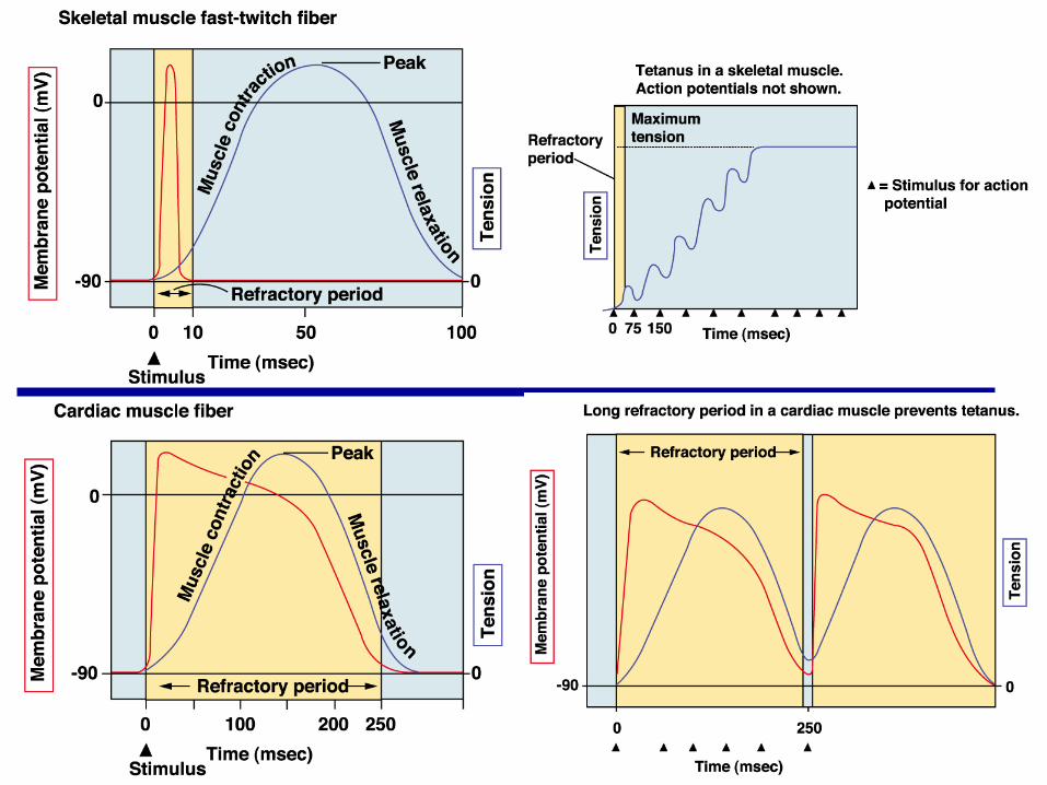
Slow response AP
Fast response AP

Transmembrane potential recorded from
a fast-response (left) and a slow-response (right) cardiac fiber
in isolated cardiac tissue immersed in an electrolyte solution.
ERP, effective refractory period;
RRP, relative refractory period.

Effect of changes in external [K+] on the transmembrane action potentials recorded from a Purkinje fiber. Stimulus artifact (St) - to the left of the upstroke of the action potential.
Fast responses may change to slow responses under certain pathological conditions (e.g., ischemia [K+] rises in the interstitial fluid because
inadequate perfusion).

atrium
Purkinje fb. ventricles
Me
mb
ran
e p
ote
ntia
l(m
V)
0
-50
-100
0
-50
-100
300 ms
• Normal atrium and ventricular myocardium
• Purkinje fibers in the specialized conductive system
Fast Action Potentials in the myocardium
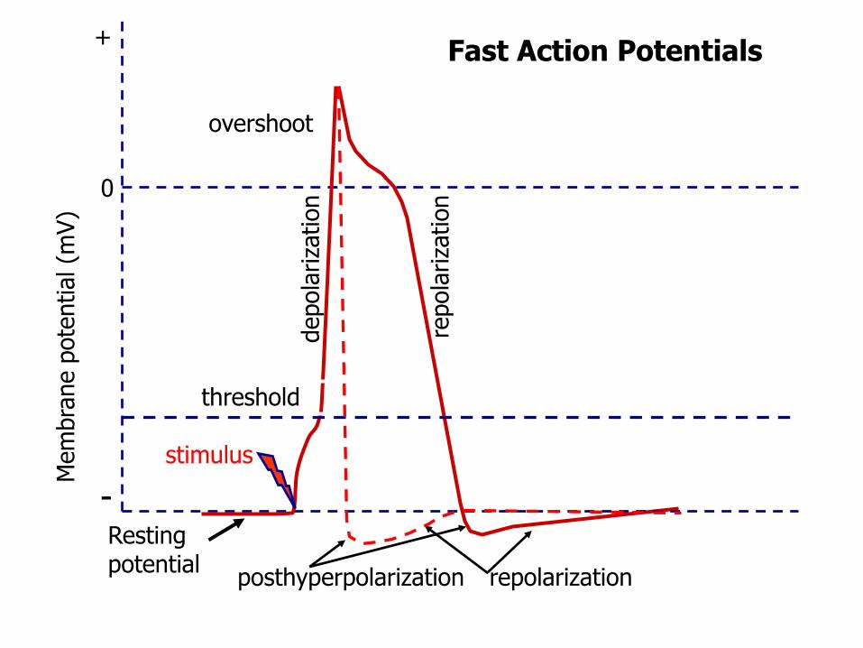
Restingpotential
Mem
bra
ne p
ote
ntial(m
V)
-
+
0
depola
riza
tion
threshold
overshoot
repolarization
repola
riza
tion
posthyperpolarization
Fast Action Potentials
stimulus

AP Phases
0 50 100 150 200 250 300 ms
Mem
bra
ne P
ote
ntial(m
V)
0
-50
-100
0
21
3
4 4
AP Phases: 0- depolarization/upstroke of the AP; 1-initial repolarization; 2-plateau; 3-repolarization; 4- resting membrane potential

0 0.15 0.30
Time (sec.)
Mem
bra
ne
Pote
nti
al
(mV
)
0
-50
-100
10
1.0
0.1
PNa+
PK+
PCa2+
AP
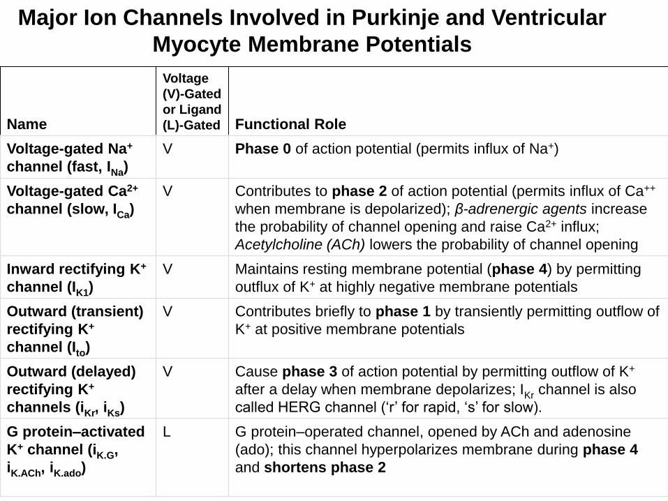
Name
Voltage
(V)-Gated
or Ligand
(L)-Gated Functional Role
Voltage-gated Na+
channel (fast, INa)
V Phase 0 of action potential (permits influx of Na+)
Voltage-gated Ca2+
channel (slow, ICa)
V Contributes to phase 2 of action potential (permits influx of Ca++
when membrane is depolarized); β-adrenergic agents increase
the probability of channel opening and raise Ca2+ influx;
Acetylcholine (ACh) lowers the probability of channel opening
Inward rectifying K+
channel (IK1)
V Maintains resting membrane potential (phase 4) by permitting
outflux of K+ at highly negative membrane potentials
Outward (transient)
rectifying K+
channel (Ito)
V Contributes briefly to phase 1 by transiently permitting outflow of
K+ at positive membrane potentials
Outward (delayed)
rectifying K+
channels (iKr, iKs)
V Cause phase 3 of action potential by permitting outflow of K+
after a delay when membrane depolarizes; IKr channel is also
called HERG channel (‘r’ for rapid, ‘s’ for slow).
G protein–activated
K+ channel (iK.G,
iK.ACh, iK.ado)
L G protein–operated channel, opened by ACh and adenosine
(ado); this channel hyperpolarizes membrane during phase 4
and shortens phase 2
Major Ion Channels Involved in Purkinje and Ventricular
Myocyte Membrane Potentials
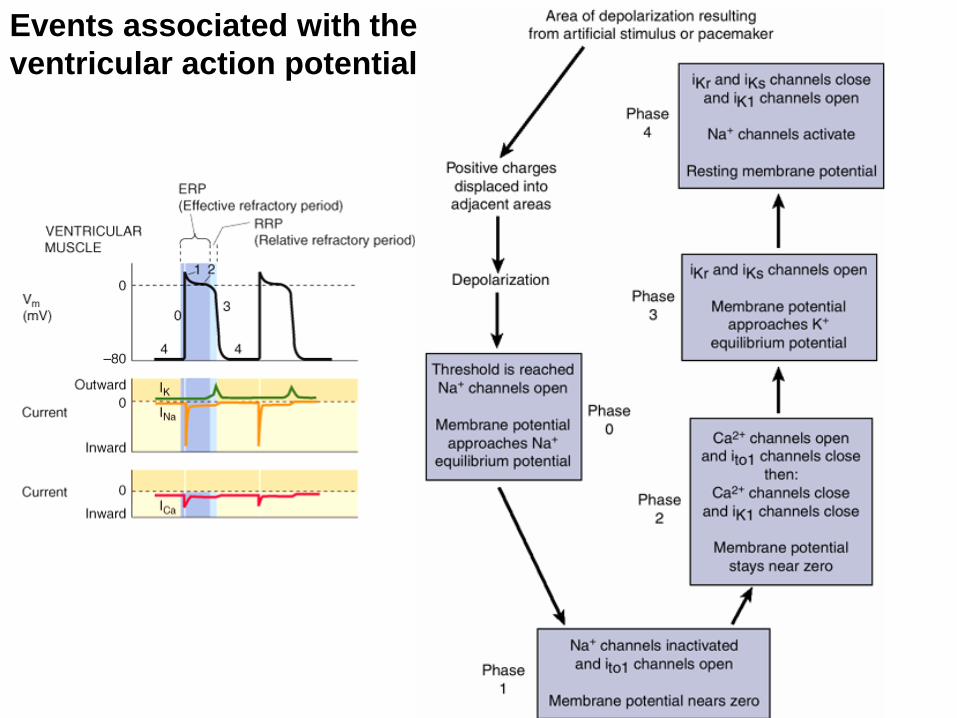
Events associated with the
ventricular action potential

0 50 100 150 200 250 300 ms
Mem
bra
ne
Pote
nti
al(m
V)
0
-50
-100
ERPRRP
Refractory periods
ERP/ARP-effective/absolute refractory period;
RRP-relative refractory period

Changes in action potential amplitude and slope of the upstroke as premature (P) action potentials are initiated at different stages of the relative refractory period of the preceding excitation in a fast-response fiber (bar = 100 msec).

Premature contraction
P
early
delayed
AP – electrical activity
Contraction – mechanical activity
P- premature contraction
Compensatory pause

Myocardial Properties
Excitability (bathmotropia)
Rhythmic activity /automaticity (chronotropia)
Conductibility (dromotropia)
Contractility (inotropia)
Relaxation (lusitropia)

Cardiac muscle cells contract without nervous stimulation
- 1% from the myocardium: pacemaker/autorhythmic cells
(99% contractile cells)
- Pacemaker cells: organized in a specialized excitatory & conductive system
- anatomically distinct
smaller, contain few contractile fibers
- electrogenic system: EXCITABILITY, CHRONOTROPISM generate AP spontaneously, rhythmically
- set the rate of the heart beat: CONDUCTIVITY
rapidly conduct APs throughout the heart generate rhythmical contraction

Specialized excitatory & conductive system
1. S-A Node: pacemaker of the heart in normal conditions;
- exhibits automaticity: generate AP at a higher rate than the AV node andHis-Purkinje system (latent pace-makers);
- located in the superior posterolat. wall of the RA; ellipsoid shape: 15/3/1 mm
- P cells: do not have a constant resting potential; membrane permeability to
Na and Ca during diastole; inward Ca current during upstroke of AP.

2. Internodal & interatrial pathways
3. A–V Node: inward Ca current during upstroke of AP;
here an AP delay of 0.1 sec (role!)
4. AV bundle (His-Purkinje)
- intercalated disks, gap junctions
- left & right branches of the AV bundle
5. Purkinje fibres: distribute to the endocardium, causes ventricles
to contract, from bottom up, pushing blood out top of heart

Action Potential (AP) in pacemaker cells
Phases of SAN action potential.
The records in this figure are idealized. IK, INa, ICa,
and If are currents through K+, Na+, Ca2+, and
nonselective cation channels, respectively.

You can dissolve an embryonic heart into its individual cell types with
trypsin, an enzyme that destroys the protein glue between the cells. Plate
these cells in a dish and you will see some cells - called myocytes - that
beat independently, some faster, some slower, as long as they do not touch
one another. The cells shown here are from the chick embryo.
http://www.cellsalive.com/myocyte.htm
Autorhythmic cells exhibit PACEMAKER POTENTIALS

Modulation of pacemaker activity
to decrease the heart rate:
A. Prolonged slow depolarization
B. Hyperpolarization (a more negative
resting potential)
C. Threshold shift towards a more
positive value

Myocardial Properties
Excitability (bathmotropia)
Rhythmic activity /automaticity (chronotropia)
Conductibility (dromotropia)
Contractility (inotropia)
Relaxation (lusitropia)

Myocardial conductivity = DROMOTROPIAin a dish… in the heart…
After two or three days in vitro, the myocytes form interconnected sheets of
cells (monolayers) that beat in unison. Pores (gap junctions) open between
adjacent touching cells, making their cytoplasms interconnected. These gap
junctions ensure the synchronous activity of the connected cells.

Myocardial conductivity = DROMOTROPIA
1. S-A Node area
2. Internodal & interatrial pathways
3. A–V Node:
inward Ca current duringupstroke of AP; AP delay 0.1 s
4. AV bundle (His-Purkinje)- intercalated disks, gapjunctions- split into left & right branches
5. Purkinje fibres: causes ventricles to contract, from bottom up, pushing blood out top of heart

1. Atria contraction precedes ventricles contraction, because of AV nodal delay:
- the impulse travels rather slowly through AV node (0.09 sec) & penetrating part of the
AV bundle (0,04 sec) (cause of the delay: less gap junctions…)
2. Both atria and ventricles should contract as a unit
-the impulse spreads so rapidly through the conducting system that all myocardial cells in
the atria and ventricles, respectively, contract at about the same time.
The myocardial conductivity is needed for an efficient pumping

The AV node
• composed of three functional regions:
1. the AN region - the transitional zone between the atrium and the remainder of the node;
2. the N region - the middle part of the AV node
3. the NH region, in which the nodal fibers gradually merge with the bundle of His
• normally, the AV node and bundle of His are the only pathways along which the cardiac impulse travels from atria to ventricles
- accessory AV pathways - reentry loops

Organization of the A-V node. The numbers represent the interval of time (sec) from the origin of the impulse in the sinus node.

Conduction velocity• Reflects the time required for excitation to spread from SAN
to the entire cardiac tissue
• Fastest in the Purkinje system, slowest in AVN (important for ventricular filling…)
– 0.02 to 0.1 m/sec in SA & AV nodes
– 1 m/sec. in internodal & interatrial anterior pathways
– 0.3-0.5 m/sec in A & V muscle
– 1.5 - 4 m/sec in Purkinje fibers
longer fibers, distributed in 1/3 of ventricular volume
gap junctions (no., permeability…),
direct connection with myocytes
fast Na currents, “regenerative spread of conducted AP”
rapid conduction of cardiac impulse

Atrioventricular and ventricular conduction system. Purkinje network distribution.

Conduction velocity
SAN / AVN: 0.02 to 0.1 m/sec
Internodal&interatrial anterior
pathways: 1m/sec.
AV delay: ~ 0.1 sec
AV bundle
Purkinje fibers: 1.5 - 4 m/sec
EndocardiumEpicardium
(in A&V mm): 0.3-0.5 m/sec
Transmission of the cardiac impulse through theheart, showing the time of appearance (infractions of a second after initial appearance atthe sinoatrial node) in different parts of the heart.
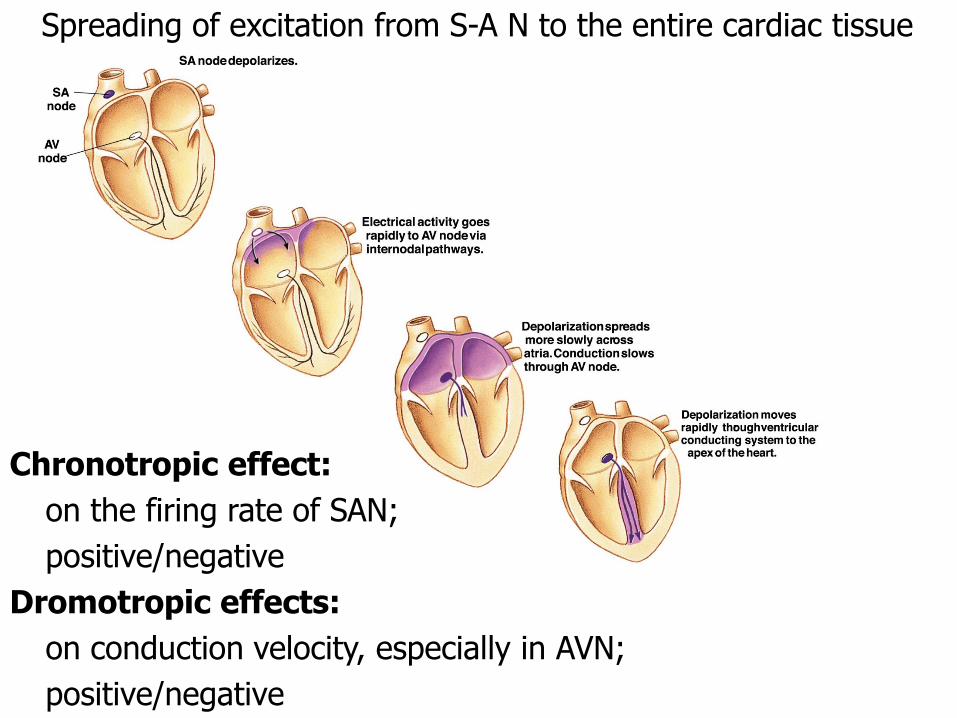
Spreading of excitation from S-A N to the entire cardiac tissue
Chronotropic effect:
on the firing rate of SAN;
positive/negative
Dromotropic effects:
on conduction velocity, especially in AVN;
positive/negative

Distribution of the nervous fibers

Modulation of the heart rate by the autonomic nervous system

PS vagal innervation Ach mediated
Muscarinic receptors on SAN, atria, AVN
Negative chronotropic effect: ↓ If , delayed slow depolarization↓ heart rate (sinus bradycardia)
Negative dromotropic effect: ↓ inward Ca current
↓ conduction velocity through AVN
AP are conducted more slowly from A to V
S innervationnorepinephrine mediated (also epinephrine - adrenal medulla)
b1 receptors
Positive chronotropic effect: ↑ If , ↑ heart rate (sinus tachycardia)
Positive dromotropic effect: ↑ inward Ca current↑ conduction velocity through AVN
! ventricular filling
Autonomic effects on automatism and
conduction velocity