Embed Size (px)
Citation preview
Developmental coxa vara is a hip deformity characterized by a defect in endochondral ossification of the medial por-tion of the femoral neck, together with the progressive ver-tical inclination of the proximal femoral physeal plate and shortening and decrease of neck shaft angle.1) The natural
history of coxa vara may be debilitating as the child devel-ops progressive limb length discrepancy, limp pain, abduc-tor weakness, and restricted motion. Secondary acetabular dysplasia and genu valgum may compound the problem. With the exception of some forms of developmental coxa vara that can resolve itself, a variety of surgical methods have been developed to deal with progressive coxa vara.2) There is a general agreement in the literature that a valgus osteotomy at a trochanteric or subtrochanteric level is the most definitive method for achieving the surgical correc-tion, which means the correction of the neck shaft angle and horizontal reorientation of the growth plate. Never-
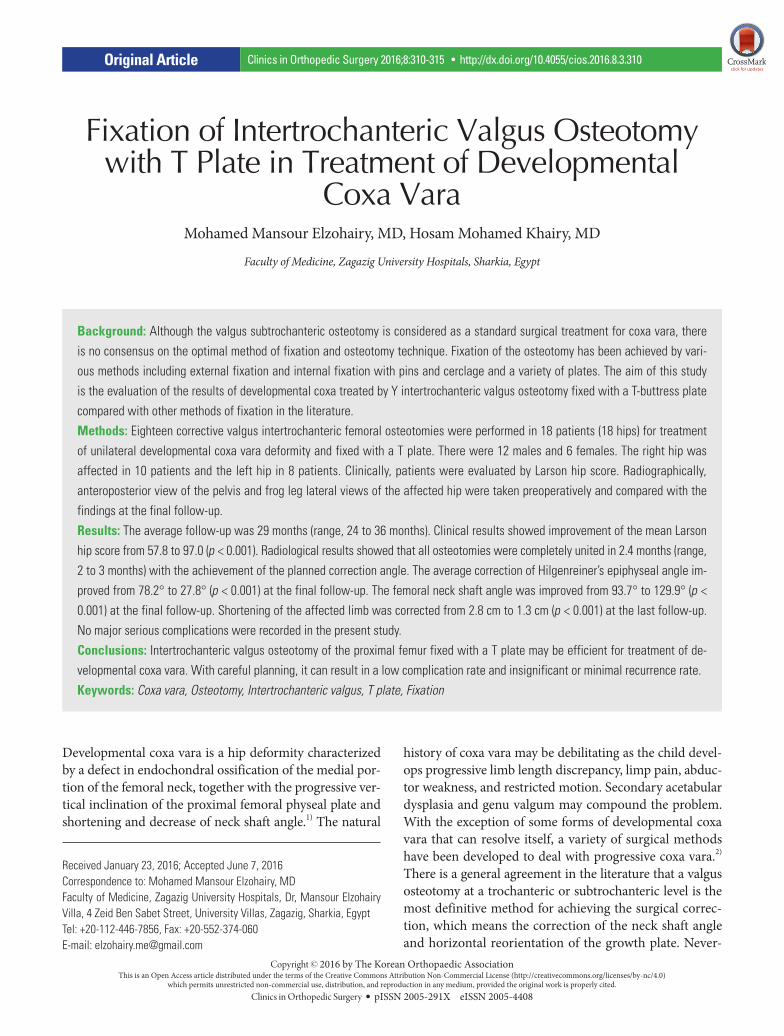
Fixation of Intertrochanteric Valgus Osteotomy with T Plate in Treatment of Developmental
Coxa VaraMohamed Mansour Elzohairy, MD, Hosam Mohamed Khairy, MD
Faculty of Medicine, Zagazig University Hospitals, Sharkia, Egypt
Background: Although the valgus subtrochanteric osteotomy is considered as a standard surgical treatment for coxa vara, there is no consensus on the optimal method of fixation and osteotomy technique. Fixation of the osteotomy has been achieved by vari-ous methods including external fixation and internal fixation with pins and cerclage and a variety of plates. The aim of this study is the evaluation of the results of developmental coxa treated by Y intertrochanteric valgus osteotomy fixed with a T-buttress plate compared with other methods of fixation in the literature. Methods: Eighteen corrective valgus intertrochanteric femoral osteotomies were performed in 18 patients (18 hips) for treatment of unilateral developmental coxa vara deformity and fixed with a T plate. There were 12 males and 6 females. The right hip was affected in 10 patients and the left hip in 8 patients. Clinically, patients were evaluated by Larson hip score. Radiographically, anteroposterior view of the pelvis and frog leg lateral views of the affected hip were taken preoperatively and compared with the findings at the final follow-up.Results: The average follow-up was 29 months (range, 24 to 36 months). Clinical results showed improvement of the mean Larson hip score from 57.8 to 97.0 (p < 0.001). Radiological results showed that all osteotomies were completely united in 2.4 months (range, 2 to 3 months) with the achievement of the planned correction angle. The average correction of Hilgenreiner’s epiphyseal angle im-proved from 78.2° to 27.8° (p < 0.001) at the final follow-up. The femoral neck shaft angle was improved from 93.7° to 129.9° (p < 0.001) at the final follow-up. Shortening of the affected limb was corrected from 2.8 cm to 1.3 cm (p < 0.001) at the last follow-up. No major serious complications were recorded in the present study. Conclusions: Intertrochanteric valgus osteotomy of the proximal femur fixed with a T plate may be efficient for treatment of de-velopmental coxa vara. With careful planning, it can result in a low complication rate and insignificant or minimal recurrence rate.Keywords: Coxa vara, Osteotomy, Intertrochanteric valgus, T plate, Fixation
Original Article Clinics in Orthopedic Surgery 2016;8:310-315 • http://dx.doi.org/10.4055/cios.2016.8.3.310
Copyright © 2016 by The Korean Orthopaedic AssociationThis is an Open Access article distributed under the terms of the Creative Commons Attribution Non-Commercial License (http://creativecommons.org/licenses/by-nc/4.0)
which permits unrestricted non-commercial use, distribution, and reproduction in any medium, provided the original work is properly cited.Clinics in Orthopedic Surgery • pISSN 2005-291X eISSN 2005-4408
Received January 23, 2016; Accepted June 7, 2016Correspondence to: Mohamed Mansour Elzohairy, MD Faculty of Medicine, Zagazig University Hospitals, Dr, Mansour Elzohairy Villa, 4 Zeid Ben Sabet Street, University Villas, Zagazig, Sharkia, EgyptTel: +20-112-446-7856, Fax: +20-552-374-060 E-mail: [email protected]
311
Elzohairy and Khairy. Intertrochanteric Valgus Osteotomy with T Plate for Developmental Coxa VaraClinics in Orthopedic Surgery • Vol. 8, No. 3, 2016 • www.ecios.org
theless, there is no consensus in the literature regarding the details of osteotomy and fixation method.1-4) The pur-pose of the present study was to evaluate the short-term results of the intertrochanteric valgus Y osteotomy with internal fixation using a contoured T plate.
METHODS
Informed consent was obtained from all parents of the individual participants included in the present study. This prospective study was conducted from January 2010 to December 2013 at Zagazig University Hospitals. Eighteen corrective intertrochanteric valgus Y femoral osteotomies were performed in 18 patients (18 hips) for treatment of unilateral developmental coxa vara deformity fixed with a
T-buttress plate for stabilization. The main indications of intertrochanteric valgus Y femoral osteotomies were the presence of shortening and the inferomedial triangular fragment of the neck that was not clear in all cases. They were 12 males and 6 females. The right hip was affected in 10 patients and the left hip in 8 patients. The average age at first presentation was 6.1 years (range, 5 to 7 years). All the patients included in the study had developmental coxa vara. Any patients with coxa vara of other etiologies, congenital, dysplastic, or acquired as infection or trauma were excluded from the study. The patient’s chief com-plaints were limping with minimal or no pain. The signs that were revealed by physical examinations in all patients were short leg gait with an abductor lurch, positive Tren-delenburg test, and limitation of abduction and internal
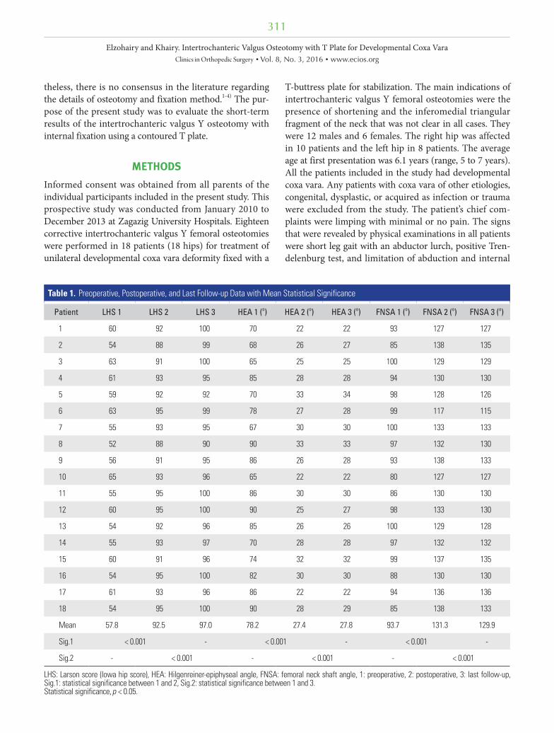
Table 1. Preoperative, Postoperative, and Last Follow-up Data with Mean Statistical Significance
Patient LHS 1 LHS 2 LHS 3 HEA 1 (o) HEA 2 (o) HEA 3 (o) FNSA 1 (o) FNSA 2 (o) FNSA 3 (o)
1 60 92 100 70 22 22 93 127 127
2 54 88 99 68 26 27 85 138 135
3 63 91 100 65 25 25 100 129 129
4 61 93 95 85 28 28 94 130 130
5 59 92 92 70 33 34 98 128 126
6 63 95 99 78 27 28 99 117 115
7 55 93 95 67 30 30 100 133 133
8 52 88 90 90 33 33 97 132 130
9 56 91 95 86 26 28 93 138 133
10 65 93 96 65 22 22 80 127 127
11 55 95 100 86 30 30 86 130 130
12 60 95 100 90 25 27 98 133 130
13 54 92 96 85 26 26 100 129 128
14 55 93 97 70 28 28 97 132 132
15 60 91 96 74 32 32 99 137 135
16 54 95 100 82 30 30 88 130 130
17 61 93 96 86 22 22 94 136 136
18 54 95 100 90 28 29 85 138 133
Mean 57.8 92.5 97.0 78.2 27.4 27.8 93.7 131.3 129.9
Sig.1 < 0.001 - < 0.001 - < 0.001 -
Sig.2 - < 0.001 - < 0.001 - < 0.001
LHS: Larson score (Iowa hip score), HEA: Hilgenreiner-epiphyseal angle, FNSA: femoral neck shaft angle, 1: preoperative, 2: postoperative, 3: last follow-up, Sig.1: statistical significance between 1 and 2, Sig.2: statistical significance between 1 and 3.Statistical significance, p < 0.05.
312
Elzohairy and Khairy. Intertrochanteric Valgus Osteotomy with T Plate for Developmental Coxa VaraClinics in Orthopedic Surgery • Vol. 8, No. 3, 2016 • www.ecios.org
rotation of the involved hip. The patients were evaluated clinically by Larson score (Iowa hip score).5) Anteropos-terior (AP) view of the pelvis was obtained to measure the Hilgenreiner-epiphyseal angle (HEA) and the femoral neck-shaft angle (FNSA) and the frog leg lateral view for the assessment of the femoral anteversion was taken pre-operatively, then monthly until 1-year postoperatively, and at the final follow-up. Limb scanograms for measurement of limb length discrepancy were done preoperatively and at the last follow-up in all patients.
All procedures performed in studies involving hu-man participants were in accordance with the ethical stan-dards of the institutional and/or national research com-mittee and with the 1964 Helsinki declaration and its later amendments or comparable ethical standards.
Statistical AnalysisStatistical analysis was performed using SPSS ver. 16.0 (SPSS Inc., Chicago, IL, USA). Wilcoxon signed-rank test was used for statistical analysis of the results and p < 0.05
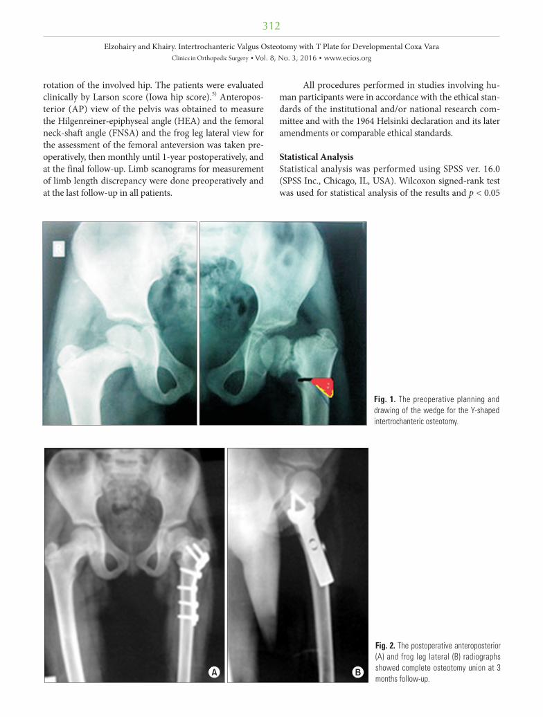
Fig. 1. The preoperative planning and drawing of the wedge for the Y-shaped inter trochanteric osteotomy.
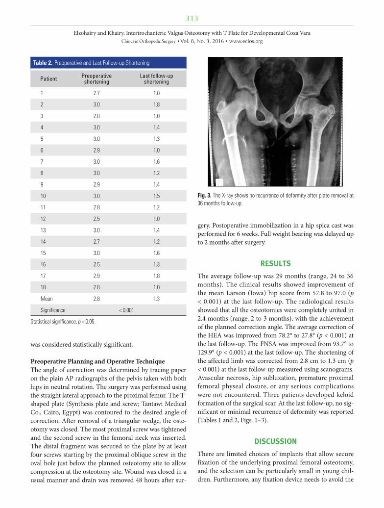
Fig. 2. The postoperative anteroposterior (A) and frog leg lateral (B) radiographs showed complete osteotomy union at 3 months follow-up.
A B
313
Elzohairy and Khairy. Intertrochanteric Valgus Osteotomy with T Plate for Developmental Coxa VaraClinics in Orthopedic Surgery • Vol. 8, No. 3, 2016 • www.ecios.org
was considered statistically significant.
Preoperative Planning and Operative Technique The angle of correction was determined by tracing paper on the plain AP radiographs of the pelvis taken with both hips in neutral rotation. The surgery was performed using the straight lateral approach to the proximal femur. The T-shaped plate (Synthesis plate and screw; Tantawi Medical Co., Cairo, Egypt) was contoured to the desired angle of correction. After removal of a triangular wedge, the oste-otomy was closed. The most proximal screw was tightened and the second screw in the femoral neck was inserted. The distal fragment was secured to the plate by at least four screws starting by the proximal oblique screw in the oval hole just below the planned osteotomy site to allow compression at the osteotomy site. Wound was closed in a usual manner and drain was removed 48 hours after sur-
gery. Postoperative immobilization in a hip spica cast was performed for 6 weeks. Full weight bearing was delayed up to 2 months after surgery.
RESULTS
The average follow-up was 29 months (range, 24 to 36 months). The clinical results showed improvement of the mean Larson (Iowa) hip score from 57.8 to 97.0 (p < 0.001) at the last follow-up. The radiological results showed that all the osteotomies were completely united in 2.4 months (range, 2 to 3 months), with the achievement of the planned correction angle. The average correction of the HEA was improved from 78.2° to 27.8° (p < 0.001) at the last follow-up. The FNSA was improved from 93.7° to 129.9° (p < 0.001) at the last follow-up. The shortening of the affected limb was corrected from 2.8 cm to 1.3 cm (p < 0.001) at the last follow-up measured using scanograms. Avascular necrosis, hip subluxation, premature proximal femoral physeal closure, or any serious complications were not encountered. Three patients developed keloid formation of the surgical scar. At the last follow-up, no sig-nificant or minimal recurrence of deformity was reported (Tables 1 and 2, Figs. 1–3).
DISCUSSION
There are limited choices of implants that allow secure fixation of the underlying proximal femoral osteotomy, and the selection can be particularly small in young chil-dren. Furthermore, any fixation device needs to avoid the
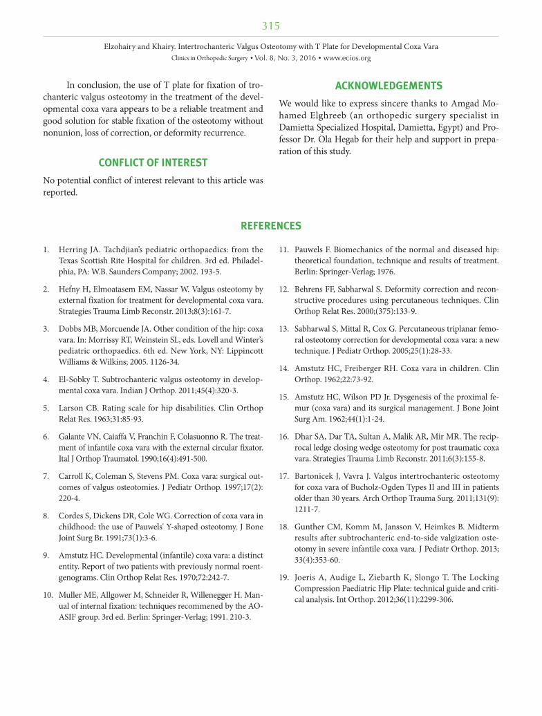
Table 2. Preoperative and Last Follow-up Shortening
Patient Preoperative shortening
Last follow-up shortening
1 2.7 1.0
2 3.0 1.8
3 2.0 1.0
4 3.0 1.4
5 3.0 1.3
6 2.9 1.0
7 3.0 1.6
8 3.0 1.2
9 2.9 1.4
10 3.0 1.5
11 2.8 1.2
12 2.5 1.0
13 3.0 1.4
14 2.7 1.2
15 3.0 1.6
16 2.5 1.3
17 2.9 1.8
18 2.8 1.0
Mean 2.8 1.3
Significance < 0.001
Statistical significance, p < 0.05.
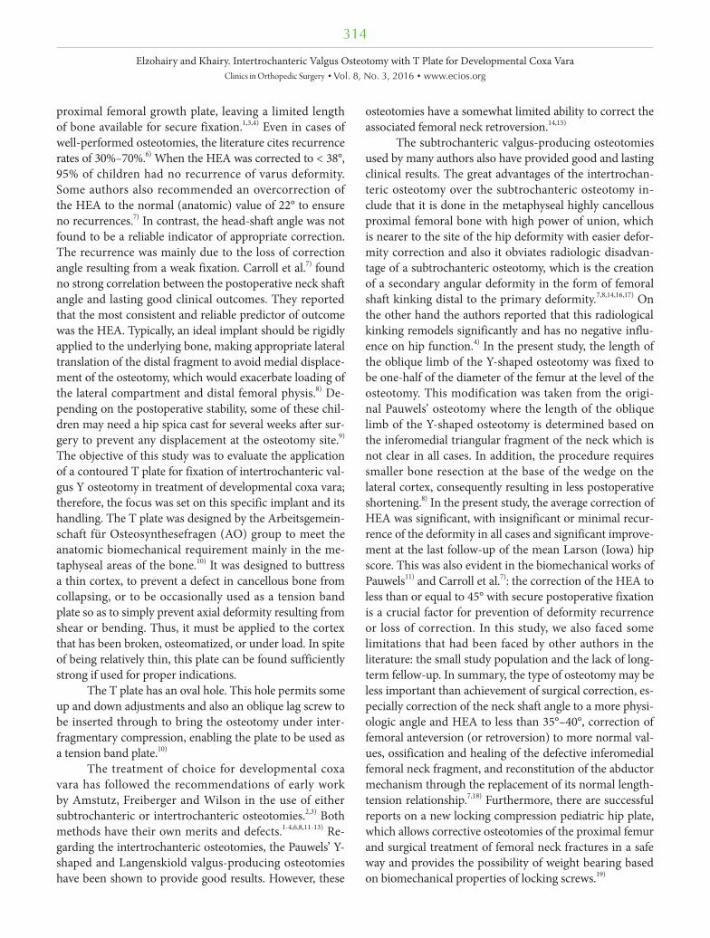
Fig. 3. The X-ray shows no recurrence of deformity after plate removal at 36 months follow-up.
314
Elzohairy and Khairy. Intertrochanteric Valgus Osteotomy with T Plate for Developmental Coxa VaraClinics in Orthopedic Surgery • Vol. 8, No. 3, 2016 • www.ecios.org
proximal femoral growth plate, leaving a limited length of bone available for secure fixation.1,3,4) Even in cases of well-performed osteotomies, the literature cites recurrence rates of 30%–70%.6) When the HEA was corrected to < 38°, 95% of children had no recurrence of varus deformity. Some authors also recommended an overcorrection of the HEA to the normal (anatomic) value of 22° to ensure no recurrences.7) In contrast, the head-shaft angle was not found to be a reliable indicator of appropriate correction. The recurrence was mainly due to the loss of correction angle resulting from a weak fixation. Carroll et al.7) found no strong correlation between the postoperative neck shaft angle and lasting good clinical outcomes. They reported that the most consistent and reliable predictor of outcome was the HEA. Typically, an ideal implant should be rigidly applied to the underlying bone, making appropriate lateral translation of the distal fragment to avoid medial displace-ment of the osteotomy, which would exacerbate loading of the lateral compartment and distal femoral physis.8) De-pending on the postoperative stability, some of these chil-dren may need a hip spica cast for several weeks after sur-gery to prevent any displacement at the osteotomy site.9) The objective of this study was to evaluate the application of a contoured T plate for fixation of intertrochanteric val-gus Y osteotomy in treatment of developmental coxa vara; therefore, the focus was set on this specific implant and its handling. The T plate was designed by the Arbeitsgemein-schaft für Osteosynthesefragen (AO) group to meet the anatomic biomechanical requirement mainly in the me-taphyseal areas of the bone.10) It was designed to buttress a thin cortex, to prevent a defect in cancellous bone from collapsing, or to be occasionally used as a tension band plate so as to simply prevent axial deformity resulting from shear or bending. Thus, it must be applied to the cortex that has been broken, osteomatized, or under load. In spite of being relatively thin, this plate can be found sufficiently strong if used for proper indications.
The T plate has an oval hole. This hole permits some up and down adjustments and also an oblique lag screw to be inserted through to bring the osteotomy under inter-fragmentary compression, enabling the plate to be used as a tension band plate.10)
The treatment of choice for developmental coxa vara has followed the recommendations of early work by Amstutz, Freiberger and Wilson in the use of either subtrochanteric or intertrochanteric osteotomies.2,3) Both methods have their own merits and defects.1-4,6,8,11-13) Re-garding the intertrochanteric osteotomies, the Pauwels’ Y-shaped and Langenskiold valgus-producing osteotomies have been shown to provide good results. However, these
osteotomies have a somewhat limited ability to correct the associated femoral neck retroversion.14,15)
The subtrochanteric valgus-producing osteotomies used by many authors also have provided good and lasting clinical results. The great advantages of the intertrochan-teric osteotomy over the subtrochanteric osteotomy in-clude that it is done in the metaphyseal highly cancellous proximal femoral bone with high power of union, which is nearer to the site of the hip deformity with easier defor-mity correction and also it obviates radiologic disadvan-tage of a subtrochanteric osteotomy, which is the creation of a secondary angular deformity in the form of femoral shaft kinking distal to the primary deformity.7,8,14,16,17) On the other hand the authors reported that this radiological kinking remodels significantly and has no negative influ-ence on hip function.4) In the present study, the length of the oblique limb of the Y-shaped osteotomy was fixed to be one-half of the diameter of the femur at the level of the osteotomy. This modification was taken from the origi-nal Pauwels’ osteotomy where the length of the oblique limb of the Y-shaped osteotomy is determined based on the inferomedial triangular fragment of the neck which is not clear in all cases. In addition, the procedure requires smaller bone resection at the base of the wedge on the lateral cortex, consequently resulting in less postoperative shortening.8) In the present study, the average correction of HEA was significant, with insignificant or minimal recur-rence of the deformity in all cases and significant improve-ment at the last follow-up of the mean Larson (Iowa) hip score. This was also evident in the biomechanical works of Pauwels11) and Carroll et al.7): the correction of the HEA to less than or equal to 45° with secure postoperative fixation is a crucial factor for prevention of deformity recurrence or loss of correction. In this study, we also faced some limitations that had been faced by other authors in the literature: the small study population and the lack of long-term fellow-up. In summary, the type of osteotomy may be less important than achievement of surgical correction, es-pecially correction of the neck shaft angle to a more physi-ologic angle and HEA to less than 35°–40°, correction of femoral anteversion (or retroversion) to more normal val-ues, ossification and healing of the defective inferomedial femoral neck fragment, and reconstitution of the abductor mechanism through the replacement of its normal length-tension relationship.7,18) Furthermore, there are successful reports on a new locking compression pediatric hip plate, which allows corrective osteotomies of the proximal femur and surgical treatment of femoral neck fractures in a safe way and provides the possibility of weight bearing based on biomechanical properties of locking screws.19)
315
Elzohairy and Khairy. Intertrochanteric Valgus Osteotomy with T Plate for Developmental Coxa VaraClinics in Orthopedic Surgery • Vol. 8, No. 3, 2016 • www.ecios.org
In conclusion, the use of T plate for fixation of tro-chanteric valgus osteotomy in the treatment of the devel-opmental coxa vara appears to be a reliable treatment and good solution for stable fixation of the osteotomy without nonunion, loss of correction, or deformity recurrence.
CONFLICT OF INTEREST
No potential conflict of interest relevant to this article was reported.
ACKNOWLEDGEMENTS
We would like to express sincere thanks to Amgad Mo-hamed Elghreeb (an orthopedic surgery specialist in Damietta Specialized Hospital, Damietta, Egypt) and Pro-fessor Dr. Ola Hegab for their help and support in prepa-ration of this study.
REFERENCES
1. Herring JA. Tachdjian’s pediatric orthopaedics: from the Texas Scottish Rite Hospital for children. 3rd ed. Philadel-phia, PA: W.B. Saunders Company; 2002. 193-5.
2. Hefny H, Elmoatasem EM, Nassar W. Valgus osteotomy by external fixation for treatment for developmental coxa vara. Strategies Trauma Limb Reconstr. 2013;8(3):161-7.
3. Dobbs MB, Morcuende JA. Other condition of the hip: coxa vara. In: Morrissy RT, Weinstein SL, eds. Lovell and Winter’s pediatric orthopaedics. 6th ed. New York, NY: Lippincott Williams & Wilkins; 2005. 1126-34.
4. El-Sobky T. Subtrochanteric valgus osteotomy in develop-mental coxa vara. Indian J Orthop. 2011;45(4):320-3.
5. Larson CB. Rating scale for hip disabilities. Clin Orthop Relat Res. 1963;31:85-93.
6. Galante VN, Caiaffa V, Franchin F, Colasuonno R. The treat-ment of infantile coxa vara with the external circular fixator. Ital J Orthop Traumatol. 1990;16(4):491-500.
7. Carroll K, Coleman S, Stevens PM. Coxa vara: surgical out-comes of valgus osteotomies. J Pediatr Orthop. 1997;17(2): 220-4.
8. Cordes S, Dickens DR, Cole WG. Correction of coxa vara in childhood: the use of Pauwels' Y-shaped osteotomy. J Bone Joint Surg Br. 1991;73(1):3-6.
9. Amstutz HC. Developmental (infantile) coxa vara: a distinct entity. Report of two patients with previously normal roent-genograms. Clin Orthop Relat Res. 1970;72:242-7.
10. Muller ME, Allgower M, Schneider R, Willenegger H. Man-ual of internal fixation: techniques recommened by the AO-ASIF group. 3rd ed. Berlin: Springer-Verlag; 1991. 210-3.
11. Pauwels F. Biomechanics of the normal and diseased hip: theoretical foundation, technique and results of treatment. Berlin: Springer-Verlag; 1976.
12. Behrens FF, Sabharwal S. Deformity correction and recon-structive procedures using percutaneous techniques. Clin Orthop Relat Res. 2000;(375):133-9.
13. Sabharwal S, Mittal R, Cox G. Percutaneous triplanar femo-ral osteotomy correction for developmental coxa vara: a new technique. J Pediatr Orthop. 2005;25(1):28-33.
14. Amstutz HC, Freiberger RH. Coxa vara in children. Clin Orthop. 1962;22:73-92.
15. Amstutz HC, Wilson PD Jr. Dysgenesis of the proximal fe-mur (coxa vara) and its surgical management. J Bone Joint Surg Am. 1962;44(1):1-24.
16. Dhar SA, Dar TA, Sultan A, Malik AR, Mir MR. The recip-rocal ledge closing wedge osteotomy for post traumatic coxa vara. Strategies Trauma Limb Reconstr. 2011;6(3):155-8.
17. Bartonicek J, Vavra J. Valgus intertrochanteric osteotomy for coxa vara of Bucholz-Ogden Types II and III in patients older than 30 years. Arch Orthop Trauma Surg. 2011;131(9): 1211-7.
18. Gunther CM, Komm M, Jansson V, Heimkes B. Midterm results after subtrochanteric end-to-side valgization oste-otomy in severe infantile coxa vara. J Pediatr Orthop. 2013; 33(4):353-60.
19. Joeris A, Audige L, Ziebarth K, Slongo T. The Locking Compression Paediatric Hip Plate: technical guide and criti-cal analysis. Int Orthop. 2012;36(11):2299-306.