Embed Size (px)
Citation preview
© Asian Journal of Biomedical and Pharmaceutical Sciences, 2015.
Five novel spectrophotometric methods for quantitative determination of Prulifloxacin in pure and pharmaceutical formulations
The main objective of this present work is to successfully develop one UV and four visible new spec-trophotometric techniques. One UV and four visible spectrophotometric methods have been devel-oped depending on the reactivity of various structural units for instance piperazine ring and tertiary amine and carbonyl group in Prulifloxacin. It is observed that the statistical analysis results of cor-relation coefficient of each method values are observed to be greater than 0.999 which speaks that the proposed methods have good linearity. The order of sensitivity of the said proposed methods are M1> M2>M5c> M5b> M5a> M4> M3. The % RSD values are observed bellow two for all methods which shows that all the proposed methods are precise. The results of analysis of the pharmaceutical formulations assert that the proposed methods are safely be adopted for routine quality c o n t r o l of Prulifloxacin in bulk and pharmaceutical preparations to yield correct and accurate analysis re-sults.Keywords: Prulifloxacin, Visible spectrophotometric methods, Bratton-Marshal reagent, Validation.
ABSTRACT :
Prulifloxacin (PRFX) pertains to prodrug of ulifloxacin which is broad spectrum fluoroquinolone an anti- bacterial agent. Prulifloxacin is metabolized in the body to the active compound in Ulifloxacin. Prulifloxacin is a prodrug and found to be effective on par with Ciprofloxacin, co-amoxiclav or Peflofloxacin in the treatment of bronchitis exacerbations and lower urinary tract infections. PRFX is not official drug in IP, BP and USP pharmacopoeia. In this connection relevant nemerous literature survey of PRFX was compiled and thoroughly examined before development of these methods to have adequate knowledge in this regard. Literature survey disclosed that very few spectrophotometric methods have been reported so far. Majority of HPLC methods were applied in determination of FQs in human plasma1-5, edible animal products, feeds and to a lesser extent in pharmaceutical formulations6-10. Capillary Zone Electrophoresis11 methods are in existence to determine the active metabolite of Prulifloxacin existed
in human plasma and some other biological fluids. It was also reported that there was a sensitive determination of Prulifloxacin owing to its fluorescence enhancement on Terbium (III)-Sodium Dodecylbenzene Sulfonate system12. There are no analytical reports available to estimate PRFX for utilizing visible spectrophotometry. Therefore the author inclined to select PRFX for the development of sensitive, precise and accurate spectrophotometric methods validated according to ICH Q2(R1) guidelines13, depending on several chemical reactions duly involving the analytically prominent functional groups existed in the structure. Thus the author chooses one ultraviolet and four visible spectrophotometric methods namely M1, M2, M3, M4, and M5a, M5b, M5c for determination of PRFX in bulk samples and pharmaceutical formulations. The broad details of different spectrophotometric methods developed are shown in Table 1.
*Corresponding author: P. RAVISANKAR, E-mail: [email protected]: +919000199106, +919059994000.
doi: 10.15272/ajbps.v5i48.724
INTRODUCTION:
Received on:11/07/2015Accepted on: 25/08/2015Published on: 15/09/2015
QR Code for mobile
Article Info:
Open Access
Literati
Research Article
Panchumarthy Ravisankar1*1Department of Pharmaceutical Analysis and Quality Assurance, Vignan Pharmacy College, Vadlamudi, Guntur (Dist.) - 522213, Andhra
Pradesh, India.1Faculty of Science, Sri Chandrasekharendra Saraswathi Viswa Maha Vidyalaya (SCSVMV University), Enathur, Kanchipuram – 631561, T.N.,
India.
Conflict of interest: Authors reported none
submit your manuscript | www.jbiopharm.com
P Ravishankar. Asian Journal of Biomedical and Pharmaceutical Sciences, 5(48), 2015, 01-13.
©Asian Journal of Biomedical and Pharmaceutical Sciences, 2015.02
Table 1. Spectrophotometric methods developed for the assay of PRFX in pharmaceutical formulations.Method No Reagent/ solvent Chemical principle involved λ max
(nm)
Beer’s law limits (µg/ml) Reference
M1 0.1N NaOH Ultra-violet absorption 275 1 - 10 Present chapter
M2 Ce (IV)/MBTH Oxidative coupling 580 1 - 5 Present chapter
M3 FC Redox reaction 700 10 - 50 Present chapter
M4 BTB Ion-association complex formation 420 5 - 25 Present
chapterM5a Fe(III)/PTL Oxidation followed by complex
formation 520 5 - 30 Present chapter
M5b Fe(III)/BPN Oxidation followed by complex formation 520 4 - 30 Present
chapterM5c Fe(III)/BPTL Oxidation followed by complex
formation 600 2 - 10 Present chapter
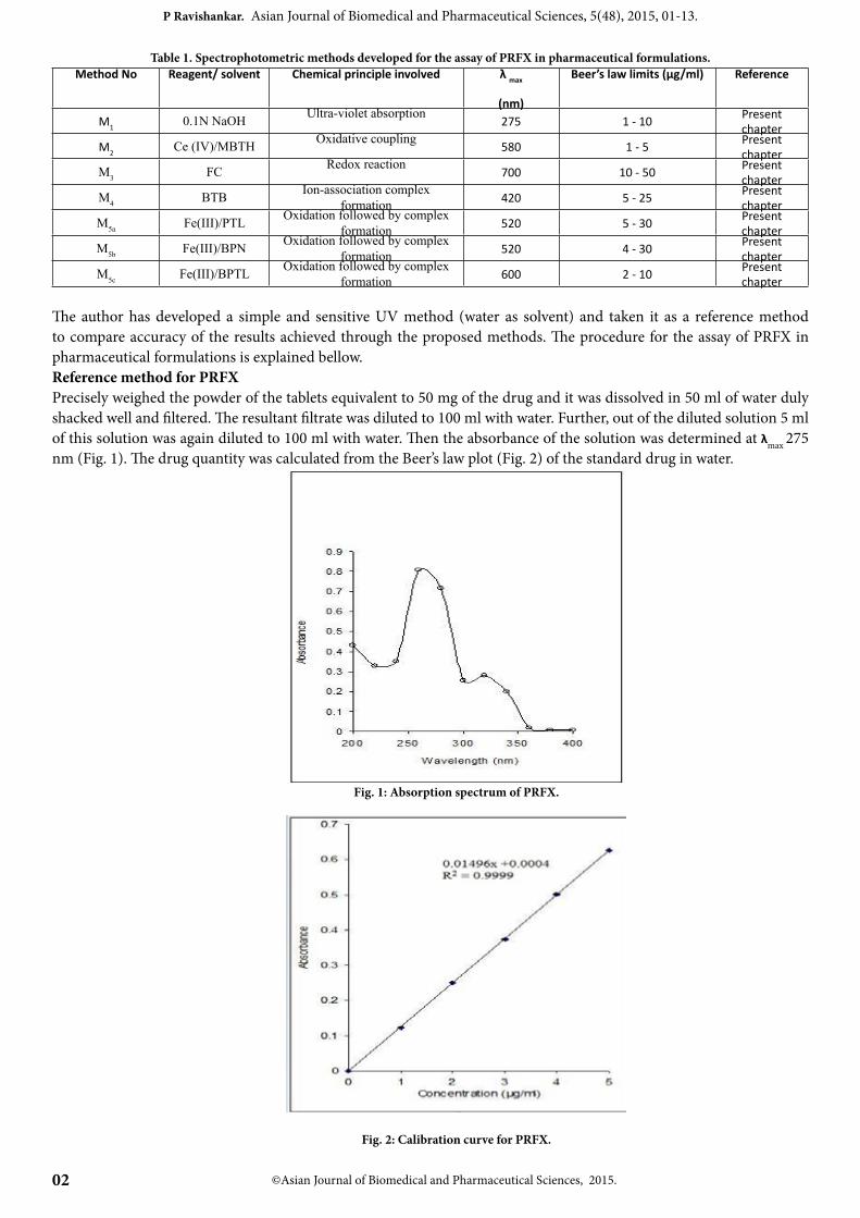
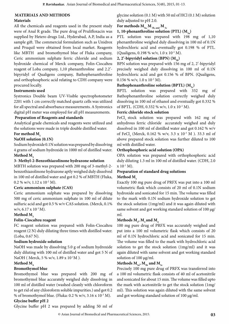
The author has developed a simple and sensitive UV method (water as solvent) and taken it as a reference method to compare accuracy of the results achieved through the proposed methods. The procedure for the assay of PRFX in pharmaceutical formulations is explained bellow.Reference method for PRFXPrecisely weighed the powder of the tablets equivalent to 50 mg of the drug and it was dissolved in 50 ml of water duly shacked well and filtered. The resultant filtrate was diluted to 100 ml with water. Further, out of the diluted solution 5 ml of this solution was again diluted to 100 ml with water. Then the absorbance of the solution was determined at λmax 275 nm (Fig. 1). The drug quantity was calculated from the Beer’s law plot (Fig. 2) of the standard drug in water.
Fig. 1: Absorption spectrum of PRFX.
Fig. 2: Calibration curve for PRFX.
03
P. Ravishankar. Asian Journal of Biomedical and Pharmaceutical Sciences, 5(48), 2015, 01-13.
© Asian Journal of Biomedical and Pharmaceutical Sciences, 2015.
MATERIALS AND METHODSMaterialsAll the chemicals and reagents used in the present study were of Anal R grade. The pure drug of Prulifloxacin was supplied by Hetero drugs Ltd., Hyderabad, A.P, India as a sample gift. The commercial formulation such as Unidrox and Pruquil were obtained from local market. Reagents like MBTH and bromothymol blue of Fluka company, Ceric ammonium sulphate ferric chloride and sodium hydroxide chemical of Merck company, Folin-Ciocalteu reagent of Loba company, 1,10 phenanthroline and 2.2’- bipyridyl of Qualigens company, Bathophenanthroline and orthophosphoric acid relating to CDH company were procured locally. Instruments usedSystronics Double beam UV-Visible spectrophotometer 2201 with 1 cm correctly matched quartz cells was utilized for all spectral and absorbance measurements. A Systronics digital pH meter was employed for all pH measurements. Preparation of Reagents and standards Analytical grade chemicals and reagents were utilized and the solutions were made in triple double distilled water.For method M1NaOH solution (0.1N)Sodium hydroxide 0.1N solution was prepared by dissolving 4 grams of sodium hydroxide in 1000 ml of distilled water.Method M23- Methyl-2-Benzothiazolinone hydrazone solution MBTH solution was prepared with 200 mg of 3-methyl-2-benzothiazolinone hydrazone aptly weighed duly dissolved in 100 ml of distilled water and got 0.2 % of MBTH (Fluka, 0.2 % w/v, 1.12 x 10⁻² M).Ceric ammonium sulphate (CAS)Ceric ammonium sulphate was prepared by dissolving 500 mg of ceric ammonium sulphate in 100 ml of dilute sulfuric acid and got 0.5 % w/v CAS solution. (Merck, 0.1% w/v, 6.17 x 10⁻³ M).Method M3 Folin-Ciocalteu reagentFC reagent solution was prepared with Folin-Ciocalteu reagent (2 N) duly diluting three times with distilled water. (Loba, 0.67 N).Sodium hydroxide solutionNaOH was made by dissolving 5.0 g of sodium hydroxide duly diluting with 100 ml of distilled water and got 5 N of NaOH ( Merck, 5 % w/v, 1.89 x 10⁻¹M ).Method M4Bromothymol blue Bromothymol blue was prepared with 200 mg of bromothymol blue accurately weighed duly dissolving in 100 ml of distilled water (washed cleanly with chloroform to get rid of any chloroform soluble impurities.) and got 0.2 % of bromothymol blue. (Fluka: 0.2 % w/v, 3.16 x 10⁻³ M).Glycine buffer pH 2Glycine buffer pH 2 was prepared by adding 50 ml of
glycine solution (0.1 M) with 50 ml of HCl (0.1 M) solution duly adjusted to pH 2.0.For methods M5a, M 5b, and M 5c1, 10-phenanthroline solution (PTL) (M5a) PTL solution was prepared with 198 mg of 1,10 phenanthroline weighed duly dissolving in 100 ml of 0.1N hydrochloric acid and eventually got 0.198 % of PTL. (Qualigens, 0.198 % w/v, 1.0 x 10⁻² M).2, 2’-bipyridyl solution (BPN) (M5b)BPN solution was prepared with 156 mg of 2, 2’-bipyridyl precisely weighed duly dissolving in 100 ml of 0.1N hydrochloric acid and got 0.156 % of BPN. (Qualigens, 0.156 % w/v, 1.0 x 10⁻² M).Bathophenanthroline solution (BPTL) (M5c)BPTL solution was prepared with 332 mg of Bathophenanthroline solution correctly weighed duly dissolving in 100 ml of ethanol and eventually got 0.332 % of BPTL. (CDH, 0.332 % w/v, 1.0 x 10⁻² M).Ferric chloride stock solutionFeCl3 stock solution was prepared with 162 mg of anhydrous ferric chloride accurately weighed and duly dissolved in 100 ml of distilled water and got 0.162 % w/v of FeCl3 (Merck, 0.162 % w/v, 3.3 x 10⁻³ M ). 33.3 ml of above prepared stock solution was further diluted to 100 ml with distilled water.Orthophosphoric acid solution (OPA)OPA solution was prepared with orthophosphoric acid duly diluting 1.3 ml in 100 ml of distilled water. (CDH, 2.0 x 10⁻¹ M).Preparation of standard drug solutionsMethod M1Exactly 100 mg pure drug of PRFX was put into a 100 ml volumetric flask which consists of 20 ml of 0.1N sodium hydroxide and sonicated for 15 min. The volume was filled to the mark with 0.1N sodium hydroxide solution to get the stock solution (1mg/ml) and it was again diluted with same solvent and got working standard solution of 100 µg/ml.Methods M2, M3 and M4100 mg pure drug of PRFX was accurately weighed and put into a 100 ml volumetric flask which consists of 20 ml of 0.1N hydrochloric acid and sonicated for 15 min. The volume was filled to the mark with hydrochloric acid solution to get the stock solution (1mg/ml) and it was again diluted with same solvent and got working standard solution of 100 µg/ml.Methods M5a, M5b and M5cPrecisely 100 mg pure drug of PRFX was transferred into a 100 ml volumetric flask consists of 40 ml of acetonitrile and sonicated for about 15 min. The volume was filled upto the mark with acetonitrile to get the stock solution (1mg/ml). This solution was again diluted with the same solvent and got working standard solution of 100 µg/ml.
©Asian Journal of Biomedical and Pharmaceutical Sciences, 2015.04
P Ravishankar. Asian Journal of Biomedical and Pharmaceutical Sciences, 5(48), 2015, 01-13.
Recommended proceduresFor bulk samplesMethod M1Standard drug aliquots of PRFX (0.1 - 0.5 ml, 100 µg/ml) solution in 0.1N sodium hydroxide were poured into a series of 10 ml volumetric flasks. The volumes were filled upto the mark with 0.1N sodium hydroxide and the absorbance of this solution was measured at 275 nm against solvent blank. The quantity of drug was calculated from calibration curve.Method M2Standard drug aliquots of PRFX (0.5 - 2.5 ml, 100 µg/ml) solution in 0.1N hydrochloric acid were shifted into a series of 10 ml volumetric flasks. To this 1.5 ml of ceric ammonium sulphate (0.5 % w/v) and 2 ml of MBTH was added and mixed thoroughly and the solution was allowed to react in the ambient temperature for 20 min. The bluish-green colored chromogen so formed was determined at 580 nm against the reagent blank. The quantity of PRFX was calculated from its linearity plot.Method M3Standard drug aliquots of PRFX (0.1 - 0.5 ml, 1000 µg/ml) solution in 0.1N hydrochloric acid were poured into a series of 10 ml volumetric flasks. To this 1ml of Folin-ciocaltaeu reagent as well as 2 ml of sodium hydroxide (5 % w/v) solution were added and shacked thoroughly and the reacted mixture was kept for 15 min. The blue colored chromogen so formed was estimated at 700 nm against the reagent blank. The quantity of PRFX was calculated from its calibration plot. Method M4Standard drug aliquots of PRFX (0.5 to 2.5 ml, 100 µg/ml ) solution in 0.1M HCl was transferred into a series of separating funnels of 125 ml and equalized with water and added 4ml of buffer pH 2.0 and 2 ml of bromothymol blue (0.2 % w/v) and then the solution was saturated with aqueous phase up to 10 ml. The reacted mixture was shaken for 5 min and kept it duly to settle. Then the drug-dye complex was extracted into 10ml of chloroform. The yellow colored so formed in organic layer was taken up for absorbance measurements and it was estimated at 420 nm against the reagent blank. The quantity of PRFX was calculated from its calibration plot. Methods M5a, M5b, and M5c:Standard drug aliquots of Prulifloxacin solution (0.5 to 2.5 ml, 100 µg/ml for M5a, 0.4 to 3.0 ml for M5b and 0.2 to1.0 ml, 100 µg/ml for M5c) transferred in to a series of 10 ml of volumetric flasks and the volume to each flask was made to 3 ml with acetonitrile. Then 1.5 ml of Fe (III) solution, 2 ml of PTL or BPN or BPTL solutions were added to each volumetric flasks. The said solutions were heated on a boiling water bath for thirty min. The above said solutions then cooled to room temperature then 2 ml of o-phosphoric acid solutions was added and the total volume was brought to 10 ml with acetonitrile. The absorbance values of
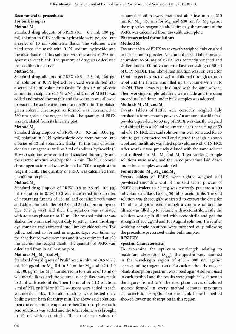
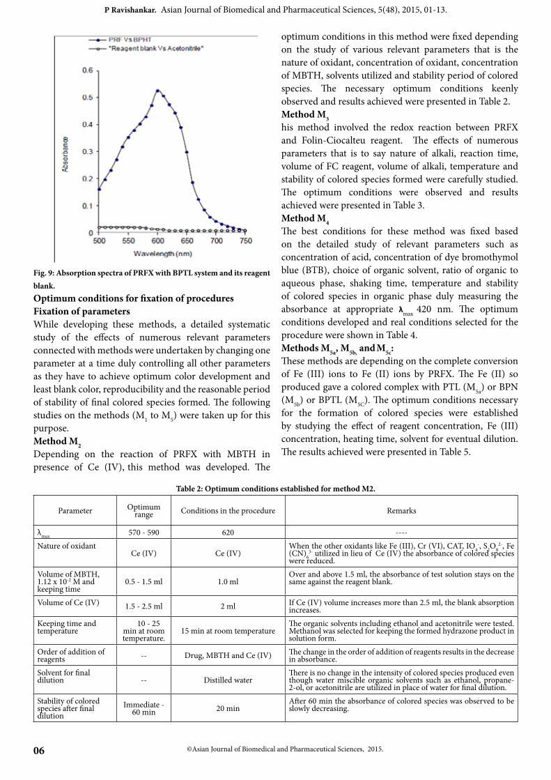
coloured solutions were measured after five min at 210 nm for M5a, 520 nm for M5b and 600 nm for M5c against the respective reagent blank. Ultimately the amount of the PRFX was calculated from the calibration plots. Pharmaceutical formulationsMethod M 1Twenty tablets of PRFX were exactly weighed duly crushed to form smooth powder. An amount of said tablet powder equivalent to 50 mg of PRFX was correctly weighed and shifted into a 100 ml volumetric flask consisting of 50 ml of 0.1N NaOH. The above said solution was sonicated for 15 min to get it extracted well and filtered through a cotton wool and the filtrate was filled up to volume with 0.1N NaOH. Then it was exactly diluted with the same solvent. Then working sample solutions were made and the same procedure laid down under bulk samples was adopted. Methods M2, M3 and M4 Twenty tablets of PRFX were correctly weighed duly crushed to form smooth powder. An amount of said tablet powder equivalent to 50 mg of PRFX was exactly weighed and shifted into a 100 ml volumetric flask consisting of 50 ml of 0.1N HCl. The said solution was well sonicated for 15 min to get it extracted well and filtered through a cotton wool and the filtrate was filled upto volume with 0.1N HCl. After words it was precisely diluted with the same solvent and utilized for M2, M3 and M4. Then working sample solutions were made and the same procedure laid down under bulk samples was adopted. For methods M5a, M5b, and M5cTwenty tablets of PRFX were rightly weighed and powdered smoothly. Out of the said tablet powder of PRFX equivalent to 50 mg was correctly put into a 100 ml volumetric flask having 50 ml of acetonitrile. The said solution was thoroughly sonicated to extract the drug for 15 min and got filtered through a cotton wool and the filtrate was filled up to volume with acetonitrile. Lastly the solution was again diluted with acetonitrile and got the strength of 100 µg/ml and 1000 µg/ml solution. There after working sample solutions were prepared duly following the procedure prescribed under bulk samples.RESULTSSpectral CharacteristicsTo determine the optimum wavelength relating to maximum absorption (λmax), the spectra were scanned in the wavelength region of 400 - 800 nm against corresponding reagent blank. For each method the reagent blank absorption spectrum was noted against solvent used in each method and the results were graphically shown in the Figures from 3 to 9. The absorption curves of colored species formed in every method denotes maximum characteristic absorption but the blank in each method showed low or no absorption in this region.
© Asian Journal of Biomedical and Pharmaceutical Sciences, 2015.05
P Ravishankar. Asian Journal of Biomedical and Pharmaceutical Sciences, 5(48), 2015, 01-13.
Fig. 3: Absorption spectra of PRFX with 0.1 N NaOH.
Fig. 4: Absorption spectra of PRFX with MBTH/Ce (IV) system and its reagent blank.
Fig. 5: Absorption spectra of PRFX with FC system and its reagent blank.
Fig. 6: Absorption spectra of PRFX with BTB system and its reagent blank.
Fig. 7: Absorption spectra of PRFX with PTL system and its reagent blank.
Fig. 8: Absorption spectra of PRFX with BPN system and its reagent blank.
©Asian Journal of Biomedical and Pharmaceutical Sciences, 2015.06
P Ravishankar. Asian Journal of Biomedical and Pharmaceutical Sciences, 5(48), 2015, 01-13.
Fig. 9: Absorption spectra of PRFX with BPTL system and its reagent blank.Optimum conditions for fixation of proceduresFixation of parametersWhile developing these methods, a detailed systematic study of the effects of numerous relevant parameters connected with methods were undertaken by changing one parameter at a time duly controlling all other parameters as they have to achieve optimum color development and least blank color, reproducibility and the reasonable period of stability of final colored species formed. The following studies on the methods (M1 to M5) were taken up for this purpose.Method M2Depending on the reaction of PRFX with MBTH in presence of Ce (IV), this method was developed. The
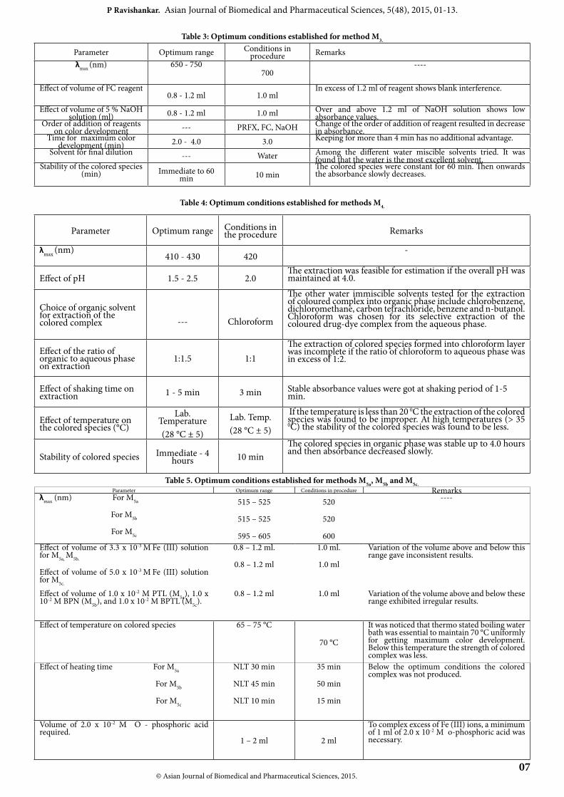
optimum conditions in this method were fixed depending on the study of various relevant parameters that is the nature of oxidant, concentration of oxidant, concentration of MBTH, solvents utilized and stability period of colored species. The necessary optimum conditions keenly observed and results achieved were presented in Table 2. Method M3his method involved the redox reaction between PRFX and Folin-Ciocalteu reagent. The effects of numerous parameters that is to say nature of alkali, reaction time, volume of FC reagent, volume of alkali, temperature and stability of colored species formed were carefully studied. The optimum conditions were observed and results achieved were presented in Table 3. Method M4The best conditions for these method was fixed based on the detailed study of relevant parameters such as concentration of acid, concentration of dye bromothymol blue (BTB), choice of organic solvent, ratio of organic to aqueous phase, shaking time, temperature and stability of colored species in organic phase duly measuring the absorbance at appropriate λmax 420 nm. The optimum conditions developed and real conditions selected for the procedure were shown in Table 4.Methods M5a, M5b, and M5c:These methods are depending on the complete conversion of Fe (III) ions to Fe (II) ions by PRFX. The Fe (II) so produced gave a colored complex with PTL (M5a) or BPN (M5b) or BPTL (M5C). The optimum conditions necessary for the formation of colored species were established by studying the effect of reagent concentration, Fe (III) concentration, heating time, solvent for eventual dilution. The results achieved were presented in Table 5.
Table 2: Optimum conditions established for method M2.
Parameter Optimum range Conditions in the procedure Remarks
λmax 570 - 590 620 ----Nature of oxidant
Ce (IV) Ce (IV)When the other oxidants like Fe (III), Cr (VI), CAT, IO4
-, S2O82-, Fe
(CN)63- utilized in lieu of Ce (IV) the absorbance of colored species
were reduced.Volume of MBTH, 1.12 x 10⁻² M and keeping time
0.5 - 1.5 ml 1.0 mlOver and above 1.5 ml, the absorbance of test solution stays on the same against the reagent blank.
Volume of Ce (IV) 1.5 - 2.5 ml 2 ml If Ce (IV) volume increases more than 2.5 ml, the blank absorption increases.
Keeping time and temperature
10 - 25 min at room temperature.
15 min at room temperatureThe organic solvents including ethanol and acetonitrile were tested. Methanol was selected for keeping the formed hydrazone product in solution form.
Order of addition of reagents -- Drug, MBTH and Ce (IV) The change in the order of addition of reagents results in the decrease
in absorbance.Solvent for final dilution -- Distilled water
There is no change in the intensity of colored species produced even though water miscible organic solvents such as ethanol, propane-2-ol, or acetonitrile are utilized in place of water for final dilution.
Stability of colored species after final dilution
Immediate - 60 min 20 min
After 60 min the absorbance of colored species was observed to be slowly decreasing.
© Asian Journal of Biomedical and Pharmaceutical Sciences, 2015. 07
P Ravishankar. Asian Journal of Biomedical and Pharmaceutical Sciences, 5(48), 2015, 01-13.
Table 3: Optimum conditions established for method M3.
Parameter Optimum range Conditions in procedure Remarks
λmax (nm) 650 - 750700
----
Effect of volume of FC reagent0.8 - 1.2 ml 1.0 ml
In excess of 1.2 ml of reagent shows blank interference.
Effect of volume of 5 % NaOH solution (ml) 0.8 - 1.2 ml 1.0 ml Over and above 1.2 ml of NaOH solution shows low absorbance values.
Order of addition of reagents on color development --- PRFX, FC, NaOH Change of the order of addition of reagent resulted in decrease
in absorbance.Time for maximum color
development (min) 2.0 - 4.0 3.0 Keeping for more than 4 min has no additional advantage.
Solvent for final dilution --- Water Among the different water miscible solvents tried. It was found that the water is the most excellent solvent.
Stability of the colored species (min) Immediate to 60
min 10 minThe colored species were constant for 60 min. Then onwards the absorbance slowly decreases.
Table 4: Optimum conditions established for methods M4.
Parameter Optimum range Conditions in the procedure Remarks
λmax (nm)410 - 430 420
-
Effect of pH 1.5 - 2.5 2.0The extraction was feasible for estimation if the overall pH was maintained at 4.0.
Choice of organic solvent for extraction of the colored complex --- Chloroform
The other water immiscible solvents tested for the extraction of coloured complex into organic phase include chlorobenzene, dichloromethane, carbon tetrachloride, benzene and n-butanol. Chloroform was chosen for its selective extraction of the coloured drug-dye complex from the aqueous phase.
Effect of the ratio of organic to aqueous phase on extraction
1:1.5 1:1The extraction of colored species formed into chloroform layer was incomplete if the ratio of chloroform to aqueous phase was in excess of 1:2.
Effect of shaking time on extraction 1 - 5 min 3 min Stable absorbance values were got at shaking period of 1-5
min.
Effect of temperature on the colored species (°C)
Lab. Temperature(28 °C ± 5)
Lab. Temp.(28 °C ± 5)
If the temperature is less than 20 0C the extraction of the colored species was found to be improper. At high temperatures (> 35 0C) the stability of the colored species was found to be less.
Stability of colored species Immediate - 4 hours 10 min
The colored species in organic phase was stable up to 4.0 hours and then absorbance decreased slowly.
Table 5. Optimum conditions established for methods M5a, M5b and M5c.Parameter Optimum range Conditions in procedure Remarks
λmax (nm) For M5a
For M5b
For M5c
515 – 525
515 – 525
595 – 605
520
520
600
----
Effect of volume of 3.3 x 10⁻³ M Fe (III) solution for M5a, M5b.
Effect of volume of 5.0 x 10⁻³ M Fe (III) solution for M5c.
0.8 – 1.2 ml.
0.8 – 1.2 ml
1.0 ml.
1.0 ml
Variation of the volume above and below this range gave inconsistent results.
Effect of volume of 1.0 x 10⁻² M PTL (M5a), 1.0 x 10⁻² M BPN (M5b), and 1.0 x 10⁻² M BPTL (M5c).
0.8 – 1.2 ml 1.0 ml Variation of the volume above and below these range exhibited irregular results.
Effect of temperature on colored species 65 – 75 °C
70 °C
It was noticed that thermo stated boiling water bath was essential to maintain 70 °C uniformly for getting maximum color development. Below this temperature the strength of colored complex was less.
Effect of heating time For M5a
For M5b
For M5c
NLT 30 min
NLT 45 min
NLT 10 min
35 min
50 min
15 min
Below the optimum conditions the colored complex was not produced.
Volume of 2.0 x 10⁻² M O - phosphoric acid required.
1 – 2 ml 2 ml
To complex excess of Fe (III) ions, a minimum of 1 ml of 2.0 x 10⁻² M o-phosphoric acid was necessary.
©Asian Journal of Biomedical and Pharmaceutical Sciences, 2015.08
P Ravishankar. Asian Journal of Biomedical and Pharmaceutical Sciences, 5(48), 2015, 01-13.
Table 5: (continued)
Parameter Optimum range Conditions in procedure Remarks
Order of addition of reagents on color development
For M5a
For M5b
For M5C
-
-
-
PRFX, Fe (III) and PTL before heating and phosphoric acid after heating.
PRFX, Fe (III) and BPN before heating and phosphoric acid after heating
PRFX, Fe (III) and BPTL before heating and phosphoric acid after heating.
Due to interchange of the order of drug, Fe (III) and PTL or BPN or BPTL no effect was found on the absorbance.
Effect of temperature (0C) 70 - 100 80 0C for 15 min. At room temperature no color was formed.
Nature of solvent for final dilution - Acetonitrile
It was observed that other solvents like methanol, acetone and dioxane did not increase the intensity of the eventual colored product.
Stability of colored species of the final dilution
Immediate 45 min
10 min
The absorbance of the colored product decreased gradually after 45 min.
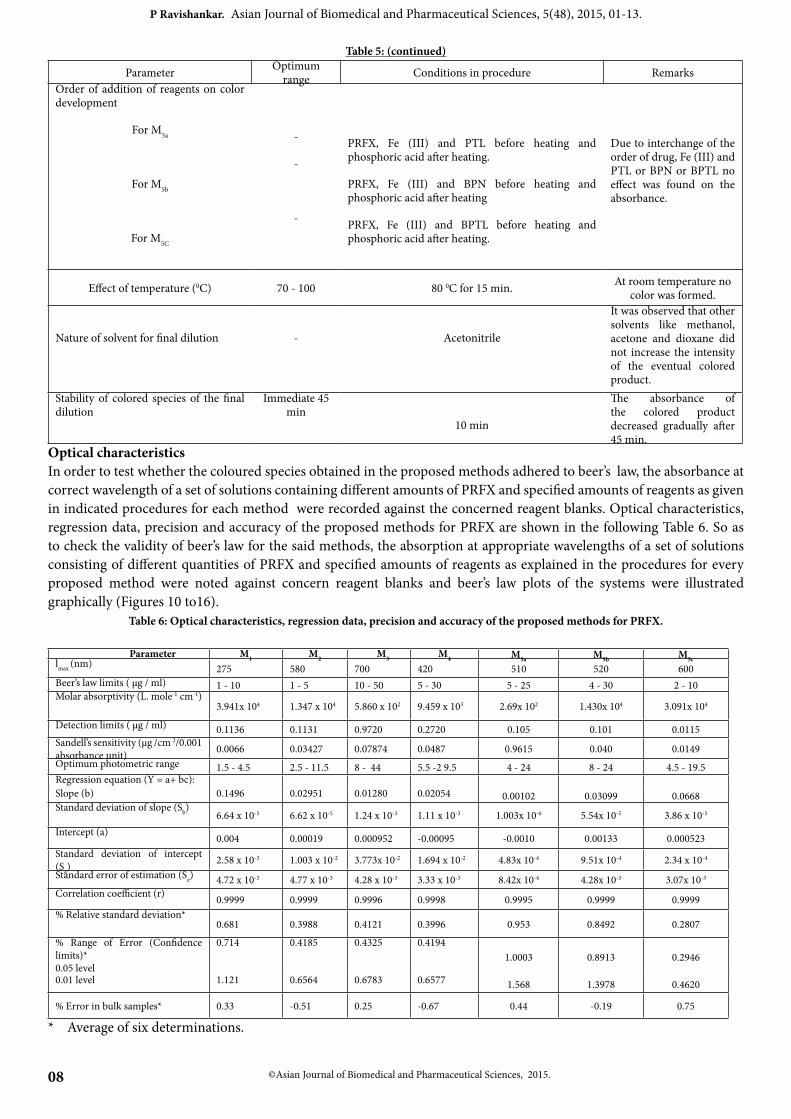
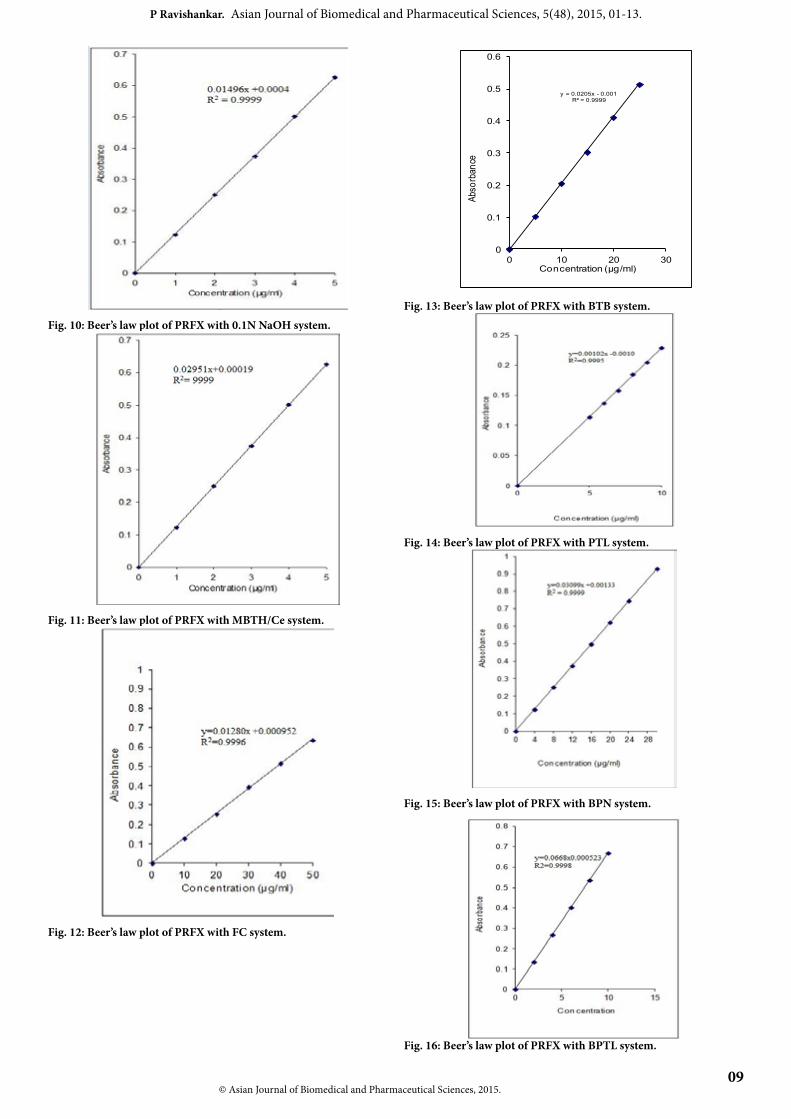
Optical characteristicsIn order to test whether the coloured species obtained in the proposed methods adhered to beer’s law, the absorbance at correct wavelength of a set of solutions containing different amounts of PRFX and specified amounts of reagents as given in indicated procedures for each method were recorded against the concerned reagent blanks. Optical characteristics, regression data, precision and accuracy of the proposed methods for PRFX are shown in the following Table 6. So as to check the validity of beer’s law for the said methods, the absorption at appropriate wavelengths of a set of solutions consisting of different quantities of PRFX and specified amounts of reagents as explained in the procedures for every proposed method were noted against concern reagent blanks and beer’s law plots of the systems were illustrated graphically (Figures 10 to16).
Table 6: Optical characteristics, regression data, precision and accuracy of the proposed methods for PRFX.
Parameter M1 M2 M3 M4 M5a M5b M5clmax (nm) 275 580 700 420 510 520 600Beer’s law limits ( μg / ml) 1 - 10 1 - 5 10 - 50 5 - 30 5 - 25 4 - 30 2 - 10Molar absorptivity (L. mole-1 cm-1)
3.941x 10⁴ 1.347 x 10⁴ 5.860 x 10² 9.459 x 10³ 2.69x 10² 1.430x 10⁴ 3.091x 10⁴
Detection limits ( μg / ml) 0.1136 0.1131 0.9720 0.2720 0.105 0.101 0.0115Sandell’s sensitivity (μg /cm 2/0.001 absorbance unit) 0.0066 0.03427 0.07874 0.0487 0.9615 0.040 0.0149Optimum photometric range 1.5 - 4.5 2.5 - 11.5 8 - 44 5.5 -2 9.5 4 - 24 8 - 24 4.5 - 19.5Regression equation (Y = a+ bc):Slope (b) 0.1496 0.02951 0.01280 0.02054 0.00102 0.03099 0.0668Standard deviation of slope (Sb) 6.64 x 10⁻⁵ 6.62 x 10⁻⁵ 1.24 x 10⁻³ 1.11 x 10⁻³ 1.003x 10⁻⁶ 5.54x 10⁻⁵ 3.86 x 10⁻⁵
Intercept (a) 0.004 0.00019 0.000952 -0.00095 -0.0010 0.00133 0.000523Standard deviation of intercept (Sa)
2.58 x 10⁻³ 1.003 x 10⁻² 3.773x 10⁻² 1.694 x 10⁻² 4.83x 10⁻⁴ 9.51x 10⁻⁴ 2.34 x 10⁻⁴
Standard error of estimation (Se) 4.72 x 10⁻³ 4.77 x 10⁻³ 4.28 x 10⁻³ 3.33 x 10⁻³ 8.42x 10⁻⁴ 4.28x 10⁻³ 3.07x 10⁻³
Correlation coefficient (r) 0.9999 0.9999 0.9996 0.9998 0.9995 0.9999 0.9999% Relative standard deviation*
0.681 0.3988 0.4121 0.3996 0.953 0.8492 0.2807
% Range of Error (Confidence limits)*0.05 level
0.714 0.4185 0.4325 0.41941.0003 0.8913 0.2946
0.01 level 1.121 0.6564 0.6783 0.6577 1.568 1.3978 0.4620
% Error in bulk samples* 0.33 -0.51 0.25 -0.67 0.44 -0.19 0.75
* Average of six determinations.
© Asian Journal of Biomedical and Pharmaceutical Sciences, 2015. 09
P Ravishankar. Asian Journal of Biomedical and Pharmaceutical Sciences, 5(48), 2015, 01-13.
Fig. 10: Beer’s law plot of PRFX with 0.1N NaOH system.
Fig. 11: Beer’s law plot of PRFX with MBTH/Ce system.
Fig. 12: Beer’s law plot of PRFX with FC system.
y = 0.0205x - 0.001R² = 0.9999
0
0.1
0.2
0.3
0.4
0.5
0.6
0 10 20 30
Abso
rban
ce
Concentration (µg/ml)
Fig. 13: Beer’s law plot of PRFX with BTB system.
Fig. 14: Beer’s law plot of PRFX with PTL system.
Fig. 15: Beer’s law plot of PRFX with BPN system.
Fig. 16: Beer’s law plot of PRFX with BPTL system.
10 ©Asian Journal of Biomedical and Pharmaceutical Sciences, 2015.
P Ravishankar. Asian Journal of Biomedical and Pharmaceutical Sciences, 5(48), 2015, 01-13.
Method validationPrecisionThe precision of every one out of 4 proposed visible spectrophotometric methods and one UV were decided individually from the absorbances values got by actual estimation of 6 replicates of a fixed quantity of PRFX in 10 ml solutions for each replicate and the precision of the method was examined. Accuracy of the methodBy getting aliquots consisting of known quantities of bulk as well as pharmaceutical formulations of PRFX duly estimating them based on the reported and proposed methods, the accuracy of the method was decided. To explain the suitability of the proposed method the recovery studies of the PRFX were held. Interference studiesThe effect of broad range of excipients and other additives generally present in the formulations of PRFX were investigated separately and were determined under best conditions.
Table 7: Assay and recovery of PRFX in dosage forms.
Method SampleLabeledAmount
(mg)
Proposed Method Found by reference
method ± S.D
% Recovery by proposed methods**
± S.DAmount found* (mg)
± S.Dt (value) F
(Value)T1 600 599.05 ± 0.013 0.652 2.125 601.03 ± 0.031 99.95 ± 0.28
M1 T2 600 598.08 ± 0.016 0.713 1.242 598.23 ± 0.011 100.12 ± 0.42T1 600 597.99 ± 0.023 0.632 1.525 597.56 ± 0.009 99.48 ± 0.74
M2 T2 600 597.98 ± 0.023 0.851 2.138 596.78 ± 0.015 100.21 ± 0.52T1 600 598.05 ± 0.012 0.572 1.893 595.82 ± 0.014 99.78 ± 0.45
M3 T2 600 600.01 ± 0.013 0.457 2.193 601.23 ± 0.016 101.97 ± 0.09T1 600 598.06 ± 0.009 0.564 1.759 596.05 ± 0.021 99.99 ± 0.41
M4 T2 600 598.04 ± 0.019 0.515 1.626 600.01 ± 0.015 101.17 ± 0.23T1 600 599.02 ± 0.012 0.719 2.531 597.45 ± 0.011 99.26 ± 0.55
M5a T2 600 600.12 ± 0.009 0.541 1.233 602.5 ± 0.084 99.04 ± 0.12T1 600 598.92 ± 0.017 1.023 1.651 599.23 ± 0.022 99.95 ± 0.11
M5b T2 600 601.52 ± 0.021 0.936 2.825 587.89 ± 0.013 98.99 ± 0.23T1 600 598.56 ± 0.009 0.672 1.249 601.85 ± 0.011 100.29 ± 0.13
M5c T2 600 602.56 ± 0.018 0.811 1.204 600.65 ± 0.026 99.98 ± 0.37
T1 and T 2 are tablets of Unidrox and Pruquil
*Average ± standard deviation of six determinations, the t and F- values refer to comparison of the proposed method with reference method. Theoretical values at
95 % confidence limits t = 2.571 and F = 5.05.
** Average of six determinations.
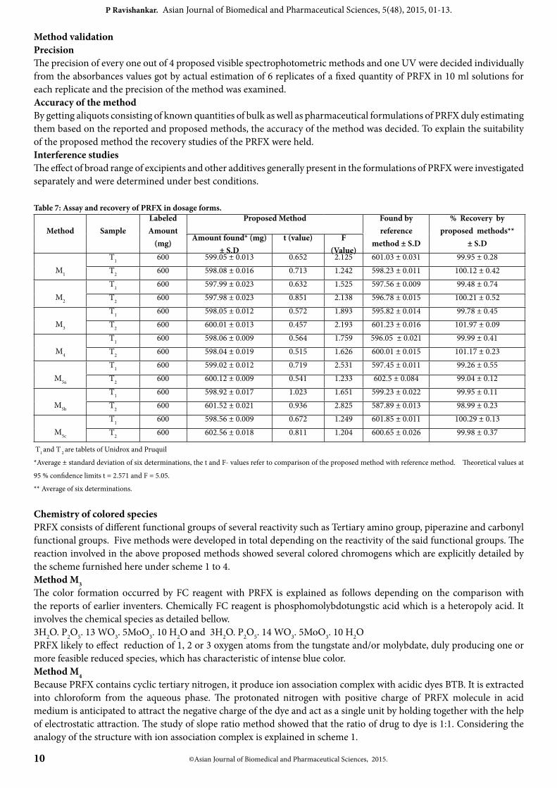
Chemistry of colored speciesPRFX consists of different functional groups of several reactivity such as Tertiary amino group, piperazine and carbonyl functional groups. Five methods were developed in total depending on the reactivity of the said functional groups. The reaction involved in the above proposed methods showed several colored chromogens which are explicitly detailed by the scheme furnished here under scheme 1 to 4. Method M3The color formation occurred by FC reagent with PRFX is explained as follows depending on the comparison with the reports of earlier inventers. Chemically FC reagent is phosphomolybdotungstic acid which is a heteropoly acid. It involves the chemical species as detailed bellow.3H2O. P2O5. 13 WO3. 5MoO3. 10 H2O and 3H2O. P2O5. 14 WO3. 5MoO3. 10 H2OPRFX likely to effect reduction of 1, 2 or 3 oxygen atoms from the tungstate and/or molybdate, duly producing one or more feasible reduced species, which has characteristic of intense blue color.Method M4Because PRFX contains cyclic tertiary nitrogen, it produce ion association complex with acidic dyes BTB. It is extracted into chloroform from the aqueous phase. The protonated nitrogen with positive charge of PRFX molecule in acid medium is anticipated to attract the negative charge of the dye and act as a single unit by holding together with the help of electrostatic attraction. The study of slope ratio method showed that the ratio of drug to dye is 1:1. Considering the analogy of the structure with ion association complex is explained in scheme 1.
© Asian Journal of Biomedical and Pharmaceutical Sciences, 2015.11
P Ravishankar. Asian Journal of Biomedical and Pharmaceutical Sciences, 5(48), 2015, 01-13.
H3C
BrOH
CH(CH3)2
CH(CH3)2
H3C Br
-O3S O
Drug-dye complex
Bromothymol blue
N
FOH
S
O
R1 =
N NR2R1
H
O
O
H2C OR2 =
N NR2R1
PRFX
O
Scheme 1: PRFX reaction with bromothymol blue Methods M5a, M5b, and M5c
PRFX shows reduced property owing to the existing of functional moieties (one or more) susceptible to oxidation selected oxidizing agents namely Fe (III) under controlled experimental conditions. When treated with known excess
of oxidant, PRFX undergoes oxidation, yielding products of oxidation apart from reduced form of oxidant, Fe (II) from Fe (III), in addition unreached oxidant. So there are feasible conditions to determine the drug content colorimetrically, which is equivalent to the reacted oxidant or reduced form. The reduced form of Fe III (Fe II) has a tendency to deliver colored complex when treated with 1, 10 - phenanthroline (M5a), 2, 2’- bipyridyl (M5b) and bathophenanthroline (M5c).
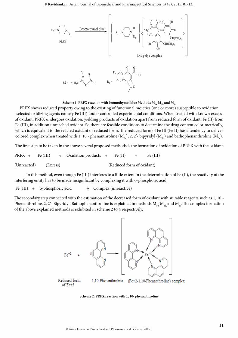
The first step to be taken in the above several proposed methods is the formation of oxidation of PRFX with the oxidant.
PRFX + Fe (III) → Oxidation products + Fe (II) + Fe (III)
(Unreacted) (Excess) (Reduced form of oxidant)
In this method, even though Fe (III) interferes to a little extent in the determination of Fe (II), the reactivity of the interfering entity has to be made insignificant by complexing it with o-phosphoric acid.
Fe (III) + o-phosphoric acid → Complex (unreactive)
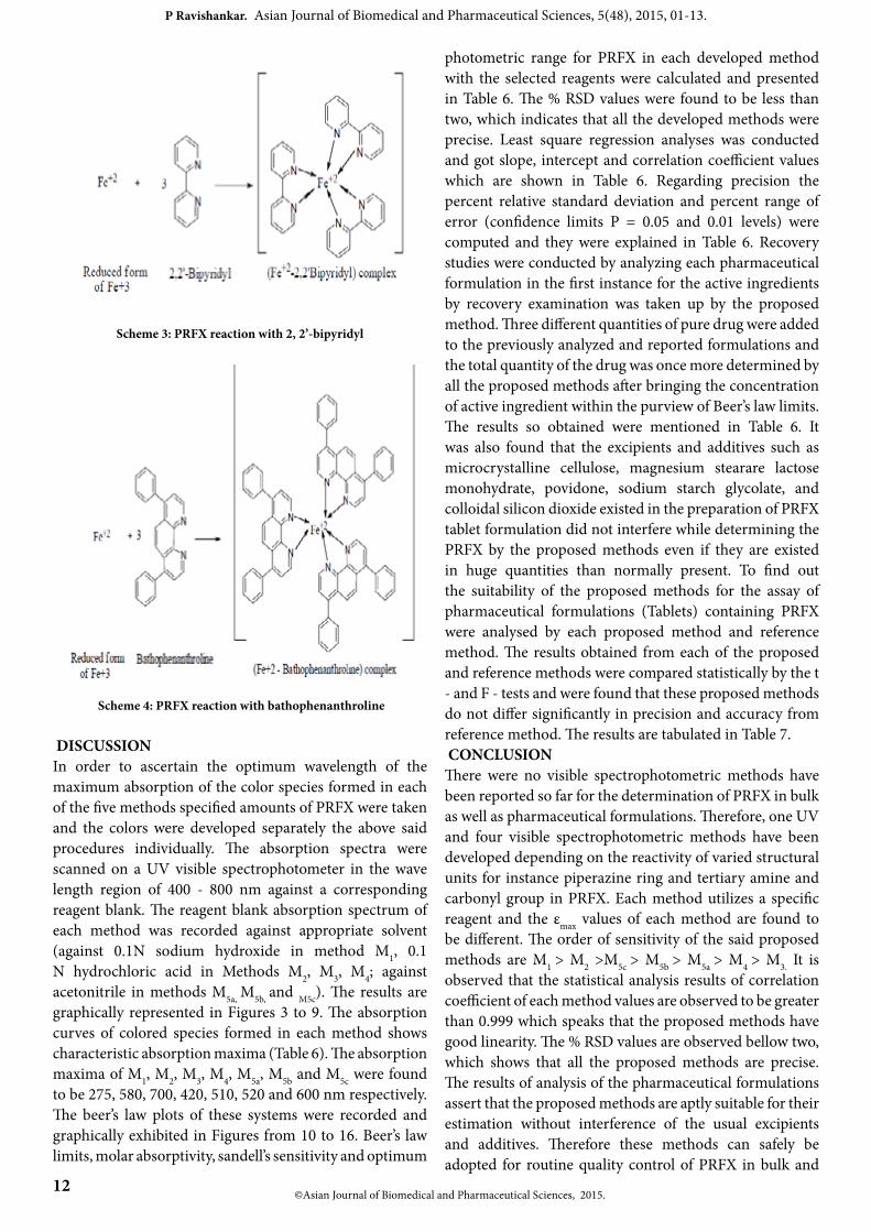
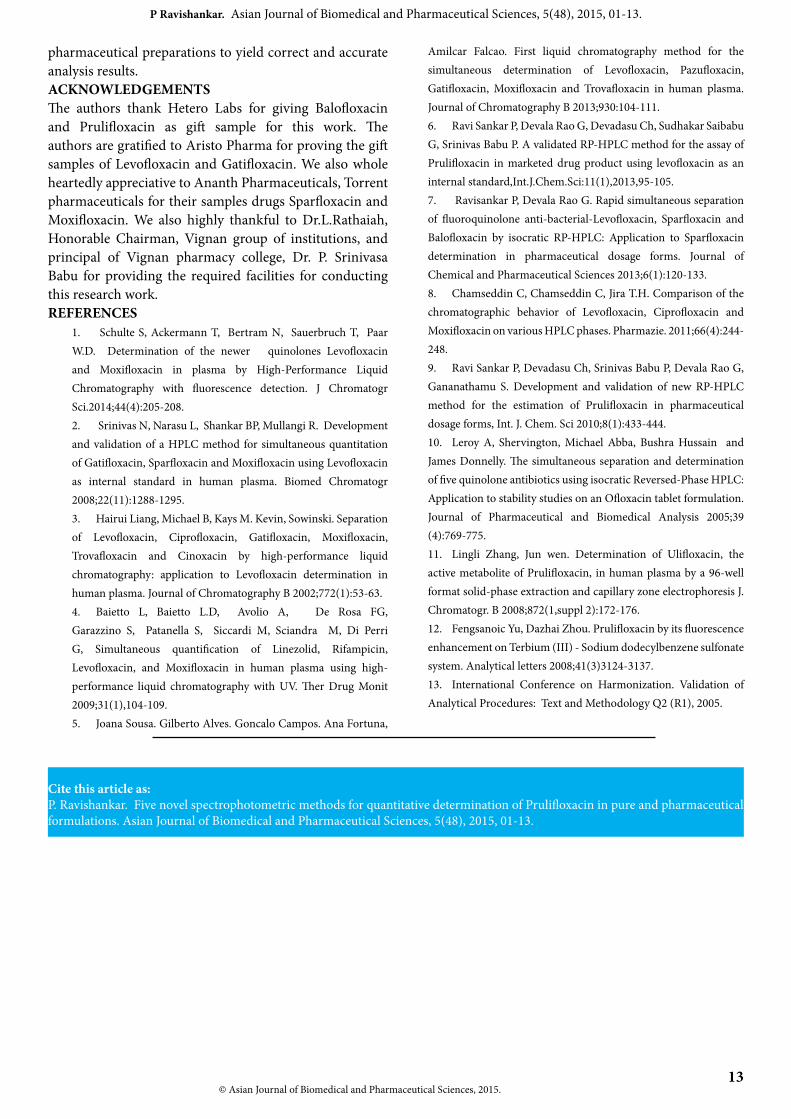
The secondary step connected with the estimation of the decreased form of oxidant with suitable reagents such as 1, 10 - Phenanthroline, 2, 2’- Bipyridyl, Bathophenanthroline is explained in methods M5a, M5b, and M5c. The complex formation of the above explained methods is exhibited in scheme 2 to 4 respectively.
Scheme 2: PRFX reaction with 1, 10- phenanthroline
12©Asian Journal of Biomedical and Pharmaceutical Sciences, 2015.
P Ravishankar. Asian Journal of Biomedical and Pharmaceutical Sciences, 5(48), 2015, 01-13.
Scheme 3: PRFX reaction with 2, 2’-bipyridyl
Scheme 4: PRFX reaction with bathophenanthroline
DISCUSSIONIn order to ascertain the optimum wavelength of the maximum absorption of the color species formed in each of the five methods specified amounts of PRFX were taken and the colors were developed separately the above said procedures individually. The absorption spectra were scanned on a UV visible spectrophotometer in the wave length region of 400 - 800 nm against a corresponding reagent blank. The reagent blank absorption spectrum of each method was recorded against appropriate solvent (against 0.1N sodium hydroxide in method M1, 0.1 N hydrochloric acid in Methods M2, M3, M4; against acetonitrile in methods M5a, M5b, and M5c). The results are graphically represented in Figures 3 to 9. The absorption curves of colored species formed in each method shows characteristic absorption maxima (Table 6). The absorption maxima of M1, M2, M3, M4, M5a, M5b and M5c were found to be 275, 580, 700, 420, 510, 520 and 600 nm respectively. The beer’s law plots of these systems were recorded and graphically exhibited in Figures from 10 to 16. Beer’s law limits, molar absorptivity, sandell’s sensitivity and optimum
photometric range for PRFX in each developed method with the selected reagents were calculated and presented in Table 6. The % RSD values were found to be less than two, which indicates that all the developed methods were precise. Least square regression analyses was conducted and got slope, intercept and correlation coefficient values which are shown in Table 6. Regarding precision the percent relative standard deviation and percent range of error (confidence limits P = 0.05 and 0.01 levels) were computed and they were explained in Table 6. Recovery studies were conducted by analyzing each pharmaceutical formulation in the first instance for the active ingredients by recovery examination was taken up by the proposed method. Three different quantities of pure drug were added to the previously analyzed and reported formulations and the total quantity of the drug was once more determined by all the proposed methods after bringing the concentration of active ingredient within the purview of Beer’s law limits. The results so obtained were mentioned in Table 6. It was also found that the excipients and additives such as microcrystalline cellulose, magnesium stearare lactose monohydrate, povidone, sodium starch glycolate, and colloidal silicon dioxide existed in the preparation of PRFX tablet formulation did not interfere while determining the PRFX by the proposed methods even if they are existed in huge quantities than normally present. To find out the suitability of the proposed methods for the assay of pharmaceutical formulations (Tablets) containing PRFX were analysed by each proposed method and reference method. The results obtained from each of the proposed and reference methods were compared statistically by the t - and F - tests and were found that these proposed methods do not differ significantly in precision and accuracy from reference method. The results are tabulated in Table 7. CONCLUSIONThere were no visible spectrophotometric methods have been reported so far for the determination of PRFX in bulk as well as pharmaceutical formulations. Therefore, one UV and four visible spectrophotometric methods have been developed depending on the reactivity of varied structural units for instance piperazine ring and tertiary amine and carbonyl group in PRFX. Each method utilizes a specific reagent and the εmax values of each method are found to be different. The order of sensitivity of the said proposed methods are M1 > M2 >M5c > M5b > M5a > M4 > M3. It is observed that the statistical analysis results of correlation coefficient of each method values are observed to be greater than 0.999 which speaks that the proposed methods have good linearity. The % RSD values are observed bellow two, which shows that all the proposed methods are precise. The results of analysis of the pharmaceutical formulations assert that the proposed methods are aptly suitable for their estimation without interference of the usual excipients and additives. Therefore these methods can safely be adopted for routine quality control of PRFX in bulk and
© Asian Journal of Biomedical and Pharmaceutical Sciences, 2015. 13
P Ravishankar. Asian Journal of Biomedical and Pharmaceutical Sciences, 5(48), 2015, 01-13.
pharmaceutical preparations to yield correct and accurate analysis results.ACKNOWLEDGEMENTSThe authors thank Hetero Labs for giving Balofloxacin and Prulifloxacin as gift sample for this work. The authors are gratified to Aristo Pharma for proving the gift samples of Levofloxacin and Gatifloxacin. We also whole heartedly appreciative to Ananth Pharmaceuticals, Torrent pharmaceuticals for their samples drugs Sparfloxacin and Moxifloxacin. We also highly thankful to Dr.L.Rathaiah, Honorable Chairman, Vignan group of institutions, and principal of Vignan pharmacy college, Dr. P. Srinivasa Babu for providing the required facilities for conducting this research work.REFERENCES
1. Schulte S, Ackermann T, Bertram N, Sauerbruch T, Paar W.D. Determination of the newer quinolones Levofloxacin and Moxifloxacin in plasma by High-Performance Liquid Chromatography with fluorescence detection. J Chromatogr Sci.2014;44(4):205-208.2. Srinivas N, Narasu L, Shankar BP, Mullangi R. Development and validation of a HPLC method for simultaneous quantitation of Gatifloxacin, Sparfloxacin and Moxifloxacin using Levofloxacin as internal standard in human plasma. Biomed Chromatogr 2008;22(11):1288-1295.3. Hairui Liang, Michael B, Kays M. Kevin, Sowinski. Separation of Levofloxacin, Ciprofloxacin, Gatifloxacin, Moxifloxacin, Trovafloxacin and Cinoxacin by high-performance liquid chromatography: application to Levofloxacin determination in human plasma. Journal of Chromatography B 2002;772(1):53-63.4. Baietto L, Baietto L.D, Avolio A, De Rosa FG, Garazzino S, Patanella S, Siccardi M, Sciandra M, Di Perri G, Simultaneous quantification of Linezolid, Rifampicin, Levofloxacin, and Moxifloxacin in human plasma using high-performance liquid chromatography with UV. Ther Drug Monit 2009;31(1),104-109.5. Joana Sousa. Gilberto Alves. Goncalo Campos. Ana Fortuna,
Amilcar Falcao. First liquid chromatography method for the simultaneous determination of Levofloxacin, Pazufloxacin, Gatifloxacin, Moxifloxacin and Trovafloxacin in human plasma. Journal of Chromatography B 2013;930:104-111.6. Ravi Sankar P, Devala Rao G, Devadasu Ch, Sudhakar Saibabu G, Srinivas Babu P. A validated RP-HPLC method for the assay of Prulifloxacin in marketed drug product using levofloxacin as an internal standard,Int.J.Chem.Sci:11(1),2013,95-105.7. Ravisankar P, Devala Rao G. Rapid simultaneous separation of fluoroquinolone anti-bacterial-Levofloxacin, Sparfloxacin and Balofloxacin by isocratic RP-HPLC: Application to Sparfloxacin determination in pharmaceutical dosage forms. Journal of Chemical and Pharmaceutical Sciences 2013;6(1):120-133.8. Chamseddin C, Chamseddin C, Jira T.H. Comparison of the chromatographic behavior of Levofloxacin, Ciprofloxacin and Moxifloxacin on various HPLC phases. Pharmazie. 2011;66(4):244-248.9. Ravi Sankar P, Devadasu Ch, Srinivas Babu P, Devala Rao G, Gananathamu S. Development and validation of new RP-HPLC method for the estimation of Prulifloxacin in pharmaceutical dosage forms, Int. J. Chem. Sci 2010;8(1):433-444.10. Leroy A, Shervington, Michael Abba, Bushra Hussain and James Donnelly. The simultaneous separation and determination of five quinolone antibiotics using isocratic Reversed-Phase HPLC: Application to stability studies on an Ofloxacin tablet formulation. Journal of Pharmaceutical and Biomedical Analysis 2005;39 (4):769-775.11. Lingli Zhang, Jun wen. Determination of Ulifloxacin, the active metabolite of Prulifloxacin, in human plasma by a 96-well format solid-phase extraction and capillary zone electrophoresis J. Chromatogr. B 2008;872(1,suppl 2):172-176.12. Fengsanoic Yu, Dazhai Zhou. Prulifloxacin by its fluorescence enhancement on Terbium (III) - Sodium dodecylbenzene sulfonate system. Analytical letters 2008;41(3)3124-3137.13. International Conference on Harmonization. Validation of Analytical Procedures: Text and Methodology Q2 (R1), 2005.
Cite this article as:P. Ravishankar. Five novel spectrophotometric methods for quantitative determination of Prulifloxacin in pure and pharmaceutical formulations. Asian Journal of Biomedical and Pharmaceutical Sciences, 5(48), 2015, 01-13.